

The Value of Optical Coherence Tomography in Patients with Pituitary Adenoma and Its Association with Clinical Features: A Pilot Study

 ,
,  ,
,
Abstract
1. Introduction
2. Materials and Methods
- established PA confirmed by MRI;
- good general condition of the patient;
- patient’s consent to participate in the study;
- age ≥ 18 years;
- no other brain tumors, intracranial infections, demyelinating lesions or cerebrovascular diseases;
- no ophthalmological eye disorders detected during a detailed ophthalmological examination.
- participants should be generally healthy, with no history of PA or major health conditions;
- age ≥ 18 years;
- no brain or systemic diseases;
- consent to participate in the study.
- individuals with major health issues, including pituitary disorders, brain tumors, or serious systemic diseases;
- those under 18, who were excluded to ensure consistency with the patient group;
- individuals who did not give informed consent.
2.1. Visual Acuity and Visual Field Evaluation
2.2. Invasiveness Evaluation
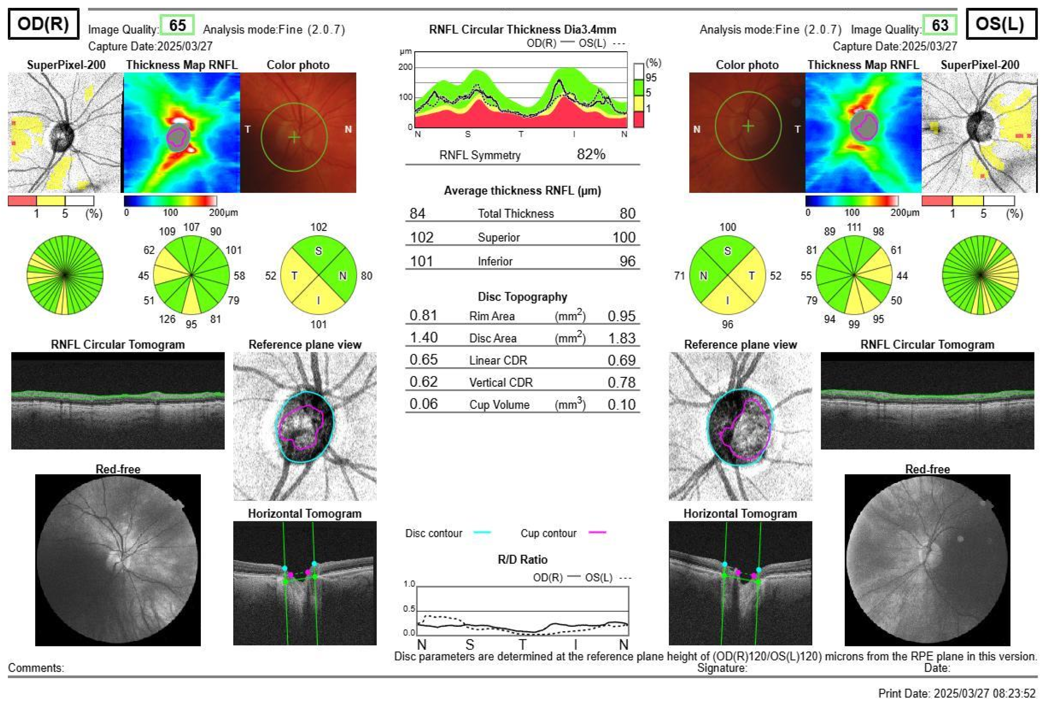
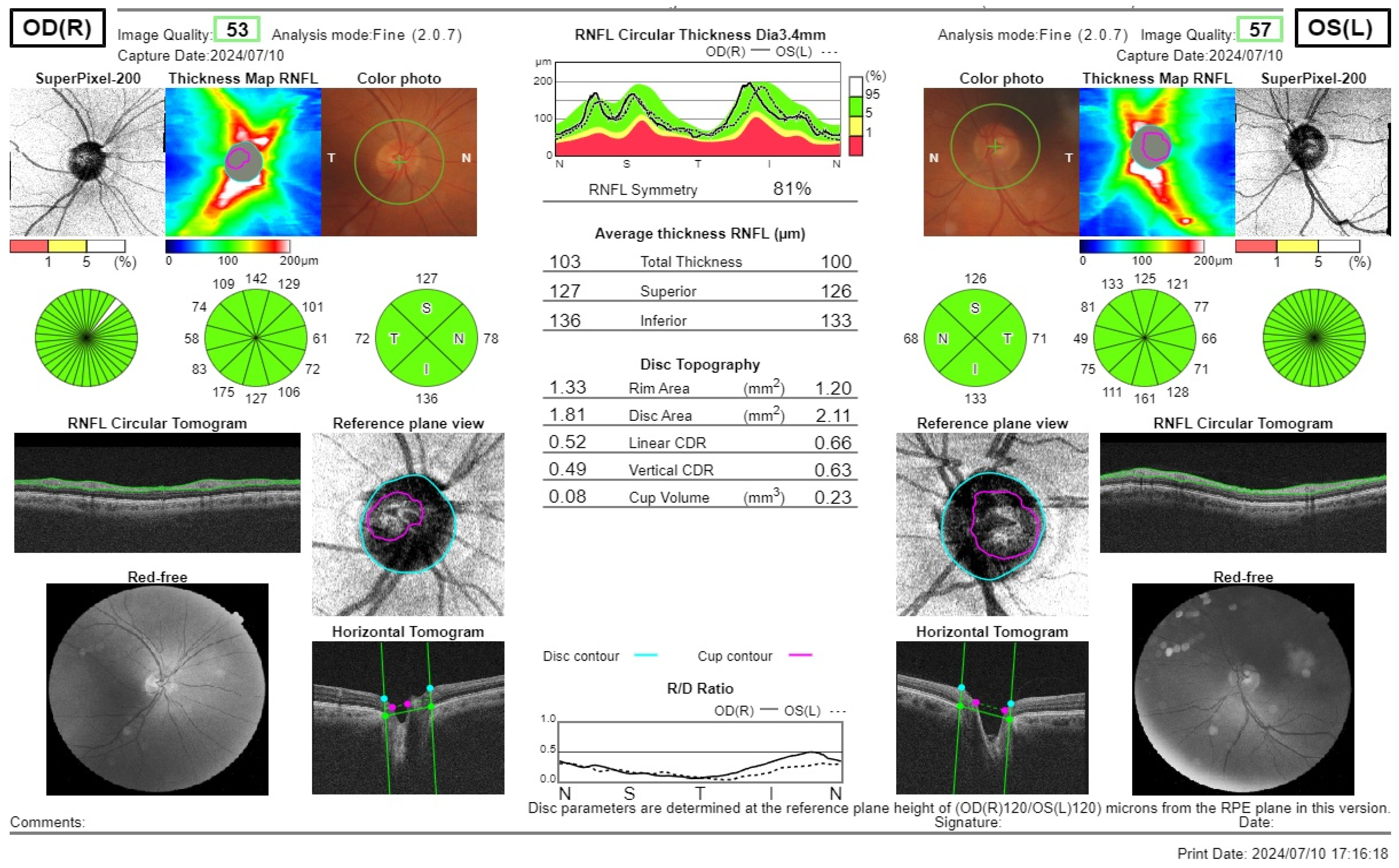
2.3. Optical Coherence Tomography
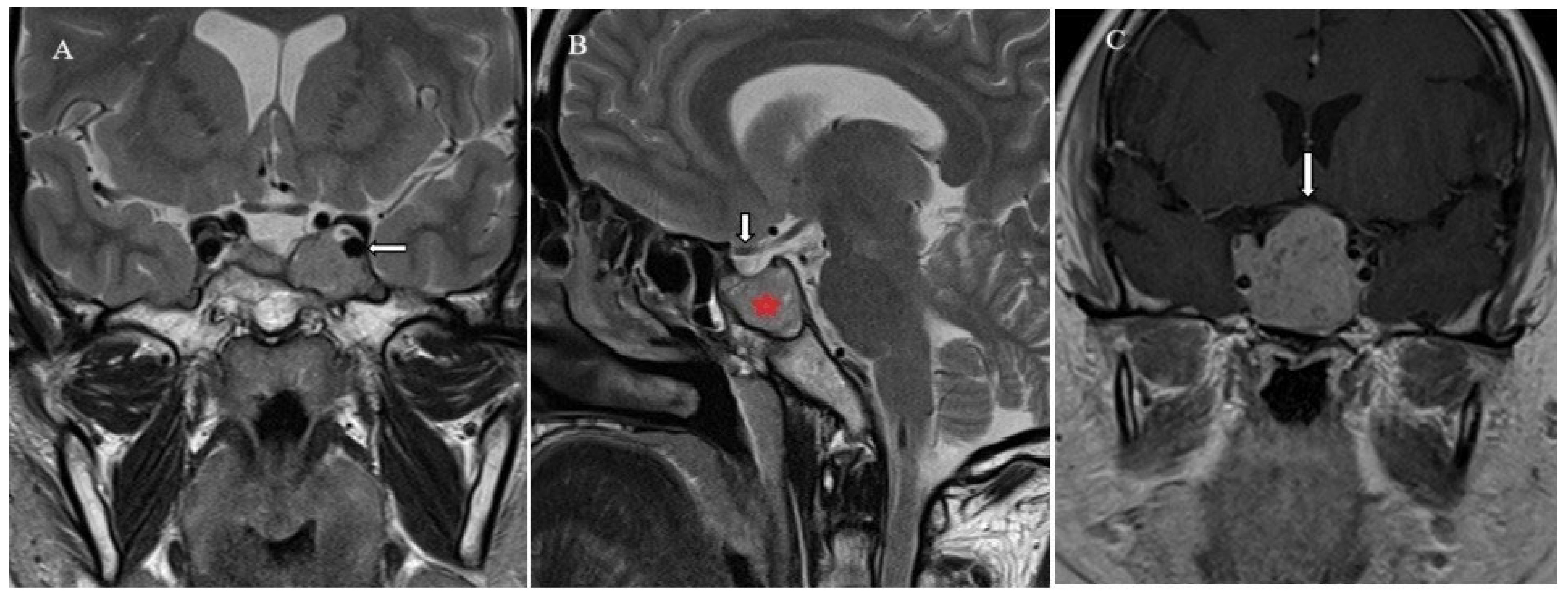
2.4. Brain Imaging
2.5. Statistical Analysis
2.6. Study Limitations
3. Results
3.1. Visual Acuity Changes
3.2. Visual Field Defects
3.3. Optical Coherent Tomography
3.4. Correlation Analysis Between Optic Disc and Rim Areas with RNFL Thickness
3.5. Visual Field Correlations with RNFL
3.6. Optic Disc Area Correlations
3.7. Optic Rim Area Correlations
4. Discussion
5. Conclusions
Author Contributions
Funding
Institutional Review Board Statement
Informed Consent Statement
Data Availability Statement
Conflicts of Interest
References
- Gonzalez-Almazan, J.A.; Cortes-Contreras, A.P.; Flores-Rabasa, R.; Mendez-Garcia, L.A.; Escobedo, G.; Navarro Olvera, J.L.; Carrillo-Ruiz, J.D. Metabolic Syndrome Components in Patients with Pituitary Adenoma. Horm. Metab. Res. 2024, 56, 118–127. [Google Scholar] [CrossRef] [PubMed]
- Graffeo, C.S.; Yagnik, K.J.; Carlstrom, L.P.; Lakomkin, N.; Bancos, I.; Davidge-Pitts, C.; Erickson, D.; Choby, G.; Pollock, B.E.; Chamberlain, A.M.; et al. Pituitary Adenoma Incidence, Management Trends, and Long-Term Outcomes: A 30-Year Population-Based Analysis. Mayo Clin. Proc. 2022, 97, 1861–1871. [Google Scholar] [CrossRef] [PubMed] [PubMed Central]
- Tsukamoto, T.; Miki, Y. Imaging of pituitary tumors: An update with the 5th WHO Classifications-part 1. Pituitary neuroendocrine tumor (PitNET)/pituitary adenoma. Jpn. J. Radiol. 2023, 41, 789–806. [Google Scholar] [CrossRef] [PubMed] [PubMed Central]
- Kim, T.G.; Jin, K.H.; Kang, J. Clinical characteristics and ophthalmologic findings of pituitary adenoma in Korean patients. Int. Ophthalmol. 2019, 39, 21–31. [Google Scholar] [CrossRef]
- De Witte, O.; Carlot, S.; Devuyst, E.; Choufani, G.; Hassid, S. Minimally invasive endoscopic unilateral transsphenoidal surgery for pituitary adenomas. B-ENT 2011, 7 (Suppl. S17), 27–32. [Google Scholar]
- Zee, C.S.; Go, J.L.; Klim, P.E.; Mitchell, D.; Ahmadi, J. Imaging of the pituitary and parasellar region. Neurosurg. Clin. N. Am. 2003, 14, 55. [Google Scholar] [CrossRef]
- Tieger, M.G.; Hedges, T.R., III; Ho, J.; Erlich-Malona, N.K.; Vuong, L.N.; Athappilly, G.K.; Mendoza-Santiesteban, C.E. Ganglion Cell Complex Loss in Chiasmal Compression by Brain Tumors. J. Neuroophthalmol. 2017, 37, 7–12. [Google Scholar] [CrossRef]
- Pang, Y.; Tan, Z.; Mo, W.; Chen, X.; Wei, J.; Guo, Q.; Zhong, Q.; Zhong, J. A pilot study of combined optical coherence tomography and diffusion tensor imaging method for evaluating microstructural change in the visual pathway of pituitary adenoma patients. BMC Ophthalmol. 2022, 22, 115. [Google Scholar] [CrossRef]
- Yum, H.R.; Park, S.H.; Park, H.Y.; Shin, S.Y. Macular ganglion cell analysis determined by cirrus HD optical coherence tomography for early detecting Chiasmal compression. PLoS ONE 2016, 11, e0153064. [Google Scholar] [CrossRef]
- Poczos, P.; Kremlacek, J.; Cesak, T.; Machackova, M.; Jiraskova, N. The use of optical coherence tomography in chiasmal compression. Cesk. Slov. Oftalmol. 2019, 75, 120–127. [Google Scholar]
- Cappabianca, P.; Alfieri, A.; Colao, A.; Ferone, D.; Lombardi, G.; de Divitiis, E. Endoscopic endonasaltranssphenoidal approach: An additional reason in support of surgery in the management of pituitary lesions. Skull Base Surg. 1999, 9, 109–117. [Google Scholar] [CrossRef] [PubMed]
- Yoneoka, Y.; Hatase, T.; Watanabe, N.; Jinguji, S.; Okada, M.; Takagi, M.; Fujii, Y. Early morphological recovery of the optic chiasm is associated with excellent visual outcome in patients with compressive chiasmal syndrome caused by pituitary tumors. Neurol. Res. 2015, 37, 1–8. [Google Scholar] [CrossRef] [PubMed]
- Zhang, J.; Zhang, S.; Song, Y.; Zhu, C.; He, M.; Ren, Q.; Shan, B.; Wang, Z.; Zeng, Y.; Xu, J. Predictive value of preoperative retinal nerve fiber layer thickness for postoperative visual recovery in patients with chiasmal compression. Oncotarget 2017, 8, 59148–59155. [Google Scholar] [CrossRef] [PubMed]
- Carpineto, P.; Ciancaglini, M.; Aharrh-Gnama, A.; Cirone, D.; Mastropasqua, L. Custom measurement of retinal nerve fiber layer thickness using STRATUS OCT in normal eyes. Eur. J. Ophthalmol. 2005, 15, 360–366. [Google Scholar] [CrossRef]
- Wang, H.; Sun, W.; Fu, Z.; Si, Z.; Zhu, Y.; Zhai, G.; Zhao, G.; Xu, S.; Pang, Q. The pattern of visual impairment in patients with pituitary adenoma. J. Int. Med. Res. 2008, 35, 1064–1069. [Google Scholar] [CrossRef]
- Glebauskiene, B.; Liutkeviciene, R.; Zlatkute, E.; Kriauciuniene, L.; Zaliuniene, D. Association of retinal nerve fibre layer thickness with quantitative magnetic resonance imaging data of the optic chiasm in pituitary adenoma patients. J. Clin. Neurosci. 2018, 50, 1–6. [Google Scholar] [CrossRef]
- Bonneville, J.F.; Bonneville, F.; Cattin, F. Magnetic resonance imaging of pituitary adenomas. Eur. Radiol. 2005, 15, 543–548. [Google Scholar] [CrossRef]
- Choi, S.H.; Kwon, B.J.; Na, D.G.; Kim, J.H.; Han, M.H.; Chang, K.H. Pituitary adenoma, craniopharyngioma, and Rathke cleft cyst involving both intrasellar and suprasellar regions: Differentiation using MRI. Clin. Radiol. 2007, 62, 453–462. [Google Scholar] [CrossRef]
- Blanch, R.J.; Micieli, J.A.; Oyesiku, N.M.; Newman, N.J.; Biousse, V. Optical coherence tomography retinal ganglion cell complex analysis for the detection of early chiasmal compression. Pituitary 2018, 21, 515–523. [Google Scholar] [CrossRef]
- Bokhari, A.R.; Davies, M.A.; Diamond, T. Endoscopic transsphenoidal pituitary surgery: A single surgeon experience and the learning curve. Br. J. Neurosurg. 2013, 27, 44–49. [Google Scholar] [CrossRef]
- Juraschka, K.; Khan, O.H.; Godoy, B.L.; Monsalves, E.; Kilian, A.; Krischek, B.; Ghare, A.; Vescan, A.; Gentili, F.; Zadeh, G. Endoscopic endonasal transsphenoidal approach to large and giant pituitary adenomas: Institutional experience and predictors of extent of resection. J. Neurosurg. 2014, 121, 75–83. [Google Scholar] [CrossRef] [PubMed]
- Lee, I.H.; Miller, N.R.; Zan, E.; Tavares, F.; Blitz, A.M.; Sung, H.; Yousem, D.M.; Boland, M.V. Visual Defects in Patients with Pituitary Adenomas: The Myth of Bitemporal Hemianopsia. AJR Am. J. Roentgenol. 2015, 205, W512–W518. [Google Scholar] [CrossRef] [PubMed]
- Anderson, D.; Faber, P.; Marcovitz, S.; Hardy, J.; Lorenzetti, D. Pituitary tumors and the ophthalmologist. Ophthalmology 1983, 90, 1265–1270. [Google Scholar] [CrossRef] [PubMed]
- Danesh-Meyer, H.V.; Wong, A.; Papchenko, T.; Matheos, K.; Stylli, S.; Nichols, A.; Frampton, C.; Daniell, M.; Savino, P.J.; Kaye, A.H. Optical coherence tomography predicts visual outcome for pituitary tumors. J. Clin. Neurosci. 2015, 22, 1098–1104. [Google Scholar] [CrossRef]
- Kawaguchi, T.; Ogawa, Y.; Tominaga, T. Retinal nerve fiber layer thickness measurement for predicting visual outcome after transsphenoidal surgery: Optic disc atrophy is not the deciding indicator. World Neurosurg. 2019, 127, e427–e435. [Google Scholar] [CrossRef]
- Garcia, T.; Sanchez, S.; Litre, C.F.; Radoi, C.; Delemer, B.; Rousseaux, P.; Ducasse, A.; Arndt, C. Prognostic value of retinal nerve fiber layer thickness for postoperative peripheral visual field recovery in optic chiasm compression. J. Neurosurg. 2014, 121, 165–169. [Google Scholar] [CrossRef]
- Monteiro, M.L.; Hokazono, K.; Fernandes, D.B.; Costa-Cunha, L.V.; Sousa, R.M.; Raza, A.S.; Wang, D.L.; Hood, D.C. Evaluation of inner retinal layers in eyes with temporal hemianopic visual loss from chiasmal compression using optical coherence tomography. Investig. Ophthalmol. Vis. Sci. 2014, 55, 3328–3336. [Google Scholar] [CrossRef]
- Al-Louzi, O.; Prasad, S.; Mallery, R.M. Utility of optical coherence tomography in the evaluation of sellar and parasellar mass lesions. Curr. Opin. Endocrinol. Diabetes Obes. 2018, 25, 274–284. [Google Scholar] [CrossRef]
- Danesh-Meyer, H.V.; Carroll, S.C.; Foroozan, R.; Savino, P.J.; Fan, J.; Jiang, Y.; Hoorn, S.V. Relationship between retinal nerve fiber layer and visual field sensitivity as measured by optical coherence tomography in chiasmal compression. Investig. Ophthalmol. Vis. Sci. 2006, 47, 4827–4835. [Google Scholar] [CrossRef]
- Iqbal, M.; Irfan, S.; Goyal, J.L.; Singh, D.; Singh, H.; Dutta, G. An Analysis of Retinal Nerve Fiber Layer Thickness Before and After Pituitary Adenoma Surgery and its Correlation with Visual Acuity. Neurol. India 2020, 68, 346–351. [Google Scholar] [CrossRef]
- Johansson, C.; Lindblom, B. The role of optical coherence tomography in the detection of pituitary adenoma. Acta Ophthalmol. 2009, 87, 776–779. [Google Scholar] [CrossRef] [PubMed]
- Park, H.H.; Oh, M.C.; Kim, E.H.; Kim, C.Y.; Kim, S.H.; Lee, K.S.; Chang, J.H. Use of optical coherence tomography to predict visual outcome in parachiasmal meningioma. J. Neurosurg. 2015, 123, 1489–1499. [Google Scholar] [CrossRef] [PubMed]
- Monteiro, M.L.; Leal, B.C.; Moura, F.C.; Vessani, R.M.; A Medeiros, F. Comparison of retinal nerve fibre layer measurements using optical coherence tomography versions 1 and 3 in eyes with band atrophy of the optic nerve and normal controls. Eye 2007, 21, 16–22. [Google Scholar] [CrossRef] [PubMed]
- Kaushik, M.; Fraser, C.L. Optical coherence tomography in compressive lesions of the anterior visual pathway. Ann. Eye Sci. 2020, 5, 15. [Google Scholar] [CrossRef]
- Lei, K.; Wang, L.N.; Wang, M.Z.; Wang, S.R.; Qu, Y.Z. Evaluation of Retinal Nerve Fiber Layer Thickness in Patients of Pituitary Adenomas With and Without Optic Chiasmal Compression. PREPRINT (Version 1). Available online: https://www.researchsquare.com/article/rs-6324/v1 (accessed on 7 April 2025).
- Menon, S.; Nair, S.; Kodnani, A.; Hegde, A.; Nayak, R.; Menon, G. Retinal nerve fiber layer thickness and its correlation with visual symptoms and radiological features in pituitary macroadenoma. J. Neurosci. Rural Pract. 2023, 14, 41–47. [Google Scholar] [CrossRef]

| Characteristics | Group | p-Value | ||
|---|---|---|---|---|
| PA Group, n = 25 | Control Group, n = 27 | |||
| Gender | Males, N (%) | 14 (28) | 12 (22.2) | 0.497 |
| Females, N (%) | 36 (72) | 42 (77.8) | ||
| Age min–max, median | 24–55, 47 | 26–70, 52 | 0.072 | |
| Size | Micro PA | 10 (40) | - | - |
| Macro PA | 15 (60) | |||
| Invasiness | Invasive PA | 15 (60) | ||
| Non-invasive PA | 10 (40) | |||
| Activeness | Active Pa | 19 (76) | ||
| Non-active PA | 6 (24) | |||
| Symptoms Per Eye | ||||
| Visual disturbances (decreased visual acuity) | ||||
| -Decreased | 5 (10) | 0 (0) | 0.017 | |
| -Not decreased | 45 (90) | 54 (100) | ||
| Visual acuity of all PA patients median (min; max); mean rank | 1 (0.01; 1); 48.18 | 1 (1; 1); 56.5 | 0.002 | |
| -PA with suprasellar extension median (min; max); mean rank | 1 (0.01; 1); 23.14 | - | 0.043 | |
| -PA without suprasellar extension median (min; max); mean rank | 1 (0.8; 1); 28.5 | |||
| Visual disturbances (visual field defects) | ||||
| -Detected | 22 (44) | 0 (0) | <0.001 | |
| -Not detected | 28 (56) | 54 (100) | ||
| RNFL Quadrants | Control Group: Median (Min; Max); Mean Rank (n = 54) | PA Group: Median (Min; Max); Mean Rank (n = 50) | p-Value * |
|---|---|---|---|
| Temporal quadrant | 73.5 (52; 109); 58.39 | 69.5 (16; 168); 46.14 | 0.038 |
| Superior quadrant | 126.5 (67; 165); 56.76 | 120 (57; 156); 47.9 | 0.134 |
| Nasal quadrant | 78 (50; 119); 56.34 | 75 (36; 119); 48.35 | 0.177 |
| Inferior quadrant | 130.5 (104; 1798); 56.28 | 129 (49; 156); 48.42 | 0.184 |
| Total | 102 (86; 125); 56.16 | 99 (40; 129); 48.55 | 0.198 |
| GCL+ (Ganglion Cell Layer [GCL] + Inner Plexiform Layer [IPL]) | Control Group: Median (Min; Max); Mean Rank (n = 54) | PA Group: Median (Min; Max); Mean Rank (n = 50) | p-Value * |
| Superior | 65 (56; 74); 56.8 | 62.5 (44; 72); 47.86 | 0.130 |
| Inferior | 64.5 (55; 77); 55.66 | 62 (42; 76); 49.09 | 0.266 |
| Total | 65 (54; 74); 56.82 | 62 (43; 71); 47.83 | 0.128 |
| GCL++ (RNFL + GCL + IPL) | Control Group: Median (Min; Max); Mean Rank (n = 54) | PA Group: Median (Min; Max); Mean Rank (n = 50) | p-Value * |
| Superior | 104 (69; 116); 54.37 | 102 (57; 115); 50.48 | 0.510 |
| Inferior | 105 (93; 126); 57.54 | 105 (61; 121); 47.06 | 0.076 |
| Total | 104 (92; 117); 56.37 | 103.5 (59; 116); 48.32 | 0.173 |
| RNFL Thickness, μm | Microadenoma: Median (Min; Max); Mean Rank (n = 20) | Macrodenoma: Median (Min; Max); Mean Rank (n = 30) | p-Value * |
|---|---|---|---|
| Temporal quadrant | 71 (58; 142); 29.38 | 69 (16; 168); 22.92 | 0.125 |
| Superior quadrant | 120 (95; 156); 26.9 | 120 (57; 144); 24.57 | 0.579 |
| Nasal quadrant | 75.5 (46; 119); 29.65 | 69.5 (36; 107); 22.73 | 0.100 |
| Inferior quadrant | 130.5 (99; 152); 27.68 | 125.5 (49; 156); 24.05 | 0.389 |
| Total | 101.5 (81;129); 28.95 | 99 (40; 118); 23.2 | 0.171 |
| GCL+ (Ganglion Cell Layer [GCL] + Inner Plexiform Layer [IPL]) | Microadenoma: Median (Min; Max); Mean Rank (n = 20) | Macrodenoma: Median (Min; Max); Mean Rank (n = 30) | p-Value * |
| Superior | 62.5 (53; 68); 26.38 | 62.5 (44; 72); 24.92 | 0.728 |
| Inferior | 63 (56; 76); 28.35 | 62 (42; 69); 23.60 | 0.258 |
| Total | 62.5 (54; 69); 26.65 | 62 (43; 71); 24.73 | 0.648 |
| GCL++ (RNFL + GCL + IPL)me | Microadenoma: Median (Min; Max); Mean Rank (n = 20) | Macrodenoma: Median (Min; Max); Mean Rank (n = 30) | p-Value * |
| Superior | 102.5 (98; 112); 28.08 | 99.5 (57; 115); 23.78 | 0.307 |
| Inferior | 106.5 (90; 121); 32.15 | 99.5 (61; 115); 21.07 | 0.008 |
| Total | 104 (95; 116); 29.65 | 100 (59; 113); 22.73 | 0.100 |
| RNFL Thickness, μm | Invasive PA: Median (Min; Max); Mean Rank (n = 30) | Non-Invasive PA: Median (Min; Max); Mean Rank (n = 20) | p-Value * |
|---|---|---|---|
| Temporal quadrant | 72.5 (16; 168); 26.5 | 68 (28; 159); 24 | 0.552 |
| Superior quadrant | 120 (57; 156); 25.3 | 120 (67; 144); 25.8 | 0.905 |
| Nasal quadrant | 74 (36; 107); 23.83 | 75.5 (46; 119); 28 | 0.322 |
| Inferior quadrant | 126.5 (49; 156); 23 | 132.5 (77; 154); 29.25 | 0.137 |
| Total | 99 (40; 118); 24.82 | 101.5 (63; 129); 26.53 | 0.684 |
| GCL+ (Ganglion Cell Layer [GCL] + Inner Plexiform Layer [IPL]) | Invasive PA: Median (Min; Max); Mean Rank (n= 30) | Non-Invasive PA: Median (Min; Max); Mean Rank (n = 20) | p-Value * |
| Superior | 63.5 (44; 72); 26.07 | 62 (50; 68); 24.65 | 0.735 |
| Inferior | 62.5 (42; 70); 24.9 | 62 (54; 76); 26.4 | 0.721 |
| Total | 63.5 (43; 71); 26.23 | 62 (52; 68); 24.4 | 0.662 |
| GCL++ (RNFL + GCL + IPL) | Invasive PA: Median (Min; Max); Mean Rank (n= 30) | Non-Invasive PA: Median (Min; Max); Mean Rank (n = 20) | p-Value * |
| Superior | 103.5 (57; 114); 26.17 | 101 (77; 115); 24.5 | 0.692 |
| Inferior | 104.5 (61; 121); 24.83 | 105.5 (78; 115); 26.5 | 0.692 |
| Total | 104 (59; 116); 26 | 102.5 (77; 113); 24.75 | 0.766 |
| RNFL Thickness, μm | Active PA: Median (Min; Max); Mean Rank (n = 38) | Non-Active PA: Median (Min; Max); Mean rank (n = 12) | p-Value * |
|---|---|---|---|
| Temporal quadrant | 69 (28; 168); 25.86 | 71.5 (16; 86); 24.38 | 0.759 |
| Superior quadrant | 121 (67; 144); 27 | 113 (57; 156); 20.75 | 0.195 |
| Nasal quadrant | 77 (37; 119); 27.51 | 69.5 (36; 87); 19.13 | 0.082 |
| Inferior quadrant | 130.5 (77; 156); 28.05 | 118.5 (49; 144); 17.42 | 0.028 |
| Total | 104 (63; 129); 27.33 | 91 (40; 118); 19.71 | 0.114 |
| GCL+ (Ganglion Cell Layer [GCL] + Inner Plexiform Layer [IPL]) | Active PA: Median (Min; Max); Mean Rank (n = 38) | Non-Active PA: Median (Min; Max); Mean Rank (n = 12) | p-Value * |
| Superior | 63.5 (46; 72); 27.2 | 61 (44; 70); 20.13 | 0.141 |
| Inferior | 62.5 (45; 70); 27.17 | 58.5 (42; 76); 20.21 | 0.148 |
| Total | 63 (46; 71); 27.64 | 56 (43; 68); 18.71 | 0.063 |
| GCL++ (RNFL + GCL + IPL) | Active PA: Median (Min; max); Mean Rank (n = 38) | Non-Active PA: Median (Min; Max); Mean Rank (n = 12) | p-Value * |
| Superior | 103 (71; 115); 27.16 | 100.5 (57; 111); 20.25 | 0.152 |
| Inferior | 105 (74; 121); 27.61 | 96.5 (61; 110); 18.83 | 0.069 |
| Total | 104 (73; 116); 27.33 | 98.5 (59; 110); 19.71 | 0.114 |
| RNFL Thickness, μm | PA with Suprasellar Extension: Mean Rank; Median (Min; Max) (n = 28) | PA Without Suprasellar Extension: Mean Rank; Median (Min; Max) (n = 22) | p-Value * |
|---|---|---|---|
| Superior quadrant | 122.5 (57; 156); 25.55 | 120 (95; 144); 25.43 | 0.977 |
| Temporal quadrant | 67.5 (16; 99); 21.66 | 72 (58; 168); 30.39 | 0.036 |
| Inferior quadrant | 125 (49; 156); 22.23 | 131 (99; 154); 29.66 | 0.074 |
| Nasal quadrant | 71.5 (36; 95); 22.64 | 75.5 (46; 119); 29.14 | 0.118 |
| Total | 99 (40; 118); 23.2 | 100.5 (81; 129); 28.43 | 0.207 |
| GCL+ (Ganglion Cell Layer [GCL] + Inner Plexiform Layer [IPL]) | PA with Suprasellar Extension: Mean Rank; Median (Min; Max) (n = 28) | PA Without Suprasellar Extension: Mean Rank; Median (Min; Max) (n = 22) | p-Value * |
| Superior | 63 (44; 70); 23.66 | 62 (53; 72); 27.84 | 0.312 |
| Inferior | 62 (42; 70); 22.46 | 63 (56; 76); 29.36 | 0.096 |
| Total | 62 (43; 69); 23.3 | 62.5 (54; 71); 28.3 | 0.228 |
| GCL++ (RNFL + GCL + IPL) | PA with Suprasellar Extension: Mean Rank; Median (Min; Max) (n = 28) | PA Without Suprasellar Extension: Mean Rank; Median (Min; Max) (n = 22) | p-Value * |
| Superior | 98.5 (57; 113); 21.8 | 103.5 (95; 115); 30.2 | 0.043 |
| Inferior | 101 (61; 121); 21.48 | 106.5 (90; 115); 30.61 | 0.028 |
| Total | 102.5 (59; 116); 21.71 | 104.5 (95; 113); 30.32 | 0.038 |
| Versus | GCL+ (Ganglion Cell Layer [GCL] + Inner Plexiform Layer [IPL]) | GCL++ (RNFL + GCL + IPL) | ||||
|---|---|---|---|---|---|---|
| Total | Superior | Inferior | Total | Superior | Inferior | |
| Correlation Coefficient of Optic Disc Area Value in PA Group | 0.008 | 0.015 | −0.025 | −0.065 | −0.129 | −0.057 |
| p-Value | 0.955 | 0.915 | 0.863 | 0.656 | 0.372 | 0.696 |
| Correlation Coefficient of Optic Disc Area Value in Control Group | 0.01 | −0.14 | 0.064 | −0.058 | −0.114 | −0.053 |
| p-Value | 0.940 | 0.921 | 0.644 | 0.675 | 0.410 | 0.702 |
| Correlation Coefficient of Optic Rim Area Value in PA Group | 0.197 | 0.212 | 0.167 | 0.201 | 0.201 | 0.193 |
| p-Value | 0.171 | 0.140 | 0.245 | 0.161 | 0.161 | 0.178 |
| Correlation Coefficient of Optic Rim Area Value in Control Group | 0.136 | 0.106 | 0.122 | −0.109 | −0.113 | −0.146 |
| p-Value | 0.328 | 0.447 | 0.379 | 0.434 | 0.417 | 0.293 |
| Versus | RNFL Thickness | ||||
|---|---|---|---|---|---|
| Total | Superior Quadrant | Temporal Quadrant | Inferior Quadrant | Nasal Quadrant | |
| Correlation Coefficient of Optic Disc Area Value in PA Group | 0.046 | 0.094 | −0.011 | −0.012 | 0.153 |
| p-Value | 0.751 | 0.716 | 0.937 | 0.935 | 0.288 |
| Correlation Coefficient of Optic Disc Area Value in Control Group | 0.440 | 0.426 | 0.268 | 0.350 | 0.203 |
| p-Value | <0.001 | 0.001 | 0.06 | 0.009 | 0.142 |
| Correlation Coefficient of Optic Rim Area Value in PA Group | 0.493 | 0.295 | 0.327 | 0.368 | 0.503 |
| p-Value | <0.001 | 0.037 | 0.020 | 0.008 | <0.001 |
| Correlation Coefficient of Optic Rim Area Value in Control Group | 0.351 | 0.333 | 0.447 | 0.334 | −0.065 |
| p-Value | 0.009 | 0.014 | <0.001 | 0.014 | 0.641 |
| Versus | RNFL Thickness | ||||
|---|---|---|---|---|---|
| Total | Superior Quadrant | Temporal Quadrant | Inferior Quadrant | Nasal Quadrant | |
| Correlation Coefficient of Visual Field Value in PA Group | −0.552 | −0.618 | −0.316 | −0.434 | −0.282 |
| p-Value | <0.001 | <0.001 | 0.025 | 0.002 | 0.047 |
Disclaimer/Publisher’s Note: The statements, opinions and data contained in all publications are solely those of the individual author(s) and contributor(s) and not of MDPI and/or the editor(s). MDPI and/or the editor(s) disclaim responsibility for any injury to people or property resulting from any ideas, methods, instructions or products referred to in the content. |
© 2025 by the authors. Licensee MDPI, Basel, Switzerland. This article is an open access article distributed under the terms and conditions of the Creative Commons Attribution (CC BY) license (https://creativecommons.org/licenses/by/4.0/).
Share and Cite
Duseikaite, M.; Vilkeviciute, A.; Dumbliauskaite, I.; Glebauskiene, B.; Zostautiene, I.; Rovite, V.; Wu, S.-N.; Tamasauskas, A.; Liutkeviciene, R. The Value of Optical Coherence Tomography in Patients with Pituitary Adenoma and Its Association with Clinical Features: A Pilot Study. J. Clin. Med. 2025, 14, 4318. https://doi.org/10.3390/jcm14124318
Duseikaite M, Vilkeviciute A, Dumbliauskaite I, Glebauskiene B, Zostautiene I, Rovite V, Wu S-N, Tamasauskas A, Liutkeviciene R. The Value of Optical Coherence Tomography in Patients with Pituitary Adenoma and Its Association with Clinical Features: A Pilot Study. Journal of Clinical Medicine. 2025; 14(12):4318. https://doi.org/10.3390/jcm14124318
Chicago/Turabian StyleDuseikaite, Monika, Alvita Vilkeviciute, Igne Dumbliauskaite, Brigita Glebauskiene, Indre Zostautiene, Vita Rovite, Sheng-Nan Wu, Arimantas Tamasauskas, and Rasa Liutkeviciene. 2025. "The Value of Optical Coherence Tomography in Patients with Pituitary Adenoma and Its Association with Clinical Features: A Pilot Study" Journal of Clinical Medicine 14, no. 12: 4318. https://doi.org/10.3390/jcm14124318
APA StyleDuseikaite, M., Vilkeviciute, A., Dumbliauskaite, I., Glebauskiene, B., Zostautiene, I., Rovite, V., Wu, S.-N., Tamasauskas, A., & Liutkeviciene, R. (2025). The Value of Optical Coherence Tomography in Patients with Pituitary Adenoma and Its Association with Clinical Features: A Pilot Study. Journal of Clinical Medicine, 14(12), 4318. https://doi.org/10.3390/jcm14124318