Incidence, Impact, and Complications of Short Cephalomedullary Nail Toggling in Patients with Wide Femoral Medullary Canal
Abstract
1. Introduction
2. Materials and Methods
2.1. Study Design
2.2. Inclusion Criteria
- Intertrochanteric fractures (AO/OTA 31, A1–A3) treated with a Short CMN, and
- Cases with a wide medullary canal that could potentially allow for nail motion/toggling and allow for easier detection/calculation of the toggling. Due to the absence of a clear definition of a wide medullary canal, and considering the risks of nail toggling in the study of George et al. [17], a medullary canal was considered wide in cases of:
- -
- Any Dorr [18] C case
- -
- Dorr B case with a medullary canal width of 15 mm or more.
2.3. Exclusion Criteria
- -
- Patients with missing immediate post-operative or follow-up X-rays.
- -
- Patients who did not complete a minimum radiographic follow-up of 6 weeks.
- -
- Tip apex distance (from the tip of the lag device to the femoral head apex), calculated from both AP and lateral radiographs.
- -
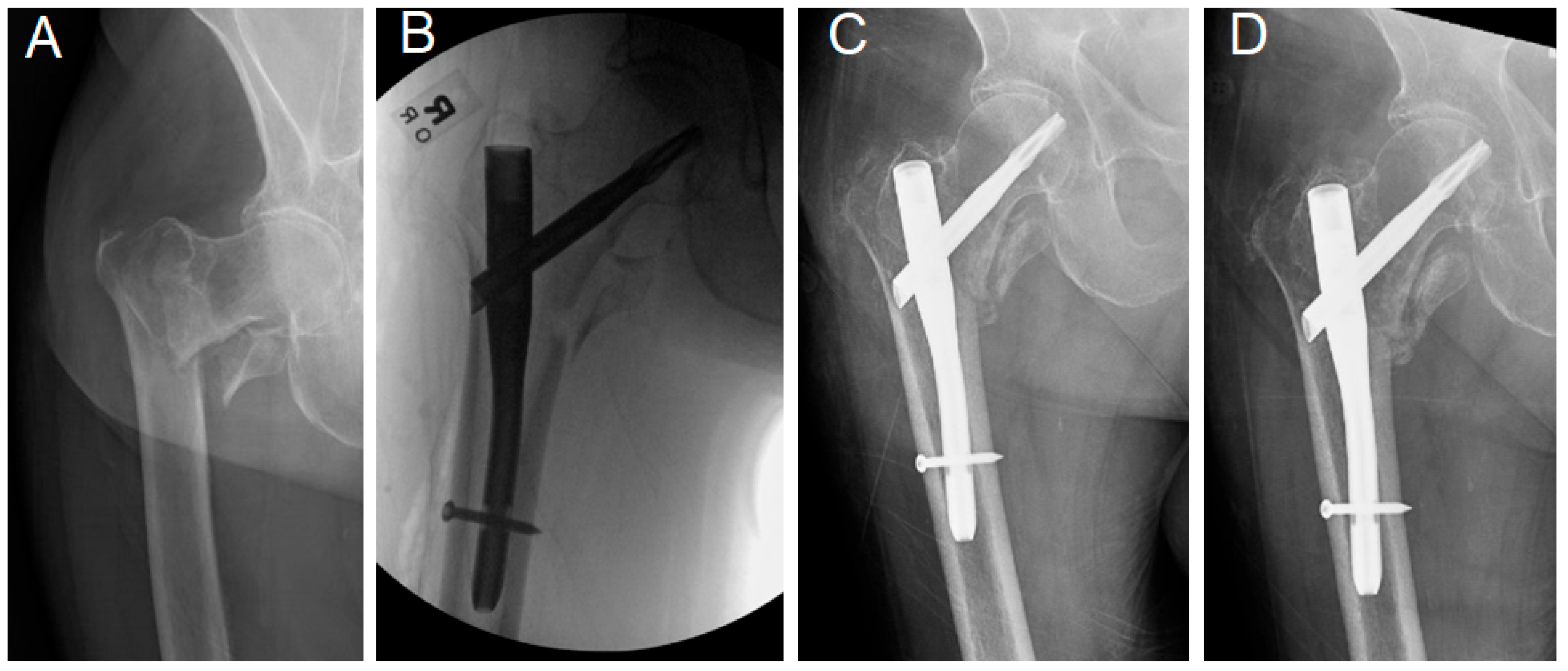
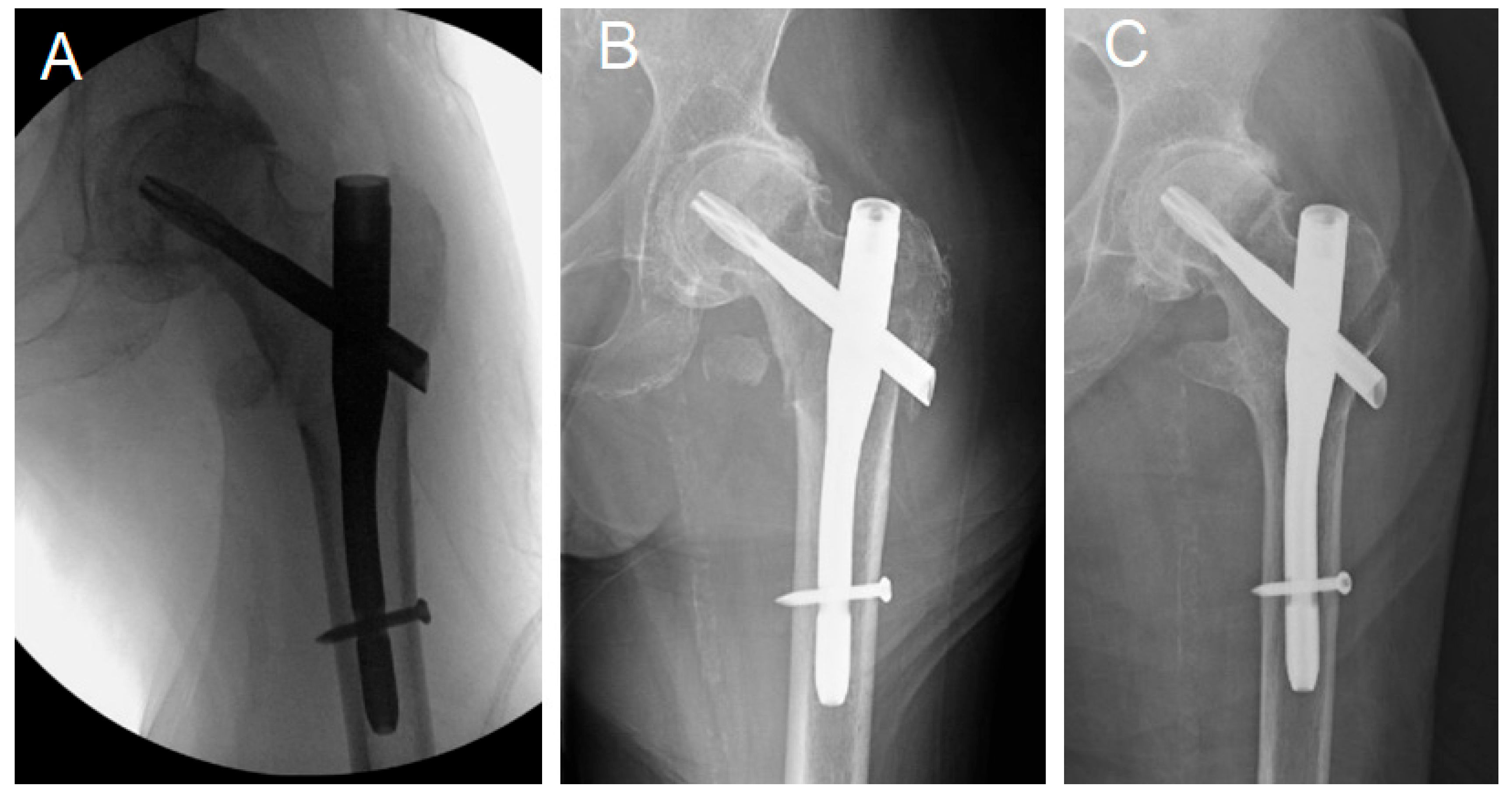
- Nail/femoral canal angle. Cases with significant toggling, as defined by George et al. [17] as a change in the nail/femur angle of more than 4 degrees, were documented.
- -
- Distance between the medial tip of the distal end of the nail and the endosteal border of the lateral femoral cortex.
- -
- Varus displacement/malunion was identified as a change in the fracture alignment with a varus change in the neck shaft angle of more than 5 degrees [17].
- -
- Medullary canal width: Calculated at 10 cm from the tip of the lesser trochanter or tangent from the other side’s lesser trochanter tip, parallel to the inter-teardrop line.
- -
- Nail Diameter.
- -
- Nail/canal ratio.
- -
- Engagement of the lateral aspect of the lag device past the lateral cortex. A lag device was identified as “engaged” when both the superolateral and inferolateral borders of the device protruded freely outside the lateral femoral cortex in the intraoperative radiographs. This measurement was particularly made in the intraoperative radiograph to document the status of lag device engagement before any weight bearing and potential fracture collapse, to judge the utilized surgical technique.
- -
- Integrity of the proximal femoral lateral cortex: The proximal femoral lateral wall, where the lateral end of the lag device engages, was assessed in the preoperative, intraoperative, and immediate postoperative X-rays or CT scan. Lateral wall incompetency was considered if any lateral wall breakage or fissure fracture was noticed.
- -
- Quality of reduction [17,20,21]: Good reductions were defined as having <4 mm of fragment displacement, a neck-shaft angle of < 5 degrees of varus or <20 degrees of valgus, and <20 degrees angulation on the lateral view radiographs. Acceptable reductions met the criteria for either alignment or displacement. Poor reductions met neither of the criteria [17]. Assessment of the reduction quality was retrospectively based on the last available C-arm radiographs.
2.4. Statistical Analysis and Data Interpretation
3. Results
3.1. Demographics, Measurements, and Overall Outcomes
3.2. Complications
4. Discussion
5. Conclusions
Author Contributions
Funding
Institutional Review Board Statement
Informed Consent Statement
Data Availability Statement
Conflicts of Interest
References
- Meinberg, E.; Agel, J.M.; Roberts, C.M.; Karam, M.D.; Kellam, J. Fracture and Dislocation Classification Compendium—2018. J. Orthop. Trauma 2018, 32, S1–S10. [Google Scholar] [CrossRef] [PubMed]
- Werner, B.C.; Fashandi, A.H.; Gwathmey, F.W.; Yarboro, S.R. Trends in the management of intertrochanteric femur fractures in the United States 2005–2011. Hip Int. 2015, 25, 270–276. [Google Scholar] [CrossRef] [PubMed]
- Horwitz, D.S.; Tawari, A.; Suk, M. Nail length in the management of intertrochanteric fracture of the femur. J. Am. Acad. Orthop. Surg. 2016, 24, e50–e58. [Google Scholar] [CrossRef]
- Lewis, S.R.; Macey, R.; Gill, J.R.; Parker, M.J.; Griffin, X.L. Cephalomedullary nails versus extramedullary implants for extracapsular hip fractures in older adults. Cochrane Database Syst. Rev. 2022, 1, CD000093. [Google Scholar] [CrossRef]
- Xu, H.; Liu, Y.; Sezgin, E.A.; Tarasevičius, Š.; Christensen, R.; Raina, D.B.; Tägil, M.; Lidgren, L. Comparative effectiveness research on proximal femoral nail versus dynamic hip screw in patients with trochanteric fractures: A systematic review and meta-analysis of randomized trials. J. Orthop. Surg. Res. 2022, 17, 292. [Google Scholar] [CrossRef]
- Matre, K.; Havelin, L.I.; Gjertsen, J.E.; Vinje, T.; Espehaug, B.; Fevang, J.M. Sliding hip screw versus IM nail in reverse oblique trochanteric and subtrochanteric fractures: A study of 2716 patients in the Norwegian Hip Fracture Register. Injury 2013, 44, 735–742. [Google Scholar] [CrossRef]
- Darbandi, A.D.; Saadat, G.H.; Siddiqi, A.; Butler, B.A. Comparison of extramedullary and intramedullary implants for stable intertrochanteric fractures: Have we swung the pendulum too far the other way? J. Am. Acad. Orthop. Surg. 2022, 30, e779–e788. [Google Scholar] [CrossRef]
- Schipper, I.B.; Marti, R.K.; van der Werken, C. Unstable trochanteric femoral fractures: Extramedullary or intramedullary fixation. Rev. Lit. Inj. 2004, 35, 142–151. [Google Scholar] [CrossRef] [PubMed]
- Sun, D.; Wang, C.; Chen, Y.; Liu, X.; Zhao, P.; Zhang, H.; Zhou, H.; Qin, C. A meta-analysis comparing intramedullary with extramedullary fixations for unstable femoral intertrochanteric fractures. Medicine 2019, 98, e17010. [Google Scholar] [CrossRef]
- Okcu, G.; Ozkayin, N.; Okta, C.; Topcu, I.; Aktuglu, K. Which implant is better for treating reverse obliquity fractures of the proximal femur: A standard or long nail? Clin. Orthop. Relat. Res. 2013, 471, 2768–2775. [Google Scholar] [CrossRef]
- Tsugeno, H.; Takegami, Y.; Tokutake, K.; Mishima, K.; Nakashima, H.; Kobayashi, K.; Imagama, S. Comparing short vs. intermediate and long nails in elderly patients with unstable multifragmental femoral trochanteric fractures (AO type A2): Multicenter (TRON group) retrospective study. Injury 2024, 55, 111420. [Google Scholar] [CrossRef] [PubMed]
- Martí-Garín, D.; Fillat-Gomà, F.; Marcano-Fernández, F.A.; Balaguer-Castro, M.; Álvarez, J.M.; Pellejero, R.; Fernández, J.S.; Torner, P.; Vives, J.M.M. Complications of standard versus long cephalomedullary nails in the treatment of unstable extracapsular proximal femoral fractures: A randomized controlled trial. Injury 2023, 54, 661–668. [Google Scholar] [CrossRef] [PubMed]
- Kleweno, C.; Morgan, J.; Redshaw, J.; Harris, M.; Rodriguez, E.; Zurakowski, D.; Vrahas, M.; Appleton, P. Short versus long cephalomedullary nails for the treatment of intertrochanteric hip fractures in patients older than 65 years. J. Orthop. Trauma 2014, 28, 391–397. [Google Scholar] [CrossRef] [PubMed]
- Krigbaum, H.; Takemoto, S.; Kim, H.T.; Kuo, A.C. Costs and complications of short versus long cephalomedullary nailing of OTA 31-A2 proximal femur fractures in U.S. veterans. J. Orthop. Trauma 2016, 30, 125–129. [Google Scholar] [CrossRef] [PubMed]
- Frisch, N.B.; Nahm, N.J.; Khalil, J.G.; Les, C.M.; Guthrie, S.T.; Charters, M.A. Short versus long cephalomedullary nails for pertrochanteric hip fracture. Orthopedics 2017, 40, 83–88. [Google Scholar] [CrossRef]
- Chang, S.M.; Hou, Z.Y.; Hu, S.J.; Du, S.C. Intertrochanteric femur fracture treatment in Asia: What we know and what the world can learn. Orthop. Clin. N. Am. 2020, 51, 189–205. [Google Scholar] [CrossRef]
- George, A.V.; Bober, K.; Eller, E.B.; Hakeos, W.M.; Hoegler, J.; Jawad, A.H.; Guthrie, S.T. Short cephalomedullary nail toggle: A closer examination. OTA Int. 2022, 5, e185. [Google Scholar] [CrossRef]
- Dorr, L.D.; Faugere, M.-C.; Mackel, A.M.; Gruen, T.A.; Bognar, B.; Malluche, H.H. Structural and cellular assessment of bone quality of proximal femur. Bone 1993, 14, 231–242. [Google Scholar] [CrossRef]
- Karayiannis, P.N.; Cassidy, R.S.; Hill, J.C.; Dorr, L.D.; Beverland, D.E. The relationship between canal diameter and the Dorr classification. J. Arthroplast. 2020, 35, 3204–3207. [Google Scholar] [CrossRef]
- Baumgaertner, M.R.; Curtin, S.L.; Lindskog, D.M.; Keggi, J.M. The value of the tip-apex distance in predicting failure of fixation of peritrochanteric fractures of the hip. J. Bone Jt. Surg. Am. 1995, 77, 1058–1064. [Google Scholar] [CrossRef]
- Parry, J.A.; Barrett, I.; Schoch, B.; Yuan, B.; Cass, J.; Cross, W. Does the angle of the nail matter for pertrochanteric fracture reduction? Matching nail angle and native neck shaft angle. J. Orthop. Trauma 2018, 32, 174–177. [Google Scholar] [CrossRef]
- Ceynowa, M.; Zerdzicki, K.; Klosowski, P.; Pankowski, R.; Rocławski, M.; Mazurek, T. The early failure of the gamma nail and the dynamic hip screw in femurs with a wide medullary canal. A biomechanical study of intertrochanteric fractures. Clin. Biomech. 2020, 71, 201–207. [Google Scholar] [CrossRef] [PubMed]
- Tisherman, R.T.; Hankins, M.L.; Moloney, G.B.; Tarkin, I.S. Distal locking of short cephalomedullary nails decreases varus collapse in unstable intertrochanteric fractures—A biomechanical analysis. Injury 2021, 52, 414–418. [Google Scholar] [CrossRef] [PubMed]
- Vopat, B.G.; Kane, P.M.; Truntzer, J.; McClure, P.; Paller, D.; Abbood, E.; Born, C. Is distal locking of long nails for intertrochanteric fractures necessary? A clinical study. J. Clin. Orthop. Trauma 2014, 5, 233–239. [Google Scholar] [CrossRef]
- Abram, S.G.; Pollard, T.C.; Andrade, A.J. Inadequate ‘three-point’ proximal fixation predicts failure of the Gamma nail. Bone Jt. J. 2013, 95-B, 825–830. [Google Scholar] [CrossRef]
- Kane, P.; Vopat, B.; Paller, D.; Koruprolu, S.; Daniels, A.H.; Born, C. A biomechanical comparison of locked and unlocked long cephalomedullary nails in a stable intertrochanteric fracture model. J. Orthop. Trauma 2014, 28, 715–720. [Google Scholar] [CrossRef] [PubMed]
- Usami, T.; Takada, N.; Kosuwon, W.; Paholpak, P.; Tokunaga, M.; Iwata, H.; Hattori, Y.; Nagaya, Y.; Murakami, H.; Kuroyanagi, G. A Lateral Fracture Line Affects Femoral Trochanteric Fracture Instability and Swing Motion of the Intramedullary Nail: A Biomechanical Study. JBJS Open Access 2024, 9, e23.00118. [Google Scholar] [CrossRef]
- Horwitz, D.S.; Mahmoud, A.N.; Suk, M. Intermediate Length Cephalomedullary Nails in Proximal Femoral Fractures: Review of Indications and Outcomes. J. Am. Acad. Orthop. Surg. 2025. [Google Scholar] [CrossRef]
- Mahmoud, A.N.; Hine, S.; Sams, K.B.; Nye, A.; Suk, M.; Horwitz, D.S. Low trochanteric fractures in the presence of knee arthroplasty: The role of intermediate length cephalomedullary nail. J. Musculoskelet. Surg. Res. 2025, 9, 70–75. [Google Scholar] [CrossRef]
- Matsumura, T.; Takahashi, T.; Nakashima, M.; Nibe, Y.; Takeshita, K. Clinical outcome of mid-length proximal femoral nail for patients with trochanteric hip fractures: Preliminary investigation in a Japanese cohort of patients more than 70 years old. Geriatr. Orthop. Surg. Rehabil. 2020, 11, 2151459320936444. [Google Scholar] [CrossRef]
- Kochar, V.; Pankaj, A.; Chadha, M.; Arora, S. Results of proximal femoral nail in intertrochanteric fractures of the hip with compromised lateral femoral wall: A clinical outcome study. J. Clin. Orthop. Trauma 2010, 1, 99–104. [Google Scholar] [CrossRef]
- Womble, T.N.; Kirk, A.; Boyle, M.; Comadoll, S.M.; Su, L.; Srinath, A.; Matuszewski, P.E.; Aneja, A. Comparison of short, intermediate, and long cephalomedullary nail length outcomes in elderly intertrochanteric femur fractures. J. Am. Acad. Orthop. Surg. Glob. Res. Rev. 2022, 6, e21.00322. [Google Scholar] [CrossRef] [PubMed]
- Lenich, A.; Mayr, E.; Rüter, A.; Möckl, C.; Füchtmeier, B. First results with the trochanter fixation nail (TFN): A report on 120 cases. Arch. Orthop. Trauma Surg. 2006, 126, 706–712. [Google Scholar] [CrossRef]
- Baldwin, P.C., III; Lavender, R.C.; Sanders, R.; Koval, K.J. Controversies in intramedullary fixation for intertrochanteric hip fractures. J. Orthop. Trauma 2016, 30, 635–641. [Google Scholar] [CrossRef] [PubMed]
- Sadeghi, C.; Prentice, H.A.; Okike, K.M.; Paxton, E.W. Treatment of intertrochanteric femur fractures with long versus short cephalomedullary nails. Perm. J. 2020, 24, 19.229. [Google Scholar] [CrossRef] [PubMed]
- Hong, C.C.; Nashi, N.; Makandura, M.C.; Tan, J.H.; Peter, L.; Murphy, D. The long and short of cephalomedullary nails in the treatment of osteoporotic pertrochanteric fracture. Singapore Med. J. 2017, 58, 85–91. [Google Scholar] [CrossRef]
- Chantarapanich, N.; Riansuwan, K. Biomechanical performance of short and long cephalomedullary nail constructs for stabilizing different levels of subtrochanteric fracture. Injury 2022, 53, 323–333. [Google Scholar] [CrossRef]
- Robinson, C.M.; Houshian, S.; Khan, L.A.K. Trochanteric-entry long cephalomedullary nailing of subtrochanteric fractures caused by low-energy trauma. J. Bone Jt. Surg. Am. 2005, 87, 2217–2226. [Google Scholar]
- Rajnish, R.K.; Srivastava, A.; Kumar, P.; Yadav, S.K.; Sharma, S.; Haq, R.U.; Aggarwal, A.N. Comparison of outcomes of long versus short cephalomedullary nails for the fixation of intertrochanteric femur fractures: A systematic review and meta-analysis of 14,547 patients. Indian J. Orthop. 2023, 57, 1165–1187. [Google Scholar] [CrossRef]
- Lee, W.C.; Chou, S.M.; Tan, C.W.; Chng, L.S.; Yam, G.J.M.; Chua, T.H.I. Intertrochanteric fracture with distal extension: When is the short proximal femoral nail antirotation too short? Injury 2021, 52, 926–932. [Google Scholar] [CrossRef]
- Longo, L.H.; Zimmermann Faggion, H.; Costa Sartor, M.; Senna Klipp, M.U.; Vogt, P.H.; Navarro Vergara, A.D.; Valenza, W.R. Factors Associated with Failure of Synthesis in the Treatment of Proximal Femur Fractures with Cephalomedullary Nails. Cureus 2024, 16, e61363. [Google Scholar] [CrossRef] [PubMed]
- Song, H.; Chang, S.M.; Hu, S.J.; Du, S.C. Low filling ratio of the distal nail segment to the medullary canal is a risk factor for loss of anteromedial cortical support: A case control study. J. Orthop. Surg. Res. 2022, 17, 27. [Google Scholar] [CrossRef] [PubMed]
- Li, S.J.; Chen, S.Y.; Chang, S.M.; Du, S.C.; Hu, S.J. Insufficient proximal medullary filling of cephalomedullary nails in intertrochanteric femur fractures predicts excessive postoperative sliding: A case-control study. BMC Musculoskelet. Disord. 2023, 24, 156. [Google Scholar] [CrossRef] [PubMed]


| Variable | Immediate Postop Tip Apex Distance | Last Follow-Up Tip Apex Distance | Immediate Postop Nail Shaft Angle | Last Follow-Up Nail Shaft Angle | Immediate. Postop Nail Tip to Medial Femoral Cortex Distance | Last Follow-Up Nail Tip to Medial femoral CORTEX Distance |
|---|---|---|---|---|---|---|
| Mean ± SD (Median) | 16 ± 4.7 (15.9) | 14.9 ± 4.8 (14.65) | −1.56 ± 2.21 (−1.35) (Negative value indicates a varus angle) | −2.56 ± 2.33 (−2.7) | 6.92 ± 2.43 (6.85) | 7.95 ± 2.67 (7.87) |
| Wilcoxon signed-rank test | p = 0.00014 | p ˂ 0.00001 | p < 0.00001 | |||
| Cases with a Toggle ≥ 4 Degrees and Varus Displacement | All Other Non-Complicated Cases, Excluding the Cases with Poor Reduction or AVN | Statistical Significance | |
|---|---|---|---|
| Number of cases | 16 | 80 | |
| Nail/canal ratio (Mean ± SD) | 0.593 ± 0.07 | 0.598 ± 0.08 | p = 0.7 (Mann-Whitney U Test) |
| Immediate postoperative TAD (Mean ± SD) | 15.7 ± 4.7 | 16 ± 4.7 | p = 0.8 (Unpaired t-Test) |
| Cases with no Proximal Fixation Defect | Cases with Only the Lag Device Not Engaging the Lateral Cortex | Cases with Only Lateral Wall Incompetency | Cases with a Combination of the Two Factors | |
|---|---|---|---|---|
| Total Number of cases (96) * | 58 | 22 | 8 | 8 |
| Cases with nail toggle >4 degrees (%) | 0 | 2 (9.09%) | 7 (87.5%) | 7 (87.5%) |
| Out of 16 cases with proximal lateral wall incompetency, 14 cases (87.5%) showed significant toggling. | ||||
| Percent of complicated cases | 0/58 (0%) | 16/38 (42.1%) | ||
| Fisher’s Exact test | p < 0.0001 (Statistically significant) | |||
| Cases with Nail Toggling and Varus Displacement (16 Cases) | Cases Without Complications (80 Cases) | |
|---|---|---|
| Number of cases with deficient proximal fixation | 16 (100%) | 22 (27.5%) |
| Fisher’s Exact test | p < 0.0001 (Statistically significant) |
Disclaimer/Publisher’s Note: The statements, opinions and data contained in all publications are solely those of the individual author(s) and contributor(s) and not of MDPI and/or the editor(s). MDPI and/or the editor(s) disclaim responsibility for any injury to people or property resulting from any ideas, methods, instructions or products referred to in the content. |
© 2025 by the authors. Licensee MDPI, Basel, Switzerland. This article is an open access article distributed under the terms and conditions of the Creative Commons Attribution (CC BY) license (https://creativecommons.org/licenses/by/4.0/).
Share and Cite
Mahmoud, A.N.; Echeverry-Martinez, M.F.; Doyle, C.M.; Bernate, J.D.; Suk, M.; Horwitz, D.S. Incidence, Impact, and Complications of Short Cephalomedullary Nail Toggling in Patients with Wide Femoral Medullary Canal. J. Clin. Med. 2025, 14, 3961. https://doi.org/10.3390/jcm14113961
Mahmoud AN, Echeverry-Martinez MF, Doyle CM, Bernate JD, Suk M, Horwitz DS. Incidence, Impact, and Complications of Short Cephalomedullary Nail Toggling in Patients with Wide Femoral Medullary Canal. Journal of Clinical Medicine. 2025; 14(11):3961. https://doi.org/10.3390/jcm14113961
Chicago/Turabian StyleMahmoud, Ahmed Nageeb, Maria F. Echeverry-Martinez, Catherine Mary Doyle, Juan David Bernate, Michael Suk, and Daniel Scott Horwitz. 2025. "Incidence, Impact, and Complications of Short Cephalomedullary Nail Toggling in Patients with Wide Femoral Medullary Canal" Journal of Clinical Medicine 14, no. 11: 3961. https://doi.org/10.3390/jcm14113961
APA StyleMahmoud, A. N., Echeverry-Martinez, M. F., Doyle, C. M., Bernate, J. D., Suk, M., & Horwitz, D. S. (2025). Incidence, Impact, and Complications of Short Cephalomedullary Nail Toggling in Patients with Wide Femoral Medullary Canal. Journal of Clinical Medicine, 14(11), 3961. https://doi.org/10.3390/jcm14113961


