Article

A β-Thalassemia Cell Biobank: Updates, Further Validation in Genetic and Therapeutic Research and Opportunities During (and After) the COVID-19 Pandemic

by
Roberto Gambari
1,*,
Maria Rita Gamberini
1,
Lucia Carmela Cosenza
2,
Cristina Zuccato
2 and
Alessia Finotti
2,*
1
Center “Chiara Gemmo and Elio Zago” for the Research on Thalassemia, Ferrara University, 44121 Ferrara, Italy
2
Department of Life Sciences and Biotechnology, Ferrara University, 44121 Ferrara, Italy
*
Authors to whom correspondence should be addressed.
J. Clin. Med. 2025, 14(1), 289; https://doi.org/10.3390/jcm14010289
Submission received: 22 November 2024 / Revised: 16 December 2024 / Accepted: 30 December 2024 / Published: 6 January 2025
(This article belongs to the Section Epidemiology & Public Health)

Abstract

:
Background: Cellular biobanks are of great interest for performing studies finalized in the development of personalized approaches for genetic diseases, including β-thalassemia and sickle cell disease (SCD), important diseases affecting the hematopoietic system. These inherited genetic diseases are characterized by a global distribution and the need for intensive health care. The aim of this report is to present an update on the composition of a cellular Thal-Biobank, to describe its utilization since 2016, to present data on its application in studies on fetal hemoglobin induction and on gene editing, and to discuss its employment as a “unique tool” during and after the COVID-19 pandemic. Methods: The methods were as follows: freezing, cryopreservation, long-term storage, and thawing of erythroid precursor cells from β-thalassemia patients; fetal hemoglobin (HbF) induction; CRISPR-Cas9 gene editing; HPLC analysis of the hemoglobin pattern. Results: The updated version of the Thal-Biobank is a cellular repository constituted of 990 cryovials from 221 β-thalassemia patients; the phenotype (pattern of hemoglobin production) is maintained after long-term storage; fetal hemoglobin induction and CRISPR-Cas9 gene editing can be performed using biobanked cells. In representative experiments using an isoxazole derivative as HbF inducer, the HbF increased from 13.36% to more than 60%. Furthermore, in CRIPR/Cas9 gene editing, de novo production of HbA was obtained (42.7% with respect to the trace amounts found in untreated cells). Conclusions: The implemented Thal-Biobank was developed before the COVID-19 outbreak and should be considered a tool of great interest for researchers working on β-thalassemia, with the aim of developing innovative therapeutic protocols and verifying the impact of the COVID-19 pandemic on erythroid precursor cells.

1. Introduction

Biobanking of biological material and construction of biorepositories are activities significantly expanding their applications in biomedicine and applied biotechnology. They are important in the fields of data sharing, shared use of precious biomaterials, and development of commonly designed and validated protocols in molecular and cellular biology [1,2,3,4]. The term “biobank” is rather general, including biological samples such as plasma, serum, whole cells and tissues, urine, buffy coats, RNA, genomic DNA, and protein extracts [5,6]. An increasing number of diverse biobanks are present worldwide, as discussed by O’Donoghue et al. [7], many of them of great interest to health researchers [8,9]. In this respect, the quality of the biobank, the updated description of its features, and access to the biological material are all crucial parameters for the possible use of the biobank in biomedicine [9,10]. In relation to these parameters, with particular reference to the availability of biological materials, the type of adopted governance strategies is a crucial step in facilitating access to biobanked material (including datasets) by the research community [11].
In this respect, one of the most appealing projects is to construct “cellular biobanks” [12,13,14,15,16] for possible future use in the post-genomic era [3]. The reason for this interest is that “cellular biobanks” are expected to be very important in developing personalized medicines [4,12,17], providing “ready-to-use” biomaterials for developing (a) therapeutic-oriented protocols based on the use of bio-drugs (low-molecular-weight compounds, microRNAs, antisense oligonucleotides, others), (b) products for gene therapy (viral and non-viral), and (c) products for gene editing. Biobanking should be considered an issue of global interest [8,18], and it is a growing field of research and biomedical applications in low- and middle-income countries (LMICs), forcing the scientific community to discuss novel legal and policy issues related to this key activity [19,20]. It can be concluded that serious attention should be given to the activity and perspectives of researchers working on biobanking-related investigations in LMICs [19]. Data sharing and exchanges of unique biobanked samples are issues of great importance to guarantee the global usability of the products and results obtained in this sector of applied research.
A biomedical area of research that needs validated biobanked collections is related to genetic diseases [21], including β-thalassemia and sickle cell disease (SCD), important diseases affecting the hematopoietic system [22,23,24]; notably, these inherited genetic diseases are characterized by a global distribution and the need for intensive health care and research efforts [25,26], with the objective of developing cost-effective treatments [27,28,29].
In this context, we produced and validated a cell biobank for β-thalassemia [30], noting that the use of cells from the biobank allows for the stratification of patients with respect to the endogenous production of fetal hemoglobin and can determine the response to treatment with fetal hemoglobin inducers and gene therapy protocols with reproducible results. In particular, we were able to demonstrate that the use of biobanked samples allows for the simultaneous comparison of many potentially therapeutic treatments across several samples grown in parallel. Furthermore, in the study reported by Cosenza et al. [30], the biobanked samples were validated for retention of the phenotype in relation to hemoglobin production after thawing frozen cells isolated from the same patient at different times. In addition, different frozen batches of the same patient’s cells retained the same hemoglobin pattern. This reproducibility was also found in cultures of the same biobanked cells that were sent to different laboratories [30]. Finally, the biobanked cells allow for stratification of the patients with respect to fetal hemoglobin (HbF) production and can be used for determining the response to HbF inducers, as discussed in a recent review article [31]. Recent reviews on HbF inducers in β-thalassemia are also available [32,33,34,35,36].
The collection of cellular samples for thalassemia research is a key issue of several EU-funded projects, such as the Thalassemia Modular Stratification System (THALAMOSS) for personalized therapy of beta-thalassemia [31]. Furthermore, this field of investigation is central in two ongoing projects related to hemoglobinopathies: the COST-action CA22119–HELIOS [37] and the International Hemoglobinopathy Research Network (INHERENT) project [38], which brings together nine existing international or regional consortia in the field of hemoglobinopathies, namely ITHANET, RADeep, ARISE, SPARCO, SADaCC, REDAC, the HVP Global Globin Network, the International Health Repository, and the ClinGen Hemoglobinopathy VCEP, as reported by Gambari et al. [31].
The Thal-Biobank reported by Cosenza et al. [30] was a cellular repository for research on thalassemia and sickle cell disease that was composed of 779 biobanked specimens from 8 healthy donors and 72 patients with sickle cell disease (SCD) and β-thalassemia. The most important difficulty of the study was related to the recruitment of patients. In fact, the distribution of the genotypes was not uniform, the most frequent genotypes being β039/β039 (29 patients), β+IVSI-110/β039 (17 patients), and β+IVSI-110/β+IVSI-110 (8 patients). On the contrary, a limited number of patients with the β+IVSI-110/β+IVSI-6 and β+IVSI-110/β+IVSI-1 genotypes were recruited. Few patients with at least one SCD allele were recruited, and their erythroid cells biobanked (7 patients, 52 vials). As far as the validation of protocols for their impact in the therapeutic field of β-thalassemia is concerned, no attempt was made to determine the phenotypic correction of biobanked erythroid cells using the gene-editing approach.
The objectives of the present report were (a) to present updates on the composition of the cell Thal-Biobank, (b) to describe its utilization since 2016, (c) to present novel data on the application of biobanked cells in studies on fetal hemoglobin induction and gene editing, and (d) to discuss the utilization of the Thal-Biobank as a “unique tool” during and after the COVID-19 pandemic, taking in consideration the fact that all the patients participating to this study were not SARS-CoV-2-infected and COVID-19-vaccinated. Therefore, the described Thal-Biobank should be considered composed of “SARS-CoV-2-free” cells to serve as precious reference control of SARS-CoV-2-infected erythroid cells in studies aimed at characterizing the effects of SARS-CoV-2 infection and COVID-19 vaccination on the hematopoietic system.
In this respect, recent reports concurrently demonstrated that (a) a perturbation of the hematopoietic system occurs in COVID-19 patients [39,40,41,42,43,44], (b) erythroid precursors can be infected by SARS-CoV-2 [45,46], and (c) infection of SARS-CoV-2 is associated with deep alteration of erythroid differentiation properties [47,48] and transcriptomic profile [49,50,51]. In particular, the multi-omics study by Bernardes et al. [49] provides strong evidence of broad cellular effects of SARS-CoV-2 infection beyond adaptive immune cells with deep involvement of the erythroid compartment. Notably, the cell Thal-Biobank described by Cosenza et al. [30] (and its implementation described in this report) was produced before the global COVID-19 pandemic outbreak. Therefore, it could represent an excellent reference for verifying possible phenotypic alterations induced by the infection by SARS-CoV-2 and by the COVID-19 vaccination in individual patients who were donors of biobanked cells and subsequently infected with SARS-CoV-2 and/or vaccinated against COVID-19.

2. Materials and Methods

2.1. Patients

Patients were recruited following all the ethical requirements and the approval of the Ethical Committees of Ferrara Hospital and Rovigo Hospital. The blood samples were collected from β-thalassemia and sickle-cell disease (SCD) patients after the informed consent form was signed.

2.2. Isolation and Culture of Peripheral Blood Cells

Peripheral blood mononuclear cells (PBMCs) (about 25 mL) were collected in Vacutainer LH-treated tubes (BD Vacutainers, Becton Dickinson, Berkshire, UK). PBMC isolation was obtained from whole blood by Ficoll–Hypaque density gradient centrifugation (Lympholyte®-H Cell Separation Media, Cedarlane, Euroclone, Italy). After the separation of the various blood components, the ring was harvested and washed once with 1X Dulbecco’s Phosphate Buffered Saline without Ca and Mg (DPBS W/O CA-MG, GIBCO, Invitrogen, Life Technologies, Carlsbad, CA, USA). CD34+ cells were selected from PBMCs using anti-CD34+ magnetic microbeads and magnetic-activated cell sorting separation LS columns (both from Miltenyi Biotec, Bergisch Gladbach, Germany) according to the manufacturer’s protocol. The separation in the column was performed twice to increase the CD34+ cells’ purification. The obtained cells were maintained in culture with two different protocols, one to obtain a differentiation phenotype (Protocol A), the other for cellular expansion before the freezing procedure (Protocol C). Protocols A and C are described in Supplementary Methods and were originally described by Cosenza et al. [30].

2.3. Freezing, Cryopreservation, and Thawing

Once achieved the maximum expansion using the Protocol C, CD34+ cells were frozen following the Naldini’s method: Iscove’s modified Dulbecco’s Medium (IMDM, Life Technologies, Carlsbad, CA, USA) with 50% FBS (Celbio, Milan, Italy) and 10% Dimethyl Sulfoxide RPE-ACS (DMSO, Carlo Erba, Italy). The cells were immediately placed at −80 °C and subsequently stored in liquid nitrogen (−147 °C). Each vial present in the cellular Thal-Biobank contains 1–5 × 10⁶ cells. The cells were thawed in 5 mL of IMDM (Life Technologies, Carlsbad, CA, USA), 5% FBS, and incubated at 37 °C with controlled humidity with 5% CO2 in an expansion medium compound as described earlier.

2.4. Quality Control Procedures

Quality control is required to ensure possible applications of biobanked cells to biomedicine [52,53,54,55]. The infrastructures and procedures of the biobank quality control system are the following: (a) the cryo room is equipped with a liquid nitrogen refrigerator dedicated to the storage of the biobanked cryovials; (b) the access to this equipment is restricted to researchers involved in the biobank management; (c) freezers and CO2 incubators are equipped with alarm systems to check temperature and CO2 variations; (d) in our cell culture laboratory two CO2 incubators are dedicated to experiments concerning the “biobank project”; (e) routine mycoplasma contamination assessment in cell cultures is performed, before the freezing and after the thawing procedures, using the Venor®GeM Classic Mycoplasma Detection Kit (cat No. 11-1050, Minerva Biolabs GmbH, Schkopauer Ring 13, D-12681 Berlin, Germany); (f) after thawing, the toxicity is routinely assayed by the Thiazolyl Blue Tetrazolium Bromide assay (cat No. M2128, Sigma-Aldrich, Saint Louis, MO, USA); (g) after thawing, induction of a fraction of the cellular sample with the reference HbF inducer mithramycin (30 nM) is routinely performed.

2.5. Treatment of Long-Storage Biobanked Cells with HbF Inducers

About four days after thawing, the CD34+ cells were treated with HbF inducers. We used 2–3 × 10⁶ cells for each experiment. We tested the cells with different types of HbF inducers, such as mithramycin, hydroxyurea, resveratrol, butyric acid, rapamycin [31,32,33,34,35,36], and isoxazole derivatives [56]. Three independent cell cultures were routinely performed in the presence of EPO and HbF inducers. Controls were constituted by EPO-cultured untreated cells. At the end of the 7-day treatment, the cells were washed twice with 1X DPBS (GIBCO, Invitrogen, Life Technologies, Carlsbad, CA, USA), and from the pellet obtained, analysis of hemoglobin production was performed by HPLC (high-performance chromatography) [57,58] as described elsewhere [59].

2.6. Treatment of Cells with the β039 CRISPR-Cas9 System

On the third day of phase II, the ErPCs were considered ready for treatment with the β039 CRISPR-Cas9 system. The CRISPR-Cas9 System was employed for correcting the β039-globin gene mutation. Regarding the gene-editing procedure, the protocol described by Cosenza et al. was followed [60]. The genomic sgRNA target sequence was 5′-TGG TCT ACC CTT GGA CCT AGA GG-3′ [60]; the gRNA complex began by joining a tracrRNA (ATTO 550 labeled Alt-R® CRISPR-Cas9 tracrRNA, IDT, Newark, NJ, USA) and the Alt-R® CRISPR-Cas9 crRNA (IDT) oligonucleotide in thermo-block at 95 °C for 5 min. In order to verify gene correction at the RNA level, the relative content of α-, β-, and γ-globin mRNAs was quantified by multiplex qPCR using primers and FAM, HEX, and Cy5/ZEN/IBFQ-labeled hydrolysis probes purchased as custom-designed PrimeTime qPCR Assays from IDT. Droplet Digital PCR (ddPCR) was employed to evaluate the efficiency of the correction of the β-globin gene at the codon 39 position [60,61].

3. Results

3.1. Characterization and Validation of the β-Thal Cell Biobank: Biobanked Samples of the Same β-Thalassemia Patient Maintain the Hemoglobin Pattern After Long-Time (6 Years) Storage

The composition of the first release of the β-Thal Cell Biobank has been reported by Cosenza et al. [30]. It was composed of 779 biobanked cellular pellets from 8 healthy donors and 72 thalassemia and sickle-cell disease (SCD) patients; the majority of recruited patients were β-thalassemia subjects with a β039/β039 genotype.
In agreement with the experiments proposed by Cosenza et al. [30], an important validation of the β-Thal Cell Biobank was the demonstration that biobanked samples of the same β-thalassemia patients maintain the hemoglobin pattern after long-term storage (see Figure 1). To this end, the HPLC pattern of biobanked cells (after thawing, expansion, and treatment with EPO as described in Section 2.5 of the Materials and Methods section) has been compared with the HPLC pattern obtained from cell cultures of biobanked cells performed before the freezing procedure.
We had the chance to compare hemoglobin patterns of different vials from the same patient (patient “A”) after a long-time (6 years) storage (from 2016 to 2022) (see the workflow reported in Figure 1A). A representative example is shown in Figure 1B,C depicting the HPLC analysis of the hemoglobin produced by cryopreserved CD34+ ErPCs from the same patient, thawed and sub-cultured in the presence of erythropoietin in 2016 (B) and in 2020 (C).
Within the “β-Thal-biobanking project”, we were interested in determining whether the peak referring to fetal hemoglobin (HbF) and the α-globin chain peak were stable over time. We, therefore, compared EPO-cultured cells from a vial of patient “A” taken from the β-Thal Cell Biobank in 2016 with EPO-cultured cells from a second vial of the same patient taken from the same Thal-Biobank in 2019 and 2020 (see the scheme of the experiment shown in Figure 1A).
The HPLC analyses shown in Figure 1B,C demonstrate that the HPLC patterns obtained are very similar when cells from the same patient were sub-cultured in 2016 and in 2020. Panels D and E of Figure 1 show the summary of the hemoglobin pattern by cells biobanked in 2016, 2019, and 2020. As is evident, the % of HbF (Figure 1D, white symbols), HbA2 (Figure 1D, black symbols), and the % of the α-globin peak (Figure 1E, grey symbols) are quite constant, confirming that the phenotype of cryo-preserved ErPCs does not change over time, even in the case of a long period of time. The novelty of this experiment with respect to similar experiments reported by Cosenza et al. [30] is that the storage period of time in the present study was much longer (6 years compared to 8 months).

3.2. Validation of the β-Thal Cell Biobank: Use of Biobanked Samples in Pre-Clinical Studies

Since 2016, the biobank has been employed in several research projects as summarized in Table 1. In total, 428 vials were used (from 106 patients) to carry out experiments related to four projects: (a) induction of HbF with low-molecular-weight compounds (a Task of the Thalamoss Project) [31], (b) induction of HbA with readthrough molecules, (c) genome editing to correct the β039 mutation, and (d) effects of the SARS-CoV-2 Spike protein on hemoglobin production by ErPCs. The results obtained are listed in Table 1. Representative examples of results obtained using materials from the β-Thal Cell Biobank are presented in Section 3.3 and Section 3.4.

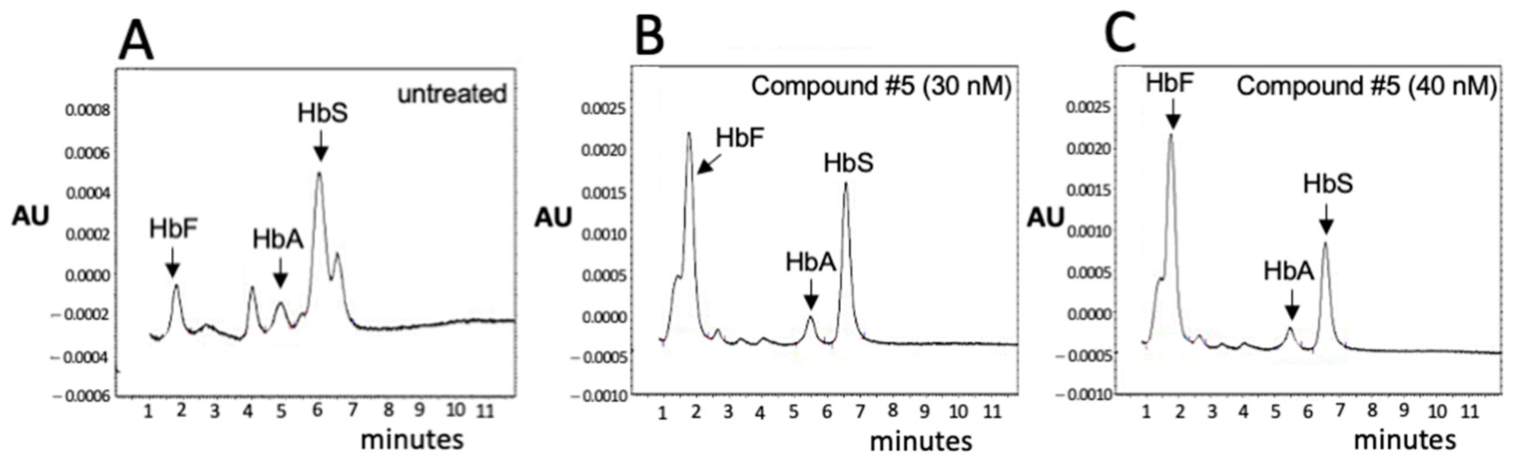
3.3. Validation of the β-Thal Cell Biobank: An Update on the Induction of Fetal Hemoglobin (HbF) Using Biobanked Samples

About four days after thawing, the CD34+ cells purchased from the β-Thal-Biobank were treated with HbF inducers. In the representative experiment shown in Figure 2, the isoxazole derivative #5 was employed [56]. This was one of the most effective isoxazoles described by Zuccato et al. [56]. For verifying the possibility of studying HbF induction, the cellular pellets obtained after 5 days of induction were used for cytoplasmic extract production for HPLC. The results obtained indicated that the % of HbF increased from 13.36% (in untreated cells, as shown in Figure 2A) to 60.92 and 69.27% in cells treated with 30 (Figure 2B) and 40 (Figure 2C) nM compound #5, respectively.
Figure 2. Compound #5 induces increased production of HbF in biobanked ErPCs from a β039/HbS patient. After thawing, EPO-cultured cells were either untreated (A) or treated for 5 days with 30 nM (B) and 40 nM (C) isoxazole derivative #5 [56].
Jcm 14 00289 g002

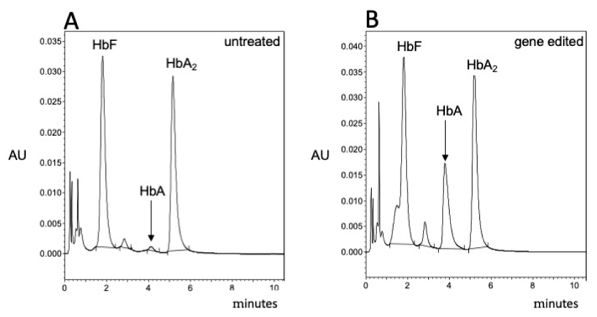
3.4. Validation of the β-Thal Cell Biobank: CRISPR-Cas9-Based Gene Editing Using Biobanked Samples

In the context of the development of personalized therapy and gene editing, the most important validation of the β-Thal Cell Biobank is the demonstration that efficient CRISPR-Cas9-based gene editing can be achieved using biobanked CD34+ ErPCs. An example of the results obtained with this approach is shown in Figure 3, which reports the HPLC pattern of untreated cells (Figure 3A) or cells edited as described by Cosenza et al. [60] (Figure 3B) using the CRISPR-Cas9 system with a forward donor DNA template.
Figure 3. CRISPR-Cas9-based gene editing of cryopreserved cells of a β039/β039 patient. Thawed cells isolated from the β-Thal Cell Biobank were cultured for 5 days and then were untreated (A) or gene-edited according to Cosenza et al. [60] using the CRISPR-Cas9 system with forward donor DNA template (B). De novo production of HbA in gene-edited cells from the β039/β039 patient is appreciable.
Jcm 14 00289 g003
The results of the experiment performed (Figure 3) clearly show that gene editing is possible after 4 years of storage of biobanked CD34+ cells of β039/β039 patients. At present, biobanked CD34+ cells from 5 β039/β039 have been thawed, gene-edited, and frozen again to be stored in the biobank as experimentally validated “gene-edited biobanked samples”.

3.5. Updates on the Composition of the Cell β-Thal Cell Biobank

Originally, the composition of the β-Thal Cell Biobank was as follows: 779 biobanked cellular pellets from 8 healthy donors and 72 thalassemia and sickle-cell disease (SCD) patients, the majority being β-thalassemia patients. Since 2016, the cellular Thal-Biobank has been implemented, and it is now composed as reported in Table 2.
The distribution of the recruited patients is, at present, still uneven, as some genotypes are frequent, such as β039/β039 (103 patients), β+IVSI-110/β039 (25 patients), and β+IVSI-110/β+IVSI-110 (9 patients), while other genotypes are rare (such as β+IVSI-110/β+IVSI-6 and β+IVSI-110/β+IVSI-1). Few sickle-cell disease patients were recruited, and their erythroid cells biobanked (10 patients, 57 vials). Regarding SARS-CoV-2 infection and COVID-19 vaccination, the analysis for future recruitment is still ongoing. During a first screening, among 74 patients from whom the erythroid cells included in the biobank were isolated, 8 were infected with SARS-CoV-2 and 30 underwent anti-COVID-19 vaccination (or with Pfizer’s BNT162b2 vaccine, or with Moderna’s mRNA-1273).

4. Discussion

The availability of biobanked cells for research on β-thalassemia is of great interest in order to perform large-scale experiments avoiding recruitment of patients for the necessary biological samples. In the case of biobanked samples, a new recruitment is not required, as we can take biobanked cells, without restrictions concerning the number of samples, and the genotype, or the basal level of fetal hemoglobin. The only issue to be considered with great attention is the quality of collected biospecimens and materials, which is crucial in the case of use by different research groups [5,6,52,53,54,55]. To ensure the highest level of quality, great care is required when handling blood sample collection, isolation of erythroid precursor cells, freezing, and long-term storage.
In this study, we report the use (Table 1) of the Thal-Biobank described by Cosenza et al. in 2016 [30] and describe representative examples demonstrating that biobanked cells can be employed to screen and characterize inducers of fetal hemoglobin (see Figure 2) and to validate protocols for gene editing (Figure 3). An important finding supporting the quality of our cellular biobank is that long-term storage (6 years) did not alter the pattern of hemoglobin production (characterized by HPLC analysis) when biobanked CD34+ cells were isolated, thawed, and sub-cultured in the presence of erythropoietin (EPO).
In this paper, we also present the updated composition of the Thal-Biobank. The comparison of the number of biobanked cells with the original Biobank described by Cosenza et al. [30] indicates an increased number of biobanked cryovials from 779 (from 80 subjects) to 990 (from 221 subjects). At the same time, a total of 428 biobanked cryovials have been used in projects related to β-thalassemia, as outlined in Table 1.
The first conclusion of our study is that the implemented Thal-Biobank should be considered a useful tool to sustain research on β-thalassemia, of potential interest for developing innovative therapeutic protocols.
A final but, at least in our opinion, very important comment is based on the fact that the biobanked cells were isolated from β-thalassemia patients before the COVID-19 pandemic outbreak [64,65,66]. Accordingly, all the patients recruited for the isolation of erythroid cells to be biobanked did not exhibit symptoms related to COVID-19. After the COVID-19 pandemic outbreak, some of them have been infected by SARS-CoV-2, and most of them have been vaccinated with anti-SARS-CoV-2 vaccines (or with Pfizer’s BNT162b2 vaccine, or with Moderna’s mRNA-1273) [67,68].
In this regard, it should be underlined that COVID-19 and the associated SARS-CoV-2 infection might have important effects on erythropoiesis. The possible impact of SARS-CoV-2 infection and COVID-19 vaccination on the hematopoietic system has been confirmed by several studies [39,40,41,42,43,44,69,70,71,72]. For instance, Ropa et al. found that SARS-CoV-2 S protein inhibits the expansion of hematopoietic stem/progenitor cells and deeply alters the colony-forming capacity. These effects were obtained by a simple exposure to S protein, even in the absence of a true SARS-CoV-2 infection [70]. The impactful results obtained by Estep et al. indicated that SARS-CoV-2 infection and COVID-19 vaccination impair the functionalities and survival of HSPCs in the umbilical cord blood [72]. In our own laboratory, a study has been published suggesting that the BNT162b2 vaccine inhibits globin gene expression when administered to erythroid cells [63]. One of the hypotheses is that the SARS-CoV-2 Spike protein might exhibit “adverse” effects on hematopoietic cells, as suggested by Trougakos et al. [73]. Since infected erythroid cells may present an altered transcriptomic and/or proteomic profile, transcriptomic and proteomic studies, as recently published [49,50,51], are crucial in order to fully understand the impact of SARS-CoV-2 infection, the Spike protein, and COVID-19 vaccination on erythroid precursor cells.
In this context, we have the unique opportunity (a) to generate post-COVID-19 biobanks consisting of erythroid cells isolated from β-thalassemia patients infected with SARS-CoV-2 and/or vaccinated against COVID-19 and (b) to compare these erythroid cells with those present within the “SARS-CoV-2 free (pre-COVID-19)” Thal-Biobank described and characterized by Cosenza et al. [30] and in this study. It should be noted that many patients who had donated the erythroid cells present within the Thal-Biobank were subsequently infected with SARS-CoV-2 (some of them developing severe forms of COVID-19) and most of them were subsequently vaccinated against COVID-19. We therefore envisage the production of other cellular biobanks containing erythroid precursors from patients who donated the cells for the pre-COVID-19 Thal-Biobank and were later infected by SARS-COV-2 or vaccinated against COVID-19. The comparison of the phenotypes of the erythroid cells obtained from these patients with those of cells of the same patients currently biobanked in our Thal-Biobank might be very helpful in the understanding person-to-person variation in the effects on erythroid cells of COVID-19, PC (post-COVID-19), PASC (Post-Acute Sequelae of COVID-19) and COVID-19 vaccination.
As a final comment, we would like to underline that our β-Thal Cell Biobank should be at present considered as a “research cellular repository” to be used for collaborative projects. Few SCD patients have been recruited for our Biobank (10 patients, 57 vials); however, a project on the construction of an SCD Biobank (CureSCi, Cure Sickle Cell Initiative) was activated in 2021-2022 to construct a Sickle Cell Hematopoietic Stem Cell Bank (SCBank) (https://biolincc.nhlbi.nih.gov/studies/curesc_scd/; accessed on 12 December 2024).
Among future perspectives, further recruitment of patients and experimental validations are necessary. Concerning the recruitment of patients, the objective is to include patients with genotypes that are still absent in the present composition of the Thal-Biobank. Concerning further experimental validation, the objective is to verify the impact of the biobank, focusing on novel applications in the biomedical research field. This research should include an analysis of possible changes in OMICS profiling in relation to the storage conditions. In the case of the positive results described here being confirmed, and depending on the available funds, infrastructures, and personnel, we will consider the possibility of creating a “fully organized” biobank [8,9,10] according to recently published guidelines (https://www.coe.int › biobanks for Europe) (accessed on 9 December 2024) [74], after confirming that (a) all the ethical, legal, and social issues (ELSIs) [75] and (b) all the regulatory and governance-related aspects [11,76] have been fully addressed. An important point will be the need for effective collaboration with external users [9,10]. This will be done in strict collaboration with the associations of thalassemia patients, since this type of intervention should consider implications related to the patients’ rights in medical research (https://www.coe.int/en/web/bioethics/biobanks; accessed on 9 December 2024). In this regard, among the legal issues to consider, we cite some examples, such as updating the “contract” between the biobank and the donors [77,78] and updating the informed consent form. Finally, the possibility of granting the biobank and its users the right to patent scientific findings obtained using the biobank resources should be considered very carefully [79,80].

5. Conclusions

In this study, we report evidence supporting the concept that the implemented β-Thal Cell Biobank (see Table 2) should be considered a tool of great interest for researchers working on β-thalassemia, with the aim of developing innovative therapeutic protocols. A second conclusion is that the implemented β-Thal Cell Biobank should be considered “SARS-CoV-2 free” and therefore could be taken into consideration for future studies aimed at comparing erythroid cells from SARS-CoV-2-infected patients with erythroid cells from the same patients isolated before the SARS-CoV-2 infection.

Supplementary Materials

The following supporting information can be downloaded at: https://www.mdpi.com/article/10.3390/jcm14010289/s1, Supplementary Method S1: Isolation and culture of peripheral blood cells (Protocol A); Supplementary Method S2: Isolation and culture of peripheral blood cells (Protocol C); Supplementary Method S3: Freezing, cryopreservation and thawing of peripheral blood cells isolated following Protocol C.

Author Contributions

Conceptualization, R.G. and M.R.G.; data curation, R.G., M.R.G., L.C.C., C.Z. and A.F.; formal analysis, R.G., M.R.G., L.C.C., C.Z. and A.F.; funding acquisition, R.G.; investigation, L.C.C., C.Z. and A.F.; methodology, L.C.C., C.Z. and A.F.; project administration, M.R.G.; resources, R.G., M.R.G. and A.F.; supervision, R.G., M.R.G. and A.F.; validation, L.C.C., C.Z. and A.F.; writing—original draft, R.G.; writing—review and editing, R.G., M.R.G., L.C.C., C.Z. and A.F. All authors have read and agreed to the published version of the manuscript.

Funding

This study was sponsored by the Wellcome Trust (Innovator Award 208872/Z/17/Z). The research leading to these results has also received funding from the UE THALAMOSS Project (Thalassemia Modular Stratification System for Personalized Therapy of Beta-Thalassemia; No. 306201-FP7-HEALTH-2012-INNOVATION-1) and FIR and FAR funds from the University of Ferrara. This research was also supported by AVLT (Associazione Veneta per la Lotta alla Talassemia “Elio Zago”–APS). C.Z. was supported by fellowships from “Tutti per Chiara Onlus” and AVLT.

Institutional Review Board Statement

The study was conducted according to the guidelines of the Declaration of Helsinki, and the procedures regarding blood sampling, blood storage and use (including the experiments on fetal hemoglobin inducers) have been approved by the Ethics Committee of Ferrara’s District, protocol names: THALAMOSS (Protocol n.93, approved on 28 June 2013) and THAL-THER (document number 533/2018/Sper/AOUFe, approved on 14 November 2018). All samples of peripheral blood were obtained after receiving written informed consent from donor patients or their legal representatives. In the accompanied explanatory document, the patients have been informed that “If useful scientific information is obtained, it will be published in medical journals, in order to make it available to the scientific community, while maintaining the commitment to publish it with the utmost confidentiality”. All the participants in this study approved and signed these documents.

Informed Consent Statement

Informed consent was obtained from all subjects involved in the study.

Data Availability Statement

Data will be made available upon reasonable request to the corresponding authors. The biological material will be made available on the basis of a scientific joint collaboration and after verification that all the ethical and legal issues are met.

Acknowledgments

We would like to thank all the patients involved in this study.

Conflicts of Interest

The authors declare no conflicts of interest. The funders had no role in the design of the study; in the collection, analyses, or interpretation of data; in the writing of the manuscript; or in the decision to publish the results.

References

  1. Al Diffalha, S.; Sexton, K.C.; Watson, P.H.; Grizzle, W.E. The importance of human tissue bioresources in advancing biomedical research. Biopreservation Biobanking 2019, 17, 209–212. [Google Scholar] [CrossRef]
  2. Paskal, W.; Paskal, A.M.; Dębski, T.; Gryziak, M.; Jaworowski, J. Aspects of modern biobank activity–comprehensive review. Pathol. Oncol. Res. 2018, 24, 771–785. [Google Scholar] [CrossRef]
  3. Compton, C.; Kelly, A. Biospecimen banking in the post-genome era. Genom. Pers. Med. 2013, 1, 229–236. [Google Scholar]
  4. Annaratone, L.; De Palma, G.; Bonizzi, G.; Sapino, A.; Botti, G.; Berrino, E.; Mannelli, C.; Arcella, P.; Di Martino, S.; Steffan, A.; et al. Basic principles of biobanking: From biological samples to precision medicine for patients. Virchows Arch. 2021, 479, 233–246. [Google Scholar] [CrossRef] [PubMed]
  5. Tang, R.; Han, C.; Yin, R.; Zhu, P.; Zhu, L.; Lu, Y.; Zheng, C. Quality Control of DNA Extracted from All-Cell Pellets After Cryopreservation for More Than 10 Years. Biopreservation Biobanking 2022, 20, 211–216. [Google Scholar] [CrossRef]
  6. Juurikka, K.; Åström, P.; Pekkala, T.; Öhman, H.; Sorsa, T.; Tervahartiala, T.; Salo, T.; Lehenkari, P.; Nyberg, P. Insights into Preservation of Blood Biomarkers in Biobank Samples. Biopreservation Biobanking 2022, 20, 297–301. [Google Scholar] [CrossRef] [PubMed]
  7. O’Donoghue, S.; Dee, S.; Byrne, J.A.; Watson, P.H. How Many Health Research Biobanks Are There? Biopreservation Biobanking 2022, 20, 224–228. [Google Scholar] [CrossRef]
  8. Coppola, L.; Cianflone, A.; Grimaldi, A.M.; Incoronato, M.; Bevilacqua, P.; Messina, F.; Baselice, S.; Soricelli, A.; Mirabelli, P.; Salvatore, M. Biobanking in health care: Evolution and future directions. J. Transl. Med. 2019, 17, 172. [Google Scholar] [CrossRef] [PubMed]
  9. Rush, A.; Catchpoole, D.R.; Reaiche-Miller, G.; Gilbert, T.; Ng, W.; Watson, P.H.; Byrne, J.A. What Do Biomedical Researchers Want from Biobanks? Results of an Online Survey. Biopreservation Biobanking 2022, 20, 271–282. [Google Scholar] [CrossRef] [PubMed]
  10. Samuel, G.; Lucassen, A. Access to Biobanks: Responsibilities Within a Research Ecosystem. Biopreservation Biobanking 2023, 21, 275–281. [Google Scholar] [CrossRef]
  11. Gille, F.; Vayena, E.; Blasimme, A. Future-proofing biobanks’ governance. Eur. J. Hum. Genet. 2020, 28, 989–996. [Google Scholar] [CrossRef] [PubMed]
  12. Roslan, F.F.; Yu, Y.; Ooi, G.C.; Then, K.L.; Then, K.Y.; Cheong, S.K.; Guo, Z.; Ab Patar, M.N.A.; Tan, J.J. From banked human cord blood to induced pluripotent stem cells: New opportunities and promise in induced pluripotent stem cell banking (Review). Int. J. Mol. Med. 2024, 54, 114. [Google Scholar] [CrossRef]
  13. Abraham, M.; Goel, S. Comprehensive characterisation and cryopreservation optimisation of buffalo (Bubalus bubalis) adipose tissue-derived mesenchymal stem cells. Cryobiology 2024, 115, 104896. [Google Scholar] [CrossRef] [PubMed]
  14. Monje, P.V. Human Schwann Cells in vitro II. Passaging, Purification, Banking, and Labeling of Established Cultures. Bio Protoc. 2023, 13, e4882. [Google Scholar] [CrossRef]
  15. D’Vaz, N.; Kidd, C.; Miller, S.; Amin, M.; Davis, J.A.; Talati, Z.; Silva, D.T.; Prescott, S.L. The ORIGINS Project Biobank: A Collaborative Bio Resource for Investigating the Developmental Origins of Health and Disease. Int. J. Environ. Res. Public Health 2023, 20, 6297. [Google Scholar] [CrossRef]
  16. Brophy, S.; Amet, R.; Foy-Stones, H.; Gardiner, N.; McElligott, A.M. Isolation and Cryopreservation of Mononuclear Cells from Peripheral Blood and Bone Marrow of Blood Cancer Patients. Methods Mol. Biol. 2023, 2645, 179–187. [Google Scholar]
  17. Degnin, M.; Agarwal, A.; Tarlock, K.; Meshinchi, S.; Druker, B.J.; Tognon, C.E. Novel Method Enabling the Use of Cryopreserved Primary Acute Myeloid Leukemia Cells in Functional Drug Screens. J. Pediatr. Hematol. Oncol. 2017, 39, e359–e366. [Google Scholar] [CrossRef] [PubMed]
  18. Harris, J.R.; Burton, P.; Knoppers, B.M.; Lindpaintner, K.; Bledsoe, M.; Brookes, A.J.; Budin-Ljøsne, I.; Chisholm, R.; Cox, D.; Deschênes, M. Toward a roadmap in global biobanking for health. Eur. J. Hum. Genet. 2012, 20, 1105–1111. [Google Scholar] [CrossRef] [PubMed]
  19. Zawati, M.H.; Tassé, A.M.; Mendy, M.; Caboux, E.; Lang, M.; on behalf of Biobank and Cohort Building Network Members. Barriers and opportunities in consent and access procedures in low- and middle-income country biobanks: Meeting notes from the BCNet training and general assembly. Biopreservation Biobanking 2018, 16, 171–178. [Google Scholar] [CrossRef] [PubMed]
  20. Vodosin, P.; Jorgensen, A.K.; Mendy, M.; Kozlakidis, Z.; Caboux, E.; Zawati, M.H. A Review of Regulatory Frameworks Governing Biobanking in the Low and Middle Income Member Countries of BCNet. Biopreservation Biobanking 2021, 19, 444–452. [Google Scholar] [CrossRef] [PubMed]
  21. Baldo, C.; Viotti, V.; Maioli, E.; Mogni, M.; Castagnetta, M.; Cavani, S.; Piombo, G.; Coviello, D. Galliera Genetic Bank: A DNA and cell line biobank from patients affected by genetic diseases. Open J. Bioresour. 2016, 3, e1–e5. [Google Scholar] [CrossRef]
  22. Bao, E.L.; Cheng, A.N.; Sankaran, V.G. The genetics of human hematopoiesis and its disruption in disease. EMBO Mol. Med. 2019, 11, e10316. [Google Scholar] [CrossRef] [PubMed]
  23. Weatherall, D.J. Phenotype-genotype relationships in monogenic disease: Lessons from the thalassaemias. Nat. Rev. Genet. 2001, 2, 245–255. [Google Scholar] [CrossRef] [PubMed]
  24. Bunn, H.F.; Noguchi, C.T.; Hofrichter, J.; Schechter, G.P.; Schechter, A.N.; Eaton, W.A. Molecular and cellular pathogenesis of hemoglobin Sickle Cell disease. Proc. Natl. Acad. Sci. USA 1982, 79, 7527–7531. [Google Scholar] [CrossRef] [PubMed]
  25. Rao, E.; Kumar Chandraker, S.; Misha Singh, M.; Kumar, R. Global distribution of β-thalassemia mutations: An update. Gene 2024, 896, 148022. [Google Scholar] [CrossRef] [PubMed]
  26. Piel, F.B.; Patil, A.P.; Howes, R.E.; Nyangiri, O.A.; Gething, P.W.; Williams, T.N.; Weatherall, D.J.; Hay, S.I. Global distribution of the sickle cell gene and geographical confirmation of the malaria hypothesis. Nat. Commun. 2010, 1, 104. [Google Scholar] [CrossRef] [PubMed]
  27. Richardson, T.; Rice, M.; Haumschild, R.; Hoffer, D.; Morana, S.; Watkins, J. Optimizing the management of inherited blood disorders in a changing market: Findings from the AMCP Market Insights Program. J. Manag. Care Spec. Pharm. 2024, 30, S1–S12. [Google Scholar] [CrossRef] [PubMed]
  28. Musallam, K.M.; Viprakasit, V.; Lombard, L.; Gilroy, K.; Rane, A.; Vinals, L.; Tam, C.; Rizzo, M.; Coates, T.D. Systematic review and evidence gap assessment of the clinical, quality of life, and economic burden of alpha-thalassemia. EJHaem. 2024, 5, 541–547. [Google Scholar] [CrossRef] [PubMed]
  29. Gamaleldin, M.; Abraham, I.; Meabed, M.; Elberry, A.; Abdelhalim, S.; Hussein, A.; Waggas, D.; Hussein, R. Cost-effectiveness analysis of Manuka honey-Omega-3 combination treatments in treating oxidative stress of pediatric β-thalassemia major. Eur. Rev. Med. Pharmacol. Sci. 2024, 28, 1144–1154. [Google Scholar] [PubMed]
  30. Cosenza, L.C.; Breda, L.; Breveglieri, G.; Zuccato, C.; Finotti, A.; Lampronti, I.; Borgatti, M.; Chiavilli, F.; Gamberini, M.R.; Satta, S.; et al. A validated cellular biobank for β-thalassemia. J. Transl. Med. 2016, 14, 255. [Google Scholar] [CrossRef]
  31. Gambari, R.; Waziri, A.D.; Goonasekera, H.; Peprah, E. Pharmacogenomics of Drugs Used in β-Thalassemia and Sickle-Cell Disease: From Basic Research to Clinical Applications. Int. J. Mol. Sci. 2024, 25, 4263. [Google Scholar] [CrossRef]
  32. Sripichai, O.; Fucharoen, S. Fetal hemoglobin regulation in β-thalassemia: Heterogeneity, modifiers and therapeutic approaches. Expert. Rev. Hematol. 2016, 9, 1129–1137. [Google Scholar] [CrossRef] [PubMed]
  33. Hashemi, Z.; Ebrahimzadeh, M.A. Hemoglobin F (HbF) Inducers; History, Structure and Efficacies. Mini Rev. Med. Chem. 2022, 22, 52–68. [Google Scholar] [CrossRef]
  34. Langer, A.L.; Esrick, E.B. β-Thalassemia: Evolving treatment options beyond transfusion and iron chelation. Hematol. Am. Soc. Hematol. Educ. Program 2021, 2021, 600–606. [Google Scholar] [CrossRef]
  35. Bou-Fakhredin, R.; De Franceschi, L.; Motta, I.; Cappellini, M.D.; Taher, A.T. Pharmacological Induction of Fetal Hemoglobin in β-Thalassemia and Sickle Cell Disease: An Updated Perspective. Pharmaceuticals 2022, 15, 753. [Google Scholar] [CrossRef] [PubMed]
  36. Foong, W.C.; Loh, C.K.; Ho, J.J.; Lau, D.S. Foetal haemoglobin inducers for reducing blood transfusion in non-transfusion-dependent beta-thalassaemias. Cochrane Database Syst. Rev. 2023, 1, CD013767. [Google Scholar] [PubMed]
  37. CA22119 - Haemoglobinopathies in European Liaison of Medicine and Science (HELIOS). Available online: https://www.cost.eu/actions/CA22119/ (accessed on 9 December 2024).
  38. Kountouris, P.; Stephanou, C.; Archer, N.; Bonifazi, F.; Giannuzzi, V.; Kuo, K.H.M.; Maggio, A.; Makani, J.; Mañú-Pereira, M.D.M.; Michailidou, K.; et al. The International Hemoglobinopathy Research Network (INHERENT): An international initiative to study the role of genetic modifiers in hemoglobinopathies. Am. J. Hematol. 2021, 96, E416–E420. [Google Scholar] [CrossRef] [PubMed]
  39. Petelina, T.I.; Musikhina, N.A.; Avdeeva, K.S.; Shcherbinina, A.E.; Leonovich, S.V.; Zueva, E.V.; Garanina, V.D.; Gultiaeva, E.P.; Yaroslavskaya, E.I.; Kalyuzhnaya, E.N.; et al. Estimation of erythrocyte parameters of general blood analysis in patients with SARS-CoV-2 -associated pneumonia. Clin. Lab. Diagn. 2022, 67, 24–30. [Google Scholar] [CrossRef] [PubMed]
  40. Delaye, J.B.; Alarcan, H.; Vallet, N.; Veyrat-Durebex, C.; Bernard, L.; Hérault, O.; Ropert, M.; Marlet, J.; Gyan, E.; Andres, C.; et al. Specific changes of erythroid regulators and hepcidin in patients infected by SARS-COV-2. J. Investig. Med. 2022, 70, 934–938. [Google Scholar] [CrossRef] [PubMed]
  41. Elahi, S. Hematopoietic responses to SARS-CoV-2 infection. Cell Mol. Life Sci. 2022, 79, 187. [Google Scholar] [CrossRef] [PubMed]
  42. Allahyani, M.A.; Aljuaid, A.A.; Almehmadi, M.M.; Alghamdi, A.A.; Halawani, I.F.; Aldairi, A.F.; Alharbi, A.M.; Albshri, M.H.; Mutwalli, A.A.; Alhazmi, A.S. Detection of erythroid progenitors and erythrocytopathies in patients with severe COVID-19 disease. Saudi Med. J. 2022, 43, 899–906. [Google Scholar] [CrossRef]
  43. Saito, S.; Shahbaz, S.; Sligl, W.; Osman, M.; Tyrrell, D.L.; Elahi, S. Differential Impact of SARS-CoV-2 Isolates, Namely, the Wuhan Strain, Delta, and Omicron Variants on Erythropoiesis. Microbiol. Spectr. 2022, 10, e0173022. [Google Scholar] [CrossRef] [PubMed]
  44. Girón-Pérez, D.A.; Nava-Piedra, U.N.; Esquivel-Esparza, Z.E.; Benitez-Trinidad, A.B.; Barcelos-Garcia, R.G.; Vázquez-Pulido, E.Y.; Toledo-Ibarra, G.A.; Ventura-Ramón, G.H.; Covantes-Rosales, C.E.; Barajas-Carrillo, V.W.; et al. Hematologic analysis of hospitalized patients and outpatients infected with SARS-CoV-2 and possible use as a prognostic biomarker. Exp. Hematol. 2023, 119–120, 21–27. [Google Scholar] [CrossRef] [PubMed]
  45. Shahbaz, S.; Xu, L.; Osman, M.; Sligl, W.; Shields, J.; Joyce, M.; Tyrrell, D.L.; Oyegbami, O.; Elahi, S. Erythroid precursors and progenitors suppress adaptive immunity and get invaded by SARS-CoV-2. Stem Cell Rep. 2021, 16, 1165–1181. [Google Scholar] [CrossRef] [PubMed]
  46. Huerga Encabo, H.; Grey, W.; Garcia-Albornoz, M.; Wood, H.; Ulferts, R.; Aramburu, I.V.; Kulasekararaj, A.G.; Mufti, G.; Papayannopoulos, V.; Beale, R.; et al. Human Erythroid Progenitors Are Directly Infected by SARS-CoV-2: Implications for Emerging Erythropoiesis in Severe COVID-19 Patients. Stem Cell Rep. 2021, 16, 428–436. [Google Scholar] [CrossRef] [PubMed]
  47. Balzanelli, M.G.; Distratis, P.; Dipalma, G.; Vimercati, L.; Inchingolo, A.D.; Lazzaro, R.; Aityan, S.K.; Maggiore, M.E.; Mancini, A.; Laforgia, R.; et al. Sars-CoV-2 Virus Infection May Interfere CD34+ Hematopoietic Stem Cells and Megakaryocyte-Erythroid Progenitors Differentiation Contributing to Platelet Defection towards Insurgence of Thrombocytopenia and Thrombophilia. Microorganisms 2021, 9, 1632. [Google Scholar] [CrossRef]
  48. Kronstein-Wiedemann, R.; Stadtmüller, M.; Traikov, S.; Georgi, M.; Teichert, M.; Yosef, H.; Wallenborn, J.; Karl, A.; Schütze, K.; Wagner, M.; et al. SARS-CoV-2 Infects Red Blood Cell Progenitors and Dysregulates Hemoglobin and Iron Metabolism. Stem Cell Rev. Rep. 2022, 18, 1809–1821. [Google Scholar] [CrossRef] [PubMed]
  49. Bernardes, J.P.; Mishra, N.; Tran, F.; Bahmer, T.; Best, L.; Blase, J.I.; Bordoni, D.; Franzenburg, J.; Geisen, U.; Josephs-Spaulding, J.; et al. Longitudinal Multi-omics Analyses Identify Responses of Megakaryocytes, Erythroid Cells, and Plasmablasts as Hallmarks of Severe COVID-19. Immunity 2020, 53, 1296–1314.e9. [Google Scholar] [CrossRef]
  50. Eltobgy, M.; Johns, F.; Farkas, D.; Leuenberger, L.; Cohen, S.P.; Ho, K.; Karow, S.; Swoope, G.; Pannu, S.; Horowitz, J.C.; et al. Longitudinal transcriptomic analysis reveals persistent enrichment of iron homeostasis and erythrocyte function pathways in severe COVID-19 ARDS. Front. Immunol. 2024, 15, 1397629. [Google Scholar] [CrossRef] [PubMed]
  51. Zhang, J.; Shu, X.; Deng, R.; Yang, Z.; Shu, M.; Ou, X.; Zhang, X.; Wu, Z.; Zeng, H.; Shao, L. Transcriptome Changes of Hematopoietic Stem and Progenitor Cells in the Peripheral Blood of COVID-19 Patients by scRNA-seq. Int. J. Mol. Sci. 2023, 24, 10878. [Google Scholar] [CrossRef]
  52. Simeon-Dubach, D.; Zeisberger, S.M.; Hoerstrup, S.P. Quality Assurance in Biobanking for Pre-Clinical Research. Transfus. Med. Hemother. 2016, 43, 353–357. [Google Scholar] [CrossRef] [PubMed]
  53. Coppola, L.; Grimaldi, A.M.; Sarnacchiaro, G.; di Fasano, M.S.; Smaldone, G.; Salvatore, M. An overview of Synlab SDN Biobank’s quality control system. Sci. Rep. 2024, 14, 19303. [Google Scholar] [CrossRef]
  54. Mouttham, L.; Garrison, S.J.; Archer, D.L.; Castelhano, M.G. A Biobank’s Journey: Implementation of a Quality Management System and Accreditation to ISO 20387. Biopreservation Biobanking 2021, 19, 163–170. [Google Scholar] [CrossRef] [PubMed]
  55. Caixeiro, N.J.; Lai, K.; Lee, C.S. Quality assessment and preservation of RNA from biobank tissue specimens: A systematic review. J. Clin. Pathol. 2016, 69, 260–265. [Google Scholar] [CrossRef] [PubMed]
  56. Zuccato, C.; Cosenza, L.C.; Tupini, C.; Finotti, A.; Sacchetti, G.; Simoni, D.; Gambari, R.; Lampronti, I. New Synthetic Isoxazole Derivatives Acting as Potent Inducers of Fetal Hemoglobin in Erythroid Precursor Cells Isolated from β-Thalassemic Patients. Molecules 2023, 29, 8. [Google Scholar] [CrossRef] [PubMed]
  57. Szuberski, J.; Oliveira, J.L.; Hoyer, J.D. A comprehensive analysis of hemoglobin variants by high-performance liquid chromatography (HPLC). Int. J. Lab. Hematol. 2012, 34, 594–604. [Google Scholar] [CrossRef] [PubMed]
  58. Joutovsky, A.; Hadzi-Nesic, J.; Nardi, M.A. HPLC retention time as a diagnostic tool for hemoglobin variants and hemoglobinopathies: A study of 60000 samples in a clinical diagnostic laboratory. Clin Chem. 2004, 50, 1736–1747. [Google Scholar] [CrossRef] [PubMed]
  59. Zuccato, C.; Cosenza, L.C.; Zurlo, M.; Gasparello, J.; Papi, C.; D’Aversa, E.; Breveglieri, G.; Lampronti, I.; Finotti, A.; Borgatti, M.; et al. Expression of γ-globin genes in β-thalassemia patients treated with sirolimus: Results from a pilot clinical trial (Sirthalaclin). Ther. Adv. Hematol. 2022, 13, 20406207221100648. [Google Scholar] [CrossRef]
  60. Cosenza, L.C.; Gasparello, J.; Romanini, N.; Zurlo, M.; Zuccato, C.; Gambari, R.; Finotti, A. Efficient CRISPR-Cas9-based genome editing of β-globin gene on erythroid cells from homozygous β039-thalassemia patients. Mol. Ther. Methods Clin. Dev. 2021, 21, 507–523. [Google Scholar] [CrossRef] [PubMed]
  61. Cosenza, L.C.; Zuccato, C.; Zurlo, M.; Gambari, R.; Finotti, A. Co-Treatment of Erythroid Cells from β-Thalassemia Patients with CRISPR-Cas9-Based β039-Globin Gene Editing and Induction of Fetal Hemoglobin. Genes 2022, 13, 1727. [Google Scholar] [CrossRef] [PubMed]
  62. Zuccato, C.; Cosenza, L.C.; Zurlo, M.; Lampronti, I.; Borgatti, M.; Scapoli, C.; Gambari, R.; Finotti, A. Treatment of Erythroid Precursor Cells from β-Thalassemia Patients with Cinchona Alkaloids: Induction of Fetal Hemoglobin Production. Int. J. Mol. Sci. 2021, 22, 13433. [Google Scholar] [CrossRef] [PubMed]
  63. Cosenza, L.C.; Marzaro, G.; Zurlo, M.; Gasparello, J.; Zuccato, C.; Finotti, A.; Gambari, R. Inhibitory effects of SARS-CoV-2 spike protein and BNT162b2 vaccine on erythropoietin-induced globin gene expression in erythroid precursor cells from patients with β-thalassemia. Exp. Hematol. 2024, 129, 104128. [Google Scholar] [CrossRef] [PubMed]
  64. Wang, C.; Horby, P.W.; Hayden, F.G.; Gao, G.F. A novel coronavirus outbreak of global health concern. Lancet 2020, 395, 470–473. [Google Scholar] [CrossRef] [PubMed]
  65. Peng, M. Outbreak of COVID-19: An emerging global pandemic threat. Biomed. Pharmacother. 2020, 129, 110499. [Google Scholar] [CrossRef]
  66. Gatto, M.; Bertuzzo, E.; Mari, L.; Miccoli, S.; Carraro, L.; Casagrandi, R.; Rinaldo, A. Spread and dynamics of the COVID-19 epidemic in Italy: Effects of emergency containment measures. Proc. Natl. Acad. Sci. USA 2020, 117, 10484–10491. [Google Scholar] [CrossRef] [PubMed]
  67. Sahin, U.; Muik, A.; Vogler, I.; Derhovanessian, E.; Kranz, L.M.; Vormehr, M.; Quandt, J.; Bidmon, N.; Ulges, A.; Baum, A.; et al. BNT162b2 vaccine induces neutralizing antibodies and poly-specific T cells in humans. Nature 2021, 595, 572–577. [Google Scholar] [CrossRef]
  68. Pegu, A.; O’Connell, S.E.; Schmidt, S.D.; O’Dell, S.; Talana, C.A.; Lai, L.; Albert, J.; Anderson, E.; Bennett, H.; Corbett, K.S.; et al. Durability of mRNA-1273 vaccine-induced antibodies against SARS-CoV-2 variants. Science 2021, 373, 1372–1377. [Google Scholar] [CrossRef] [PubMed]
  69. Ratajczak, M.Z.; Kucia, M. SARS-CoV-2 infection and overactivation of Nlrp3 inflammasome as a trigger of cytokine "storm" and risk factor for damage of hematopoietic stem cells. Leukemia 2020, 34, 1726–1729. [Google Scholar] [CrossRef] [PubMed]
  70. Ropa, J.; Cooper, S.; Capitano, M.L.; Van’t Hof, W.; Broxmeyer, H.E. Human Hematopoietic Stem, Progenitor, and Immune Cells Respond Ex Vivo to SARS-CoV-2 Spike Protein. Stem Cell Rev. Rep. 2021, 17, 253–265. [Google Scholar] [CrossRef]
  71. Kucia, M.; Ratajczak, J.; Bujko, K.; Adamiak, M.; Ciechanowicz, A.; Chumak, V.; Brzezniakiewicz-Janus, K.; Ratajczak, M. An evidence that SARS-CoV-2/COVID-19 spike protein (SP) damages hematopoietic stem/progenitor cells in the mechanism of pyroptosis in Nlrp3 inflammasome-dependent manner. Leukemia 2021, 35, 3026–3029. [Google Scholar] [CrossRef]
  72. Estep, B.K.; Kuhlmann, C.J.; Osuka, S.; Suryavanshi, G.W.; Nagaoka-Kamata, Y.; Samuel, C.N.; Blucas, M.T.; Jepson, C.E.; Goepfert, P.A.; Kamata, M. Skewed fate and hematopoiesis of CD34+ HSPCs in umbilical cord blood amid the COVID-19 pandemic. iScience 2022, 25, 105544. [Google Scholar] [CrossRef] [PubMed]
  73. Trougakos, I.P.; Terpos, E.; Alexopoulos, H.; Politou, M.; Paraskevis, D.; Scorilas, A.; Kastritis, E.; Andreakos, E.; Dimopoulos, M.A. Adverse effects of COVID-19 mRNA vaccines: The spike hypothesis. Trends Mol. Med. 2022, 28, 542–554. [Google Scholar] [CrossRef]
  74. Zika, E.; Paci, D.; Schulte in den Bäumen, T.; Braun, A.; Rijkers-Defrasne, S.; Deschênes, M.; Fortier, I.; Laage-Hellman, J.; A Scerri, C.; Ibarreta Ruiz, D. Biobanks in Europe: Prospects for Harmonisation and Networking; JRC57831; Publications Office of the European Union: Luxembourg, 2010. [Google Scholar]
  75. Tzortzatou-Nanopoulou, O.; Akyüz, K.; Goisauf, M.; Kozera, Ł.; Mežinska, S.; Th Mayrhofer, M.; Slokenberga, S.; Reichel, J.; Croxton, T.; Ziaka, A.; et al. Ethical, legal, and social implications in research biobanking: A checklist for navigating complexity. Dev. World Bioeth. 2024, 24, 139–150. [Google Scholar] [CrossRef] [PubMed]
  76. Gottweis, H.; Zatloukal, K. Biobank governance: Trends and perspectives. Pathobiology 2007, 74, 206–211. [Google Scholar] [CrossRef] [PubMed]
  77. Ducournau, P.; Strand, R. Trust, Distrust and Co-production: The Relationship Between Research Biobanks and Donors. In The Ethics of Research Biobanking; Solbakk, J., Holm, S., Hofmann, B., Eds.; Springer: Boston, MA, USA, 2009. [Google Scholar] [CrossRef]
  78. Hoeyer, K.; Olofsson, B.; Mjörndal, T.; Lynöe, N. The Ethics of Research Using Biobanks: Reason to Question the Importance Attributed to Informed Consent. Arch. Intern. Med. 2005, 165, 97–100. [Google Scholar] [CrossRef] [PubMed]
  79. McMahon, A.M.; Kolawole, O.I. Biobank donation in search of public benefits and the potential impact of intellectual property rights over access to health-technologies developed: A focus on the bioethical implications. Med. Law Rev. 2024, 32, 205–228. [Google Scholar] [CrossRef] [PubMed]
  80. Jordan, M.; Liddicoat, J.; Liddell, K. An empirical study of large, human biobanks: Intellectual property policies and financial conditions for access. J. Law. Biosci. 2021, 8, lsab018. [Google Scholar] [CrossRef]
Figure 1. Biobanked samples of the same β-thalassemia patient maintain the hemoglobin pattern after long-term storage. (A) The experimental plan of the present study. Results concerning response to HbF inducers are presented in Figure 2; results concerning CRISPR-Cas9 gene editing are presented in Figure 3. (BE) Pattern of hemoglobin production when cryo-preserved cells were thawed and sub-cultured in 2016 (A) and in 2020 (B). (D,E) Percentage of HbF (D, white circles), HbA2 (D, black circles), and percentage of the α-globin peak (E, grey circles) in cells of different vials from the same patient, thawed and sub-cultured in EPO in 2016, 2019, and 2020, as indicated.
Jcm 14 00289 g001
Table 1. Use of biobanked samples (vials) to sustain research on Thalassemia.
THALAMOSS Project: Induction of HbF with LMW DrugsProject: Induction of HbA with Readthrough-Based MoleculesProject: β039 CRISPR-Cas9 Genome Editing SystemProject: Effects of
SARS-CoV-2 Spike Protein on ErPCs
Number of patientsNumber of VialsNumber of patientsNumber of VialsNumber
of patients
Number
of Vials
Number of
patients
Number of Vials
843795101127612
Publications: Zuccato et al., 2021 [62]; Zuccato et al., 2022 [59]; Zuccato et al., 2023 [56]UnpublishedPublications: Cosenza et al., 2021 [60]; Cosenza et al., 2022 [61]Publications: Cosenza et al., 2024 [63]
Table 2. Implementation of the β-Thal Cell Biobank from 2016.
Cosenza et al., 2016 [26]Present Composition of the Biobank
Genotype/PhenotypeNumber of PatientsNumber of VialsNumber of PatientsNumber of Vials
β039/β03929260103421
β+IVSI-110/β+IVSI-110881965
β+IVSI-110/β039171912551
β+IVSI-6/β+IVSI-6215328
β+IVSI-6/β039467524
β+IVSI-110/β+IVSI-611118
β+IVSI-110/β+IVSI-11717
SCD/SCD213317
SCD/β03917210
SCD/β+IVSI-1101528
SCD/β+IVSI-61717
SCD/β+IVSI-1220215
Others119564329
Total80779 221990
Disclaimer/Publisher’s Note: The statements, opinions and data contained in all publications are solely those of the individual author(s) and contributor(s) and not of MDPI and/or the editor(s). MDPI and/or the editor(s) disclaim responsibility for any injury to people or property resulting from any ideas, methods, instructions or products referred to in the content.

