Abstract
Spinal injuries, including cervical and thoracolumbar fractures, continue to be a major public health concern. Recent advancements in machine learning and deep learning technologies offer exciting prospects for improving both diagnostic and prognostic approaches in spinal injury care. This narrative review systematically explores the practical utility of these computational methods, with a focus on their application in imaging techniques such as computed tomography (CT) and magnetic resonance imaging (MRI), as well as in structured clinical data. Of the 39 studies included, 34 were focused on diagnostic applications, chiefly using deep learning to carry out tasks like vertebral fracture identification, differentiation between benign and malignant fractures, and AO fracture classification. The remaining five were prognostic, using machine learning to analyze parameters for predicting outcomes such as vertebral collapse and future fracture risk. This review highlights the potential benefit of machine learning and deep learning in spinal injury care, especially their roles in enhancing diagnostic capabilities, detailed fracture characterization, risk assessments, and individualized treatment planning.
1. Introduction
Spinal injuries, encompassing both cervical and thoracolumbar regions, present a significant health concern. Traumatic cervical spine fractures have an incidence rate ranging from 4 to 17 per 100,000 person years in Western populations [1]. Historically, these fractures were more prevalent in younger males due to high-energy traumas like road accidents. However, recent trends show a shift toward elderly individuals, both male and female, sustaining these injuries from common low-energy traumas, such as falls [1]. Thoracolumbar fractures, including osteoporotic vertebral fractures (OVFs), are another key area of focus. The rate of thoracolumbar fractures in blunt trauma patients is 6.9%, with motor vehicle collisions and high-energy falls being the leading causes [2]. OVFs are particularly prevalent in the elderly, often resulting from osteoporosis and caused by low-energy traumas like falls from standing height or less. These fractures can lead to various morbidities, including back pain and a decline in the quality of life [3]. Treatment is primarily conservative but may require surgical intervention in severe cases. Despite the high prevalence of OVFs, osteoporosis is often undertreated, emphasizing the need for improved management and prevention strategies. Given the significant impact on patients’ quality of life and broader societal implications, ensuring accurate and timely diagnosis and prognosis of spinal injuries is crucial for effective treatment without complications. This is especially crucial in an aging society where the prevalence of such injuries, including OVFs, is increasing.
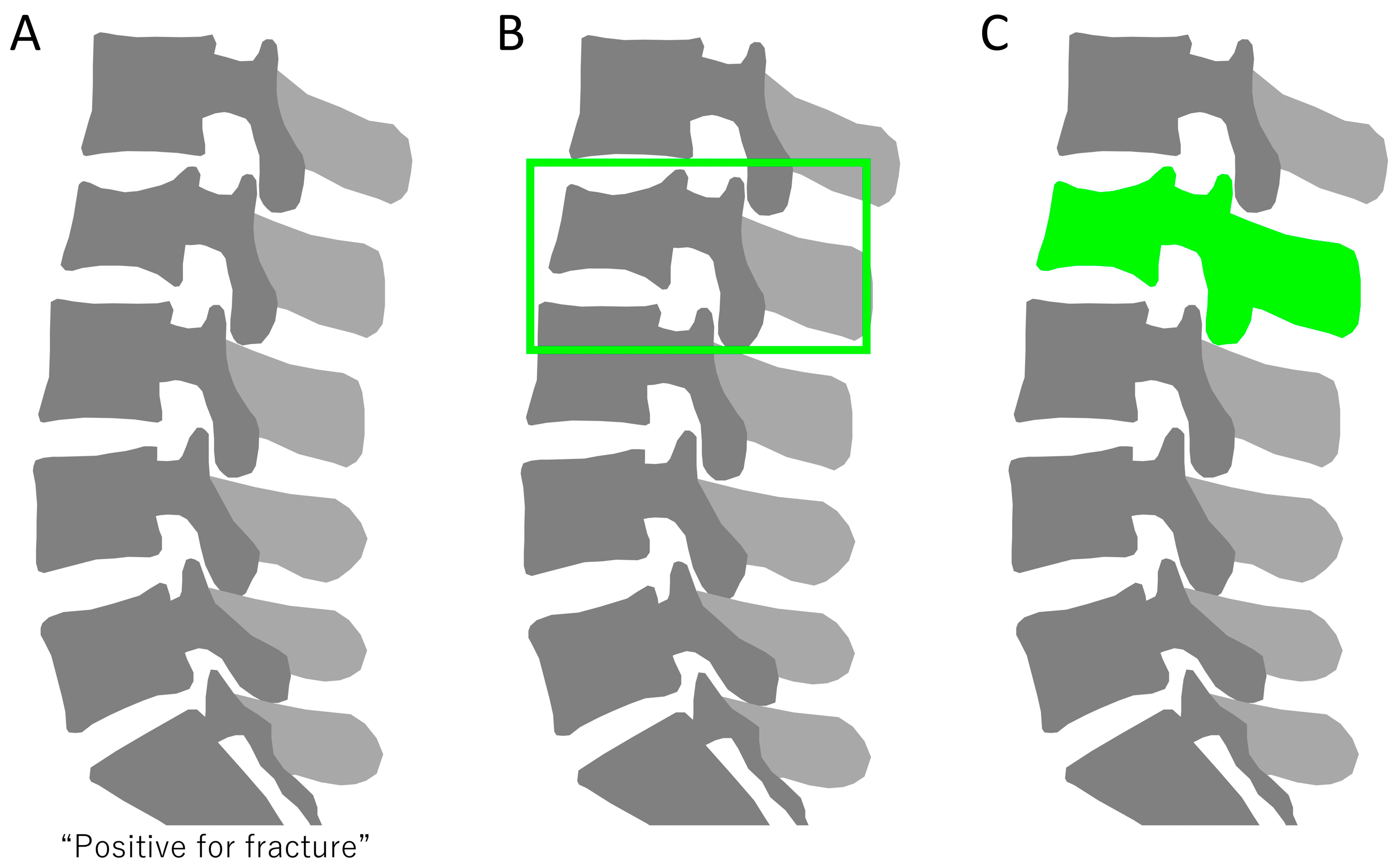
Artificial intelligence (AI) is the broadest term, encompassing the creation of machines or software that can perform tasks requiring human-like intelligence. This includes reasoning, learning, problem solving, perception, and language understanding. Machine learning is a subset of AI and involves training algorithms to learn from and make predictions or decisions based on data. It is often used with structured data, such as tables in databases, where the algorithm learns to identify patterns and relationships. The basic principle is to feed data into an algorithm, allowing it to learn from that data, and then use the learned model to make predictions or decisions on new, unseen data. Deep learning is a subset of machine learning that uses neural networks with many layers to analyze complex patterns in data (Figure 1). It is particularly effective for the following: classification problems: assigning data into predefined categories, like diagnosing diseases from medical images; object detection: identifying and locating objects within images, vital for applications like medical imaging to pinpoint abnormalities; and segmentation: dividing an image into segments to identify and locate different parts, such as different tissues in a medical scan (Figure 2). Machine learning and deep learning, both integral components of artificial intelligence (AI), are more than just trending topics; they are technologies that bring about significant change and have begun to bring about major changes in multiple areas, including healthcare [4]. In spine care, advancements in computational methods, especially machine learning and deep learning, have demonstrated significant potential in enhancing the diagnosis and prognosis of spine lesions [5,6,7,8,9]. These technologies have been particularly effective in analyzing various types of data, including imaging data from CT scans and MRI, as well as structured clinical data. Machine learning and deep learning have not only shown promise in improving surgical planning, patient selection, and postoperative care but also in driving personalized medicine approaches [5,6,7,8,9]. The integration of machine learning and deep learning in spine care holds the potential to elevate patient outcomes and reduce healthcare costs. Notably, while there exist several studies that review the application of machine learning and deep learning in spinal surgeries and spinal cord injuries (SCIs), relatively little focus has been given to their role in spinal injuries [5,6,7,8,9]. Questions regarding their robustness and generalizability in spinal injury care across different settings remain largely unexplored. This lack of comprehensive understanding motivates the need for a narrative review that explores both the diagnostic and prognostic aspects of machine learning and deep learning in spinal injury.
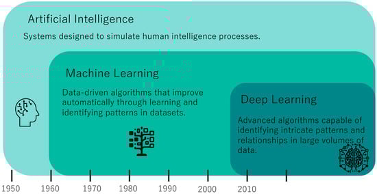
Figure 1.
Diagram illustrating the relationship and distinctions between artificial intelligence, machine learning, and deep learning.
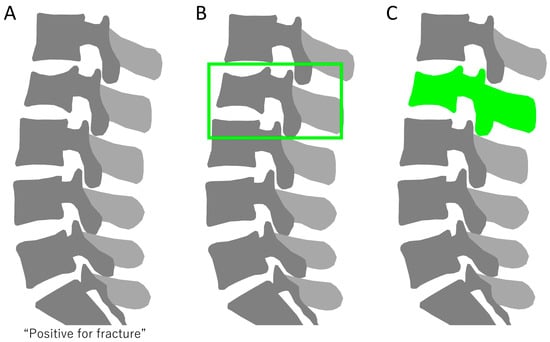
Figure 2.
(A) Classification of vertebral fractures, identifying the presence or absence of fractures in radiographs. (B) Object detection in spine radiographs, with bounding boxes indicating fracture locations. (C) Segmentation of vertebral fractures, delineating fractured areas.
The purpose of this narrative review is to explore the scope and clinical applicability of machine learning and deep learning algorithms in the diagnosis and prognosis of spinal injuries. We aim to provide a comprehensive overview of the existing literature, focusing on studies that utilize either imaging or tabulated data, to offer valuable insights that could guide future research and clinical practice in the diagnosis and treatment of spinal injury.
2. Materials and Methods
2.1. Search Criteria
We searched the PubMed database using the terms (“Cervical Fracture” OR “Thoracic Fracture” OR “Lumbar Fracture” OR “Spinal Fracture” OR “Spine Fracture” OR “Vertebral Fractures” OR “Spinal Trauma” OR “Vertebral Injury” OR “Fracture Screening”) AND (“Artificial Intelligence” OR “Machine Learning” OR “Deep Learning” OR “Neural Network” OR “Computer Vision” OR “Automated”) AND (“2013/01/01”[Date-Publication]: “2023/09/11”[Date-Publication]). Studies published in English from 1 January 2013 to 11 September 2023 were considered. The starting point of 2013 was specifically chosen to include research that utilizes modern deep learning techniques. No limitations were set on the geographical locations of the studies.
2.2. Eligibility Criteria
To be included in the review, studies needed to implement machine learning or deep learning algorithms, focus on the diagnosis or prognosis of spinal injuries, and utilize either imaging or tabulated clinical data. On the other hand, we excluded studies that specifically focused on spinal cord injuries, those that aimed to estimate fracture risk based on bone density, as well as reviews and systematic reviews. Studies published in languages other than English, case studies, and studies without full text available were also excluded.
2.3. Study Selection
The initial search yielded 86 papers. Titles and abstracts were initially screened for relevance by the first author (SM) of this review. In cases where eligibility was uncertain, two independent reviewers (TF and MI) were consulted. Any disagreement between the reviewers and the first author was resolved through discussion.
2.4. Review Framework and Components
Our narrative review was organized to comprehensively cover the application of machine learning and deep learning in the diagnosis and prognosis of spinal injuries. To ensure a full scope of information, we focused on the following components:
- Diagnosis/Prognosis: The role the algorithms played in diagnosing or predicting the outcome of the spinal injuries.
- Target Pathology: The specific type of spinal injury being investigated in each study.
- Patients Studied: Number of the patients who participated in the studies.
- Images Studied: Number of images analyzed in the studies.
- Type of Data: Categories of data, such as imaging or tabulated clinical data, used in the study.
- Computational Task: The specific computational goal for which machine learning or deep learning algorithms were employed, such as object detection, segmentation, or classification.
- Machine Learning Models: Types of machine learning and deep learning algorithms used, such as decision trees, random forests, etc.
- Summaries: Summaries of the key findings and essential takeaways from each individual study.
3. Results
Out of the initial 86 papers identified, 39 met the inclusion criteria and were included in the final analysis (Figure 3). These studies were divided into two main categories: those focusing on diagnostic algorithms (34 articles) (Table 1) and those dedicated to prognostic evaluations (5 articles) (Table 2).
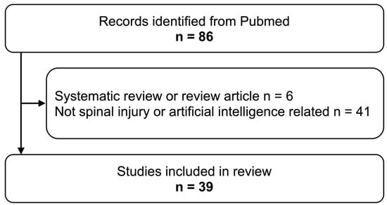
Figure 3.
Flowchart of study selection criteria for the review.

Table 1.
List of diagnostic studies selected for final analysis.
Table 2.
List of prognostic studies selected for final analysis.
3.1. Algorithms for Diagnosing Spinal Injuries
The majority of the 34 diagnostic studies employed deep learning algorithms using imaging modalities such as CT, MRI, DXA, and X-ray. The algorithms used in these studies were mainly convolutional neural networks.
3.2. Algorithms for Predicting Spinal Injury Outcomes
In contrast, four out of the five prognostic studies used machine learning algorithms such as the decision tree model, random forest, and XGBoost, relying on tabulated clinical data. These prognostic studies covered topics such as the progression of vertebral collapse, imminent new vertebral fractures after vertebral augmentation, future vertebral fractures, treatment-related risk factors, and predictions of nonunion. Much like the diagnostic studies, the majority of these prognostic studies also focused on thoracolumbar injuries.
3.3. Types of Data and Study Design
Both imaging data and tabulated clinical data were employed across the selected studies, though each type of data was more prevalent in one of the two main focus areas—imaging in diagnosis and tabulated data in prognosis. These studies predominantly targeted thoracolumbar injuries, with only five focusing on cervical spinal injuries. The retrospective design was dominant in both categories, accounting for 37 of the 39 studies, indicating a gap in prospective research in this field.
4. Discussion
4.1. Overview of Machine Learning Applications in Spinal Care
In this review, we explored a diverse range of objectives in the studies reviewed, encompassing everything from the diagnostic precision in morphometric vertebral fracture analysis to the prediction of imminent and future vertebral fractures. This range highlights the expansive yet focused applications of machine learning and deep learning in spinal injury care. Deep learning techniques, especially CNNs, have been at the forefront in refining diagnostic approaches. These include enhancing the accuracy in the detection and classification of fractures, as well as in the critical task of differentiating between benign and malignant fractures using complex imaging data. On the other hand, machine learning methods, such as random forests and SVMs, have primarily been applied for prognostic purposes, offering valuable insights into the progression of vertebral collapse and identifying treatment-related risk factors using structured clinical data. While prognostic studies utilizing machine learning are fewer, they provide significant contributions to understanding spinal injury outcomes. This distinction highlights the tailored application of each technology to the unique challenges presented by spinal injury diagnosis and prognosis. The diversity and complexity of spinal injuries, along with their varied treatments, underscore the utility of machine learning and deep learning in facilitating more individualized care approaches.
4.2. Diagnostic Approaches
4.2.1. X-ray
Diagnostic Accuracy Using Entire X-rays
The accuracy of determining the presence of vertebral fractures using plain radiographs of the entire thoracolumbar spine can be challenging because identifying vertebral fractures in a broad region of interest might be a complex task for neural networks. A study by Chen et al. using a CNN reported an accuracy of around 73.59% for vertebral fracture identification with these X-ray images [15]. Murata et al. trained a CNN to identify thoracolumbar vertebral fractures using X-ray images and achieved accuracy, sensitivity, and specificity rates of 86.0%, 84.7%, and 87.3%, respectively [41].
Evaluation of Manually Cropped Regions
Rosenberg et al. focused on using deep learning models to diagnose traumatic thoracolumbar fractures by analyzing individual vertebra regions that were manually cropped from the lateral radiographs, finding that the ResNet18 architecture outperformed VGG16 with higher sensitivity (91%), specificity (89%), and accuracy (88%) [22].
Object Detection and Ensemble Approaches
An alternative and potentially more accurate approach may involve first using object detection algorithms to identify individual vertebrae, followed by classification or segmentation to assess their condition [16,42]. Li et al. used a deep learning ensemble on 941 lateral spine radiographs to detect osteoporotic lumbar vertebral fractures, achieving 93% accuracy, 91% sensitivity, and 93% specificity [42]. Chou et al. employed an ensemble model on 1305 thoracolumbar X-rays to identify vertebral fractures, attaining similar performance metrics—93.36% accuracy, 88.97% sensitivity, and 94.26% specificity [16]. Shen et al. developed and validated a deep-learning-based system (AI_OVF_SH) for diagnosing and grading osteoporotic vertebral fractures using plain radiographs [25]. In external validation, the system demonstrated high performance, achieving an accuracy of 96.85%, sensitivity of 83.35%, and specificity of 94.70% for detecting all types of fractures [25].
Cervical Spine
Naguib et al. developed a computer-aided diagnosis system based on deep learning algorithms, specifically AlexNet and GoogleNet, to classify cervical spine injuries as either fractures or dislocations using X-ray images [18].
Age-Specific Algorithm Performance
Alqahtani et al. reported that a software program initially trained on adult spinal fracture data, using DXA and X-ray for diagnosis, showed poor sensitivity and specificity when applied to pediatric cases. This underscores the need for algorithms to consider the age of the patient for the target condition in their training data [10,11,12].
4.2.2. DEXA
Mehta et al. demonstrated that using ancillary numerical data obtained from routine DEXA scans, support vector machine (SVM) analysis can accurately identify lumbar spine fractures. The method, specifically with a linear kernel SVM, yielded a high area under the curve (AUC = 0.9258), indicating that it can detect fractures without the need for additional imaging or radiation exposure [43]. Derkatch et al. found that CNNs, specifically InceptionResNetV2 and DenseNet, can accurately identify vertebral fractures in thoracolumbar regions using images from DXA scans. The CNN ensemble achieved an area under the receiver operating characteristic curve of 0.94, with a sensitivity of 87.4% and a specificity of 88.4% [17]. Monchka et al. showed that CNNs can accurately identify vertebral fractures in images from multiple types of DXA scanners [44]. The final CNN ensemble model achieved a sensitivity of 91.9%, a specificity of 99.0%, and an F1-score of 86.1% [44]. The F1-score is the harmonic mean of precision and recall, providing a balance between the two.
4.2.3. CT
Automated Detection Algorithms
Roux et al.’s study employed software from Zebra Medical Vision to perform opportunistic screening for vertebral fractures and osteoporosis in more than 150,000 routine lumbar spine CT scans, achieving success rates of 82% for vertebral fracture assessment and 87% for Hounsfield Unit (HU) measurements, showing the potential to enhance diagnostic accuracy in large-scale screening [23]. Tomita et al. utilized deep neural networks for the automatic detection of osteoporotic vertebral fractures in CT scans, achieving an accuracy of 89.2% and an F1-score of 90.8% [27]. The study by Inoue et al. used Faster R-CNN to detect fractures in the pelvis, ribs, and spine, offering a comprehensive approach for trauma cases [45]. However, the model’s sensitivity is lower compared to specialized spinal fracture models, suggesting room for improvement. Rueckel et al. investigated the utility of AI assistance in detecting missed thoracic findings, including vertebral fractures, in emergency whole-body CT scans [24]. The study found that 57.8% of suspicious vertebral bodies were identified solely by the AI, while radiologists alone detected 29.7%, and both AI and radiologists detected 12.5%, suggesting that AI assistance can significantly reduce the rate of missed thoracic findings in emergency settings [24].
Fracture Classification
Some models not only diagnose the presence of fractures but also classify the type of fracture. For example, Chen et al. used deep learning for high-accuracy AO (Arbeitsgemeinschaft für Osteosynthesefragen) classification of thoracolumbar fractures [14]. On the other hand, Doerr et al. developed a model to categorize vertebral morphology and determine posterior ligamentous complex integrity for the purpose of assigning Thoracolumbar Injury Classification and Severity Score (TLICS), both using CT scans [46]. Zhang et al. developed a multistage system using CNNs (U-net, GCN, 3D-ResNet) that can automatically detect and classify acute thoracolumbar vertebral body fractures on CT images with high-accuracy AO classification—achieving a sensitivity of 95.23%, an overall accuracy of 97.93%, a specificity of 98.35%, and balanced accuracy rates ranging from 79.56% to 94.5% for different fracture types according to AO classification [34].
Opportunistic Screening and Fracture Liaison
The ability to screen for vertebral fractures from CT scans taken for other purposes highlights the strength of AI in this context. Nicolaes et al. used a 3D CNN to identify thoracolumbar vertebral fractures from CT scans with 94% sensitivity and 93% specificity. The algorithm showed an AUC of 0.94, making it highly effective in opportunistically identifying vertebral fractures in routine CT scans [19]. Ong et al. evaluated the efficacy of a machine learning algorithm in identifying vertebral fractures from CT scans, revealing that the algorithm detected fractures in 19.1% of 4461 patients, outperforming hospital radiologists whose reports only mentioned 49% of these fractures [20]. Valentinitsch et al. used a random forest classifier and 3D texture features for opportunistic osteoporosis screening in thoracolumbar spine multi-detector CT scans, demonstrating high discriminatory power (AUC = 0.88) and outperforming global vertebral bone mineral density (vBMD) alone in identifying vertebral fractures [28].
3D-Based Algorithms
While 3D volume data may have higher computational costs, they hold the potential for a more accurate assessment of fracture morphology. Burns et al. used 3D volume data from CT scans to achieve high sensitivity and low false-positive rates in detecting and anatomically localizing thoracic and lumbar vertebral body fractures [13]. Zakharov et al. developed an anchor-free vertebra detection network using convolutional neural networks that effectively localizes the vertebral column in 3D CT images and simultaneously detects individual vertebrae and quantifies fractures in 2D, demonstrating strong performance with an AUC of 0.95, sensitivity of 0.85, and specificity of 0.9 on the challenging VerSe dataset containing various unseen vertebra fracture types [33]. Zhang et al. introduced a novel multi-scale attention-guided network (MAGNet) for diagnosing thoracolumbar vertebral fractures and three-column injuries, achieving an AUC of 0.884 for vertebral fracture diagnosis and 0.920 for three-column injury diagnosis, both with high precision on CT images [35].
Distinguishing Benign from Malignant Vertebral Fractures
Differentiating between osteoporotic and malignant vertebral fractures is crucial, especially among elderly patients, for appropriate treatment planning and prognosis. Goller et al. in 2023 used a CNN-based framework to distinguish between benign and malignant thoracolumbar vertebral fractures using CT-based texture features. Their study found statistically significant differences in these features between benign and malignant fractures [47]. Park et al. developed an automated segmentation algorithm for CT scans of thoracolumbar fractures, showing that its performance in predicting fracture malignancy was comparable to human expert segmentation [21].
Cervical Spine
Research on detecting cervical fractures is comparatively less abundant than studies on thoracolumbar fractures. Golla et al. in 2023 used convolutional neural networks to detect cervical spine fractures from CT scans. Their algorithm detected 87.2% of fractures with an average of 3.5 false positives per case, using a spinal canal aligned volumes of interest (VOI) approach [48]. Small et al. evaluated a CNN developed by Aidoc for detecting cervical spine fractures using CT scans, reporting a 92% accuracy rate with 76% sensitivity and 97% specificity [26]. Voter et al. evaluated the diagnostic performance of a deep learning algorithm by Aidoc for detecting cervical spine fractures in CT scans, finding a relatively low sensitivity of 54.9% but a high specificity of 94.1% [29].
4.2.4. MRI
Diagnosing Fresh Osteoporotic Vertebral Fracture
Yabu et al. used an ensemble method comprising VGG16, VGG19, DenseNet201, and ResNet50 to diagnose fresh osteoporotic vertebral fractures in thoracolumbar MRI scans, finding that the CNN’s performance metrics were comparable to those of two spine surgeons [30].
Distinguishing Benign from Malignant Vertebral Fractures
Yoda et al. employed a deep convolutional neural network using Xception architecture to differentiate osteoporotic and malignant vertebral fractures using MRI [32]. The model’s accuracy was statistically equal or superior to that of spine surgeons. Yeh et al. applied a ResNet50 deep learning algorithm to MRI scans of the whole spine for distinguishing between benign and malignant vertebral fractures, achieving an accuracy of 92% and potentially improving diagnostic performance for less experienced clinicians [31].
4.3. Prognostic Approaches
Research focused on predicting prognosis is relatively scarce compared to diagnosis and classification. Most of these few studies do not directly predict from images but use machine learning on parameters extracted from images. For instance, Cho et al. worked on predicting the progression of vertebral collapse in osteoporotic vertebral fractures. Using manually extracted parameters from X-ray and MRI images, they employed machine learning techniques like decision trees and random forests [36]. Jiang et al. employed machine learning models, specifically random survival forest and COX proportional hazard analysis, to predict new osteoporotic vertebral compression fractures after vertebral augmentation using T1W MR images [37]. Kong et al. focused on predicting osteoporotic fractures in the lumbosacral spine using X-ray images and deep learning in a longitudinal cohort study [38]. Their DeepSurv model, trained on both images and clinical features, outperformed traditional methods like FRAX and CoxPH in terms of C-index values, suggesting its potential for more accurate fracture prognosis [38]. Takahashi et al. employed machine learning models including logistic regression, decision trees, XGBoost, and RF to improve the prediction of nonunion following osteoporotic vertebral fractures in the thoracolumbar region, using MRI data from 505 patients [40]. The study found high prognostic accuracy with AUC scores of 0.860 and 0.845 for RF and XGBoost, respectively [40]. Leister et al. aimed to identify treatment-related risk factors for nonunion of odontoid fractures in the cervical spine using machine learning models, specifically XGBoost and binary logistic regression [39]. The study found moderate predictive power, with an AUC of 0.68 for the XGBoost model and 0.71 for the binary logistic regression model, suggesting their potential utility in understanding treatment-related risks [39].
4.4. Advantages and Disadvantages of Each Model
Machine learning and deep learning methods play a crucial role in diagnosing and predicting spinal injuries, each with its own strengths and drawbacks. Techniques such as CNNs, random forests, SVMs, XGBoost, and recurrent neural networks (RNNs) are important in this field. CNNs are particularly good at recognizing patterns in images, including object detection and segmentation, which is helpful for examining complex data like X-ray, CT, and MRI scans [49,50]. Random forests are strong at analyzing big datasets, SVMs work well in handling data with many features, and XGBoost is known for its execution speed. RNNs are especially useful for data that are ordered over time. These methods have their own limitations regarding data requirements, computing power, and how easy it is to understand their results [51]. For the advantages and disadvantages of each model related to spinal injury care, please see Table 3. Choosing the right method depends on the specific needs of spinal injury data, the computing resources available, and finding a balance between precision and interpretability.
Table 3.
Advantages and disadvantages of machine learning models in the context of spinal trauma.
4.5. Future Direction
Future research in spinal fracture care should prioritize the development and refinement of predictive algorithms offered by machine learning to enhance both preoperative and postoperative care. The following specific directions are worth considering:
Integration of diverse data for tailored treatment: Emphasis should be placed on integrating clinical and radiological data to tailor treatment plans to individual patient profiles. This approach has the potential to significantly improve diagnosis accuracy and the efficacy of interventions like vertebral augmentation and surgical stabilization [52].
Focus on cervical spine fractures: Given the relative underrepresentation of cervical spine fractures in the current literature, future studies should particularly focus on this area. Research could explore new diagnostic techniques and frameworks for treatment decision making.
Application of large language models: The use of large language models in analyzing medical texts, such as patient records and radiology reports, presents a promising avenue for improving diagnosis accuracy and treatment personalization. Future studies should explore the potential of these models, as highlighted in the recent study [53].
Development of multimodal language models: Research should be directed toward developing multimodal models that process and integrate different types of data (imaging, clinical notes, sensor data) for a comprehensive understanding of the patient’s condition [54]. This approach aligns with the personalized and data-driven healthcare trend.
Collaboration and regulatory compliance: The effective translation of these technologies into clinical practice necessitates collaboration between engineers, clinicians, and regulatory agencies in model validation, auditing, quality monitoring, and updates. Clinical deployment protocols must delineate the role of algorithmic assistance and human clinical judgment, ensuring accountability.
Expanding prognostic research: There is a need to expand research into machine learning algorithms for predicting outcomes like vertebral collapse, future fracture risk, and nonunion. This expansion is crucial for more informed surgical decision making and aligns with ethical and practical considerations for clinical safety and effectiveness.
4.6. Limitations
While our review provides a broad overview, it is important to recognize certain inherent limitations that should be addressed in future studies:
Retrospective nature of studies: A significant limitation of our review is the reliance on retrospective studies. Predictive modeling, a key focus of our research tends to yield more robust and generalizable results when based on prospectively collected data. This limitation underscores the need for future studies to prioritize prospective data collection.
Lack of external validation: Another limitation is the absence of external validation in most of the machine learning models discussed. External validation is essential to confirm the models’ reliability, efficacy, and safety in diverse clinical settings.
Explainability in deep learning: The lack of explainability in many deep learning studies presents a barrier to their acceptance and practical utility in clinical settings. Clinicians require transparent and understandable models to make informed decisions.
These limitations not only highlight areas for improvement but also guide future research directions. Prospective studies, rigorous external validation processes, and the development of explainable AI models should be the focus of subsequent investigations in spinal injury care. By addressing these limitations, future research can significantly contribute to advancing the field.
5. Conclusions
In conclusion, this review underscores the growing importance of machine learning and deep learning in spinal injury care. These technologies are not just enhancing diagnostic methods for morphometric vertebral fractures but are also widening the scope for near and long-term fracture prediction. Their potential is most apparent in offering more precise diagnoses and differentiating between benign and malignant fractures. Despite being less prevalent, prognostic studies offer important insights, such as vertebral collapse progression and treatment-related risk factors. These computational approaches seem well positioned to manage the complexity and diversity of spinal injuries, suggesting they may play a meaningful role in facilitating more individualized care strategies. As these technologies continue to develop, they hold the potential to make significant contributions to both the scientific understanding and clinical management of spinal injuries. By aligning data-driven analyses with individual patient needs, machine learning and deep learning could become increasingly relevant in the field of spinal injury care.
Author Contributions
Conceptualization, S.M.; methodology, S.M.; investigation, S.M., T.F. and M.I.; writing—original draft preparation, S.M.; writing—review and editing, Y.S., K.I. and Y.E.; supervision, S.O. (Sumihisa Orita) and S.O. (Seiji Ohtori). All authors have read and agreed to the published version of the manuscript.
Funding
This research received no external funding.
Institutional Review Board Statement
Not applicable to this study as it is a review article and does not involve any new studies with human or animal subjects.
Informed Consent Statement
Not applicable.
Data Availability Statement
Not applicable.
Acknowledgments
The authors acknowledge the use of ChatGPT (GPT-4 September 25 version) for editing and proofreading this manuscript. All data interpretations and final manuscript revisions were performed by human researchers.
Conflicts of Interest
The authors declare no conflicts of interest.
References
- Utheim, N.C.; Helseth, E.; Stroem, M.; Rydning, P.; Mejlænder-Evjensvold, M.; Glott, T.; Hoestmaelingen, C.T.; Aarhus, M.; Roenning, P.A.; Linnerud, H. Epidemiology of Traumatic Cervical Spinal Fractures in a General Norwegian Population. Inj. Epidemiol. 2022, 9, 10. [Google Scholar] [CrossRef]
- Katsuura, Y.; Osborn, J.M.; Cason, G.W. The Epidemiology of Thoracolumbar Trauma: A Meta-Analysis. J. Orthop. 2016, 13, 383–388. [Google Scholar] [CrossRef]
- Zileli, M.; Sharif, S.; Fornari, M. Incidence and Epidemiology of Thoracolumbar Spine Fractures: WFNS Spine Committee Recommendations. Neurospine 2021, 18, 704–712. [Google Scholar] [CrossRef] [PubMed]
- Davenport, T.; Kalakota, R. The Potential for Artificial Intelligence in Healthcare. Future Healthc. J. 2019, 6, 94–98. [Google Scholar] [CrossRef]
- McDonnell, J.M.; Evans, S.R.; McCarthy, L.; Temperley, H.; Waters, C.; Ahern, D.; Cunniffe, G.; Morris, S.; Synnott, K.; Birch, N.; et al. The Diagnostic and Prognostic Value of Artificial Intelligence and Artificial Neural Networks in Spinal Surgery: A Narrative Review. Bone Jt. J. 2021, 103, 1442–1448. [Google Scholar] [CrossRef]
- Dietz, N.; Jaganathan, V.; Alkin, V.; Mettille, J.; Boakye, M.; Drazin, D. Machine Learning in Clinical Diagnosis, Prognostication, and Management of Acute Traumatic Spinal Cord Injury (SCI): A Systematic Review. J. Clin. Orthop. Trauma 2022, 35, 102046. [Google Scholar] [CrossRef] [PubMed]
- Hornung, A.L.; Hornung, C.M.; Mallow, G.M.; Barajas, J.N.; Rush, A.; Sayari, A.J.; Galbusera, F.; Wilke, H.-J.; Colman, M.; Phillips, F.M.; et al. Artificial Intelligence in Spine Care: Current Applications and Future Utility. Eur. Spine J. 2022, 31, 2057–2081. [Google Scholar] [CrossRef]
- Khan, O.; Badhiwala, J.H.; Grasso, G.; Fehlings, M.G. Use of Machine Learning and Artificial Intelligence to Drive Personalized Medicine Approaches for Spine Care. World Neurosurg. 2020, 140, 512–518. [Google Scholar] [CrossRef]
- Galbusera, F.; Casaroli, G.; Bassani, T. Artificial Intelligence and Machine Learning in Spine Research. JOR Spine 2019, 2, e1044. [Google Scholar] [CrossRef] [PubMed]
- Alqahtani, F.F.; Crabtree, N.J.; Bromiley, P.A.; Cootes, T.; Broadley, P.; Lang, I.; Offiah, A.C. Diagnostic Performance of Morphometric Vertebral Fracture Analysis (MXA) in Children Using a 33-Point Software Program. Bone 2020, 133, 115249. [Google Scholar] [CrossRef]
- Alqahtani, F.F.; Messina, F.; Offiah, A.C. Are Semi-Automated Software Program Designed for Adults Accurate for the Identification of Vertebral Fractures in Children? Eur. Radiol. 2019, 29, 6780–6789. [Google Scholar] [CrossRef] [PubMed]
- Alqahtani, F.F.; Messina, F.; Kruger, E.; Gill, H.; Ellis, M.; Lang, I.; Broadley, P.; Offiah, A.C. Evaluation of a Semi-Automated Software Program for the Identification of Vertebral Fractures in Children. Clin. Radiol. 2017, 72, 904.e11–904.e20. [Google Scholar] [CrossRef] [PubMed]
- Burns, J.E.; Yao, J.; Muñoz, H.; Summers, R.M. Automated Detection, Localization, and Classification of Traumatic Vertebral Body Fractures in the Thoracic and Lumbar Spine at CT. Radiology 2016, 278, 64–73. [Google Scholar] [CrossRef]
- Chen, X.; Liu, Y. A Classification Method for Thoracolumbar Vertebral Fractures Due to Basketball Sports Injury Based on Deep Learning. Comput. Math. Methods Med. 2022, 2022, 8747487. [Google Scholar] [CrossRef] [PubMed]
- Chen, H.-Y.; Hsu, B.W.-Y.; Yin, Y.-K.; Lin, F.-H.; Yang, T.-H.; Yang, R.-S.; Lee, C.-K.; Tseng, V.S. Application of Deep Learning Algorithm to Detect and Visualize Vertebral Fractures on Plain Frontal Radiographs. PLoS ONE 2021, 16, e0245992. [Google Scholar] [CrossRef]
- Chou, P.-H.; Jou, T.H.-T.; Wu, H.-T.H.; Yao, Y.-C.; Lin, H.-H.; Chang, M.-C.; Wang, S.-T.; Lu, H.H.-S.; Chen, H.-H. Ground Truth Generalizability Affects Performance of the Artificial Intelligence Model in Automated Vertebral Fracture Detection on Plain Lateral Radiographs of the Spine. Spine J. 2022, 22, 511–523. [Google Scholar] [CrossRef]
- Derkatch, S.; Kirby, C.; Kimelman, D.; Jozani, M.J.; Davidson, J.M.; Leslie, W.D. Identification of Vertebral Fractures by Convolutional Neural Networks to Predict Nonvertebral and Hip Fractures: A Registry-Based Cohort Study of Dual X-Ray Absorptiometry. Radiology 2019, 293, 405–411. [Google Scholar] [CrossRef]
- Naguib, S.M.; Hamza, H.M.; Hosny, K.M.; Saleh, M.K.; Kassem, M.A. Classification of Cervical Spine Fracture and Dislocation Using Refined Pre-Trained Deep Model and Saliency Map. Diagnostics 2023, 13, 1273. [Google Scholar] [CrossRef]
- Nicolaes, J.; Liu, Y.; Zhao, Y.; Huang, P.; Wang, L.; Yu, A.; Dunkel, J.; Libanati, C.; Cheng, X. External Validation of a Convolutional Neural Network Algorithm for Opportunistically Detecting Vertebral Fractures in Routine CT Scans. Osteoporos. Int. 2023, 35, 143–152. [Google Scholar] [CrossRef]
- Ong, T.; Copeland, R.; Thiam, C.N.; Cerda Mas, G.; Marshall, L.; Sahota, O. Integration of a Vertebral Fracture Identification Service into a Fracture Liaison Service: A Quality Improvement Project. Osteoporos. Int. 2021, 32, 921–926. [Google Scholar] [CrossRef]
- Park, T.; Yoon, M.A.; Cho, Y.C.; Ham, S.J.; Ko, Y.; Kim, S.; Jeong, H.; Lee, J. Automated Segmentation of the Fractured Vertebrae on CT and Its Applicability in a Radiomics Model to Predict Fracture Malignancy. Sci. Rep. 2022, 12, 6735. [Google Scholar] [CrossRef]
- Rosenberg, G.S.; Cina, A.; Schiró, G.R.; Giorgi, P.D.; Gueorguiev, B.; Alini, M.; Varga, P.; Galbusera, F.; Gallazzi, E. Artificial Intelligence Accurately Detects Traumatic Thoracolumbar Fractures on Sagittal Radiographs. Medicina 2022, 58, 998. [Google Scholar] [CrossRef]
- Roux, C.; Rozes, A.; Reizine, D.; Hajage, D.; Daniel, C.; Maire, A.; Bréant, S.; Taright, N.; Gordon, R.; Fechtenbaum, J.; et al. Fully Automated Opportunistic Screening of Vertebral Fractures and Osteoporosis on More than 150 000 Routine Computed Tomography Scans. Rheumatology 2022, 61, 3269–3278. [Google Scholar] [CrossRef]
- Rueckel, J.; Sperl, J.I.; Kaestle, S.; Hoppe, B.F.; Fink, N.; Rudolph, J.; Schwarze, V.; Geyer, T.; Strobl, F.F.; Ricke, J.; et al. Reduction of Missed Thoracic Findings in Emergency Whole-Body Computed Tomography Using Artificial Intelligence Assistance. Quant. Imaging Med. Surg. 2021, 11, 2486–2498. [Google Scholar] [CrossRef]
- Shen, L.; Gao, C.; Hu, S.; Kang, D.; Zhang, Z.; Xia, D.; Xu, Y.; Xiang, S.; Zhu, Q.; Xu, G.; et al. Using Artificial Intelligence to Diagnose Osteoporotic Vertebral Fractures on Plain Radiographs. J. Bone Miner. Res. 2023, 38, 1278–1287. [Google Scholar] [CrossRef] [PubMed]
- Small, J.E.; Osler, P.; Paul, A.B.; Kunst, M. CT Cervical Spine Fracture Detection Using a Convolutional Neural Network. Am. J. Neuroradiol. 2021, 42, 1341–1347. [Google Scholar] [CrossRef] [PubMed]
- Tomita, N.; Cheung, Y.Y.; Hassanpour, S. Deep Neural Networks for Automatic Detection of Osteoporotic Vertebral Fractures on CT Scans. Comput. Biol. Med. 2018, 98, 8–15. [Google Scholar] [CrossRef] [PubMed]
- Valentinitsch, A.; Trebeschi, S.; Kaesmacher, J.; Lorenz, C.; Löffler, M.T.; Zimmer, C.; Baum, T.; Kirschke, J.S. Opportunistic Osteoporosis Screening in Multi-Detector CT Images via Local Classification of Textures. Osteoporos. Int. 2019, 30, 1275–1285. [Google Scholar] [CrossRef]
- Voter, A.F.; Larson, M.E.; Garrett, J.W.; Yu, J.-P.J. Diagnostic Accuracy and Failure Mode Analysis of a Deep Learning Algorithm for the Detection of Cervical Spine Fractures. Am. J. Neuroradiol. 2021, 42, 1550–1556. [Google Scholar] [CrossRef]
- Yabu, A.; Hoshino, M.; Tabuchi, H.; Takahashi, S.; Masumoto, H.; Akada, M.; Morita, S.; Maeno, T.; Iwamae, M.; Inose, H.; et al. Using Artificial Intelligence to Diagnose Fresh Osteoporotic Vertebral Fractures on Magnetic Resonance Images. Spine J. 2021, 21, 1652–1658. [Google Scholar] [CrossRef]
- Yeh, L.-R.; Zhang, Y.; Chen, J.-H.; Liu, Y.-L.; Wang, A.-C.; Yang, J.-Y.; Yeh, W.-C.; Cheng, C.-S.; Chen, L.-K.; Su, M.-Y. A Deep Learning-Based Method for the Diagnosis of Vertebral Fractures on Spine MRI: Retrospective Training and Validation of ResNet. Eur. Spine J. 2022, 31, 2022–2030. [Google Scholar] [CrossRef] [PubMed]
- Yoda, T.; Maki, S.; Furuya, T.; Yokota, H.; Matsumoto, K.; Takaoka, H.; Miyamoto, T.; Okimatsu, S.; Shiga, Y.; Inage, K.; et al. Automated Differentiation Between Osteoporotic Vertebral Fracture and Malignant Vertebral Fracture on MRI Using a Deep Convolutional Neural Network. Spine 2022, 47, E347–E352. [Google Scholar] [CrossRef] [PubMed]
- Zakharov, A.; Pisov, M.; Bukharaev, A.; Petraikin, A.; Morozov, S.; Gombolevskiy, V.; Belyaev, M. Interpretable Vertebral Fracture Quantification via Anchor-Free Landmarks Localization. Med. Image Anal. 2023, 83, 102646. [Google Scholar] [CrossRef] [PubMed]
- Zhang, J.; Liu, F.; Xu, J.; Zhao, Q.; Huang, C.; Yu, Y.; Yuan, H. Automated Detection and Classification of Acute Vertebral Body Fractures Using a Convolutional Neural Network on Computed Tomography. Front. Endocrinol. 2023, 14, 1132725. [Google Scholar] [CrossRef] [PubMed]
- Zhang, S.; Zhao, Z.; Qiu, L.; Liang, D.; Wang, K.; Xu, J.; Zhao, J.; Sun, J. Automatic Vertebral Fracture and Three-Column Injury Diagnosis with Fracture Visualization by a Multi-Scale Attention-Guided Network. Med. Biol. Eng. Comput. 2023, 61, 1661–1674. [Google Scholar] [CrossRef]
- Cho, S.T.; Shin, D.-E.; Kim, J.-W.; Yoon, S.; Il Lee, H.; Lee, S. Prediction of Progressive Collapse in Osteoporotic Vertebral Fractures Using Conventional Statistics and Machine Learning. Spine 2023, 48, 1535–1543. [Google Scholar] [CrossRef] [PubMed]
- Jiang, Y.; Cai, J.; Zeng, Y.; Ye, H.; Yang, T.; Liu, Z.; Liu, Q. Development and Validation of a Machine Learning Model to Predict Imminent New Vertebral Fractures after Vertebral Augmentation. BMC Musculoskelet. Disord. 2023, 24, 472. [Google Scholar] [CrossRef]
- Kong, S.H.; Lee, J.-W.; Bae, B.U.; Sung, J.K.; Jung, K.H.; Kim, J.H.; Shin, C.S. Development of a Spine X-Ray-Based Fracture Prediction Model Using a Deep Learning Algorithm. Endocrinol. Metab. 2022, 37, 674–683. [Google Scholar] [CrossRef]
- Leister, I.; Haider, T.; Vogel, M.; Vastmans, J.; Langthaler, P.; Mattiassich, G.; Christ, A.; Etschmaier, M.; Eijkenboom, A.; Burghuber, J.; et al. A Predictive Model to Identify Treatment-Related Risk Factors for Odontoid Fracture Nonunion Using Machine Learning. Spine 2023, 48, 164–171. [Google Scholar] [CrossRef]
- Takahashi, S.; Terai, H.; Hoshino, M.; Tsujio, T.; Kato, M.; Toyoda, H.; Suzuki, A.; Tamai, K.; Yabu, A.; Nakamura, H. Machine-Learning-Based Approach for Nonunion Prediction Following Osteoporotic Vertebral Fractures. Eur. Spine J. 2022, 32, 3788–3796. [Google Scholar] [CrossRef]
- Murata, K.; Endo, K.; Aihara, T.; Suzuki, H.; Sawaji, Y.; Matsuoka, Y.; Nishimura, H.; Takamatsu, T.; Konishi, T.; Maekawa, A.; et al. Artificial Intelligence for the Detection of Vertebral Fractures on Plain Spinal Radiography. Sci. Rep. 2020, 10, 20031. [Google Scholar] [CrossRef] [PubMed]
- Li, Y.-C.; Chen, H.-H.; Horng-Shing Lu, H.; Hondar Wu, H.-T.; Chang, M.-C.; Chou, P.-H. Can a Deep-Learning Model for the Automated Detection of Vertebral Fractures Approach the Performance Level of Human Subspecialists? Clin. Orthop. Relat. Res. 2021, 479, 1598–1612. [Google Scholar] [CrossRef]
- Mehta, S.D.; Sebro, R. Computer-Aided Detection of Incidental Lumbar Spine Fractures from Routine Dual-Energy X-Ray Absorptiometry (DEXA) Studies Using a Support Vector Machine (SVM) Classifier. J. Digit. Imaging 2020, 33, 204–210. [Google Scholar] [CrossRef]
- Monchka, B.A.; Schousboe, J.T.; Davidson, M.J.; Kimelman, D.; Hans, D.; Raina, P.; Leslie, W.D. Development of a Manufacturer-Independent Convolutional Neural Network for the Automated Identification of Vertebral Compression Fractures in Vertebral Fracture Assessment Images Using Active Learning. Bone 2022, 161, 116427. [Google Scholar] [CrossRef] [PubMed]
- Inoue, T.; Maki, S.; Furuya, T.; Mikami, Y.; Mizutani, M.; Takada, I.; Okimatsu, S.; Yunde, A.; Miura, M.; Shiratani, Y.; et al. Automated Fracture Screening Using an Object Detection Algorithm on Whole-Body Trauma Computed Tomography. Sci. Rep. 2022, 12, 16549. [Google Scholar] [CrossRef]
- Doerr, S.A.; Weber-Levine, C.; Hersh, A.M.; Awosika, T.; Judy, B.; Jin, Y.; Raj, D.; Liu, A.; Lubelski, D.; Jones, C.K.; et al. Automated Prediction of the Thoracolumbar Injury Classification and Severity Score from CT Using a Novel Deep Learning Algorithm. Neurosurg. Focus 2022, 52, E5. [Google Scholar] [CrossRef]
- Goller, S.S.; Foreman, S.C.; Rischewski, J.F.; Weißinger, J.; Dietrich, A.-S.; Schinz, D.; Stahl, R.; Luitjens, J.; Siller, S.; Schmidt, V.F.; et al. Differentiation of Benign and Malignant Vertebral Fractures Using a Convolutional Neural Network to Extract CT-Based Texture Features. Eur. Spine J. 2023, 32, 4314–4320. [Google Scholar] [CrossRef]
- Golla, A.-K.; Lorenz, C.; Buerger, C.; Lossau, T.; Klinder, T.; Mutze, S.; Arndt, H.; Spohn, F.; Mittmann, M.; Goelz, L. Cervical Spine Fracture Detection in Computed Tomography Using Convolutional Neural Networks. Phys. Med. Biol. 2023, 68, 115010. [Google Scholar] [CrossRef]
- Cui, Y.; Zhu, J.; Duan, Z.; Liao, Z.; Wang, S.; Liu, W. Artificial Intelligence in Spinal Imaging: Current Status and Future Directions. Int. J. Environ. Res. Public Health 2022, 19, 11708. [Google Scholar] [CrossRef] [PubMed]
- Baur, D.; Kroboth, K.; Heyde, C.-E.; Voelker, A. Convolutional Neural Networks in Spinal Magnetic Resonance Imaging: A Systematic Review. World Neurosurg. 2022, 166, 60–70. [Google Scholar] [CrossRef] [PubMed]
- Ren, G.; Yu, K.; Xie, Z.; Wang, P.; Zhang, W.; Huang, Y.; Wang, Y.; Wu, X. Current Applications of Machine Learning in Spine: From Clinical View. Global Spine J. 2022, 12, 1827–1840. [Google Scholar] [CrossRef] [PubMed]
- Huang, S.-C.; Pareek, A.; Seyyedi, S.; Banerjee, I.; Lungren, M.P. Fusion of Medical Imaging and Electronic Health Records Using Deep Learning: A Systematic Review and Implementation Guidelines. NPJ Digit. Med. 2020, 3, 136. [Google Scholar] [CrossRef]
- Pagano, S.; Holzapfel, S.; Kappenschneider, T.; Meyer, M.; Maderbacher, G.; Grifka, J.; Holzapfel, D.E. Arthrosis Diagnosis and Treatment Recommendations in Clinical Practice: An Exploratory Investigation with the Generative AI Model GPT-4. J. Orthop. Traumatol. 2023, 24, 61. [Google Scholar] [CrossRef] [PubMed]
- Meskó, B. The Impact of Multimodal Large Language Models on Health Care’s Future. J. Med. Internet Res. 2023, 25, e52865. [Google Scholar] [CrossRef] [PubMed]

Disclaimer/Publisher’s Note: The statements, opinions and data contained in all publications are solely those of the individual author(s) and contributor(s) and not of MDPI and/or the editor(s). MDPI and/or the editor(s) disclaim responsibility for any injury to people or property resulting from any ideas, methods, instructions or products referred to in the content. |
© 2024 by the authors. Licensee MDPI, Basel, Switzerland. This article is an open access article distributed under the terms and conditions of the Creative Commons Attribution (CC BY) license (https://creativecommons.org/licenses/by/4.0/).