Article

Haemodynamic Assessment and Outcomes of Aortic Valvuloplasty for Aortic Regurgitation in Patients with Bicuspid Aortic Valve

1
Department of Cardiac Surgery, The Jikei University School of Medicine, 3-25-8 Nishishinbashi, Minato-ku, Tokyo 105-8461, Japan
2
Biostatistics Center, Kurume University School of Medicine, 67 Asahimachi, Kurume 830-0011, Japan
3
Department of Cardiac Surgery, The Cardiovascular Institute, 3-2-19 Nishiazabu, Minato-ku, Tokyo 106-0031, Japan
*
Author to whom correspondence should be addressed.
J. Clin. Med. 2024, 13(24), 7544; https://doi.org/10.3390/jcm13247544
Submission received: 8 November 2024 / Revised: 5 December 2024 / Accepted: 6 December 2024 / Published: 11 December 2024

Abstract

:
Background: Aortic valvuloplasty for bicuspid aortic valve carries a risk of postoperative stenosis. We evaluated the haemodynamic differences between aortic valvuloplasty for bicuspid aortic valve, tricuspid aortic valve, and aortic valve replacement by echocardiography. We also assessed whether a higher postoperative pressure gradient affects the outcomes of aortic valvuloplasty for bicuspid aortic valve. Methods: From 2014 to 2021, patients undergoing aortic valvuloplasty were classified into aortic valvuloplasty for bicuspid aortic valve (Group-PB) and aortic valvuloplasty for tricuspid aortic valve (Group-PT). We also enrolled patients undergoing aortic valve replacement (Group-R) between 2002 and 2021. Mid-term outcomes were compared within Group-PB based on peak pressure gradients of ≥20 mmHg (subgroup-H) and <20 mmHg (subgroup-L). Results: Group-PB included 42 patients and Group-PT included 70 patients. Both 7-day and 1-year echocardiography showed the highest peak/mean pressure gradients in Group-PB (n = 41) and the lowest values in Group-PT (n = 67). Propensity scoring analysis yielded similar results to an unadjusted analysis. The mid-term outcomes were not significantly different between subgroup-H (n = 20) and subgroup-L (n = 22), with rates of freedom from aortic regurgitation >II at 5 years of 94.4% vs. 94.4% (p = 0.749) and freedom from reoperation of 94.4% vs. 100.0% (p = 0.317), respectively. Conclusions: Aortic valvuloplasty for tricuspid aortic valve shows favourable valve function in the early postoperative period, whereas aortic valvuloplasty for bicuspid aortic valve has a risk of postoperative stenosis. However, a high pressure gradient (peak pressure gradient of ≥20 mmHg) after aortic valvuloplasty for bicuspid aortic valve does not impact mid-term outcomes.

1. Introduction

Aortic valvuloplasty (AVP) has emerged as an alternative surgical option for aortic regurgitation (AR), and satisfactory long-term outcomes have been reported [1,2]. However, AVP for bicuspid aortic valve (BAV) is associated with a risk of postoperative stenosis [3]. On the other hand, aortic valve replacement (AVR) results in a smaller aortic valve area (AVA) than that before treatment, regardless of the type of prosthetic valve used [4]. We speculated that AVP for tricuspid aortic valve would show favourable valve function.
A high postoperative peak pressure gradient (PG), defined as ≥20 mmHg, negatively affects valve durability [5]. Therefore, it may be important to prevent residual postoperative PG.
In the present study, we evaluated the haemodynamic differences between AVP for BAV, AVP for tricuspid aortic valve, and AVR. We also investigated the risk factors for residual relevant PG in patients with BAV. Furthermore, we compared the mid-term outcomes between a high-PG subgroup and low-PG subgroup after AVP for BAV.

2. Materials and Methods

2.1. Patients

Patients who underwent AVP at two institutions (the Jikei University School of Medicine and The Cardiovascular Institute) between March 2014 and October 2021 were divided into the following two groups: AVP for BAV (Group-PB) and AVP for tricuspid aortic valve (Group-PT). Patients who underwent valve-sparing aortic root replacement (VSRR) were included in this study as AVP. Patients undergoing emergent AVP for acute aortic dissection were excluded. Patients with unicuspid or quadricuspid aortic valve were also excluded. We also enrolled patients undergoing AVR for AR (Group-R) between November 2002 and October 2021. To compare the results of postoperative echocardiographic findings at 7 days and 1 year between each group, patients who underwent reoperation on the aortic valve within one year were excluded from the analysis. However, in the evaluation of postoperative outcomes in Group-PB, events that led to AVR through reoperation were not excluded from the analysis of mid-term outcomes.

2.2. Ethical Approval

The Jikei University School of Medicine and The Cardiovascular Institute approved the anonymous use of patient data in this retrospective study (ID:33-497 on 4 April 2022 and ID: 436 on 1 February 2023, respectively).

2.3. Operative Techniques

Our routine AVP and VSRR technique was described in detail previously [6]. Briefly, when the aortic root was dilated (≥45 mm), VSRR using a tube graft (Intergard; Maquet, Rastatt, Germany) and external suture annuloplasty with a CV-0 expanded polytetrafluoroethylene suture (Gore-Tex; W.L. Gore & Associates, Inc., Flagstaff, AZ, USA) were performed. In cases with a more than moderate degree of AR with a normal-size aortic root, AVP with double annuloplasty (external suture annuloplasty and sinotubular junction (STJ) remodelling using a tube graft) was performed. When external suture annuloplasty was performed, the suture was circularly passed around the outside of the root at the level of the basal ring and tied around a Hegar dilator (TK (Takanashi-Kunihara)-sizer; MA Corp., Chiba, Japan, distributed by JP Creed Corp., Tokyo, Japan) [7].
Aortic cusp configuration was evaluated carefully, and the effective height (<9 mm was defined as cusp prolapse [1]), geometric height, and free margin length (since 2018) were measured intraoperatively [8]. A glutaraldehyde-soaked autologous pericardial patch (aPP) (0.6% for 5 min) was used to fill perforation site or defects, or to perform the tricuspidization of BAV.

2.4. Echocardiographic Assessments

The diameter of the ventriculoaortic junction, sinus of Valsalva, and STJ were measured by preoperative transoesophageal echocardiography. Preoperative or postoperative AVA was calculated by using the continuity equation method.
Based on a previous study [5], mid-term outcomes were compared within Group-PB according to the peak PG—high-PG (subgroup-H) ≥20 mmHg and low-PG (subgroup-L) <20 mmHg. In Group-PB, the left ventricular (LV) reverse remodelling rate (%) (=[postoperative LV dimensions − preoperative LV dimensions]/[preoperative LV dimensions] × 100) was also calculated.

2.5. Clinical End Points

The primary end point was to reveal the haemodynamic differences between AVP for BAV, AVP for tricuspid aortic valve, and AVR. The secondary end points were the identification of risk factors associated with a higher PG after AVP in BAV patients and freedom from AR >II recurrence and reoperation on the aortic valve within Group-PB.

2.6. Statistical Analysis

All continuous values are expressed as the median (interquartile range) and all categorical values are expressed as the number (percentage). The patient characteristics and operative details among the three groups were compared by analysis of variance. The Kruskal–Wallis test was used for the comparison of the continuous variables, and the χ2 test was used for the comparison of the frequencies between groups.
We used propensity scoring analysis by the inverse probability of treatment weighting (IPTW) using the average treatment effect (ATE) weight method. The propensity score was calculated using logistic regression adjusted for age, sex, body surface area, LV ejection fraction (LVEF), LV diastolic diameter (LVDd), and LV systolic diameter (LVDs) in Group-PT vs. Group-R and Group-PB vs. Group-R, and age, sex, body surface area, LVEF, LVDd, LVDs, mean PG, peak PG, peak jet velocity (Vmax), and AVA in Group-PT and Group-PB. A covariate balance plot was created to examine the covariate balance before and after weighting based on propensity score.
Additionally, we explored associated factors using univariable and multivariable linear regression modelling to determine coefficients, including 95% confidence intervals (CIs) for peak PG at 7 days. The variables included in the multivariable models were determined using a stepwise method. These analyses were performed within the cases since 2018 due to missing free margin length values. The Kaplan–Meier method was used to evaluate time-dependent variables, and comparisons were performed using the log-rank test of equality. If the revision of AVP was performed during the same period of hospitalisation, freedom from reoperation and AR >II were defined according to the day of the second operation.
In post hoc subgroup analyses, no statistical hypothesis was set, and multiplicity was not accounted for, because all analyses were performed in an exploratory manner.
All statistical tests were two-sided, and statistical significance was set at p < 0.05. Statistical analysis was performed with JMP 15.0 and SAS version 9.4 (SAS Institute Inc., Cary, NC, USA) and R version 4.1.2.

3. Results

3.1. Baseline Characteristics

In this analysis, patients who underwent reoperation on the aortic valve within one year (Group-PB: one patient and Group-PT: three patients) were excluded. All excluded cases developed AR after surgery, with one case in Group-PB who had aortitis and another case in Group-PT who had Loeys–Dietz syndrome. A total of 108 patients who underwent AVP, 41 patients were classified into Group-PB, and 67 patients were classified into Group-PT. There were 132 patients in Group-R. The patient characteristics are shown in Table 1. The peak/mean PG and Vmax were significantly higher in Group-PB than in Group-PT.
Operative details are also shown in Table 1. There was no significant difference in the type of AVP (AVP or VSRR) between Group-PB and Group-PT. Cusp plication, external suture annuloplasty, and STJ remodelling were performed frequently in both groups. An aPP was also used in both groups. Tricupidization was performed in two patients in Group-PB. The majority of patients in Group-R received a bioprosthetic valve (70.5%).

3.2. Postoperative Echocardiographic Findings

Follow-up transthoracic echocardiography (TTE) was completed at 7 days in all patients and performed at 1 year in 193 patients (79.1%). The postoperative echocardiographic findings are shown in Table 2.
At 7-day follow-up (Table 3), the results of the two-group comparison between Group-PB and -R, Group-PT and -R, and Group-PT and -PB in terms of Vmax, peak PG, and mean PG showed that the Vmax and PG were lower in Group-PT compared to in -R and -PB. Conversely, the Vmax and PG in Group-PB were higher than those in -PT and -R. Regarding AVA, the two-group comparison results for each group indicated that the AVA of Group-PT was larger than that of -R and -PB. Conversely, the AVA of Group-PB was smaller than that of -R and -PT. The findings at 1 year were similar to those at 7 days.

3.3. Postoperative Echocardiographic Findings Using the IPTW Method

We used the propensity scoring analysis by the IPTW method to balance the data (Figure S1).
The postoperative echocardiographic findings using the IPTW method showed that the Vmax and PG were lower in Group-PT compared to -R and -PB at 7 days (Table 3). Conversely, the Vmax and PG in Group-PB were higher than those in -PT and -R at 7 days. However, there were no significant differences in the haemodynamic parameters between Group-PB and -PT at 1 year.

3.4. Risk Factors for Higher Peak PG After AVP in BAV Patients

A preoperative higher Vmax, higher peak PG, smaller ventriculoaortic junction, shorter geometric height, shorter free margin length, smaller external suture annuloplasty size/body surface area (external suture annuloplasty size index), cusp plication, and use of the aPP were significantly correlated with a higher postoperative peak PG in BAV patients (Table 4). Multiple stepwise regression analysis showed that a shorter geometric height (p = 0.046), shorter free margin length (p = 0.010), and use of the aPP (p < 0.001) were independently correlated with a higher postoperative peak PG (postoperative peak PG = 83.307 − 1.315 × geometric height − 0.806 × free margin length − 25.290 × use of the aPP) (use of the aPP: yes = 1, no = 0).
In Group-PB, cusp plication was performed on the fused cusp in 38 patients (92.7%), while cusp plication on the non-fused cusp was performed in 25 patients (61.0%). However, there were no significant differences in peak PG (p = 0.802), mean PG (p = 0.635), Vmax (p = 0.835), or AVA (p = 0.454), regardless of whether cusp plication was performed on the non-fused cusp or not.

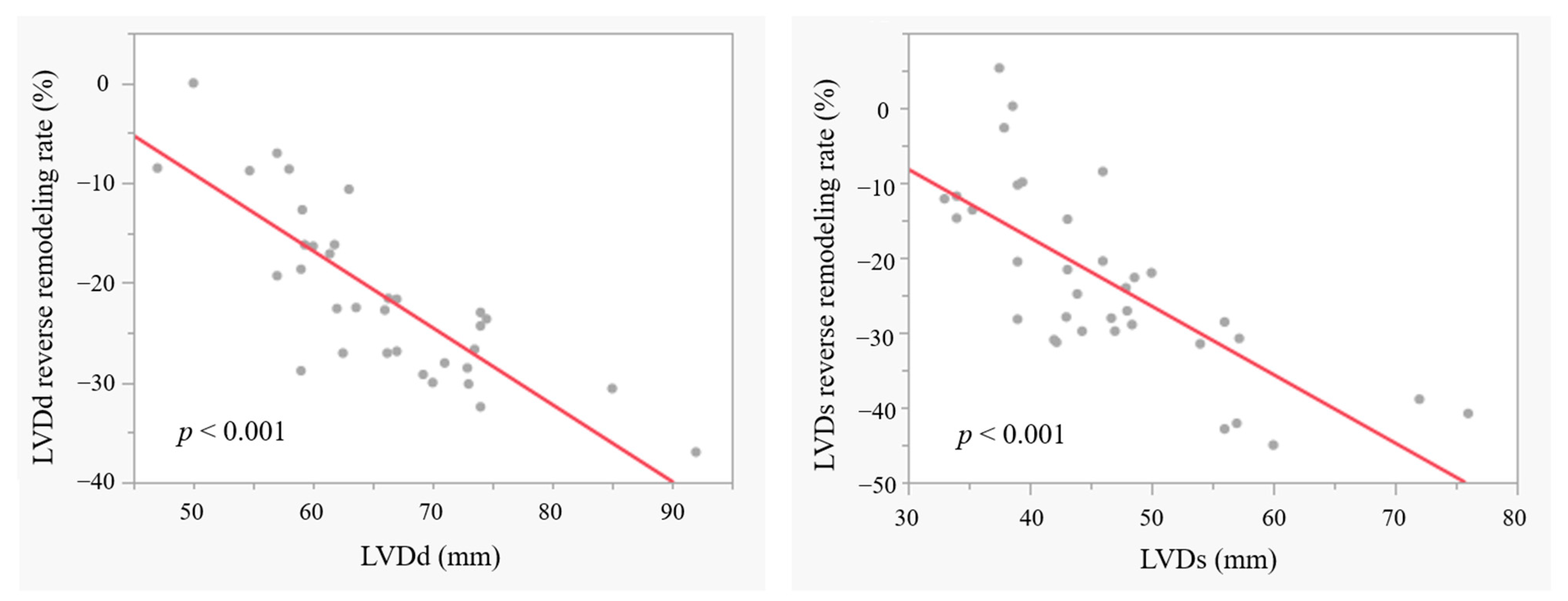
3.5. LV Reverse Remodelling in Patients with BAV

In Group-PB, the mean LV reverse remodelling rates were −22.6 (−28.0–−16.2)% (LVDd) and −24.8 (−30.8–−14.0)% (LVDs) at 1 year. The LV reverse remodelling rates in LVDd and LVDs were greater in patients with a larger preoperative LV dimension (both p < 0.001) (Figure 1); however, the postoperative peak/mean PG at 7-day TTE did not affect the LV reverse remodelling rate (LVDd: p = 0.815/0.778, LVDs: p = 0.993/0.772).

3.6. Mid-Term Outcomes in AVP for BAV

In the analysis of postoperative outcomes in Group-PB, we included a total of 42 patients. The mean follow-up duration for Group-PB was 27.5 (13.2–46.0) months. No patients died within 30 days after surgery. During the follow-up period, only one patient in Group-PB with coexisting aortitis syndrome died of acute myocardial infarction at 28.2 months after surgery. The actuarial survival rate was 95.2% at both 3 and 5 years (Figure S2a). AR >II developed in three patients during follow-up, two of whom required reoperation. The first of these patients developed AR due to aortic root destruction and multiple holes in all cusps after isolated BAV repair due to aortitis syndrome and underwent aortic root replacement at 7.1 months after surgery. This was the only patient who died, as described above. The second patient had recurrent AR again due to annular dilatation and fused cusp prolapse, despite re-AVP due to a tear in the fused cusp during the same period of hospitalisation after first AVP. This patient underwent AVR at 87.2 months after the initial surgery. The third patient developed AR due to an inadequate leaflet coaptation of the commissure at 63.0 months after the tricuspidization of very asymmetrical BAV using the aPP, and has been followed-up annually. The overall actuarial freedom from AR >II was 94.2% at both 3 and 5 years (Figure S2b) and the overall actuarial freedom from reoperation on the aortic valve was 97.2% at both 3 and 5 years (Figure S2c). Neither risk factors for AR recurrence nor reoperation could be detected.

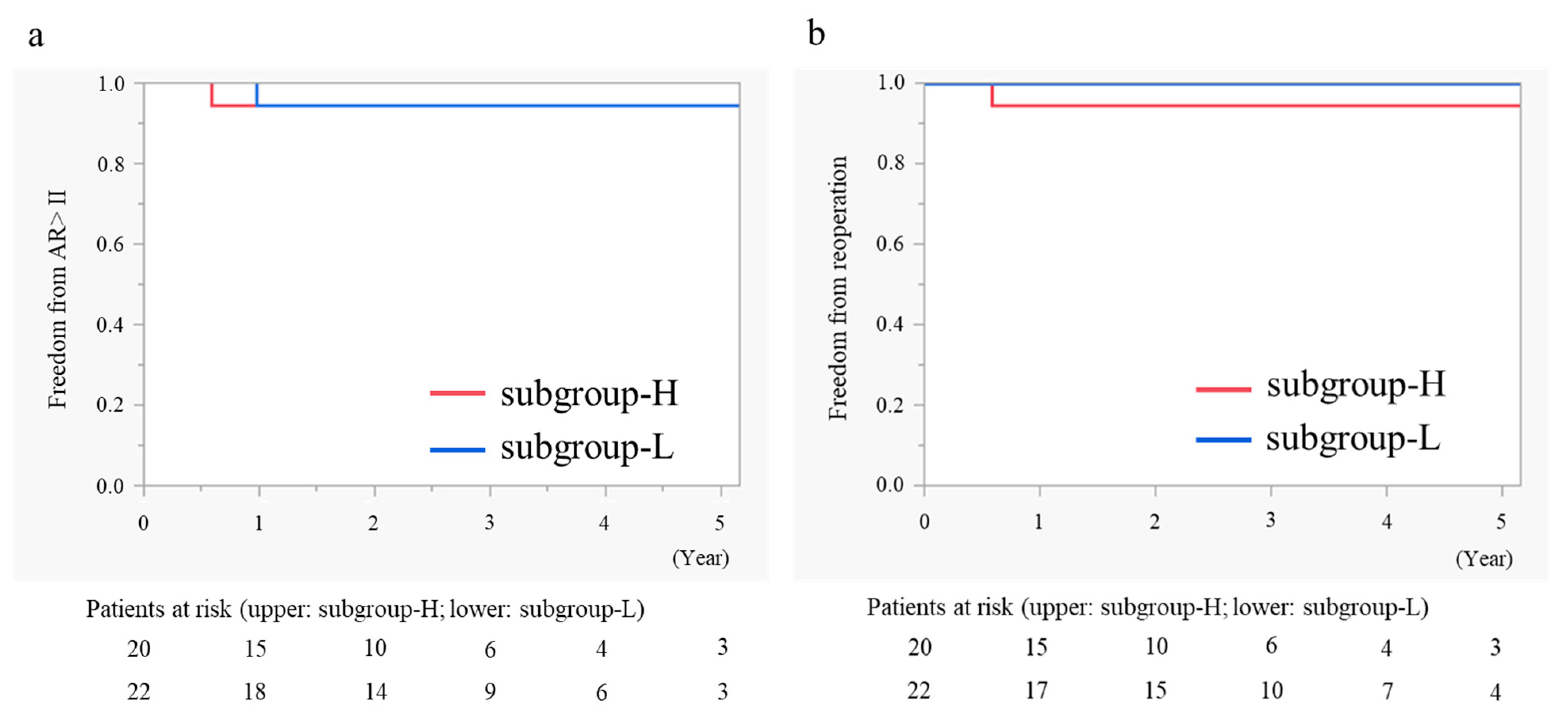
3.7. Subgroup Analysis in AVP for BAV According to Postoperative Peak PG

The mid-term outcomes were compared within Group-PB according to the postoperative peak PG—subgroup-H (n = 20) vs. subgroup-L (n = 22) (Table 5). The preoperative peak/mean PG, Vmax, and AVA were not different between the two subgroups. However, the preoperative aortic root size and external suture annuloplasty size index were larger in subgroup-L. Subgroup-H showed satisfactory LV reverse remodelling in LVDd (p < 0.001 at 1 year) and LVDs (p < 0.001 at 1 year) (Table 5).
The overall actuarial freedom from AR >II at 5 years was 94.4% in both subgroups (Figure 2a) and freedom from reoperation on the aortic valve at 5 years was 94.4% in subgroup-H and 100% in subgroup-L (Figure 2b); there were no differences between the groups (p = 0.749 and p = 0.317, respectively).

4. Discussion

This study showed that AVP for tricuspid aortic valve was more advantageous with regard to valve function compared to AVP for BAV and AVR in the early postoperative period, while AVP for BAV was associated with the risk of a higher postoperative PG compared to the other procedures. Multivariable analysis showed that a shorter geometric height, shorter free margin length, and use of the aPP were correlated with a higher postoperative PG. However, a higher postoperative PG after AVP for BAV did not affect the mid-term outcomes in this study.

4.1. Advantages of AVP with Regard to Valve Function

With increasing numbers of studies on AVP and its long-term outcomes [9], AVP has recently emerged as a reliable and reproducible technique. AVP has received class I recommendation in the current guidelines [10], thus providing a reasonable alternative to AVR, especially for young patients [11]. The majority of AR candidates for surgery are relatively young, and are, therefore, more likely to have complications related to the prosthetic valve if AVR is performed [12]. A recent meta-analysis showed that VSRR was associated with lower incidences of late death, thromboembolism, and bleeding events and had a similar valve durability to the Bentall operation [13]. Stent-posts supporting prosthetic valves were reported to obstruct blood flow, with a reduction in the effective orifice area by 40–70% of the total area occupied by the native valve [4]. Our results supported these findings and suggested that AVP for tricuspid aortic valve may be preferable to AVR with regard to valve function.

4.2. Higher PG After AVP in BAV Patients

The long-term outcomes of AVP for BAV are still controversial compared to those for tricuspid aortic valve [14]. Patlolla et al. reported that disease progression with calcification or fibrosis is the most common cause of valve failure after AVP for BAV [15]. Vohra et al. reported that leaflet calcification and BAV were risk factors for a residual higher PG, which affected valve durability [5]. Patients with BAV are more prone to a residual higher PG because they require complex repair more frequently than patients with tricuspid aortic valve [16,17,18]. Our BAV patients also required significantly more cusp plication than the tricuspid aortic valve patients, and, therefore, PG after AVP was significantly higher, although the rate of aPP use was lower in the BAV patients. Although there were no significant differences between Group-PT and Group-PB in peak/mean PG, Vmax, or AVA at 1 year in the propensity scoring analysis, these parameters may increase over time in Group-PB.
We identified short cusp geometry as a risk factor for postoperative stenosis. Schäfers et al. suggested that a geometric height of at least 16 mm in tricuspid aortic valve and 20 mm in BAV are necessary to achieve durable repair [19]. Pettersson et al. reported that shortening the free margin length (overcorrection) reduced regurgitation, but caused AVA reduction and a higher PG [16]. These results can be explained simply as BAV requires cusp plication to the fused cusp with reference to the non-fused cusp, and, therefore, a short free margin length of the non-fused cusp may lead to restriction of the motion of the fused cusp, resulting in a higher PG. To resolve this issue, the aPP may be used to extend and augment the cusp (e.g., tricuspidization). However, the use of the aPP was shown previously and in our study to be a negative prognostic factor for the long-term durability of repair [20]. These results suggest that a shorter geometric height and shorter free margin length should be managed carefully, and the use of the aPP should be avoided as much as possible.

4.3. Mid-Term Outcomes After AVP in BAV Patients

Postoperative residual PG should be prevented, because a higher PG negatively affects long-term outcomes, although only a few studies have discussed the impact of residual PG on clinical outcomes [5]. Vohra et al. reported that patients with a postoperative PG of ≥20 mmHg were more likely to have AR recurrence and require aortic valve reintervention during follow-up [5]. In the present study, however, PG ≥20 mmHg did not affect the mid-term outcomes and provided satisfactory LV reverse remodelling in BAV patients. Some studies have reported that postoperative LV reverse remodelling was one of the predictors of long-term outcomes, such as cardiac death or hospitalisation due to heart failure [21]. Amano et al. demonstrated that a late recurrence of LV dysfunction often occurred after AVR, and preoperative and follow-up echocardiographic parameters were predictors of the recurrence of LV dysfunction [22]. However, recent studies have shown that cardioprotective drugs play a significant role in the treatment of heart failure [23]. Mylonas et al. reported that Sodium–Glucose Cotransporter 2 Inhibitors were detected in the myocardial tissue of patients who underwent valvular surgery and were associated with myocardial remodelling and inflammation [23]. Unfortunately, our study did not account for the history of medication. The introduction and continuation of cardioprotective drugs after surgery may influence the LV reverse remodelling rate and long-term prognosis. Further studies in this area are warranted.
The inconsistency between previous studies and our study can be explained by the following reasons: Vohra et al. included both BAV and tricuspid aortic valve patients in their study. Additionally, the high-PG group (PG ≥ 20 mmHg) in their study had a significantly higher proportion of BAV patients compared to the low-PG group (PG < 20 mmHg) and more frequently underwent decalcification and shaving procedures (29.2%) than in our study. These suggest that BAV and advanced valve degeneration influenced their results. In contrast, our study focused on a younger cohort with only BAV patients and included fewer decalcification or shaving procedures (two patients in subgroup-H and two patients in subgroup-L). However, our study had a shorter follow-up period than Vohra et al.’s study. In patients with a high PG, the repair may not remain durable in the long term, potentially leading to insufficiency or the progression of valve stenosis and calcification [5]. Patlolla et al. also demonstrated that disease progression with calcification or fibrosis is the most common cause of valve failure after initial AVP for BAV, and the cumulative incidence of reoperation after initial valve repair was 11.1%, 35.9%, and 57.5% at 5 years, 10 years, and 15 years, respectively [15]. Their findings suggest that dysfunction due to valve degeneration progressively increases in the long term. Further studies with a longer follow-up are needed to validate our findings.

4.4. Limitations

This study had some limitations. First, the patient cohorts were not coincident in time. All cases with AR underwent AVR before 2014, when AVP had not been introduced in our facilities. In addition, there was only one surgeon for AVP (T.K.), while different surgeons performed AVR depending on the time period. Moreover, there were variations in the types of prosthetic valves used. These factors may introduce some degree of bias in the results. Second, this was a retrospective study with a small sample size. Third, this study consisted of three groups, Group-PB, Group-PT, and Group-R. BAV patients are younger and anatomically distinctive, thus, we employed propensity score analysis in the echocardiographic analysis; however, significant differences in backgrounds may introduce bias. Fourth, preoperative haemodynamic evaluations were not listed for Group-R (Table 1), because routine evaluations were not previously performed in AR patients and there were many missing haemodynamic values for Group-R. Therefore, haemodynamic variables were also excluded when adjusting between Group-R and the other groups. In addition, anatomical measurements of the aortic root (STJ, Valsalva, and ventriculoaortic junction) were collected in Group-PB, but not in Group-R. In Group-PT, we were unable to collect data from all cases. Then, in the comparison between Group-PB and -PT, we matched haemodynamic parameters, allowing for a reasonably accurate comparison. Fifth, we counted VSRR as AVP, while the type of procedure (isolated AVP or VSRR) may affect the postoperative haemodynamics. Therefore, we included the type of AVP (isolated AVP or VSRR) as a clinical variable in the analysis (Table 4). Sixth, atrial fibrillation may impact haemodynamics. While there were no cases of atrial fibrillation in Group-PB, there were seven cases (10.4%) in Group-PT. There were no data for comorbidities in Group-R. This introduces a potential source of bias. Seventh, this was a mid-term follow-up study, which should be validated in further studies with longer follow-ups.

5. Conclusions

AVP for tricuspid aortic valve showed favourable valve function in the early postoperative period compared to that of AVP for BAV or AVR.
AVP for BAV is associated with the risk of a higher PG after surgery. A shorter geometric height, shorter free margin length, and use of the aPP carry risks of a higher PG. A higher PG (≥20 mmHg) after AVP for BAV did not affect the mid-term outcomes. However, further studies with longer follow-ups are needed to validate our findings.

Supplementary Materials

The following supporting information can be downloaded at: https://www.mdpi.com/article/10.3390/jcm13247544/s1.

Author Contributions

Conceptualisation, data curation, methodology, investigation, writing—original draft Preparation, writing—review and editing, K.S.; conceptualisation, data curation, methodology, investigation, S.A.; data curation, investigation, T.T.; data curation, investigation, A.M.; data curation, investigation, Y.M.; data curation, investigation, M.Y.; data curation, investigation, R.N.; formal analysis, K.M.; project administration, supervision, writing—review and editing, T.K. All authors have read and agreed to the published version of the manuscript.

Funding

This research received no external funding.

Institutional Review Board Statement

The study protocol was approved by ethics committee of the Jikei University School of Medicine and (No. 33−497; date of approval: 4 April 2022) and the Cardiovascular Institute (No: 436; date of approval: 1 February 2023).

Informed Consent Statement

Informed consent was obtained from all subjects involved in the study.

Data Availability Statement

The original contributions presented in this study are included in the article/Supplementary Material. Further inquiries can be directed to the corresponding author.

Conflicts of Interest

The authors declare no conflicts of interest.

Correction Statement

This article has been republished with a minor correction to the readability of Table 3. This change does not affect the scientific content of the article.

References

  1. Schäfers, H.J.; Raddatz, A.; Schmied, W.; Takahashi, H.; Miura, Y.; Kunihara, T.; Aicher, D. Reexamining remodeling. J. Thorac. Cardiovasc. Surg. 2015, 149, S30–S36. [Google Scholar] [CrossRef] [PubMed]
  2. Tamer, S.; Mastrobuoni, S.; Lemaire, G.; Jahanyar, J.; Navarra, E.; Poncelet, A.; Astarci, P.; El Khoury, G.; de Kerchove, L. Two decades of valve-sparing root reimplantation in tricuspid aortic valve: Impact of aortic regurgitation and cusp repair. Eur. J. Cardiothorac. Surg. 2021, 59, 1069–1076. [Google Scholar] [CrossRef]
  3. de Kerchove, L.; Mastrobuoni, S.; Froede, L.; Tamer, S.; Boodhwani, M.; van Dyck, M.; EL Khoury, G.; Schäfers, H.J. Variability of repairable bicuspid aortic valve phenotypes: Towards an anatomical and repair-oriented classification. Eur. J. Cardiothorac. Surg. 2019, 56, 351–359. [Google Scholar] [CrossRef]
  4. Pibarot, P.; Dumesnil, J.G. Hemodynamic and clinical impact of prosthesis-patient mismatch in the aortic valve position and its prevention. J. Am. Coll. Cardiol. 2000, 36, 1131–1141. [Google Scholar] [CrossRef] [PubMed]
  5. Vohra, H.A.; Whistance, R.N.; de Kerchove, L.; Glineur, D.; Noirhomme, P.; El Khoury, G. Influence of higher valve gradient on long-term outcome after aortic valve repair. Ann. Cardiothorac. Surg. 2013, 2, 30–39. [Google Scholar]
  6. Kunihara, T.; Arimura, S.; Sata, F.; Giebels, C.; Schneider, U.; Schäfers, H.J. Aortic annulus does not dilate over time after aortic root remodeling with or without annuloplasty. J. Thorac. Cardiovasc. Surg. 2018, 155, 885–894. [Google Scholar] [CrossRef]
  7. Kunihara, T. Toward standardization of valve-sparing root replacement and annuloplasty. Gen. Thorac. Cardiovasc. Surg. 2018, 66, 685–691. [Google Scholar] [CrossRef] [PubMed]
  8. Schäfers, H.J.; Bierbach, B.; Aicher, D. A new approach to the assessment of aortic cusp geometry. J. Thorac. Cardiovasc. Surg. 2006, 132, 436–438. [Google Scholar] [CrossRef]
  9. Klotz, S.; Stock, S.; Sievers, H.H.; Diwoky, M.; Petersen, M.; Stierle, U.; Richardt, D. Survival and reoperation pattern after 20 years of experience with aortic valve-sparing root replacement in patients with tricuspid and bicuspid valves. J. Thorac. Cardiovasc. Surg. 2018, 155, 1403–1411. [Google Scholar] [CrossRef]
  10. Vahanian, A.; Beyersdorf, F.; Praz, F.; Milojevic, M.; Baldus, S.; Bauersachs, J.; Capodanno, D.; Conradi, L.; De Bonis, M.; De Paulis, R.; et al. 2021 ESC/EACTS Guidelines for the management of valvular heart disease. Eur. J. Cardiothorac. Surg. 2021, 60, 727–800. [Google Scholar]
  11. Gocoł, R.; Bis, J.; Malinowski, M.; Ciosek, J.; Hudziak, D.; Morkisz, Ł.; Jasiński, M.; Deja, M.A. Comparison of bicuspid and tricuspid aortic valve repair. Eur. J. Cardiothorac. Surg. 2021, 59, 1183–1190. [Google Scholar] [CrossRef] [PubMed]
  12. Korteland, N.M.; Etnel, J.R.G.; Arabkhani, B.; Mokhles, M.M.; Mohamad, A.; Roos-Hesselink, J.W.; Bogers, A.J.J.C.; Takkenberg, J.J.M. Mechanical aortic valve replacement in non-elderly adults: Meta-analysis and microsimulation. Eur. Heart. J. 2017, 38, 3370–3377. [Google Scholar] [CrossRef] [PubMed]
  13. Elbatarny, M.; Tam, D.Y.; Edelman, J.J.; Rocha, R.V.; Chu, M.W.A.; Peterson, M.D.; El-Hamamsy, I.; Appoo, J.J.; Friedrich, J.O.; Boodhwani, M.; et al. Valve-Sparing Root Replacement Versus Composite Valve Grafting in Aortic Root Dilation: A Meta-Analysis. Ann. Thorac. Surg. 2020, 110, 296–306. [Google Scholar] [CrossRef] [PubMed]
  14. Fattouch, K.; Murana, G.; Castrovinci, S.; Nasso, G.; Mossuto, C.; Corrado, E.; Ruvolo, G.; Speziale, G. Outcomes of aortic valve repair according to valve morphology and surgical techniques. Interact. Cardiovasc. Thorac. Surg. 2012, 15, 644–650. [Google Scholar] [CrossRef]
  15. Patlolla, S.H.; Schaff, H.V.; Stulak, J.M.; Michelena, H.I.; Saran, N.; King, K.S. Bicuspid Aortic Valve Repair: Causes of Valve Failure and Long-Term Outcomes. Ann. Thorac. Surg. 2021, 111, 1225–1233. [Google Scholar] [CrossRef]
  16. Pettersson, G.B.; Crucean, A.C.; Savage, R.; Halley, C.M.; Grimm, R.A.; Svensson, L.G.; Naficy, S.; Gillinov, A.M.; Feng, J.; Blackstone, E.H. Toward Predictable Repair of Regurgitant Aortic Valves. J. Am. Coll. Cardiol. 2008, 52, 40–49. [Google Scholar] [CrossRef]
  17. Bavaria, J.E.; Desai, N.; Szeto, W.Y.; Komlo, C.; Rhode, T.; Wallen, T.; Vallabhajosyula, P. Valve-sparing root reimplantation and leaflet repair in a bicuspid aortic valve: Comparison with the 3-cusp David procedure. J. Thorac. Cardiovasc. Surg. 2015, 149, S22–S28. [Google Scholar] [CrossRef] [PubMed]
  18. Prinzing, A.; Boehm, J.; Erlebach, M.; Sideris, K.; Lange, R.; Krane, M. Comparison of outcomes following isolated repair of tricuspid versus bicuspid aortic valves. J. Thorac. Dis. 2020, 12, 3514–3533. [Google Scholar] [CrossRef]
  19. Schäfers, H.J.; Schmied, W.; Marom, G.; Aicher, D. Cusp height in aortic valves. J. Thorac. Cardiovasc. Surg. 2013, 146, 269–274. [Google Scholar] [CrossRef]
  20. Karliova, I.; Schneider, U.; Ehrlich, T.; Schäfers, H.J. Results of Pericardial Patches in Tricuspid and Bicuspid Aortic Cusp Repair. Ann. Thorac. Surg. 2020, 109, 728–735. [Google Scholar] [CrossRef]
  21. Izumi, C.; Kitai, T.; Kume, T.; Onishi, T.; Yuda, S.; Hirata, K.; Yamashita, E.; Kawata, T.; Nishimura, K.; Takeuchi, M.; et al. Effect of Left Ventricular Reverse Remodeling on Long-term Outcomes After Aortic Valve Replacement. Am. J. Cardiol. 2019, 124, 105–112. [Google Scholar] [CrossRef] [PubMed]
  22. Amano, M.; Izumi, C.; Imamura, S.; Onishi, N.; Tamaki, Y.; Enomoto, S.; Miyake, M.; Tamura, T.; Kondo, H.; Kaitani, K.; et al. Late recurrence of left ventricular dysfunction after aortic valve replacement for severe chronic aortic regurgitation. Int. J. Cardiol. 2016, 224, 240–244. [Google Scholar] [CrossRef] [PubMed]
  23. Mylonas, N.; Nikolaou, P.E.; Karakasis, P.; Stachteas, P.; Fragakis, N.; Andreadou, I. Endothelial Protection by Sodium-Glucose Cotransporter 2 Inhibitors: A Literature Review of In Vitro and In Vivo Studies. Int. J. Mol. Sci. 2024, 25, 7274. [Google Scholar] [CrossRef] [PubMed]
Figure 1. The LV reverse remodelling rates in LVDd and LVDs. LV: left ventricular; LVDd: left ventricular diastolic diameter; LVDs: left ventricular systolic diameter.
Jcm 13 07544 g001
Figure 2. Kaplan–Meier curves showing the differences in freedom from AR >II (a) and freedom from reoperation on the aortic valve (b) between subgroup-H and subgroup-L. AR: aortic regurgitation.
Jcm 13 07544 g002
Table 1. Patient characteristics and operative details.
AVP (n = 108)AVR (n = 132)p-Value
Group-PB (n = 41)Group-PT (n = 67)Group-R (n = 132)
Patient characteristics
Age40.0 (26.0–49.0)57.0 (50.0–66.0)65.5 (55.0–73.0)<0.001
Body surface area (m2)1.81 (1.72–1.91)1.78 (1.62–1.90)1.71 (1.56–1.85)0.002
Male sex (%)40 (97.6)58 (87.9)106 (80.3)0.019
Preoperative echocardiography
LVDd (mm)65.8 (59.1–72.9)59.4 (53.0–67.0)65.0 (60.0–69.0)0.002
LVDs (mm)46.5 (39.4–52.5)40.0 (35.6–48.3)47.0 (41.0–52.0)0.001
LVEF (%)57.6 (51.0–60.2)58.3 (52.2–62.1)53.0 (47.0–61.0)0.119
Peak PG (mmHg)17.0 (11.8–23.2)8.6 (5.1–12.7) <0.001
Mean PG (mmHg)9.1 (6.5–12.3)4.4 (2.8–7.1) 0.001
Vmax (m/s)2.2 (1.8–2.4)1.6 (1.2–2.0) <0.001
AVA (cm2)3.4 (2.9–4.2)3.5 (2.9–4.1) 0.804
Operations
VSRR (remodelling) (%)16 (39.0)29 (43.3) 0.663
AVP (%)25 (61.0)38 (56.7)
AVP details
Cusp plication (%)38 (92.7)46 (68.7) 0.004
External suture annuloplasty (%)41 (100)59 (88.1) 0.022
External suture annuloplasty size (mm)18 mm:1
20 mm: 5
22 mm: 19
24 mm: 16
18 mm: 4
20 mm: 21
22 mm: 29
24 mm: 4
30 mm: 1
0.002
STJ remodelling (%)36 (87.8)59 (88.1) 0.969
STJ remodelling size (mm)20 mm: 1
22 mm: 4
24 mm: 18
26 mm: 13
20 mm: 1
22 mm: 12
24 mm: 38
26 mm: 7
30 mm: 1
0.064
Use of the aPP (%)6 (14.6)15 (22.4) 0.323
AVR operation 132
Bioprosthetic valve (%) 93 (70.5)
Mechanical valve (%) 39 (29.5)
Aortic prosthetic valve size (mm) 19 mm: 5
21 mm: 9
23 mm: 34
25 mm: 53
27 mm: 31
All continuous values are expressed as the median (interquartile range). aPP: autologous pericardial patch; AVP: aortic valvuloplasty; AVR: aortic valve replacement; LVDd: left ventricular diastolic diameter; LVDs: left ventricular systolic diameter; LVEF: left ventricular ejection fraction; Vmax: peak jet velocity; VSRR: valve-sparing aortic root replacement.
Table 2. Postoperative echocardiographic findings.
Group-PB (n = 41)Group-PT (n = 67)Group-R (n = 132)
7-day TTE
Aortic regurgitation grade (>II) (%)0 (0)0(0)0 (0)
LVDd (mm)54.0 (50.3–58.4)49.8 (47.0–55.0)52.0 (48.0–56.2)
LVDs (mm)41.0 (35.0–47.3)36.5 (32.0–43.0)39.0 (33.8–45.0)
LVEF (%)45.0 (35.1–58.0)54.0 (41.0–60.0)50.0 (37.0–59.0)
Peak PG (mmHg)19.1 (13.2–25.3)8.8 (6.2–12.2)13.6 (10.7–17.9)
Mean PG (mmHg)11.7 (7.6–15.0)5.1 (3.4–6.4)7.5 (6.0–10.7)
Vmax (m/s)2.2 (1.9–2.5)1.5 (1.4–1.9)1.9 (1.6–2.3)
AVA (cm2)1.7 (1.4–2.1)2.6 (2.1–3.1)1.8 (1.5–2.2)
1-year TTE
Aortic regurgitation grade (>II) (%)1 (2.9)2 (3.3)0 (0.0)
LVDd (mm)51.0 (49.0–53.0)48.7 (44.5–51.5)48.0 (44.0–51.0)
LVDs (mm)34.4 (31.0–37.6)30.5 (28.0–34.0)31.9 (27.3–35.0)
LVEF (%)59.0 (56.5–64.0)63.0 (60.3–67.7)64.0 (58.3–69.0)
Peak PG (mmHg)20.0 (13.0–28.9)8.4 (6.0–12.0)15.6 (10.5–20.2)
Mean PG (mmHg)11.6 (7.7–15.8)4.4 (3.0–6.1)8.2 (5.7–11.3)
Vmax (m/s)2.4 (1.9–2.9)1.7 (1.4–1.8)2.1 (1.6–2.3)
AVA (cm2)2.1 (1.5–2.6)2.6 (2.1–3.0)2.0 (1.6–2.3)
All continuous values are expressed as the median (interquartile range). AVA: aortic valve area; LVEF: left ventricular ejection fraction; LVDd: left ventricular diastolic diameter; LVDs: left ventricular systolic diameter; PG: pressure gradient; TTE: transthoracic echocardiography; Vmax: transaortic velocity.
Table 3. Comparison of postoperative echocardiography findings between groups.
<Group-PB vs. Group-R (Reference)>
Evaluated Time PointOutcome TypeOutcomeUnadjusted (n = 173)IPTW (n = 169)
7-day TTEOrdered categoryOdds ratio95% CIs p-valueOdds ratio95% CIsp-value
Aortic regurgitation grade1.440.6913.0040.3311.2780.7762.1050.335
ContinuousCoefficient95% CIsp-valueCoefficient95% CIsp-value
Peak PG (mmHg)5.373 2.488 8.258 <0.0015.467 3.016 7.918 <0.001
Mean PG (mmHg)3.373 1.702 5.043 <0.0013.413 1.999 4.826 <0.001
Vmax (m/s)0.303 0.116 0.489 0.0020.345 0.171 0.520 <0.001
AVA (cm2)−0.168 −0.397 0.060 0.147−0.322 −0.534 −0.111 0.003
1-year TTEOrdered categoryOdds ratio95% CIsp-valueOdds ratio95% CIsp-value
Aortic regurgitation grade0.356 0.164 0.770 0.0090.476 0.283 0.799 0.005
ContinuousCoefficient95% CIsp-valueCoefficient95% CIsp-value
Peak PG (mmHg)5.402 2.146 8.658 0.0015.647 2.833 8.462 <0.001
Mean PG (mmHg)3.250 1.367 5.133 <0.0013.504 1.923 5.084 <0.001
Vmax (m/s)0.357 0.048 0.667 0.0250.588 0.298 0.878 <0.001
AVA (cm2)0.335 0.036 0.633 0.0280.105 −0.214 0.423 0.517
<Group-PT vs. Group-R (Reference)>
Evaluated time pointOutcome typeOutcomeUnadjusted (n = 199)IPTW (n = 192)
7-day TTEOrdered categoryOdds ratio95% CIsp-valueOdds ratio95% CIsp-value
Aortic regurgitation grade0.5950.3251.0910.0940.4370.290.661<0.001
ContinuousCoefficient95% CIsp-valueCoefficient95% CIsp-value
Peak PG (mmHg)−4.810 −7.111 −2.509 <0.001−4.827−6.887−2.768<0.001
Mean PG (mmHg)−2.980 −4.261 −1.699 <0.001−2.899−4.014−1.785<0.001
Vmax (m/s)−0.282 −0.448 −0.116 0.001−0.261−0.431−0.0910.003
AVA (cm2)0.618 0.419 0.817 <0.0010.4530.2680.638<0.001
1-year TTEOrdered categoryOdds ratio95% CIsp-valueOdds ratio95% CIsp-value
Aortic regurgitation grade0.234 0.117 0.465 <0.0010.2810.1780.445<0.001
ContinuousCoefficient95% CIsp-valueCoefficient95% CIsp-value
Peak PG (mmHg)−6.451 −8.671 −4.230 <0.001−6.417−8.476−4.357<0.001
Mean PG (mmHg)−3.603 −4.868 −2.338 <0.001−3.636−4.811−2.460<0.001
Vmax (m/s)−0.291 −0.569 −0.014 0.040−0.172−0.4900.1470.285
AVA (cm2)0.659 0.449 0.869 <0.0010.6360.4340.837<0.001
<Group-PB vs. Group-PT (Reference)>
Evaluated time pointOutcome typeOutcomeUnadjusted (n = 108)IPTW (n = 52)
7-day TTEOrdered categoryOdds ratio95% CIsp-valueOdds ratio95% CIsp-value
Aortic regurgitation grade2.2851.0225.1080.0440.7080.3081.6290.417
ContinuousCoefficient95% CIsp-valueCoefficient95% CIsp-value
Peak PG (mmHg)10.184 7.00413.363<0.0015.586 −0.106 11.277 0.054
Mean PG (mmHg)6.353 4.4758.232<0.0013.827 0.663 6.992 0.019
Vmax (m/s)0.585 0.4040.765<0.0010.337 0.028 0.647 0.033
AVA (cm2)−0.786 −1.050−0.522<0.001−0.264 −0.659 0.132 0.187
1-year TTEOrdered categoryOdds ratio95% CIsp-valueOdds ratio95% CIsp-value
Aortic regurgitation grade1.272 0.5712.8350.5560.582 0.268 1.263 0.171
ContinuousCoefficient95% CIsp-valueCoefficient95% CIsp-value
Peak PG (mmHg)11.853 8.34715.358<0.0013.323 −2.536 9.182 0.259
Mean PG (mmHg)6.853 4.8298.876<0.0012.893 −0.413 6.200 0.085
Vmax (m/s)0.649 0.3860.912<0.0010.148 −0.239 0.536 0.442
AVA (cm2)−0.324 −0.7130.0650.1010.335 −0.256 0.925 0.259
AVA: aortic valve area; IPWT: inverse probability of treatment weighting; PG: pressure gradient; TTE: transthoracic echocardiography; Vmax: peak jet velocity; 95% CIs: 95%Confidence Intervals.
Table 4. Correlation between pre- and intraoperative valuables and postoperative pressure gradient (mmHg).
Univariable Linear RegressionStepwise Multivariable Linear Regression (R2 = 0.806)
Coefficient95%CIsp ValueCoefficient95%CIsp ValueStd CoefficientVIF
Age−0.073−0.3890.2440.638
Body surface area18.693−6.56243.9470.139
Male sex8.268−11.87628.4130.403
Ventriculoaortic junction−1.843−3.310−0.3760.016
Sinus of Valsalva−0.352−0.8100.1060.125
STJ −0.289−0.9280.3500.356
LVDd−0.352−0.9920.2880.266
LVDs−0.362−0.8380.1130.128
LVEF0.218−0.4650.9000.515
Peak PG0.3300.0500.6110.023
Vmax6.2050.21912.1910.043
AVP or VSRR−7.218−16.8052.3690.132
Cusp plication−14.667−27.919−1.4140.032
External suture annuloplasty size index−3.426−6.826−0.0270.048
Geometric height−1.805−3.462−0.1490.034−1.315−2.605−0.0250.046−0.3031.502
Free margin length−0.896−1.663−0.1300.024−0.806−1.388−0.2250.010−0.4151.524
Use of the aPP27.90511.79544.0140.00225.29014.68935.891<0.0010.5861.024
aPP: autologous pericardial patch; AVP: aortic valvuloplasty; External suture annuloplasty size index: external suture annuloplasty size/body surface area; LVDd: left ventricular diastolic diameter; LVDs: left ventricular systolic diameter; LVEF: left ventricular ejection fraction; PG; pressure gradient; STJ: sinotubular junction; VIF: Variance inflation factor; Vmax: peak jet velocity; VSRR: valve-sparing aortic root replacement; 95%CIs: 95% Confidence Intervals.
Table 5. Comparison of patient characteristics, preoperative and postoperative echocardiography findings between peak PG ≥20 mmHg (subgroup-H) and peak PG <20 mmHg (subgroup-L) at 7-day TTE in patients with BAV.
Subgroup-H (n = 20)Subgroup-L (n = 22)p-Value
         Peak PG at 7-day TTE (mmHg)25.5 (22.4–35.0)13.3 (10.8–18.1)
Patient characteristics
         Age (year)41.0 (22.8–49.0)39.0 (28.0–49.3)0.804
         Body surface area (m2)1.8 (1.7–2.0)1.8 (1.7–1.9)0.431
         Male (%)20 (100)21 (95.5)0.335
         Hypertension9 (45.0)5 (22.7)0.126
         Dyslipidaemia2 (10.0)00.129
         Diabetes mellitus00
         Haemodialysis00
         Atrial fibrillation00
Preoperative transthoracic echocardiography
         LVDd (mm)65.9 (59.2–73.0)64.5 (59.0–73.0)0.791
         LVDs (mm)46.9 (40.1–51.9)46.0 (39.0–56.3)0.738
         LVEF (%)58.2 (51.1–60.7)57.5 (50.0–60.7)0.622
         Peak PG (mmHg)17.2 (12.2–23.9)17.5 (8.6–23.5)0.525
         Mean PG (mmHg)9.6 (7.5–12.4)7.5 (4.4–16.4)0.480
         Vmax (m/s)2.2 (1.8–2.7)2.2 (1.8–2.7)0.571
         AVA (cm2)3.2 (2.2–3.9)3.5 (3.0–4.3)0.092
Preoperative transoesophageal echocardiography
         Ventriculoaortic junction (mm)29.0 (28.0–30.0)30.0 (28.0–33.0)0.322
         Sinus of Valsalva (mm)34.0 (33.0–39.0)38.0 (35.0–40.0)0.019
         STJ (mm)29.0 (27.0–31.8)30.0 (25.0–35.5)0.200
Intraoperative cusp measurement
         Geometric height (mm)21.5 (20.0–24.0)24.5 (22.3–25.0)0.017
         Free margin length (mm)38.0 (35.0–40.0)44.5 (38.5–46.3)0.020
Operations
         AVP: Remodelling16: 410: 120.021
         Cusp plication (%)18 (90.0)21 (95.5)0.493
         External suture annuloplasty size index11.4 (10.9–13.0)12.6 (12.4–13.4)0.003
         Use of the aPP (%)4 (20.0)2 (9.1)0.313
         Decalcification and shaving (%)2 (10.0)2 (9.1)0.920
1-year transthoracic echocardiography
         Aortic regurgitation grade (>II) (%)01 (4.5)0.324
         LVDd (mm)52.0 ± 3.450.3 ± 4.10.174
         Reverse remodelling rate (%) in LVDd−20.6 ± 8.7−21.9 ± 8.60.684
         LVDs (mm)35.1 ± 1.034.7 ± 1.00.757
         Reverse remodelling rate (%) in LVDs−24.7 ± 12.2−23.1 ± 10.90.699
         LVEF (%)59.7 ± 5.858.7 ± 9.80.717
         Peak PG (mmHg)25.3 ± 11.117.4 ± 8.70.029
         Mean PG (mmHg)13.8 ± 6.59.7 ± 5.1<0.001
         Vmax (m/s)2.5 ± 0.62.2 ± 0.50.094
         AVA (cm2)2.0 ± 0.92.7 ± 1.20.094
All continuous values are expressed as the median (interquartile range). aPP: autologous pericardial patch; AVA: aortic valve area; AVP: aortic valvuloplasty; external suture annuloplasty size index: external suture annuloplasty size/body surface area; LVEF: left ventricular ejection fraction; LVDd: left ventricular diastolic diameter; LVDs: left ventricular systolic diameter; PG: pressure gradient; STJ: sinotubular junction; Vmax: peak jet velocity.
Disclaimer/Publisher’s Note: The statements, opinions and data contained in all publications are solely those of the individual author(s) and contributor(s) and not of MDPI and/or the editor(s). MDPI and/or the editor(s) disclaim responsibility for any injury to people or property resulting from any ideas, methods, instructions or products referred to in the content.

