Ultrasonographic Measurement of Anterior Fontanelle Size in Infants with Deformational Plagiocephaly
Abstract
1. Introduction
2. Materials and Methods
3. Results
4. Discussion
5. Conclusions
Author Contributions
Funding
Institutional Review Board Statement
Informed Consent Statement
Data Availability Statement
Conflicts of Interest
References
- Unnithan, A.K.A.; De Jesus, O. Plagiocephaly. In StatPearls; StatPearls Publishing LLC.: Treasure Island, FL, USA, 2024. [Google Scholar]
- Bialocerkowski, A.E.; Vladusic, S.L.; Wei Ng, C. Prevalence, risk factors, and natural history of positional plagiocephaly: A systematic review. Dev. Med. Child. Neurol. 2008, 50, 577–586. [Google Scholar] [CrossRef] [PubMed]
- Miller, R.I.; Clarren, S.K. Long-term developmental outcomes in patients with deformational plagiocephaly. Pediatrics 2000, 105, E26. [Google Scholar] [CrossRef]
- Collett, B.R.; Wallace, E.R.; Kartin, D.; Cunningham, M.L.; Speltz, M.L. Cognitive Outcomes and Positional Plagiocephaly. Pediatrics 2019, 143, e20182373. [Google Scholar] [CrossRef] [PubMed]
- Collett, B.R.; Starr, J.R.; Kartin, D.; Heike, C.L.; Berg, J.; Cunningham, M.L.; Speltz, M.L. Development in toddlers with and without deformational plagiocephaly. Arch. Pediatr. Adolesc. Med. 2011, 165, 653–658. [Google Scholar] [CrossRef]
- Boere-Boonekamp, M.M.; van der Linden-Kuiper, L.L. Positional preference: Prevalence in infants and follow-up after two years. Pediatrics 2001, 107, 339–343. [Google Scholar] [CrossRef] [PubMed]
- de Chalain, T.M.; Park, S. Torticollis associated with positional plagiocephaly: A growing epidemic. J. Craniofac Surg. 2005, 16, 411–418. [Google Scholar] [CrossRef]
- Knight, S.J.; Anderson, V.A.; Meara, J.G.; Da Costa, A.C. Early neurodevelopment in infants with deformational plagiocephaly. J. Craniofac Surg. 2013, 24, 1225–1228. [Google Scholar] [CrossRef] [PubMed]
- Mawji, A.; Vollman, A.R.; Fung, T.; Hatfield, J.; McNeil, D.A.; Sauvé, R. Risk factors for positional plagiocephaly and appropriate time frames for prevention messaging. Paediatr. Child. Health 2014, 19, 423–427. [Google Scholar] [CrossRef]
- Rousslang, L.K.; Rooks, E.A.; Smith, A.C.; Wood, J.R. Fibromatosis colli leading to positional plagiocephaly with gross anatomical and sonographic correlation. BMJ Case Rep. 2021, 14, e239236. [Google Scholar] [CrossRef]
- Jung, B.K.; Yun, I.S. Diagnosis and treatment of positional plagiocephaly. Arch. Craniofac Surg. 2020, 21, 80–86. [Google Scholar] [CrossRef] [PubMed]
- Miyabayashi, H.; Nagano, N.; Hashimoto, S.; Saito, K.; Kato, R.; Noto, T.; Sasano, M.; Sumi, K.; Yoshino, A.; Morioka, I. Evaluating Cranial Growth in Japanese Infants Using a Three-dimensional Scanner: Relationship between Growth-related Parameters and Deformational Plagiocephaly. Neurol. Med. Chir. 2022, 62, 521–529. [Google Scholar] [CrossRef] [PubMed]
- Sze, R.W.; Hopper, R.A.; Ghioni, V.; Gruss, J.S.; Ellenbogen, R.G.; King, D.; Hing, A.V.; Cunningham, M.L. MDCT diagnosis of the child with posterior plagiocephaly. AJR Am. J. Roentgenol. 2005, 185, 1342–1346. [Google Scholar] [CrossRef] [PubMed]
- Kim, J.K.; Kwon, D.R.; Park, G.Y. A new ultrasound method for assessment of head shape change in infants with plagiocephaly. Ann. Rehabil. Med. 2014, 38, 541–547. [Google Scholar] [CrossRef]
- Marino, S.; Ruggieri, M.; Marino, L.; Falsaperla, R. Sutures ultrasound: Useful diagnostic screening for posterior plagiocephaly. Childs Nerv. Syst. 2021, 37, 3715–3720. [Google Scholar] [CrossRef] [PubMed]
- Regelsberger, J.; Delling, G.; Tsokos, M.; Helmke, K.; Kammler, G.; Kränzlein, H.; Westphal, M. High-frequency ultrasound confirmation of positional plagiocephaly. J. Neurosurg. 2006, 105, 413–417. [Google Scholar] [CrossRef] [PubMed]
- Soboleski, D.; McCloskey, D.; Mussari, B.; Sauerbrei, E.; Clarke, M.; Fletcher, A. Sonography of normal cranial sutures. AJR Am. J. Roentgenol. 1997, 168, 819–821. [Google Scholar] [CrossRef][Green Version]
- Soboleski, D.; Mussari, B.; McCloskey, D.; Sauerbrei, E.; Espinosa, F.; Fletcher, A. High-resolution sonography of the abnormal cranial suture. Pediatr. Radiol. 1998, 28, 79–82. [Google Scholar] [CrossRef]
- Proisy, M.; Riffaud, L.; Chouklati, K.; Tréguier, C.; Bruneau, B. Ultrasonography for the diagnosis of craniosynostosis. Eur. J. Radiol. 2017, 90, 250–255. [Google Scholar] [CrossRef]
- Sze, R.W.; Parisi, M.T.; Sidhu, M.; Paladin, A.M.; Ngo, A.V.; Seidel, K.D.; Weinberger, E.; Ellenbogen, R.G.; Gruss, J.S.; Cunningham, M.L. Ultrasound screening of the lambdoid suture in the child with posterior plagiocephaly. Pediatr. Radiol. 2003, 33, 630–636. [Google Scholar] [CrossRef]
- D’Antoni, A.V.; Donaldson, O.I.; Schmidt, C.; Macchi, V.; De Caro, R.; Oskouian, R.J.; Loukas, M.; Shane Tubbs, R. A comprehensive review of the anterior fontanelle: Embryology, anatomy, and clinical considerations. Childs Nerv. Syst. 2017, 33, 909–914. [Google Scholar] [CrossRef]
- Popich, G.A.; Smith, D.W. Fontanels: Range of normal size. J. Pediatr. 1972, 80, 749–752. [Google Scholar] [CrossRef] [PubMed]
- Oumer, M.; Tazebew, A.; Alemayehu, M. Anterior Fontanel Size Among Term Newborns: A Systematic Review and Meta-Analysis. Public. Health Rev. 2021, 42, 1604044. [Google Scholar] [CrossRef] [PubMed]
- Sarigecili, E.; Makharoblidze, K.; Çobanogullari, M.D.; Yildirim, D.D.; Komur, M.; Okuyaz, C. Neurodevelopmental risk evaluation of premature closure of the anterior fontanelle. Childs Nerv. Syst. 2021, 37, 561–566. [Google Scholar] [CrossRef]
- Direk, M.; Makharoblıdze, K.; Polat, B.G.; Özdemir, A.A.; Okuyaz, Ç. The neurodevelopmental profile of healthy children with premature anterior fontanel closure. Turk. J. Med. Sci. 2022, 52, 934–941. [Google Scholar] [CrossRef] [PubMed]
- Kim, D.G.; Lee, J.S.; Lee, J.W.; Yang, J.D.; Chung, H.Y.; Cho, B.C.; Choi, K.Y. The Effects of Helmet Therapy Relative to the Size of the Anterior Fontanelle in Nonsynostotic Plagiocephaly: A Retrospective Study. J. Clin. Med. 2019, 8, 1977. [Google Scholar] [CrossRef] [PubMed]
- Mortenson, P.A.; Steinbok, P. Quantifying positional plagiocephaly: Reliability and validity of anthropometric measurements. J. Craniofac Surg. 2006, 17, 413–419. [Google Scholar] [CrossRef] [PubMed]
- van Vlimmeren, L.A.; Takken, T.; van Adrichem, L.N.; van der Graaf, Y.; Helders, P.J.; Engelbert, R.H. Plagiocephalometry: A non-invasive method to quantify asymmetry of the skull; a reliability study. Eur. J. Pediatr. 2006, 165, 149–157. [Google Scholar] [CrossRef]
- Argenta, L.; David, L.; Thompson, J. Clinical classification of positional plagiocephaly. J. Craniofac Surg. 2004, 15, 368–372. [Google Scholar] [CrossRef]
- Jaffe, M.; Harel, J.; Goldberg, A.; Rudolph-Schnitzer, M.; Winter, S.T. The use of the Denver Developmental Screening Test in infant welfare clinics. Dev. Med. Child. Neurol. 1980, 22, 55–60. [Google Scholar] [CrossRef]
- Mirrett, P.L.; Bailey, D.B., Jr.; Roberts, J.E.; Hatton, D.D. Developmental screening and detection of developmental delays in infants and toddlers with fragile X syndrome. J. Dev. Behav. Pediatr. 2004, 25, 21–27. [Google Scholar] [CrossRef]
- Nasiri, J.; Madihi, Y.; Mirzadeh, A.S.; Mohammadzadeh, M. Neurodevelopmental Outcomes of Infants with Benign Enlargement of the Subarachnoid Space. Iran. J. Child. Neurol. 2021, 15, 33–40. [Google Scholar] [PubMed]
- Rah, S.S.; Jung, M.; Lee, K.; Kang, H.; Jang, S.; Park, J.; Yoon, J.Y.; Hong, S.B. Systematic Review and Meta-analysis: Real-World Accuracy of Children’s Developmental Screening Tests. J. Am. Acad. Child. Adolesc. Psychiatry 2023, 62, 1095–1109. [Google Scholar] [CrossRef] [PubMed]
- Frankenburg, W.K.; Dodds, J.; Archer, P.; Shapiro, H.; Bresnick, B. The Denver II: A major revision and restandardization of the Denver Developmental Screening Test. Pediatrics 1992, 89, 91–97. [Google Scholar] [CrossRef] [PubMed]
- Frankenburg, W.K.; Dodds, J.B. The Denver developmental screening test. J. Pediatr. 1967, 71, 181–191. [Google Scholar] [CrossRef] [PubMed]
- Kim, D.H.; Kwon, D.R. Neurodevelopmental delay according to severity of deformational plagiocephaly in children. Medicine 2020, 99, e21194. [Google Scholar] [CrossRef] [PubMed]
- Kiesler, J.; Ricer, R. The abnormal fontanel. Am. Fam. Physician 2003, 67, 2547–2552. [Google Scholar]
- Wendling-Keim, D.S.; Macé, Y.; Lochbihler, H.; Dietz, H.G.; Lehner, M. A new parameter for the management of positional plagiocephaly: The size of the anterior fontanelle matters. Childs Nerv. Syst. 2020, 36, 363–371. [Google Scholar] [CrossRef]
- Esmaeili, M.; Esmaeili, M.; Ghane Sharbaf, F.; Bokharaie, S. Fontanel Size from Birth to 24 Months of Age in Iranian Children. Iran. J. Child. Neurol. 2015, 9, 15–23. [Google Scholar]
- Pedroso, F.S.; Rotta, N.; Quintal, A.; Giordani, G. Evolution of anterior fontanel size in normal infants in the first year of life. J. Child. Neurol. 2008, 23, 1419–1423. [Google Scholar] [CrossRef]
- Sasani, H.; Tüfekçi, S.; Haksayar, A. A morphometric evaluation of anterior fontanel and cranial sutures in infants using computed tomography. J. Exp. Clin. Med. 2022, 39, 321–326. [Google Scholar] [CrossRef]
- Idriz, S.; Patel, J.H.; Ameli Renani, S.; Allan, R.; Vlahos, I. CT of Normal Developmental and Variant Anatomy of the Pediatric Skull: Distinguishing Trauma from Normality. Radiographics 2015, 35, 1585–1601. [Google Scholar] [CrossRef] [PubMed]
- Furtado, L.M.F.; Filho, J.; Freitas, L.S.; Dantas Dos Santos, A.K. Anterior fontanelle closure and diagnosis of non-syndromic craniosynostosis: A comparative study using computed tomography. J. Pediatr. 2022, 98, 413–418. [Google Scholar] [CrossRef] [PubMed]
- Duc, G.; Largo, R.H. Anterior fontanel: Size and closure in term and preterm infants. Pediatrics 1986, 78, 904–908. [Google Scholar] [CrossRef] [PubMed]
- Kwon, D.R.; Kim, Y. Sternocleidomastoid size and upper trapezius muscle thickness in congenital torticollis patients: A retrospective observational study. Medicine 2021, 100, e28466. [Google Scholar] [CrossRef] [PubMed]
- Park, G.Y.; Kwon, D.R.; Kwon, D.G. Shear wave sonoelastography in infants with congenital muscular torticollis. Medicine 2018, 97, e9818. [Google Scholar] [CrossRef] [PubMed]
- Kwon, D.R. Sonographic Analysis of Changes in Skull Shape After Cranial Molding Helmet Therapy in Infants with Deformational Plagiocephaly. J. Ultrasound Med. 2016, 35, 695–700. [Google Scholar] [CrossRef]


| Age Group (n) | Mean Age (Days) | Mean Anterior Fontanelle Size (cm, Range) |
|---|---|---|
| 0–1 month (n = 13) | 19.77 ± 9.38 | 2.23 ± 0.25 (1.81–2.52) |
| 1–2 month (n = 19) | 50.37 ± 9.25 | 2.26 ± 0.29 (1.82–3.07) |
| 2–3 month (n = 16) | 79.25 ± 8.61 | 2.31 ± 0.53 (1.14–3.12) |
| 3–4 month (n = 47) | 106.15 ± 9.15 | 2.34 ± 0.48 (0.9–3.43) |
| 4–5 month (n = 50) | 132.94 ± 7.92 | 2.12 ± 0.52 (0.75–3.3) |
| 5–6 month (n = 19) | 165.74 ± 9.13 | 2.06 ± 0.45 (0.83–2.52) |
| >6 month (n = 25) | 214.92 ± 31.33 | 1.86 ± 0.79 (0–3.44) |
| Age Group (n) | Detection Rate (n, %) | Mean Anterior Fontanelle Size (cm, Range) * |
|---|---|---|
| 0–1 month (n = 13) | 0/13 (0%) | - |
| 1–2 month (n = 19) | 6/19 (31.6%) | 2.50 ± 0.83 (1.69–3.98) |
| 2–3 month (n = 16) | 7/16 (43.8%) | 2.83 ± 0.95 (1.51–4.13) |
| 3–4 month (n = 47) | 31/47 (66%) | 3.02 ± 0.84 (1.00–5.26) |
| 4–5 month (n = 50) | 35/50 (70%) | 2.53 ± 0.85 (1.01–4.95) |
| 5–6 month (n = 19) | 13/19 (68.4%) | 2.51 ± 0.99 (0.92–4.54) |
| >6 month (n = 25) | 21/25 (84%) | 2.39 ± 1.21 (0–4.63) |
| Total (n = 189) | 113/189 (59.8%) |
| Variable | Group 1 (n = 43) (CVA ≤ 5 mm) | Group 2 (n = 105) (5 mm < CVA < 10 mm) | Group 3 (n = 41) (CVA ≥ 10 mm) | p-Value |
|---|---|---|---|---|
| Age (days) | 114.58 ± 54.30 | 115.28 ± 53.82 | 136.80 ± 56.36 | 0.080 |
| Gender (male/female) | 26:17 | 66:39 | 30:11 | 0.411 |
| Affected side (right/left) | 20:23 | 65:40 | 22:19 | 0.209 |
| Risk factors | ||||
| Oligohydramnios | 0 | 4 | 2 | 0.380 |
| Breech delivery | 4 | 9 | 0 | 0.143 |
| Twin baby | 3 | 10 | 3 | 0.842 |
| Scale (grade) | 2.00 ± 0.44 †♦ | 2.48 ± 0.67 †‡ | 2.78 ± 0.57 ‡♦ | 0.000 * |
| CVA (mm) | 4.28 ± 0.88 †♦ | 7.34 ± 1.08 †‡ | 11.61 ± 1.77 ‡♦ | 0.000 * |
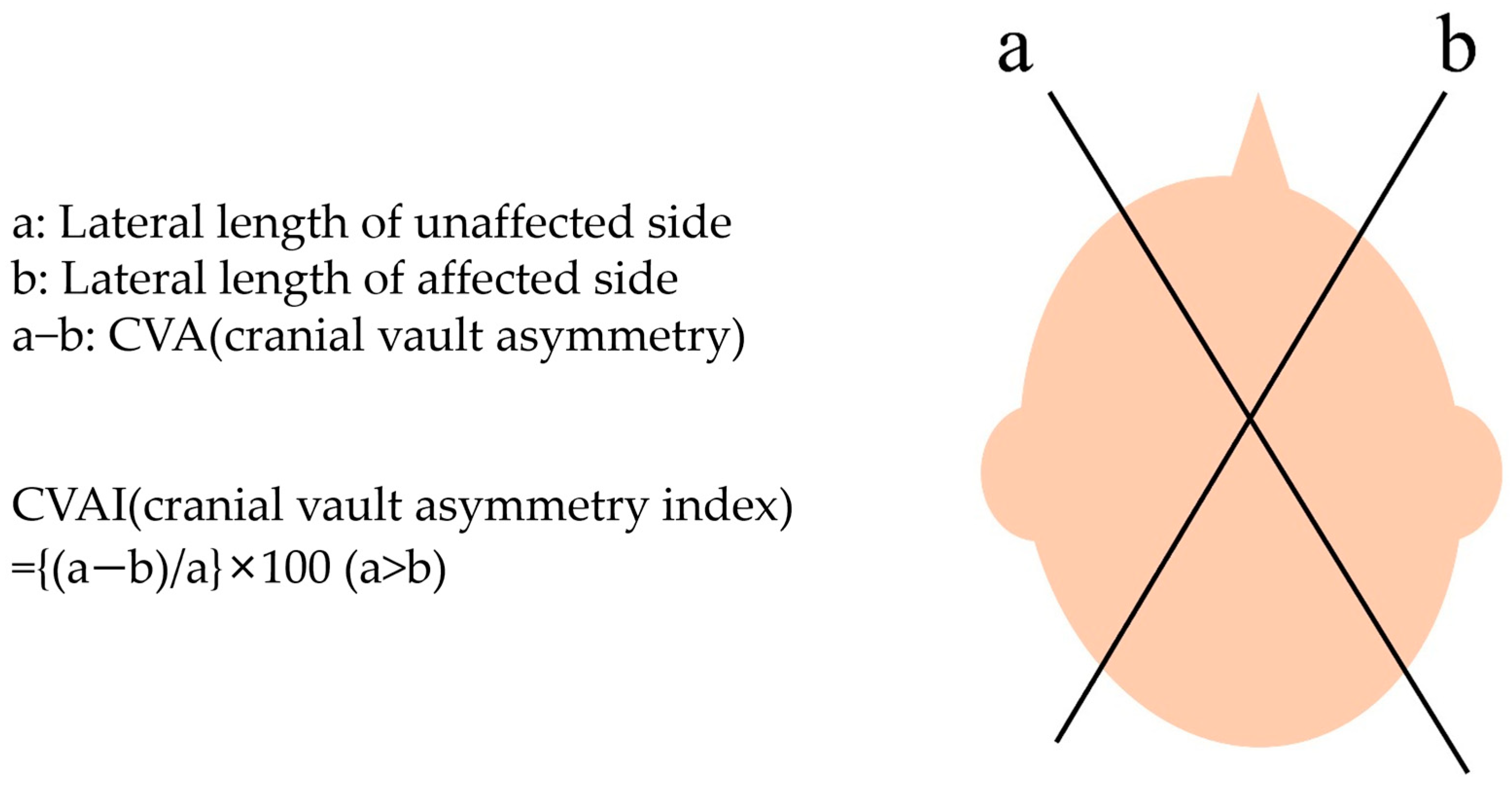
| CVAI (%) | 3.13 ± 0.61 †♦ | 5.28 ± 0.74 †‡ | 8.18 ± 1.23 ‡♦ | 0.000 * |
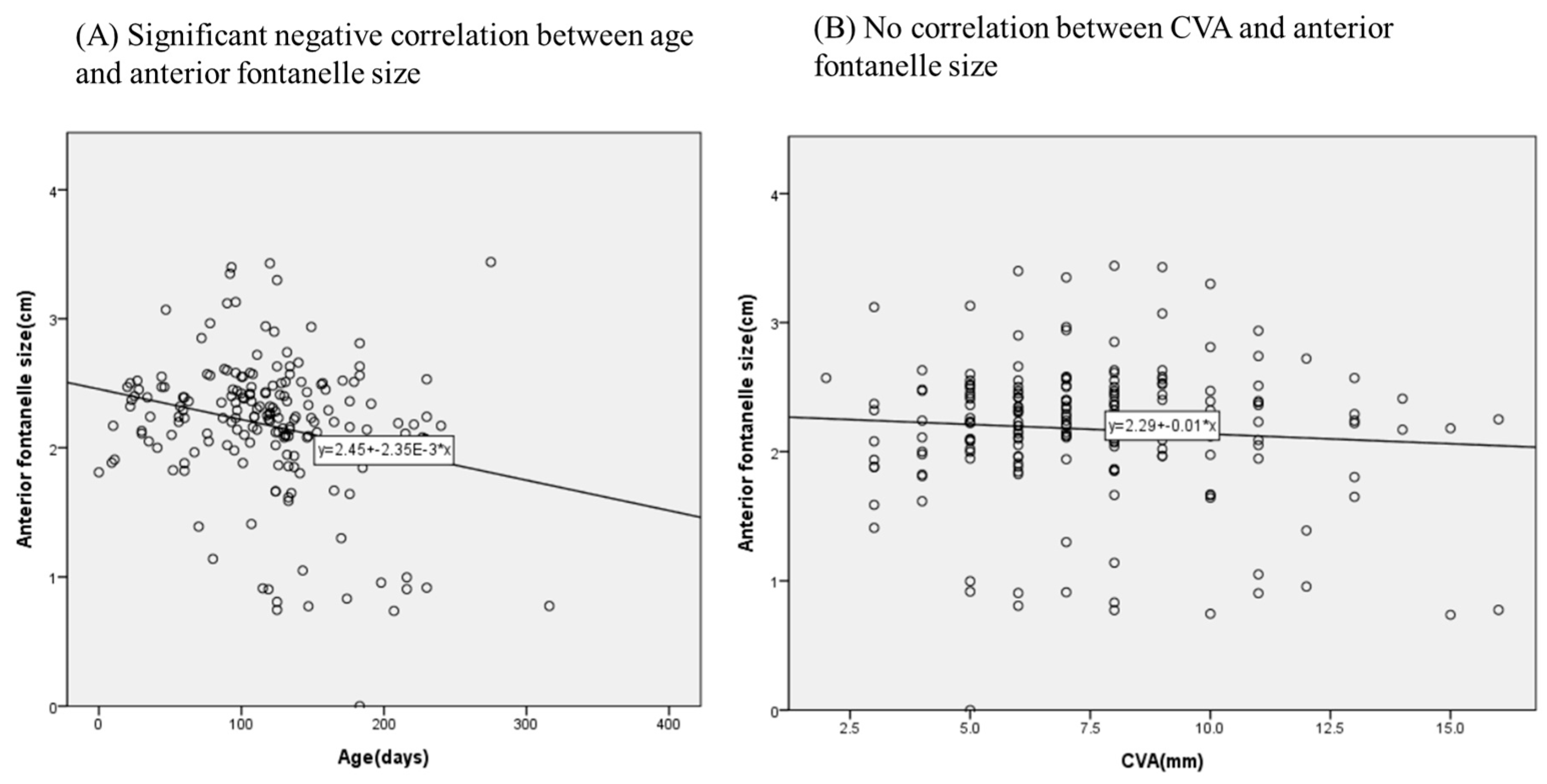
| Anterior fontanelle size (cm) | 2.11 ± 0.55 | 2.25 ± 0.48 | 2.04 ± 0.62 | 0.074 |
| Variable | Developmental Delay (−) (n = 21) | Developmental Delay (+) (n = 19) | p-Value |
|---|---|---|---|
| Scale (grade) | 2.52 ± 0.51 | 2.32 ± 0.67 | 0.421 |
| CVA (mm) | 7.05 ± 2.18 | 6.79 ± 2.90 | 0.486 |
| CVAI (%) | 5.02 ± 1.55 | 4.91 ± 1.94 | 0.688 |
| Anterior fontanelle size (cm) | 2.05 ± 0.44 | 1.48 ± 0.51 | 0.09 |
Disclaimer/Publisher’s Note: The statements, opinions and data contained in all publications are solely those of the individual author(s) and contributor(s) and not of MDPI and/or the editor(s). MDPI and/or the editor(s) disclaim responsibility for any injury to people or property resulting from any ideas, methods, instructions or products referred to in the content. |
© 2024 by the authors. Licensee MDPI, Basel, Switzerland. This article is an open access article distributed under the terms and conditions of the Creative Commons Attribution (CC BY) license (https://creativecommons.org/licenses/by/4.0/).
Share and Cite
Lee, J.H.; Park, G.-Y.; Kwon, D.R. Ultrasonographic Measurement of Anterior Fontanelle Size in Infants with Deformational Plagiocephaly. J. Clin. Med. 2024, 13, 5012. https://doi.org/10.3390/jcm13175012
Lee JH, Park G-Y, Kwon DR. Ultrasonographic Measurement of Anterior Fontanelle Size in Infants with Deformational Plagiocephaly. Journal of Clinical Medicine. 2024; 13(17):5012. https://doi.org/10.3390/jcm13175012
Chicago/Turabian StyleLee, Jae Hee, Gi-Young Park, and Dong Rak Kwon. 2024. "Ultrasonographic Measurement of Anterior Fontanelle Size in Infants with Deformational Plagiocephaly" Journal of Clinical Medicine 13, no. 17: 5012. https://doi.org/10.3390/jcm13175012
APA StyleLee, J. H., Park, G.-Y., & Kwon, D. R. (2024). Ultrasonographic Measurement of Anterior Fontanelle Size in Infants with Deformational Plagiocephaly. Journal of Clinical Medicine, 13(17), 5012. https://doi.org/10.3390/jcm13175012


