Association of Microvasculature Changes with Visual Outcomes in Idiopathic Epiretinal Membrane Surgery: A Clinical Trial
Abstract
1. Introduction
2. Methods
2.1. Patients and Study Design
2.2. Surgical Technique
2.3. Pre-, Intra-, and Postoperative Data
2.4. Main Outcome Measures
2.5. Statistical Analysis
3. Results
3.1. Preoperative and Intraoperative Data
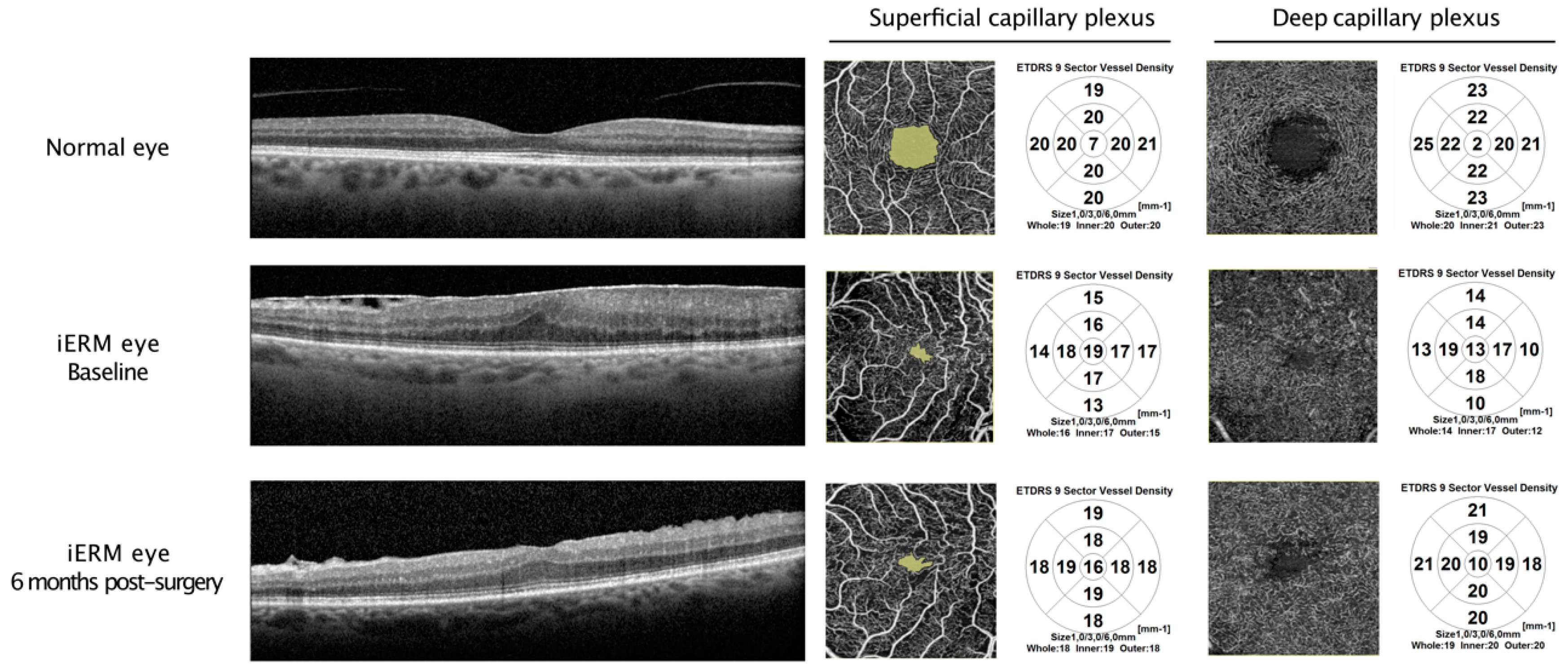
3.2. OCTA Parameters in iERM Eyes and Comparison with Those of Fellow Eyes
3.3. Correlation between OCT Parameters, Visual Acuity, and Macular Morphology in iERM Eyes
3.4. Predictive Factors for Postoperative Visual Outcomes
4. Discussion
Author Contributions
Funding
Institutional Review Board Statement
Informed Consent Statement
Data Availability Statement
Conflicts of Interest
References
- Fung, A.T.; Galvin, J.; Tran, T. Epiretinal membrane: A review. Clin. Exp. Ophthalmol. 2021, 49, 289–308. [Google Scholar] [CrossRef] [PubMed]
- Steel, D.H.W.; Lotery, A.J. Idiopathic vitreomacular traction and macular hole: A comprehensive review of pathophysiology, diagnosis, and treatment. Eye 2013, 27 (Suppl. S1), S1–S21. [Google Scholar] [CrossRef] [PubMed]
- McCarty, D.J.; Mukesh, B.N.; Chikani, V.; Wang, J.J.; Mitchell, P.; Taylor, H.R.; McCarty, C.A. Prevalence and associations of epiretinal membranes in the visual impairment project. Am. J. Ophthalmol. 2005, 140, 288–294. [Google Scholar] [CrossRef] [PubMed]
- Iuliano, L.; Fogliato, G.; Gorgoni, F.; Corbelli, E.; Bandello, F.; Codenotti, M. Idiopathic epiretinal membrane surgery: Safety, efficacy and patient related outcomes. Clin. Ophthalmol. 2019, 13, 1253–1265. [Google Scholar] [CrossRef]
- Wong, J.G.; Sachdev, N.; Beaumont, P.E.; Chang, A.A. Visual outcomes following vitrectomy and peeling of epiretinal membrane. Clin. Exp. Ophthalmol. 2005, 33, 373–378. [Google Scholar] [CrossRef] [PubMed]
- Kauffmann, Y.; Ramel, J.-C.; Lefebvre, A.; Isaico, R.; De Lazzer, A.; Bonnabel, A.; Bron, A.M.; Creuzot-Garcher, C. Preoperative Prognostic Factors and Predictive Score in Patients Operated On for Combined Cataract and Idiopathic Epiretinal Membrane. Am. J. Ophthalmol. 2015, 160, 185–192.e5. [Google Scholar] [CrossRef] [PubMed]
- Scheerlinck, L.M.E.; van der Valk, R.; van Leeuwen, R. Predictive factors for postoperative visual acuity in idiopathic epiretinal membrane: A systematic review. Acta Ophthalmol. 2015, 93, 203–212. [Google Scholar] [CrossRef]
- Wang, L.-C.; Lo, W.-J.; Huang, Y.-Y.; Chou, Y.-B.; Li, A.-F.; Chen, S.-J.; Chou, T.-Y.; Lin, T.-C. Correlations between Clinical and Histopathologic Characteristics in Idiopathic Epiretinal Membrane. Ophthalmology 2022, 129, 1421–1428. [Google Scholar] [CrossRef]
- Govetto, A.; Lalane, R.A.; Sarraf, D.; Figueroa, M.S.; Hubschman, J.P. Insights Into Epiretinal Membranes: Presence of Ectopic Inner Foveal Layers and a New Optical Coherence Tomography Staging Scheme. Am. J. Ophthalmol. 2017, 175, 99–113. [Google Scholar] [CrossRef]
- Zur, D.; Iglicki, M.; Feldinger, L.; Schwartz, S.; Goldstein, M.; Loewenstein, A.; Barak, A. Disorganization of Retinal Inner Layers as a Biomarker for Idiopathic Epiretinal Membrane After Macular Surgery-The DREAM Study. Am. J. Ophthalmol. 2018, 196, 129–135. [Google Scholar] [CrossRef] [PubMed]
- Inoue, M.; Morita, S.; Watanabe, Y.; Kaneko, T.; Yamane, S.; Kobayashi, S.; Arakawa, A.; Kadonosono, K. Preoperative inner segment/outer segment junction in spectral-domain optical coherence tomography as a prognostic factor in epiretinal membrane surgery. Retina 2011, 31, 1366–1372. [Google Scholar] [CrossRef] [PubMed]
- Koo, H.C.; Rhim, W.I.; Lee, E.K. Morphologic and functional association of retinal layers beneath the epiretinal membrane with spectral-domain optical coherence tomography in eyes without photoreceptor abnormality. Graefes Arch. Clin. Exp. Ophthalmol. 2012, 250, 491–498. [Google Scholar] [CrossRef] [PubMed]
- Spaide, R.F.; Fujimoto, J.G.; Waheed, N.K.; Sadda, S.R.; Staurenghi, G. Optical coherence tomography angiography. Prog. Retin. Eye Res. 2018, 64, 1–55. [Google Scholar] [CrossRef] [PubMed]
- Shiihara, H.; Sakamoto, T.; Yamashita, T.; Kakiuchi, N.; Otsuka, H.; Terasaki, H.; Sonoda, S. Reproducibility and differences in area of foveal avascular zone measured by three different optical coherence tomographic angiography instruments. Sci. Rep. 2017, 7, 9853. [Google Scholar] [CrossRef] [PubMed]
- Kim, G.-H.; Hwang, B.-E.; Chun, H.; Kim, J.Y.; Kim, R.Y.; Kim, M.; Park, Y.-G.; Park, Y.-H. Morphologic analysis of the foveal avascular zone for prediction of postoperative visual acuity in advanced idiopathic epiretinal membrane. Sci. Rep. 2023, 13, 10400. [Google Scholar] [CrossRef] [PubMed]
- Chen, H.; Chi, W.; Cai, X.; Deng, Y.; Jiang, X.; Wei, Y.; Zhang, S. Macular microvasculature features before and after vitrectomy in idiopathic macular epiretinal membrane: An OCT angiography analysis. Eye 2019, 33, 619–628. [Google Scholar] [CrossRef] [PubMed]
- Mao, J.; Xu, Z.; Lao, J.; Chen, Y.; Xu, X.; Wu, S.; Zheng, Z.; Liu, B.; Shen, L. Assessment of macular microvasculature features before and after vitrectomy in the idiopathic macular epiretinal membrane using a grading system: An optical coherence tomography angiography study. Acta Ophthalmol. 2021, 99, e1168–e1175. [Google Scholar] [CrossRef] [PubMed]
- Mao, J.; Lao, J.; Liu, C.; Zhang, C.; Chen, Y.; Tao, J.; Shen, L. A study analyzing macular microvasculature features after vitrectomy using OCT angiography in patients with idiopathic macular epiretinal membrane. BMC Ophthalmol. 2020, 20, 165. [Google Scholar] [CrossRef] [PubMed]
- Told, R.; Georgopoulos, M.; Reiter, G.S.; Wassermann, L.; Aliyeva, L.; Baumann, L.; Abela-Formanek, C.; Pollreisz, A.; Schmidt-Erfurth, U.; Sacu, S. Intraretinal microvascular changes after ERM and ILM peeling using SSOCTA. PLoS ONE 2020, 15, e0242667. [Google Scholar] [CrossRef]
- Xu, Z.; Mao, J.; Lao, J.; Deng, X.; Liu, C.; Xu, J.; Wu, S.; Chen, Y.; Shen, L. Macular Retinal Sensitivity and Microvasculature Changes before and after Vitrectomy in Idiopathic Macular Epiretinal Membrane with Classification. Ophthalmologica 2021, 244, 569–580. [Google Scholar] [CrossRef]
- Yoshida, H.; Terashima, H.; Ueda, E.; Hasebe, H.; Matsuoka, N.; Nakano, H.; Fukuchi, T. Relationship between morphological changes in the foveal avascular zone of the epiretinal membrane and postoperative visual function. BMJ Open Ophthalmol. 2020, 5, e000636. [Google Scholar] [CrossRef]
- Isik-Ericek, P.; Sizmaz, S.; Esen, E.; Demircan, N. The effect of epiretinal membrane surgery on macular microvasculature: An optical coherence tomography angiography study. Int. Ophthalmol. 2021, 41, 777–786. [Google Scholar] [CrossRef] [PubMed]
- Hirata, A.; Nakada, H.; Mine, K.; Masumoto, M.; Sato, T.; Hayashi, K. Relationship between the morphology of the foveal avascular zone and the degree of aniseikonia before and after vitrectomy in patients with unilateral epiretinal membrane. Graefes Arch. Clin. Exp. Ophthalmol. 2019, 257, 507–515. [Google Scholar] [CrossRef]
- Okawa, Y.; Maruko, I.; Kawai, M.; Hasegawa, T.; Arakawa, H.; Iida, T. Foveal structure and vasculature in eyes with idiopathic epiretinal membrane. PLoS ONE 2019, 14, e0214881. [Google Scholar] [CrossRef] [PubMed]
- Bacherini, D.; Dragotto, F.; Caporossi, T.; Lenzetti, C.; Finocchio, L.; Savastano, A.; Savastano, M.C.; Barca, F.; Dragotto, M.; Vannozzi, L.; et al. The Role of OCT Angiography in the Assessment of Epiretinal Macular Membrane. J. Ophthalmol. 2021, 2021, 8866407. [Google Scholar] [CrossRef]
- Feng, J.; Yang, X.; Xu, M.; Wang, Y.; Shi, X.; Zhang, Y.; Huang, P. Association of Microvasculature and Macular Sensitivity in Idiopathic Macular Epiretinal Membrane: Using OCT Angiography and Microperimetry. Front. Med. 2021, 8, 655013. [Google Scholar] [CrossRef] [PubMed]
- Nelis, P.; Alten, F.; Clemens, C.R.; Heiduschka, P.; Eter, N. Quantification of changes in foveal capillary architecture caused by idiopathic epiretinal membrane using OCT angiography. Graefes Arch. Clin. Exp. Ophthalmol. 2017, 255, 1319–1324. [Google Scholar] [CrossRef]
- Kim, Y.J.; Kim, S.; Lee, J.Y.; Kim, J.-G.; Yoon, Y.H. Macular capillary plexuses after epiretinal membrane surgery: An optical coherence tomography angiography study. Br. J. Ophthalmol. 2018, 102, 1086–1091. [Google Scholar] [CrossRef]
- Lee, E.K.; Yu, H.G. Ganglion cell-inner plexiform layer thickness after epiretinal membrane surgery: A spectral-domain optical coherence tomography study. Ophthalmology 2014, 121, 1579–1587. [Google Scholar] [CrossRef]
- Lee, J.; Moon, B.G.; Cho, A.R.; Yoon, Y.H. Optical Coherence Tomography Angiography of DME and Its Association with Anti-VEGF Treatment Response. Ophthalmology 2016, 123, 2368–2375. [Google Scholar] [CrossRef]
- Okamoto, F.; Sugiura, Y.; Okamoto, Y.; Hiraoka, T.; Oshika, T. Inner nuclear layer thickness as a prognostic factor for metamorphopsia after epiretinal membrane surgery. Retina 2015, 35, 2107–2114. [Google Scholar] [CrossRef] [PubMed]
- Cho, K.H.; Park, S.J.; Cho, J.H.; Woo, S.J.; Park, K.H. Inner-Retinal Irregularity Index Predicts Postoperative Visual Prognosis in Idiopathic Epiretinal Membrane. Am. J. Ophthalmol. 2016, 168, 139–149. [Google Scholar] [CrossRef] [PubMed]
- Kim, J.H.; Kang, S.W.; Kong, M.G.; Ha, H.S. Assessment of retinal layers and visual rehabilitation after epiretinal membrane removal. Graefes Arch. Clin. Exp. Ophthalmol. 2013, 251, 1055–1064. [Google Scholar] [CrossRef] [PubMed]
- Chua, P.Y.; Sandinha, M.T.; Steel, D.H. Idiopathic epiretinal membrane: Progression and timing of surgery. Eye 2022, 36, 495–503. [Google Scholar] [CrossRef] [PubMed]
- Elhusseiny, A.M.; Flynn, H.W.; Smiddy, W.E. Long-Term Outcomes After Idiopathic Epiretinal Membrane Surgery. Clin. Ophthalmol. 2020, 14, 995–1002. [Google Scholar] [CrossRef] [PubMed]


| Baseline Data | Values |
|---|---|
| Number of eyes, n | 47 |
| Age, years (median (IQR)) | 71.0 (8.0) |
| Sex, n (%) | |
| 18 (38.3) |
| 29 (61.7) |
| Axial length, mm (median (IQR)) | 23.7 (1.1) |
| Preoperative lens status, n (%) | |
| 36 (76.6) |
| 11 (23.4) |
| Preoperative BCVA, logMAR (median (IQR)) | 0.3 (0.2) |
| iERM grade, n (%) | |
| 0 |
| 12 (25.5) |
| 23 (49) |
| 12 (25.5) |
| Central macular thickness, µm (median (IQR)) | 480.0 (106.0) |
| Surgical procedure, n (%) | |
| Combined cataract extraction | 36 (75.6) |
| OCTA Parameters | Fellow Eyes | iERM Eyes | p-Value * | |||
|---|---|---|---|---|---|---|
| M0 | M6 | p a | p b | p c | ||
| FAZ area, mm2 | 0.3(0.2) | 0.03(0.03) | 0.08(0.05) | <0.001 | <0.001 | <0.001 |
| SCP, mm2 | ||||||
| Whole VD | 18.0(1.5) | 16.0(2.5) | 18.0(1.0) | <0.001 | <0.001 | 0.004 |
| Foveal VD | 8.0(2.0) | 16.0(2.5) | 13.0(3.0) | <0.001 | <0.001 | <0.001 |
| Parafoveal VD | 18.0(2.0) | 16.0(3.0) | 18.0(2.0) | <0.001 | <0.001 | 0.043 |
| Perifoveal VD | 20.0(2.5) | 16.0(2.5) | 16.0(3.0) | <0.001 | <0.001 | <0.001 |
| DCP, mm2 | ||||||
| Whole VD | 17.0(6.0) | 13.0(5.0) | 17.0(3.5) | <0.001 | <0.001 | 0.202 |
| Foveal VD | 5.0(2.0) | 11.0(3.0) | 7.0(2.5) | <0.001 | <0.001 | <0.001 |
| Parafoveal VD | 17.0(6.0) | 14.0(6.0) | 18.0(4.5) | <0.001 | <0.001 | 0.402 |
| Perifoveal VD | 18.0(7.0) | 13.0(6.0) | 18.0(5.0) | <0.001 | <0.010 | 0.709 |
| Baseline Parameters | Preoperative logMAR BCVA | Postoperative logMAR BCVA | ||
|---|---|---|---|---|
| r | p-Value | r | p-Value | |
| FAZ area, mm2 | −0.499 | <0.001 | −0.059 | 0.689 |
| SCP, mm2 | ||||
| Whole VD | −0.276 | 0.059 | −0.394 | 0.006 |
| DCP, mm2 | ||||
| Whole VD | −0.422 | 0.003 | −0.569 | <0.001 |
| Baseline Parameters | iERM Stage | CMT | ||
|---|---|---|---|---|
| r | p-Value | r | p-Value | |
| FAZ area, mm2 | −0.267 | 0.069 | −0.143 | 0.336 |
| SCP, mm2 | ||||
| Whole VD | 0.288 | 0.049 | 0.164 | 0.270 |
| DCP, mm2 | ||||
| Whole VD | 0.033 | 0.825 | −0.138 | 0.352 |
| Bivariate Analysis | Multivariate Analysis | |||
|---|---|---|---|---|
| Factors | Beta Coefficient (Standard Deviation) | p | Beta Coefficient (Standard Deviation) | p |
| Preoperative BCVA | 0.475 (0.052) | <0.001 | 0.457 (0.064) | <0.001 |
| iERM stage | 0.088 (0.040) | 0.034 | 0.037 (0.029) | 0.210 |
| Age | 0.008 (0.0038) | 0.026 | 0.002 (0.003) | 0.498 |
| Axial length | −0.012 (0.019) | 0.528 | ||
| CMT | 0.0001 (0.0004) | 0.675 | ||
| Vascular parameters | ||||
| Whole VD in the SCP | −0.024 (0.012) | 0.042 | 0.008 (0.023) | 0.728 |
| Whole VD in the DCP | −0.013 (0.008) | 0.095 | 0.005 (0.008) | 0.550 |
| FAZ area | −0.513 (0.393) | 0.199 | 0.137 (0.153) | 0.375 |
Disclaimer/Publisher’s Note: The statements, opinions and data contained in all publications are solely those of the individual author(s) and contributor(s) and not of MDPI and/or the editor(s). MDPI and/or the editor(s) disclaim responsibility for any injury to people or property resulting from any ideas, methods, instructions or products referred to in the content. |
© 2024 by the authors. Licensee MDPI, Basel, Switzerland. This article is an open access article distributed under the terms and conditions of the Creative Commons Attribution (CC BY) license (https://creativecommons.org/licenses/by/4.0/).
Share and Cite
Henry, M.; Ndiaye, N.C.; Angioi-Duprez, K.; Berrod, J.-P.; Conart, J.-B. Association of Microvasculature Changes with Visual Outcomes in Idiopathic Epiretinal Membrane Surgery: A Clinical Trial. J. Clin. Med. 2024, 13, 4748. https://doi.org/10.3390/jcm13164748
Henry M, Ndiaye NC, Angioi-Duprez K, Berrod J-P, Conart J-B. Association of Microvasculature Changes with Visual Outcomes in Idiopathic Epiretinal Membrane Surgery: A Clinical Trial. Journal of Clinical Medicine. 2024; 13(16):4748. https://doi.org/10.3390/jcm13164748
Chicago/Turabian StyleHenry, Marie, Ndeye Coumba Ndiaye, Karine Angioi-Duprez, Jean-Paul Berrod, and Jean-Baptiste Conart. 2024. "Association of Microvasculature Changes with Visual Outcomes in Idiopathic Epiretinal Membrane Surgery: A Clinical Trial" Journal of Clinical Medicine 13, no. 16: 4748. https://doi.org/10.3390/jcm13164748
APA StyleHenry, M., Ndiaye, N. C., Angioi-Duprez, K., Berrod, J.-P., & Conart, J.-B. (2024). Association of Microvasculature Changes with Visual Outcomes in Idiopathic Epiretinal Membrane Surgery: A Clinical Trial. Journal of Clinical Medicine, 13(16), 4748. https://doi.org/10.3390/jcm13164748


