
Reduced Incidence of Pneumothorax and Chest Tube Placement following Transthoracic CT-Guided Lung Biopsy with Gelatin Sponge Torpedo Track Embolization: A Propensity Score–Matched Study

 ,
,
Abstract
1. Introduction
2. Materials and Methods
- Study population
- Demographic data
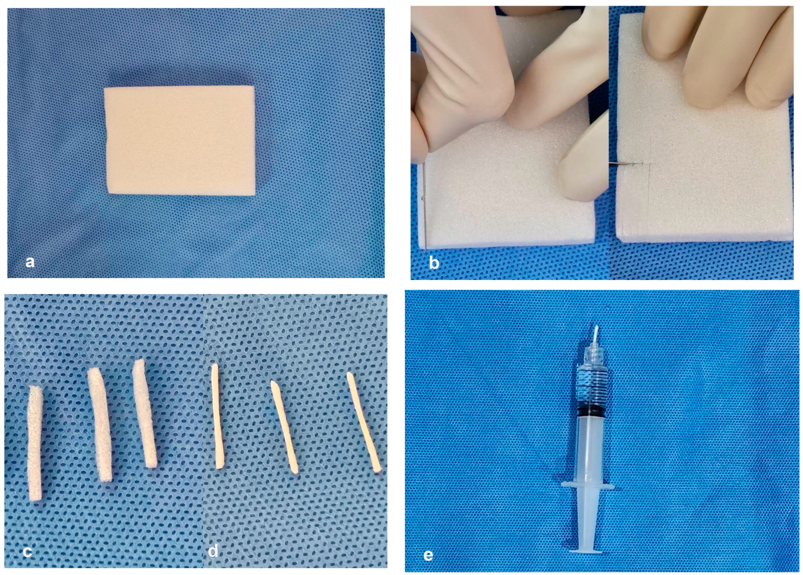
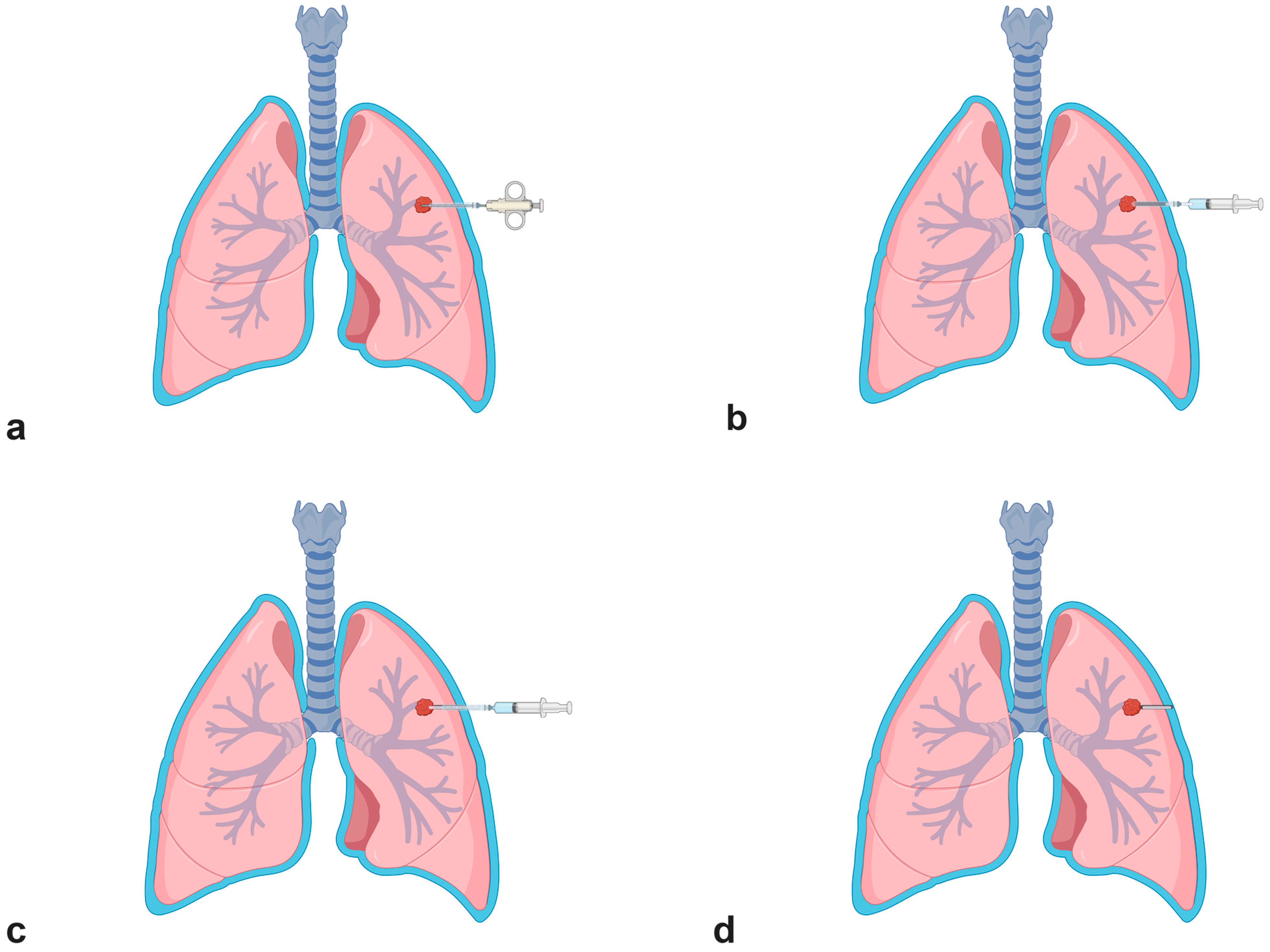
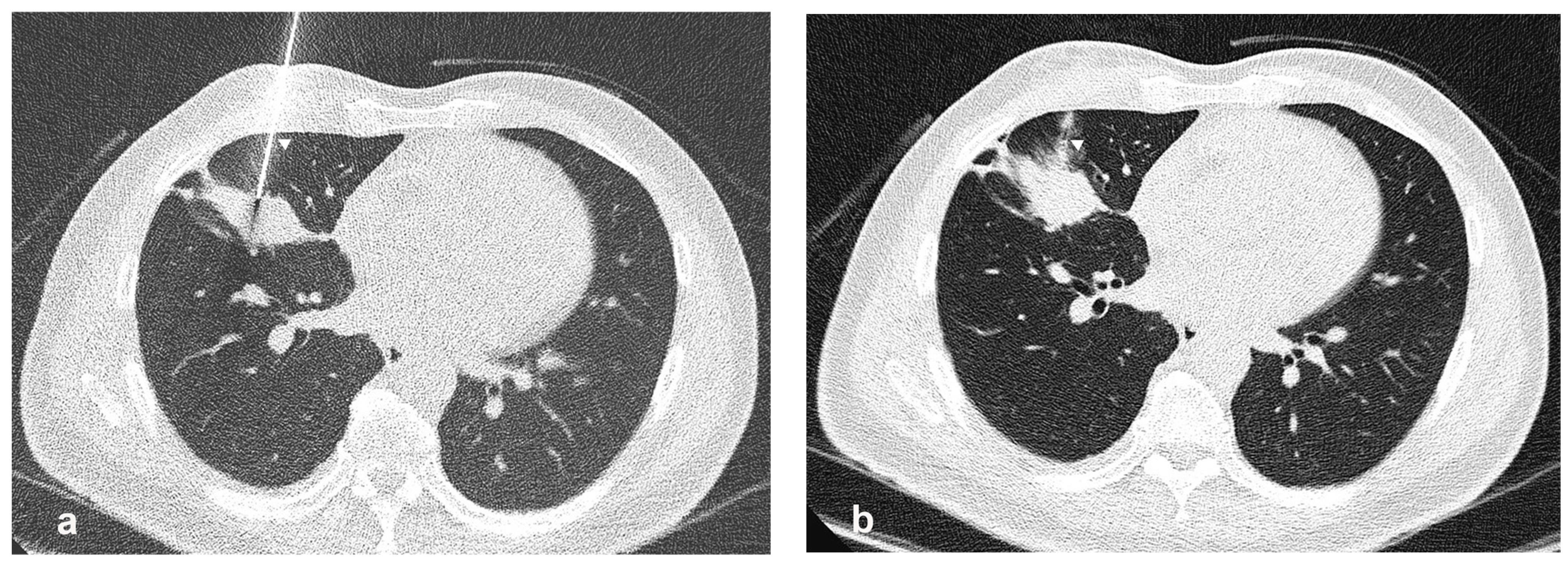
- Procedure technique
- Statistical analysis
3. Results
3.1. Comparison of Pneumothorax
3.2. Chest Tube Insertion Rate
3.3. Variable Factors Affecting Pneumothorax
4. Discussion
5. Conclusions
Author Contributions
Funding
Institutional Review Board Statement
Informed Consent Statement
Data Availability Statement
Conflicts of Interest
References
- Manhire, A.; Charig, M.; Clelland, C.; Gleeson, F.; Miller, R.; Moss, H.; Pointon, K.; Richardson, C.; Sawicka, E. Guidelines for radiologically guided lung biopsy. Thorax 2003, 58, 920–936. [Google Scholar] [CrossRef] [PubMed]
- Li, Y.; Du, Y.; Yang, H.; Yu, J.; Xu, X. CT-guided percutaneous core needle biopsy for small (≤20 mm) pulmonary lesions. Clin. Radiol. 2013, 68, e43–e48. [Google Scholar] [CrossRef] [PubMed]
- Wang, Y.; Jiang, F.; Tan, X.; Tian, P. CT-guided percutaneous transthoracic needle biopsy for paramediastinal and nonparamediastinal lung lesions: Diagnostic yield and complications in 1484 patients. Medicine 2016, 95, e4460. [Google Scholar] [CrossRef] [PubMed]
- Drumm, O.; Joyce, E.A.; de Blacam, C.; Gleeson, T.; Kavanagh, J.; McCarthy, E.; McDermott, R.; Beddy, P. CT-guided lung biopsy: Effect of biopsy-side down position on pneumothorax and chest tube placement. Radiology 2019, 292, 190–196. [Google Scholar] [CrossRef] [PubMed]
- Veltri, A.; Bargellini, I.; Giorgi, L.; Almeida, P.A.M.S.; Akhan, O. CIRSE guidelines on percutaneous needle biopsy (PNB). Cardiovasc. Interv. Radiol. 2017, 40, 1501–1513. [Google Scholar] [CrossRef] [PubMed]
- Zhu, J.; Qu, Y.; Wang, X.; Jiang, C.; Mo, J.; Xi, J.; Wen, Z. Risk factors associated with pulmonary hemorrhage and hemoptysis following percutaneous CT-guided transthoracic lung core needle biopsy: A retrospective study of 1,090 cases. Quant. Imaging Med. Surg. 2020, 10, 1008. [Google Scholar] [CrossRef] [PubMed]
- Heerink, W.J.; de Bock, G.H.; de Jonge, G.J.; Groen, H.J.; Vliegenthart, R.; Oudkerk, M. Complication rates of CT-guided transthoracic lung biopsy: Meta-analysis. Eur. Radiol. 2017, 27, 138–148. [Google Scholar] [CrossRef] [PubMed]
- Huo, Y.R.; Chan, M.V.; Habib, A.-R.; Lui, I.; Ridley, L. Post-biopsy manoeuvres to reduce pneumothorax incidence in CT-guided transthoracic lung biopsies: A systematic review and meta-analysis. Cardiovasc. Interv. Radiol. 2019, 42, 1062–1072. [Google Scholar] [CrossRef] [PubMed]
- Engeler, C.E.; Hunter, D.W.; Castaneda-Zuniga, W.; Tashjian, J.H.; Yedlicka, J.W.; Amplatz, K. Pneumothorax after lung biopsy: Pre-vention with transpleural placement of compressed collagen foam plugs. Radiology 1992, 184, 787–789. [Google Scholar] [CrossRef]
- Huo, Y.R.; Chan, M.V.; Habib, A.-R.; Lui, I.; Ridley, L. Pneumothorax rates in CT-Guided lung biopsies: A comprehensive systematic review and meta-analysis of risk factors. Br. J. Radiol. 2020, 93, 20190866. [Google Scholar] [CrossRef]
- Sheth, R.A.; Baerlocher, M.O.; Connolly, B.L.; Dariushnia, S.R.; Shyn, P.B.; Vatsky, S.; Tam, A.L.; Gupta, S. Society of interventional radiology quality improvement standards on percutaneous needle biopsy in adult and pediatric patients. J. Vasc. Interv. Radiol. 2020, 31, 1840–1848. [Google Scholar] [CrossRef] [PubMed]
- Billich, C.; Muche, R.; Brenner, G.; Schmidt, S.A.; Krüger, S.; Brambs, H.-J.; Pauls, S. CT-guided lung biopsy: Incidence of pneumothorax after instillation of NaCl into the biopsy track. Eur. Radiol. 2008, 18, 1146–1152. [Google Scholar] [CrossRef] [PubMed]
- Li, Y.; Du, Y.; Luo, T.; Yang, H.; Yu, J.; Xu, X.; Zheng, H.; Li, B. Usefulness of normal saline for sealing the needle track after CT-guided lung biopsy. Clin. Radiol. 2015, 70, 1192–1197. [Google Scholar] [CrossRef]
- Bourgeais, G.; Frampas, E.; Liberge, R.; Nicolas, A.; Defrance, C.; Blanc, F.-X.; Coudol, S.; Morla, O. Pneumothorax Incidence with Normal Saline Instillation for Sealing the Needle Track After Computed Tomography-Guided Percutaneous Lung Biopsy. CardioVascular Interv. Radiol. 2024, 47, 604–612. [Google Scholar] [CrossRef] [PubMed]
- Wagner, J.M.; Hinshaw, J.L.; Lubner, M.G.; Robbins, J.B.; Kim, D.H.; Pickhardt, P.J.; Lee, F.T., Jr. CT-guided lung biopsies: Pleural blood patching reduces the rate of chest tube placement for postbiopsy pneumothorax. Am. J. Roentgenol. 2011, 197, 783–788. [Google Scholar] [CrossRef] [PubMed]
- Malone, L.J.; Stanfill, R.M.; Wang, H.; Fahey, K.M.; Bertino, R.E. Effect of intraparenchymal blood patch on rates of pneumothorax and pneumothorax requiring chest tube placement after percutaneous lung biopsy. Am. J. Roentgenol. 2013, 200, 1238–1243. [Google Scholar] [CrossRef]
- Clayton, J.D.; Elicker, B.M.; Ordovas, K.G.; Kohi, M.P.; Nguyen, J.; Naeger, D.M. Nonclotted blood patch technique reduces pneumothorax and chest tube placement rates after percutaneous lung biopsies. J. Thorac. Imaging 2016, 31, 243–246. [Google Scholar] [CrossRef] [PubMed]
- Türk, Y.; Devecioğlu, İ. A retrospective analysis of the effectiveness of extrapleural autologous blood patch injection on pneumothorax and intervention need in CT-guided lung biopsy. CardioVascular Interv. Radiol. 2021, 44, 1223–1230. [Google Scholar] [CrossRef] [PubMed]
- Zaetta, J.M.; Licht, M.O.; Fisher, J.S.; Avelar, R.L.; Group, B.-S.S. A lung biopsy tract plug for reduction of postbiopsy pneumothorax and other complications: Results of a prospective, multicenter, randomized, controlled clinical study. J. Vasc. Interv. Radiol. 2010, 21, 1235–1243.e3. [Google Scholar] [CrossRef]
- Ahrar, J.U.; Gupta, S.; Ensor, J.E.; Mahvash, A.; Sabir, S.H.; Steele, J.R.; McRae, S.E.; Avritscher, R.; Huang, S.Y.; Odisio, B.C. Efficacy of a self-expanding tract sealant device in the reduction of pneumothorax and chest tube placement rates after percutaneous lung biopsy: A matched controlled study using propensity score analysis. Cardiovasc. Interv. Radiol. 2017, 40, 270–276. [Google Scholar] [CrossRef]
- Tran, A.A.; Brown, S.B.; Rosenberg, J.; Hovsepian, D.M. Tract embolization with gelatin sponge slurry for prevention of pneumothorax after percutaneous computed tomography-guided lung biopsy. Cardiovasc. Interv. Radiol. 2014, 37, 1546–1553. [Google Scholar] [CrossRef]
- Baadh, A.S.; Hoffmann, J.C.; Fadl, A.; Danda, D.; Bhat, V.R.; Georgiou, N.; Hon, M. Utilization of the track embolization technique to improve the safety of percutaneous lung biopsy and/or fiducial marker placement. Clin. Imaging 2016, 40, 1023–1028. [Google Scholar] [CrossRef] [PubMed]
- Renier, H.; Gérard, L.; Lamborelle, P.; Cousin, F. Efficacy of the tract embolization technique with gelatin sponge slurry to reduce pneumothorax and chest tube placement after percutaneous CT-guided lung biopsy. Cardiovasc. Interv. Radiol. 2020, 43, 597–603. [Google Scholar] [CrossRef]
- Grange, R.; Di Bisceglie, M.; Habert, P.; Resseguier, N.; Sarkissian, R.; Ferre, M.; Dassa, M.; Grange, S.; Izaaryene, J.; Piana, G. Evaluation of preventive tract embolization with standardized gelatin sponge slurry on chest tube placement rate after CT-guided lung biopsy: A propensity score analysis. Insights Into Imaging 2023, 14, 212. [Google Scholar] [CrossRef] [PubMed]
- Izaaryene, J.; Mancini, J.; Louis, G.; Chaumoitre, K.; Bartoli, J.-M.; Vidal, V.; Gaubert, J.-Y. Embolisation of pulmonary radio frequency pathway–a randomised trial. Int. J. Hyperth. 2017, 33, 814–819. [Google Scholar] [CrossRef] [PubMed]
- Graveleau, P.; Frampas, É.; Perret, C.; Volpi, S.; Blanc, F.-X.; Goronflot, T.; Liberge, R. Chest tube placement incidence when using gelatin sponge torpedoes after pulmonary radiofrequency ablation. Res. Diagn. Interv. Imaging 2024, 10, 100047. [Google Scholar] [CrossRef]
- de Baère, T. Pneumothorax and lung thermal ablation: Is it a complication? Is it only about tract sealing? Cardiovasc. Interv. Radiol. 2021, 44, 911–912. [Google Scholar] [CrossRef]


| Non-Track Embolization Group (n = 667) | Track Embolization Group (n = 217) | p-Value | STD | |
|---|---|---|---|---|
| Age (years) | 64.0 ± 12.7 | 64.9 ± 12.8 | 0.318 | 0.08 |
| Sex (n,%) | 0.236 | 0.09 | ||
| Male | 329 (49) | 97 (45) | ||
| Female | 338 (51) | 120 (55) | ||
| Underlying lung disease (n,%) | 0.844 | 0.08 | ||
| None | 528 (79) | 178 (82) | ||
| Effusion | 53 (8) | 16 (7) | ||
| Emphysematous lung | 42 (6) | 12 (6) | ||
| Reticular fibrosis | 44 (7) | 11 (5) | ||
| Morphology (n,%) | ||||
| Solid | 472 (71) | 172 (79) | 0.019 | 0.16 |
| Subsolid/Subgroundglass | 44 (7) | 6 (3) | ||
| Ground glass opacity | 12 (2) | 8 (4) | ||
| Cavity | 28 (4) | 6 (3) | ||
| Consolidation | 63 (9) | 11 (5) | ||
| Internal air bronchogram | 48 (7) | 14 (6) | ||
| Location (n,%) | 0.525 | 0.07 | ||
| RUL | 180 (27) | 57 (26) | ||
| RML | 40 (6) | 9 (4) | ||
| RLL | 160 (24) | 46 (21) | ||
| LUL | 152 (23) | 60 (28) | ||
| LLL | 132 (20) | 45 (21) | ||
| Maximal diameter (mm) | 32.1 ± 21.0 | 32.9 ± 20.9 | 0.623 | 0.04 |
| Maximal diameter from needle projection (mm) | 27.3 ± 17.8 | 28.3 ± 16.6 | 0.469 | 0.06 |
| Depth from pleura (mm) | 13.8 ± 13.8 | 16.5 ± 13.2 | 0.01 | 0.2 |
| Patient position, (n,%) | 0.892 | 0.06 | ||
| Supine | 239 (36) | 84 (39) | ||
| Prone | 326 (49) | 103 (47) | ||
| Right lateral decubitus | 44 (7) | 13 (6) | ||
| Left lateral decubitus | 58 (9) | 17 (8) | ||
| Coaxial needle (Gauge) | 0 | 0.32 | ||
| 16 | 90 (13) | 7 (3) | ||
| 17 | 574 (86) | 210 (97) | ||
| 19 | 3 (0) | 0 (0) | ||
| Passing fissure | 0.343 | 0.07 | ||
| No | 648 (97) | 208 (96) | ||
| Yes | 19 (3) | 9 (4) | ||
| Number of cutting attempts | 3.3 ± 1.3 | 3.2 ± 1.3 | 0.798 | 0.02 |
| Total cutting length (cm) | 5.7 ± 2.6 | 5.2 ± 2.3 | 0.015 | 0.2 |
| Non-Track Embolization Group (n = 217) | Track Embolization Group (n = 217) | p-Value | STD | |
|---|---|---|---|---|
| Age (years) | 64.4 ± 11.9 | 64.9 ± 12.8 | 0.7 | 0.00 |
| Sex (n,%) | 1.0 | 0.00 | ||
| Male | 97 (45) | 97 (45) | ||
| Female | 120 (55) | 120 (55) | ||
| Underlying lung disease (n,%) | 0.8 | −0.1 | ||
| None | 185 (85) | 178 (82) | ||
| Effusion | 13 (6) | 16 (7) | ||
| Emphysematous lung | 11 (5) | 12 (6) | ||
| Reticular fibrosis | 8 (4) | 11 (5) | ||
| Morphology (n,%) | ||||
| Solid | 169 (78) | 172 (79) | 0.7 | 0.00 |
| Subsolid/Subgroundglass | 8 (4) | 6 (3) | ||
| Ground glass opacity | 4 (2) | 8 (4) | ||
| Cavity | 6 (3) | 6 (3) | ||
| Consolidation | 17 (8) | 11 (5) | ||
| Internal air bronchogram | 13 (6) | 14 (6) | ||
| Location (n,%) | 0.6 | 0.00 | ||
| RUL | 54 (25) | 57 (26) | ||
| RML | 17 (8) | 9 (4) | ||
| RLL | 48 (22) | 46 (21) | ||
| LUL | 54 (25) | 60 (28) | ||
| LLL | 44 (20) | 45 (21) | ||
| Maximal diameter (mm) | 28.9 ± 20.6 | 32.9 ± 20.9 | 0.1 | −0.2 |
| Maximal diameter from needle projection (mm) | 25.3 ± 17.6 | 28.3 ± 16.6 | 0.1 | −0.2 |
| Depth from pleura (mm) | 14.5 ± 14.0 | 16.5 ± 13.2 | 0.1 | −0.1 |
| Patient position, (n,%) | 0.8 | 0.1 | ||
| Supine | 83 (38) | 84 (39) | ||
| Prone | 97 (45) | 103 (47) | ||
| Right lateral decubitus | 17 (8) | 13 (6) | ||
| Left lateral decubitus | 20 (9) | 17 (8) | ||
| Coaxial needle (Gauge) | 0.6 | −0.1 | ||
| 16 | 10 (5) | 7 (3) | ||
| 17 | 207 (95) | 210 (97) | ||
| Passing fissure | 1.0 | 0.0 | ||
| No | 208 (96) | 208 (96) | ||
| Yes | 9 (4) | 9 (4) | ||
| Number of cutting attempts | 3.1 ± 1.3 | 3.2 ± 1.3 | 0.4 | −0.1 |
| Total cutting length (cm) | 4.8 ± 2.3 | 5.2 ± 2.3 | 0.1 | 0.0 |
| Non-Track Embolization Group (n = 217) | Track Embolization Group (n = 217) | p-Value | |
|---|---|---|---|
| Pneumothorax (n,%) | |||
| Early (within 4 h) | 52 (24) | 29 (13) | 0.001 * |
| Late (4 to 24 h) | 40 (18) | 23 (11) | 0.009 * |
| Chest tube placement | 10 (5) | 2 (1) | 0.036 * |
| Univariate Analysis | |||
|---|---|---|---|
| Variables | Odds Ratio | 95%CI | p Value |
| Age (years) | 1.00 | 0.98–1.02 | 0.69 |
| Sex | 1.32–3.59 | 0.89 | |
| Male | 2.18 | ||
| Female | 1.00 | ||
| Underlying lung disease | 0.89 | ||
| None | 1.48 | 0.42–5.23 | |
| Effusion | 1.12 | 0.22–5.78 | |
| Emphysematous lung | 1.42 | 0.27–7.44 | |
| Reticular fibrosis | 1.00 | ||
| Morphology | 0.27 | ||
| Solid | 2.83 | 0.65–12.37 | |
| Subsolid/Subgroundglass | 7.89 | 1.24–49.83 | |
| Ground glass opacity | 3.67 | 0.52–25.77 | |
| Cavity | 3.67 | 0.52–25.77 | |
| Consolidation | 1.00 | ||
| Internal air bronchogram | 4.28 | 0.79–23.19 | |
| Location | 0.40 | ||
| RUL | 1.76 | 0.89–3.51 | |
| RML | 2.01 | 0.72–5.59 | |
| RLL | 1.26 | 0.60–2.65 | |
| LUL | 1.00 | ||
| LLL | 1.07 | 0.49–2.33 | |
| Maximal diameter (mm) | 0.98 | 0.96–0.99 | 0.00 * |
| Maximal diameter from needle projection (mm) | 0.98 | 0.96–1.00 | 0.03 * |
| Depth from pleura (mm) | 1.01 | 1.00–1.03 | 0.12 |
| Patient position | 0.16 | ||
| Supine | 1.71 | 0.99–2.95 | |
| Prone | 1.00 | ||
| Right lateral decubitus | 1.81 | 0.70–4.67 | |
| Left lateral decubitus | 2.02 | 0.85–4.82 | |
| Coaxial needle (Gauge) | 0.91 | ||
| 16 | 1.00 | ||
| 17 | 1.08 | 0.30–3.91 | |
| Passing fissure | 0.06 | ||
| No | 1.00 | ||
| Yes | 2.76 | 1.02–7.50 | |
| Number of cutting attempts | 0.97 | 0.80–1.17 | 0.73 |
| Total cutting length (cm) | 1.00 | 0.89–1.11 | 0.95 |
Disclaimer/Publisher’s Note: The statements, opinions and data contained in all publications are solely those of the individual author(s) and contributor(s) and not of MDPI and/or the editor(s). MDPI and/or the editor(s) disclaim responsibility for any injury to people or property resulting from any ideas, methods, instructions or products referred to in the content. |
© 2024 by the authors. Licensee MDPI, Basel, Switzerland. This article is an open access article distributed under the terms and conditions of the Creative Commons Attribution (CC BY) license (https://creativecommons.org/licenses/by/4.0/).
Share and Cite
Feinggumloon, S.; Radchauppanone, P.; Panpikoon, T.; Buangam, C.; Pichitpichatkul, K.; Treesit, T. Reduced Incidence of Pneumothorax and Chest Tube Placement following Transthoracic CT-Guided Lung Biopsy with Gelatin Sponge Torpedo Track Embolization: A Propensity Score–Matched Study. J. Clin. Med. 2024, 13, 4666. https://doi.org/10.3390/jcm13164666
Feinggumloon S, Radchauppanone P, Panpikoon T, Buangam C, Pichitpichatkul K, Treesit T. Reduced Incidence of Pneumothorax and Chest Tube Placement following Transthoracic CT-Guided Lung Biopsy with Gelatin Sponge Torpedo Track Embolization: A Propensity Score–Matched Study. Journal of Clinical Medicine. 2024; 13(16):4666. https://doi.org/10.3390/jcm13164666
Chicago/Turabian StyleFeinggumloon, Sasikorn, Panupong Radchauppanone, Tanapong Panpikoon, Chinnarat Buangam, Kaewpitcha Pichitpichatkul, and Tharintorn Treesit. 2024. "Reduced Incidence of Pneumothorax and Chest Tube Placement following Transthoracic CT-Guided Lung Biopsy with Gelatin Sponge Torpedo Track Embolization: A Propensity Score–Matched Study" Journal of Clinical Medicine 13, no. 16: 4666. https://doi.org/10.3390/jcm13164666
APA StyleFeinggumloon, S., Radchauppanone, P., Panpikoon, T., Buangam, C., Pichitpichatkul, K., & Treesit, T. (2024). Reduced Incidence of Pneumothorax and Chest Tube Placement following Transthoracic CT-Guided Lung Biopsy with Gelatin Sponge Torpedo Track Embolization: A Propensity Score–Matched Study. Journal of Clinical Medicine, 13(16), 4666. https://doi.org/10.3390/jcm13164666