

Incidence and Causes of Tubal Occlusion in Infertility: A Retrospective Cohort Study

 , ,
, , 
Abstract
1. Introduction
2. Materials and Methods
2.1. Patient Population
2.2. Parameters Analyzed
2.3. Statistical Analysis
3. Results
3.1. Basic Patient Characteristics
3.2. Main Findings about Tubal Patency Testing
3.3. Other Causes for Infertility and Their Association with Tubal Occlusion
4. Discussion
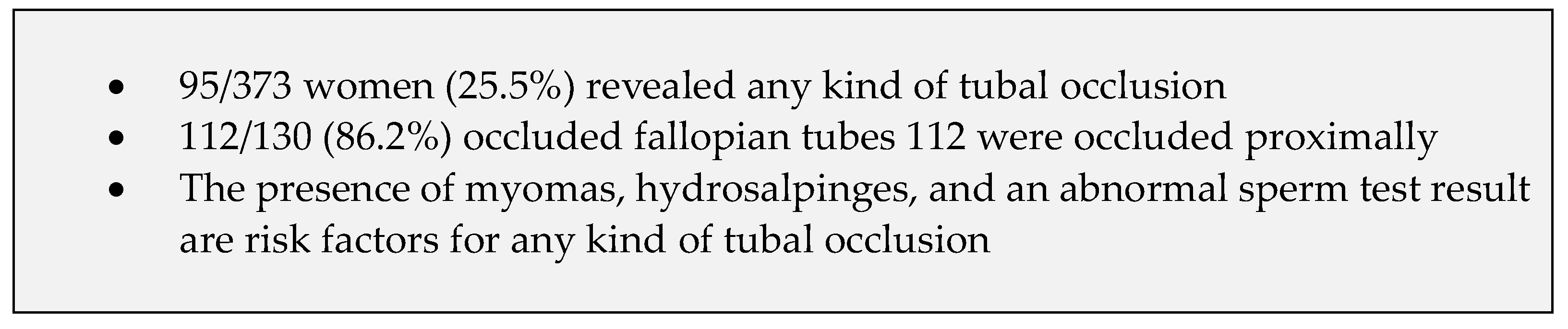
5. Conclusions
Author Contributions
Funding
Institutional Review Board Statement
Informed Consent Statement
Data Availability Statement
Conflicts of Interest
Abbreviations
| ART | assisted reproductive technologies |
| BMI | body mass index |
| FHA | functional hypothalamic amenorrhea |
| FSH | follicle-stimulating hormone |
| HSG | hysterosalpingography |
| HyCoSy | hysterosalpingo-contrast sonography |
| IVF | in vitro fertilization |
| PCOS | polycystic ovarian syndrome |
| POI | premature ovarian insufficiency |
| IUI | intrauterine insemination |
References
- Vander Borght, M.; Wyns, C. Fertility and Infertility: Definition and Epidemiology. Clin. Biochem. 2018, 62, 2–10. [Google Scholar] [CrossRef]
- Penzias, A.; Bendikson, K.; Falcone, T.; Hansen, K.; Hill, M.; Jindal, S.; Mersereau, J.; Racowsky, C.; Rebar, R.; Steiner, A.Z.; et al. Evidence-Based Treatments for Couples with Unexplained Infertility: A Guideline. Fertil. Steril. 2020, 113, 305–322. [Google Scholar] [CrossRef]
- Carson, S.A.; Kallen, A.N. Diagnosis and Management of Infertility: A Review. JAMA 2021, 326, 65–76. [Google Scholar] [CrossRef]
- Infertility Workup for the Women’s Health Specialist: ACOG Committee Opinion, Number 781. Obstet. Gynecol. 2019, 133, e377. [CrossRef]
- Kamphuis, D.; van Eekelen, R.; van Welie, N.; Dreyer, K.; van Rijswijk, J.; van Hooff, M.H.A.; de Bruin, J.P.; Verhoeve, H.R.; Mol, F.; van Baal, W.M.; et al. Hysterosalpingo-Foam Sonography versus Hysterosalpingography during Fertility Work-up: An Economic Evaluation alongside a Randomized Controlled Trial. Hum. Reprod. 2024, 39, 1222–1230. [Google Scholar] [CrossRef]
- Hager, M.; Ott, J.; Göbl, C.; Holzer, I.; Seemann, R.; Kurz, C.; Parry, J.P. Detection of Hysteroscopic Fluid in the Pouch of Douglas: A Prospective Cohort Study about the Predictability of Bilateral Tubal Occlusion. Arch. Gynecol. Obs. 2021, 304, 1073–1080. [Google Scholar] [CrossRef]
- Saunders, R.D.; Shwayder, J.M.; Nakajima, S.T. Current Methods of Tubal Patency Assessment. Fertil. Steril. 2011, 95, 2171–2179. [Google Scholar] [CrossRef]
- Ott, J.; Hager, M.; Nouri, K.; Marschalek, J.; Kurz, C. Assessment of Tubal Patency: A Prospective Comparison of Diagnostic Hysteroscopy and Laparoscopic Chromopertubation. J. Minim. Invasive Gynecol. 2020, 27, 135–140. [Google Scholar] [CrossRef]
- Hager, M.; Ott, J.; Holzer, I.; Seemann, R.; Kurz, C.; Parry, J.P. Hysteroscopic Assessment of Tubal Patency: A Randomized Comparison between the Flow and Parryscope Techniques. J. Minim. Invasive Gynecol. 2020, 27, 1552–1557.e1. [Google Scholar] [CrossRef] [PubMed]
- Alcázar, J.L.; Martinez, A.; Duarte, M.; Welly, A.; Marín, A.; Calle, A.; Garrido, R.; Pascual, M.A.; Guerriero, S. Two-Dimensional Hysterosalpingo-Contrast-Sonography Compared to Three/Four-Dimensional Hysterosalpingo-Contrast-Sonography for the Assessment of Tubal Occlusion in Women with Infertility/Subfertility: A Systematic Review with Meta-Analysis. Hum. Fertil. 2022, 25, 43–55. [Google Scholar] [CrossRef] [PubMed]
- Luciano, D.E.; Exacoustos, C.; Luciano, A.A. Contrast Ultrasonography for Tubal Patency. J. Minim. Invasive Gynecol. 2014, 21, 994–998. [Google Scholar] [CrossRef] [PubMed]
- Goldberg, J.M.; Falcone, T.; Diamond, M.P. Current Controversies in Tubal Disease, Endometriosis, and Pelvic Adhesion. Fertil. Steril. 2019, 112, 417–425. [Google Scholar] [CrossRef]
- Kurz, C.; Ott, J.; Parry, J.P.; Janjic, N.; Hager, M.; Mauer-Gesek, B.; Petrozza, J.C.; Weninger, W.J. Is There a Fallopian Tube Sphincter That Causes Tubal Spasm? An Anatomic Pilot Study in Transmen. Fertil. Steril. 2023, 119, 883–885. [Google Scholar] [CrossRef]
- Ng, K.Y.B.; Cheong, Y. Hydrosalpinx—Salpingostomy, Salpingectomy or Tubal Occlusion. Best Pract. Res. Clin. Obstet. Gynaecol. 2019, 59, 41–47. [Google Scholar] [CrossRef]
- Holzer, I.; Ott, J.; Kurz, C.; Hofstetter, G.; Hager, M.; Kuessel, L.; Parry, J.P. Is Chronic Endometritis Associated with Tubal Infertility? A Prospective Cohort Study. J. Minim. Invasive Gynecol. 2021, 28, 1876–1881. [Google Scholar] [CrossRef] [PubMed]
- Ghobrial, S.; Parry, J.P.; Holzer, I.; Aschauer, J.; Selzer, C.; Brezina, A.; Helmy-Bader, S.; Ott, J. The Prevalence of Fallopian Tube Occlusion in Women with Polycystic Ovary Syndrome Seems Similar to Non-Subfertile Women: A Retrospective Cohort Study. J. Clin. Med. 2022, 11, 5610. [Google Scholar] [CrossRef] [PubMed]
- Lo Monte, G.; Capobianco, G.; Piva, I.; Caserta, D.; Dessole, S.; Marci, R. Hysterosalpingo Contrast Sonography (HyCoSy): Let’s Make the Point! Arch. Gynecol. Obs. 2015, 291, 19–30. [Google Scholar] [CrossRef] [PubMed]
- Baramki, T.A. Hysterosalpingography. Fertil. Steril. 2005, 83, 1595–1606. [Google Scholar] [CrossRef]
- Hager, M.; Wenzl, R.; Riesenhuber, S.; Marschalek, J.; Kuessel, L.; Mayrhofer, D.; Ristl, R.; Kurz, C.; Ott, J. The Prevalence of Incidental Endometriosis in Women Undergoing Laparoscopic Ovarian Drilling for Clomiphene-Resistant Polycystic Ovary Syndrome: A Retrospective Cohort Study and Meta-Analysis. J. Clin. Med. 2019, 8, 1210. [Google Scholar] [CrossRef]
- The Rotterdam ESHRE/ASRM-sponsored PCOS consensus workshop group Revised 2003 Consensus on Diagnostic Criteria and Long-term Health Risks Related to Polycystic Ovary Syndrome (PCOS). Hum. Reprod. 2004, 19, 41–47. [CrossRef]
- Beitl, K.; Dewailly, D.; Seemann, R.; Hager, M.; Bünker, J.; Mayrhofer, D.; Holzer, I.; Ott, J. Polycystic Ovary Syndrome Phenotype D Versus Functional Hypothalamic Amenorrhea With Polycystic Ovarian Morphology: A Retrospective Study About a Frequent Differential Diagnosis. Front. Endocrinol. 2022, 13, 904706. [Google Scholar] [CrossRef] [PubMed]
- The ESHRE Guideline Group on POI; Webber, L.; Davies, M.; Anderson, R.; Bartlett, J.; Braat, D.; Cartwright, B.; Cifkova, R.; de Muinck Keizer-Schrama, S.; Hogervorst, E.; et al. ESHRE Guideline: Management of Women with Premature Ovarian Insufficiency. Hum. Reprod. 2016, 31, 926–937. [Google Scholar] [CrossRef]
- Boitrelle, F.; Shah, R.; Saleh, R.; Henkel, R.; Kandil, H.; Chung, E.; Vogiatzi, P.; Zini, A.; Arafa, M.; Agarwal, A. The Sixth Edition of the WHO Manual for Human Semen Analysis: A Critical Review and SWOT Analysis. Life 2021, 11, 1368. [Google Scholar] [CrossRef]
- Ledger, W.L. Demographics of Infertility. Reprod. BioMedicine Online 2009, 18, S11–S14. [Google Scholar] [CrossRef]
- Mascarenhas, M.N.; Flaxman, S.R.; Boerma, T.; Vanderpoel, S.; Stevens, G.A. National, Regional, and Global Trends in Infertility Prevalence Since 1990: A Systematic Analysis of 277 Health Surveys. PLoS Med. 2012, 9, e1001356. [Google Scholar] [CrossRef]
- Hu, H.; Kirby, A.; Dowthwaite, S.; Mizia, K.; Zen, M. Lipiodol Flushing under Ultrasound Guidance at Time of Hystero-Salpingo Contrast Sonography (HyCoSy): A Retrospective Observational Study. Aust. N. Z. J. Obstet. Gynaecol. 2022, 62, 755–760. [Google Scholar] [CrossRef] [PubMed]
- Gunn, D.D.; Bates, G.W. Evidence-Based Approach to Unexplained Infertility: A Systematic Review. Fertil. Steril. 2016, 105, 1566–1574.e1. [Google Scholar] [CrossRef] [PubMed]
- Hull, M.G.; Glazener, C.M.; Kelly, N.J.; Conway, D.I.; Foster, P.A.; Hinton, R.A.; Coulson, C.; Lambert, P.A.; Watt, E.M.; Desai, K.M. Population Study of Causes, Treatment, and Outcome of Infertility. Br. Med. J. (Clin. Res. Ed.) 1985, 291, 1693–1697. [Google Scholar] [CrossRef]
- Tan, J.; Tannus, S.; Taskin, O.; Kan, A.; Albert, A.; Bedaiwy, M. The Effect of Unilateral Tubal Block Diagnosed by Hysterosalpingogram on Clinical Pregnancy Rate in Intrauterine Insemination Cycles: Systematic Review and Meta-Analysis. BJOG Int. J. Obstet. Gynaecol. 2019, 126, 227–235. [Google Scholar] [CrossRef]
- Honoré, G.M.; Holden, A.E.C.; Schenken, R.S. Pathophysiology and Management of Proximal Tubal Blockage. Fertil. Steril. 1999, 71, 785–795. [Google Scholar] [CrossRef]
- Papaioannou, S. A Hypothesis for the Pathogenesis and Natural History of Proximal Tubal Blockage. Hum. Reprod. 2004, 19, 481–485. [Google Scholar] [CrossRef] [PubMed]
- Hillier, S.L.; Bernstein, K.T.; Aral, S. A Review of the Challenges and Complexities in the Diagnosis, Etiology, Epidemiology, and Pathogenesis of Pelvic Inflammatory Disease. J. Infect. Dis. 2021, 224, S23–S28. [Google Scholar] [CrossRef] [PubMed]
- Mayrhofer, D.; Parry, J.P.; Hager, M.; Beitl, K.; Kurz, C.; Ott, J. Are the Stage and the Incidental Finding of Endometriosis Associated with Fallopian Tube Occlusion? A Retrospective Cohort Study on Laparoscopic Chromopertubation in Infertile Women. J. Clin. Med. 2022, 11, 3750. [Google Scholar] [CrossRef] [PubMed]


| Number of women | 373 | |
| Age (years) * | 33 (29; 36) | |
| BMI (kg/m2) * | 23.9 (21.1; 28.0) | |
| Number of tubes | 735 | |
| Women with one tube # | 11 (2.9) | |
| Secondary infertility # | 167 (44.8) | |
| Gravidity # | 0 | 208 (55.8) |
| 1 | 94 (25.2) | |
| ≥2 | 71 (19.0) | |
| Parity # | 0 | 270 (72.4) |
| 1 | 75 (20.1) | |
| ≥2 | 28 (7.5) | |
| Previous first-trimester miscarriage # | 66 (17.7) | |
| Previous extrauterine pregnancy # | 18 (4.8) | |
| Previous termination of pregnancy # | 25 (6.7) | |
| Previous intrauterine fetal death # | 3 (0.8) | |
| Current smoking # | 45 (12.1) | |
| Previous abdominal surgery # | Cesarean section | 41 (11.0) |
| Curettage | 49 (13.1) | |
| Laparoscopy: unilateral salpingectomy | 10 (2.7) | |
| Laparoscopy: ovarian cyst removal | 25 (6.7) | |
| Laparoscopy: endometriosis | 14 (3.8%) | |
| Laparoscopy or laparotomy: myoma enucleation | 9 (2.4) | |
| Laparoscopy: ovarian drilling | 9 (2.4) | |
| Diagnostic laparoscopy | 36 (9.7) | |
| Appendectomy | 33 (8.8) | |
| Other intraperitoneal procedures | 14 (3.8) | |
| Previous hysteroscopic myoma resection | 4 (1.1) | |
| Number of Evaluated Women | Unilateral Occlusion | Bilateral Occlusion | p | Number of Evaluated Tubes | Number of Occluded Tubes | p | |
|---|---|---|---|---|---|---|---|
| Hysterosalpingography | 186 | 23 (12.4) | 12 (6.5) | 0.019 | 368 | 47 (12.8) | <0.001 |
| HyCoSy | 56 | 14 (25.0) | 8 (14.3) | 109 | 30 (27.5) | ||
| Chromopertubation | 131 | 23 (17.6) | 15 (11.5) | 258 | 53 (20.5) |
| Number of Women (n, %) | Unilateral Occlusion | Bilateral Occlusion | Total Number of Tubes | Occluded Tubes | |
|---|---|---|---|---|---|
| Previously known or newly diagnosed endometriosis * | 54 (14.5) | 9 (16.7) | 6 (11.1) | 108 | 21 (19.4) § |
| PCOS * | 72 (19.3) | 9 (12.5) | 3 (4.2) | 143 | 15 (10.5) § |
| Hypogonadotropic hypogonadism * | 10 (2.7) | 1 (10.0) | 0 | 20 | 1 (5.0) § |
| Premature ovarian insufficiency * | 5 (1.3) | 1 (20.0) | 0 | 10 | 1 (10.0) § |
| Myoma * | 58 (15.5) | 14 (24.1) | 9 (15.5) | 113 | 32 (28.3) § |
| Endometrial polyp * | 25 (6.7) | 3 (12.0) | 3 (12.0) | 49 | 9 (18.4) § |
| Male factor * | 168 (45.0) | 38 (22.6) | 20 (11.9) | 335 | 78 (23.3) § |
| One tube missing * | 11 (2.9) | 4 (36.4) | - | 11 | 4 (36.4) § |
| Otherwise unexplained infertility # | 68 (18.2) | 1 (1.5) | 2 (2.9) | 136 | 5 (3.7) § |
| Women with Uni- or Bilateral Occlusion (n = 95) | Women with Bilateral Patency (n = 278) | OR (95%CI) | p | Adj. OR (95%CI) | Adj. p | ||
|---|---|---|---|---|---|---|---|
| Age (years) * | 34 (30; 38) | 32 (29; 36) | 1.059 (1.009; 1.112) | 0.020 | 1.055 (1.000; 1.113) | 0.051 | |
| BMI (kg/m2) * | 24.5 (21.3; 28.4) | 23.4 (21.0; 27.9) | 1.019 (0.976; 1.064) | 0.392 | - | - | |
| Smoking # | 13 (13.7) | 32 (11.5) | 1.219 (0.610; 2.433) | 0.575 | - | - | |
| Secondary infertility # | 47 (49.5) | 114 (41.0) | 1.388 (0.869; 2.215) | 0.170 | - | - | |
| Parity | 0 | 69 (72.6) | 201 (72.3) | reference | 0.892 | - | - |
| 1 | 18 (18.9) | 57 (20.5) | 0.920 (0.507; 1.670) | - | |||
| ≥2 | 8 (8.4) | 20 (7.2) | 1.165 (0.491; 2.766) | - | |||
| Previous extrauterine pregnancy without salpingectomy # | 4 (4.2) | 6 (2.2) | 2.519 (0.751; 8.450) | 0.135 | - | - | |
| Previous salpingectomy # | 5 (5.3) | 6 (2.2) | 1.993 (0.550; 7.219) | 0.294 | - | - | |
| Previous Cesarean section # | 13 (13.7) | 28 (10.1) | 1.416 (0.700; 2.860) | 0.333 | - | - | |
| Previous other intraperitoneal surgery # | 36 (37.9) | 77 (27.7) | 1.593 (0.975; 2.602) | 0.063 | - | - | |
| Previously known or newly diagnosed endometriosis # | 15 (15.8) | 39 (14.0) | 1.149 (0.602; 2.195) | 0.674 | - | - | |
| PCOS # | 12 (12.6) | 60 (21.6) | 0.525 (0.269; 1.026) | 0.059 | - | - | |
| Hypogonadotropic hypogonadism # | 1 (1.1) | 9 (3.2) | 0.318 (0.040; 2.543) | 0.280 | - | - | |
| Premature ovarian insufficiency # | 1 (1.1) | 4 (1.4) | 0.729 (0.080; 6.602) | 0.778 | - | - | |
| Myoma # | 23 (24.2) | 35 (12.6) | 2.218 (1.232; 3.994) | 0.008 | 2.108 (1.008; 4.409) | 0.048 | |
| Endometrial polyp # | 6 (6.3) | 19 (6.8) | 0.919 (0.356; 2-374) | 0.861 | - | - | |
| Male factor # | 58 (61.1) | 110 (39.6) | 2.394 (1.485; 3.859) | <0.001 | 2.105 (1.156; 3.833) | 0.015 | |
| Otherwise unexplained infertility # | 3 (3.2) | 65 (23.4) | 0.107 (0.033; 0.349) | <0.001 | 0.204 (0.057; 0.733) | 0.015 | |
| Presence of any hydrosalpinx # | 9 (9.5) | 2 (0.7) | 14.442 (3.062; 68.122) | <0.001 | 13.323 (2.679; 66.253) | 0.002 | |
Disclaimer/Publisher’s Note: The statements, opinions and data contained in all publications are solely those of the individual author(s) and contributor(s) and not of MDPI and/or the editor(s). MDPI and/or the editor(s) disclaim responsibility for any injury to people or property resulting from any ideas, methods, instructions or products referred to in the content. |
© 2024 by the authors. Licensee MDPI, Basel, Switzerland. This article is an open access article distributed under the terms and conditions of the Creative Commons Attribution (CC BY) license (https://creativecommons.org/licenses/by/4.0/).
Share and Cite
Mayrhofer, D.; Holzer, I.; Aschauer, J.; Selzer, C.; Parry, J.P.; Ott, J. Incidence and Causes of Tubal Occlusion in Infertility: A Retrospective Cohort Study. J. Clin. Med. 2024, 13, 3961. https://doi.org/10.3390/jcm13133961
Mayrhofer D, Holzer I, Aschauer J, Selzer C, Parry JP, Ott J. Incidence and Causes of Tubal Occlusion in Infertility: A Retrospective Cohort Study. Journal of Clinical Medicine. 2024; 13(13):3961. https://doi.org/10.3390/jcm13133961
Chicago/Turabian StyleMayrhofer, Daniel, Iris Holzer, Judith Aschauer, Clara Selzer, John Preston Parry, and Johannes Ott. 2024. "Incidence and Causes of Tubal Occlusion in Infertility: A Retrospective Cohort Study" Journal of Clinical Medicine 13, no. 13: 3961. https://doi.org/10.3390/jcm13133961
APA StyleMayrhofer, D., Holzer, I., Aschauer, J., Selzer, C., Parry, J. P., & Ott, J. (2024). Incidence and Causes of Tubal Occlusion in Infertility: A Retrospective Cohort Study. Journal of Clinical Medicine, 13(13), 3961. https://doi.org/10.3390/jcm13133961