Article

Amiodarone Administration during Cardiopulmonary Resuscitation Is Not Associated with Changes in Short-Term Mortality or Neurological Outcomes in Cardiac Arrest Patients with Shockable Rhythms

1
Department of Cardiology, Pulmonology and Angiology, Medical Faculty, Heinrich Heine University of Düsseldorf, 40225 Düsseldorf, Germany
2
Cardiovascular Research Institute Düsseldorf (CARID), 40225 Düsseldorf, Germany
3
Department of Cardiology and Electrophysiology, St. Agnes-Hospital Bocholt, 46397 Bocholt, Germany
*
Authors to whom correspondence should be addressed.
These authors contributed equally to this work.
J. Clin. Med. 2024, 13(13), 3931; https://doi.org/10.3390/jcm13133931
Submission received: 17 June 2024 / Revised: 2 July 2024 / Accepted: 3 July 2024 / Published: 4 July 2024
(This article belongs to the Special Issue Cardiac Arrest in Intensive Care: Management and Prognosis)

Abstract

:
Background: The search for the best therapeutic approach in cardiopulmonary resuscitations (CPR) remains open to question. In this study, we evaluated if Amiodarone administration during CPR was associated with short-term mortality or neurological development. Methods: A total of 232 patients with sudden cardiac arrest (CA) with shockable rhythms were included in our analysis. Propensity score matching based on age, gender, type of CA, and CPR duration was used to stratify between patients with and without Amiodarone during CPR. Primary endpoints were short-term mortality (30-day) and neurological outcomes assessed by the cerebral performance category. Secondary endpoints were plasma lactate, phosphate levels at hospital admission, and the peak Neuron-specific enolase. Results: Propensity score matching was successful with a caliper size used for matching of 0.089 and a sample size of n = 82 per group. The 30-day mortality rates were similar between both groups (p = 0.24). There were no significant differences in lactate levels at hospital admission and during the following five days between the groups. Patients receiving Amiodarone showed slightly higher phosphate levels at hospital admission, while the levels decreased to a similar value during the following days. Among CA survivors to hospital discharge, no differences between the proportion of good neurological outcomes were detected between the two groups (p = 0.58), despite slightly higher peak neuron-specific enolase levels in CA patients receiving Amiodarone (p = 0.03). Conclusions: Amiodarone administration is not associated with short-term mortality or neurological outcomes in CA patients with shockable rhythms receiving CPR.

1. Introduction

Sudden cardiac arrest (CA) is a leading cause of death worldwide [1], accounting for up to 20% of deaths in Western societies [2]. Despite significant improvements in cardiopulmonary resuscitation (CPR) algorithms and post-resuscitation care, mortality post-CA remains high, with mortality rates as high as 90% for out-of-hospital cardiac arrest (OHCA) [3] and 75% for in-hospital cardiac arrest (IHCA) patients [4]. In surviving patients, only a minority display good neurological outcomes [4]. Several individual traits during CPR, such as time to ROSC, initial rhythm, cause of CA, or bystander CPR, increase the chance of survival and, in survivors, the good neurological outcomes [5,6,7]. Interestingly, most of the characteristics that influence the outcomes seem to be those of the initial presentation of the CA, reinforcing the idea that the preclinical care of the patients is, in fact, the most essential part of the treatment [8]. Therefore, treatment algorithms rely heavily on initially rapidly available and cost-effective markers. Lactate and phosphate are feasible laboratory markers used at bedside to assess disease severity and, in the absence of more precise prognostication markers, to forecast survival chances [9,10,11,12]. In the current era, the use of digitalization, data extraction, artificial intelligence, and machine learning algorithms has already provided accurate risk score models such as the SCARS model [13,14,15,16]. Very interestingly, most of these models incorporate traits from initial presentation and prehospital intervention (i.e., accounting for >55% of the relative importance of the SCARS model [13]), further validating the importance of structured preclinical care.
Unlike factors that cannot be directly influenced, CPR treatment algorithms have been subjected to numerous changes following hundreds of studies searching for the best therapeutic approach. In shockable rhythms, i.e., ventricular fibrillation or tachycardia (VF/VT), the European Resuscitation Council Guidelines recommend using Adrenaline and Amiodarone after the third unsuccessful electrical shock [17]. In the PARAMEDIC2 trial, Adrenaline improved 30 days survival but not the neurological outcome [18]. Amiodarone blocks the potassium currents and modulates sympathetic activity due to interactions with α and β receptors supporting VF/VT termination [19]. In CPR, Amiodarone influences the response to defibrillation when administered for VF or hemodynamically unstable VT [20,21,22].
In older randomized controlled trials (RCT), Amiodarone improved survival chances to hospital admission, proving superior to Lidocaine in terms of survival to hospital admission [23,24]. In newer RCT with OHCA patients suffering from VT or pulseless ventricular tachycardia, neither Amiodarone nor Lidocaine improved survival or neurological outcomes [25,26,27]. Based on these and other trials, Amiodarone administration remains an IIb recommendation and, therefore, an individual physician choice [28]. While these studies displayed the effects of solitary Amiodarone administration, the current guidelines recommend its use along with Adrenaline. Some recent trials have reported a benefit from early Amiodarone administrations in OHCA patients with shockable rhythms [29,30]. However, the full extent of Amiodarone administration supplementary to Adrenaline during CPR on mortality and neurological outcomes, as occurring in a real-world setting, remains unknown. Therefore, the current study aimed to examine whether Amiodarone administered along with Adrenaline in shockable heart rhythms was associated with lower in-hospital mortality and improved neurological outcomes.

2. Methods

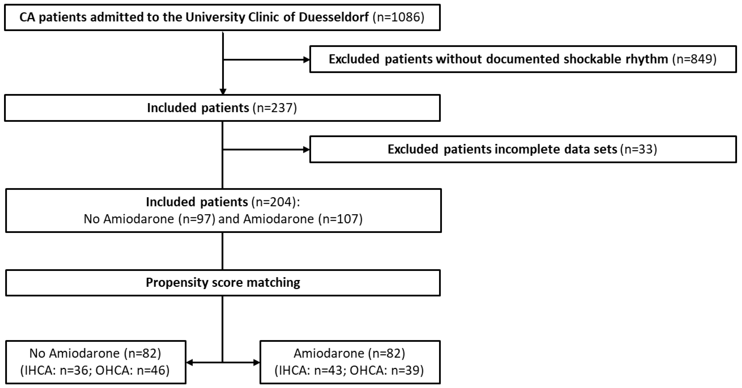
2.1. Study Setting and Population

This study retrospectively analyzed IHCA and OHCA patients who received CPR and were ultimately admitted to the internal medicine Intensive Care Units of the University Hospital of Duesseldorf (UKD) between 2013 and 2017. The database had been previously presented extensively by us [11]. The analyzed data covered the report of the emergency physician and all data documented at the hospital, including laboratory parameters, medical imaging, and documentation until discharge from the hospital. Pre-existing illnesses were segmented into cardiac (heart failure, cardiomyopathies, coronary heart disease, valvular diseases) and pulmonic diseases (chronic obstructive pulmonic disease, bronchial asthma, lung cancer, pulmonic hypertension).
Initially, all patients were treated in accordance with the guidelines applicable at the time for IHCA and OHCA [31,32]. All patients received Adrenaline and one or more defibrillation attempts by electrical shock. Following ROSC, the patients were admitted to the non-surgical intensive care unit of the University Hospital of Duesseldorf, where target temperature management with a target of 34 °C was established for 24 h, followed by 72 h of normal body temperature.
Group stratification was according to Amiodarone administration. Exclusion criteria were non-shockable rhythms, patients with incomplete data sets, or patients younger than eighteen years. The primary endpoints of this study were the 30-day mortality and neurological outcomes among survivors until hospital discharge. We used the cerebral performance category (CPC) to evaluate the neurological outcome. The CPC is a five-point scale used to categorize the neurological outcome after cardiac arrest, ranging from good cerebral performance (1) to brain death (5) [33]. It is dichotomized, with 1–2 as good and 3–4 as bad neurological outcomes [34]. Secondary endpoints were the plasma lactate and phosphate levels at hospital admission, the peak neuron-specific enolase (NSE), and cranial imaging results, which were assessed by computed tomography or cranial magnetic resonance imaging.
This study was conducted according to the guidelines of the Declaration of Helsinki and approved by the Medical Faculty of the Heinrich-Heine-University Ethics Committee in Duesseldorf, Germany, with the reference number 2018-109-RetroDEuA. Due to the retrospective nature of our study, informed consent was waived.

2.2. Recording of Data

Data were extracted from the hospital information systems Medico (Cerner GmbH, Berlin, Germany) and PEGASOS (Nexus/Marabu GmbH, Berlin, Germany). For data acquisition, we analyzed all existing physician letters, clinical investigations, the patients’ documentation curves, and laboratory values. Data on the patients’ course before hospitalization were obtained from the ambulance call report, which included documentation of the initial and all subsequent cardiac rhythms during treatment, the drugs administered, and the duration of resuscitation.

2.3. Propensity Score Matching

Propensity score matching (PPM) was performed using a logistic regression model. The treatment variable was the administration of Amiodarone during CPR, which acted as the dependent variable. Covariates were age, gender, type of cardiac arrest, and duration of CPR, which acted as independent variables. Propensity scores were calculated using logistic regression. Patients were matched using the nearest neighbor matching algorithm. The maximum caliper size was set at 0.5 and iteratively adjusted to ensure both treated and control groups had at least 75 patients each. The final caliper size used was approximately 0.089. All analyses were performed in R-Studio with ‘MatchIt’, ‘tidyverse’, ‘openxlsx’, and ‘cobalt’ packages.

2.4. Statistics

Continuous data are presented as mean ± standard deviation (SD), and categorical data as absolute numbers and percentages. Data were compared with the Student’s t-test for normally distributed continuous variables and the Mann–Whitney test for non-normally distributed continuous variables. The Chi-square test was used for categorical data. Differences in survival of the groups were assessed by Kaplan–Meier analysis with Log-rank (Mantel–Cox) test. p-values < 0.05 were considered as statistically significant. Statistical analyses were performed using Prism 9.3.0 (GraphPad Software, San Diego, CA, USA). Propensity score matching was performed in R-Studio (R version 4.1.2 (2021-11-01), R Foundation for Statistical Computing, Vienna, Austria).

3. Results

3.1. Study Patients

Between 2013 and 2017, 1086 patients with cardiac arrest were admitted to the Intensive Care Units of the University Hospital of Duesseldorf. Of these, 237 patients presented a shockable initial heart rhythm. To achieve robust PPM, all patients without documented characteristics used for matching (i.e., age, gender, type of CA, CPR duration) were excluded from analysis (n = 33). The remaining 204 patients were used for PPM (Figure 1).
PPM was successful with the final caliper size used for matching of 0.089, resulting in two groups of n = 82 being created. The overlap coefficient was 0. The standardized mean differences (Table 1) highlighted balanced comparison groups with substantial improvements after matching. In addition to the variables used for matching, there were no significant differences in CPR cause, it being primarily cardiac in both groups (p = 0.86). Notably, the returns to spontaneous circulation rates were nearly equal (84% in patients without Amiodarone versus 83% in patients receiving Amiodarone, p = 0.83). The complete baseline comparison between the groups is shown in Table 2.

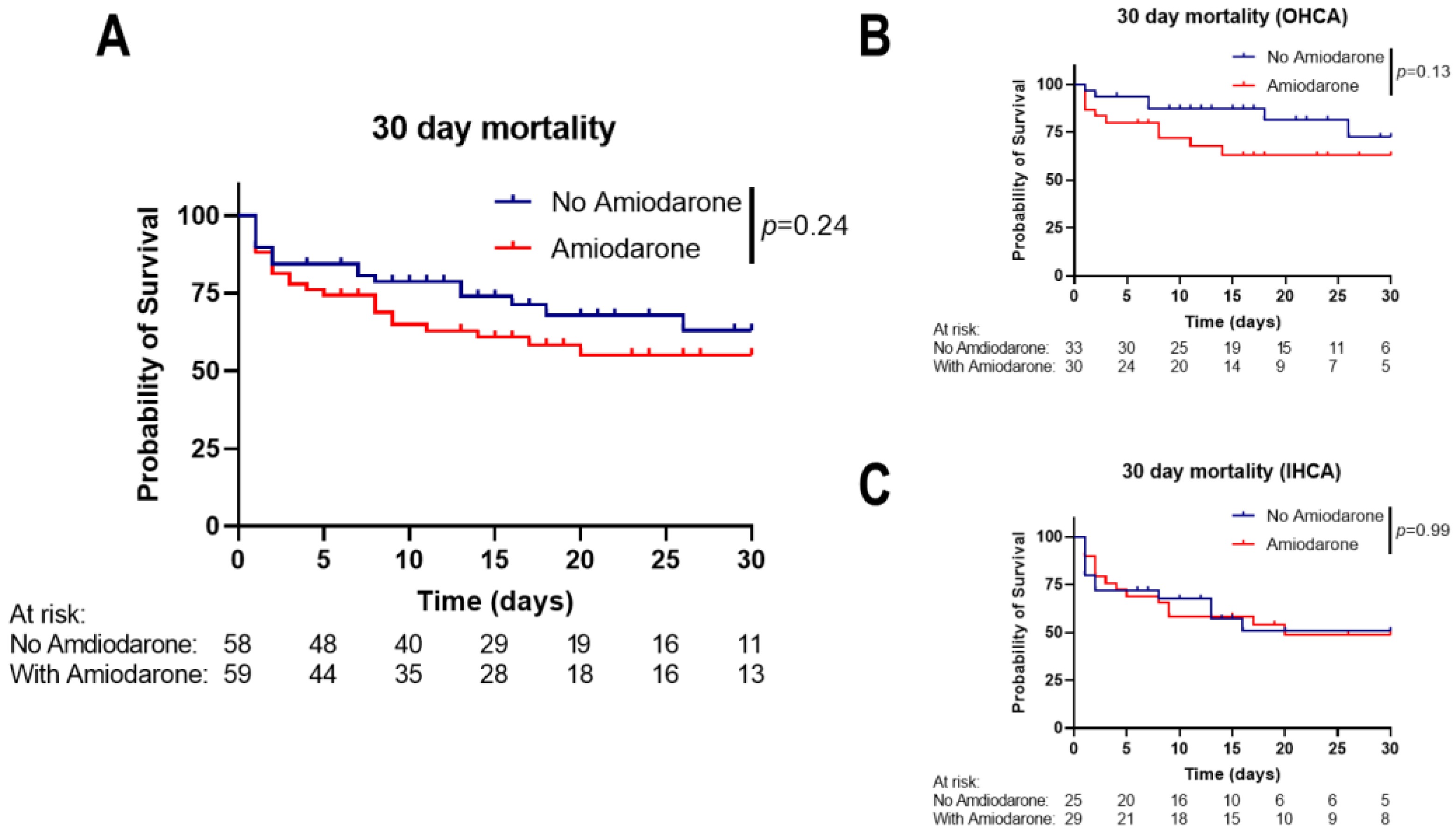
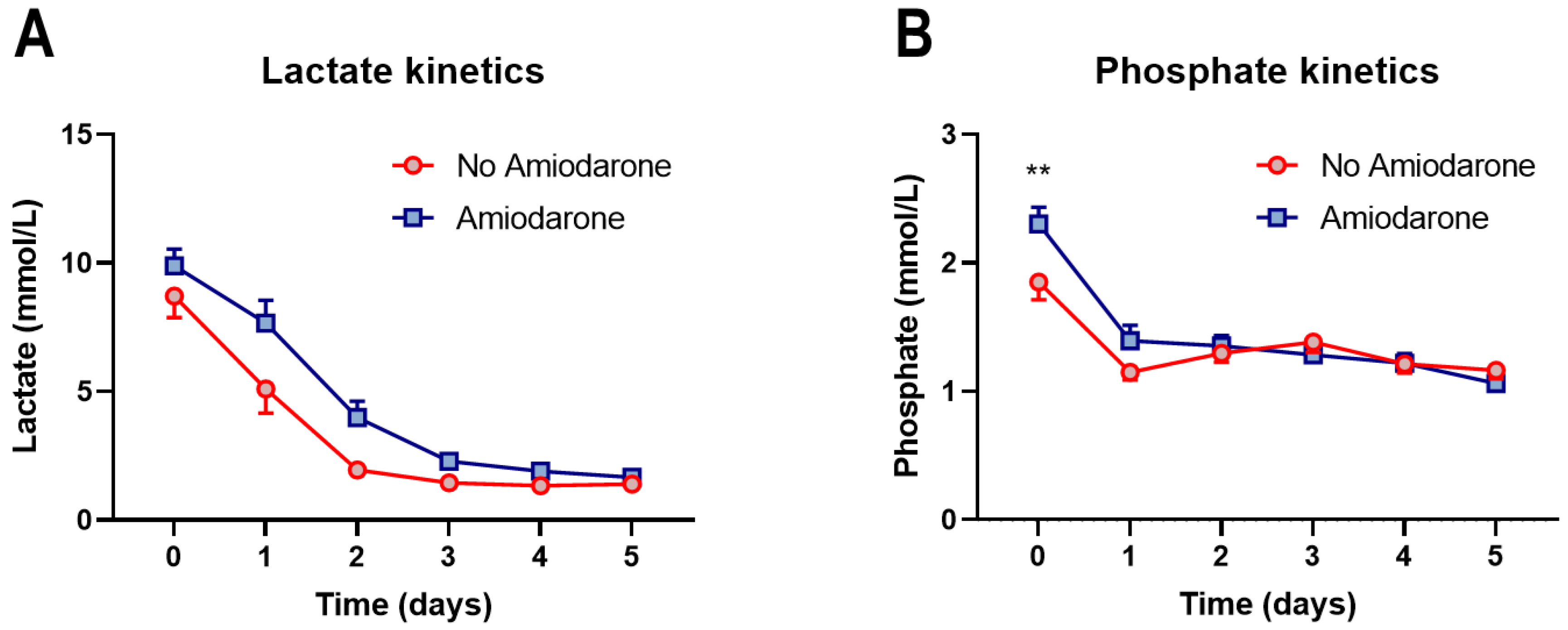
3.2. Amiodarone Administration during CPR Is Not Associated with Short-Term Mortality

Comparison of the 30-day mortality between the matched groups showed no significant difference (p = 0.24, hazard ratio (Mantel–Haenszel) for administering Amiodarone: 1.46, 95% CI: 0.78–2.74; Figure 2A). A similar 30-day mortality for administering Amiodarone was seen in the subgroups of OHCA and IHCA patients (Figure 2B,C). Lactate levels, which are well-established prognostic markers in CA patients and after CPR [35], were similar from hospital admission and during the following five days of hospitalization (p-value for interaction: 0.14, Figure 3A). We recently showed that serum phosphate levels can be used as prognostic markers [11,36]. We saw a slight phosphate level elevation at hospital admission in patients receiving Amiodarone (1.85 ± 0.96 mmol/L vs. 2.3 ± 0.99 mmol/L, p = 0.0038), while the levels were similar during the following days between the groups (Figure 3B). In a Cox regression analysis adjusted for initial lactate and phosphate levels, administering Amiodarone was not significantly associated with survival to hospital discharge (Hazard Ratio: 1.23, 95% CI: 0.67–2.32, p = 0.52).

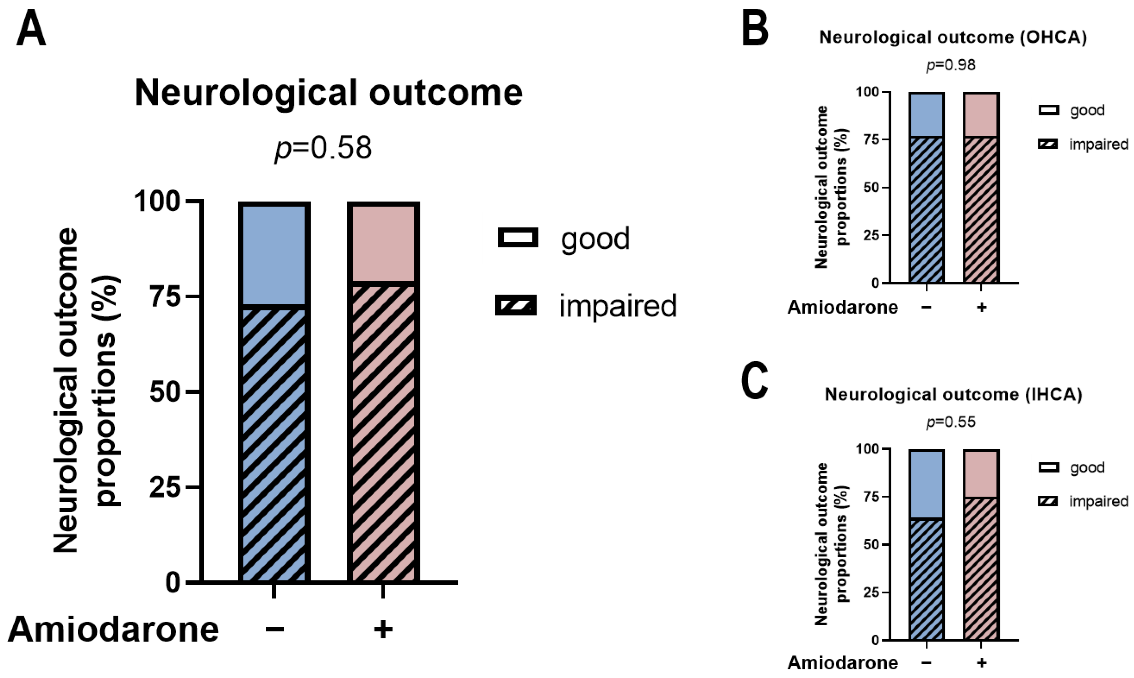
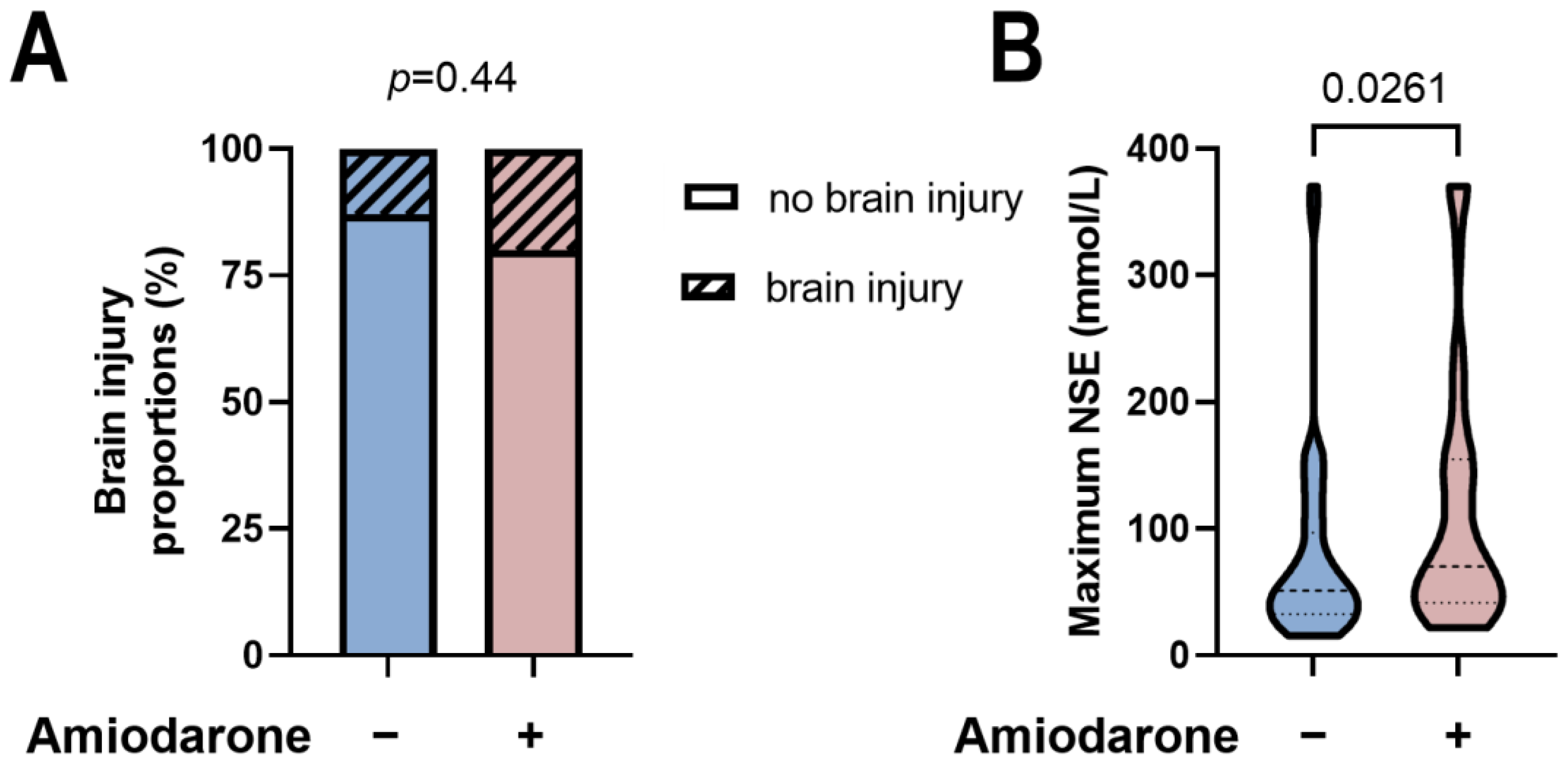
3.3. Amiodarone Administration during CPR Is Not Associated with Neurological Development

In survivors to hospital discharge, no difference was detected between initial Amiodarone administration during CPR and later neurological development. There was a proportion of ~20–25% of patients with good neurological development, highlighted by a CPC score of 1–2 in each group, whereas the vast majority suffered impairment of neurological function, characterized by CPC scores of 3–4 (Figure 4A, p = 0.58). Subgroup stratification according to the type of CA showed similar results for treatment with or without Amiodarone during the initial CPR (Figure 4B,C). In line, in patients receiving cardiac imaging (i.e., cranial computer tomography or cranial magnetic resonance imaging), the proportion of those with new cranial lesions was similar between the groups (p = 0.44, Figure 5A). Despite the comparable outcome rates, there was a higher peak NSE in the group of patients receiving Amiodarone (p = 0.0261, Figure 5B).

4. Discussion

The current study provides evidence that Amiodarone administration supplementary to Adrenaline during CPR of CA patients with shockable rhythms was not associated with changes in short-term mortality. In addition, analysis of survivors to hospital discharge showed that the initial administration of Amiodarone during CPR had no association with the neurological outcome at hospital discharge. Our analysis was carried out in two subgroups established by propensity score matching and adjusted to resemble each other closely in age, gender, type of CA, and CPR duration, all key factors contributing to prognosis after CPR [5,37]. In fact, newer reports show that the female sex is associated with a lower rate of bystander CPR [38,39], which in turn worsens outcomes of OHCA [3,7]. Similarly, administering Amiodarone during CPR had no link to initial lactate levels, but it might have led to a degree of phosphate and NSE level elevations.
The high mortality and the low rates of good neurological outcomes following CPR highlight the need for research in this field [3,4]. From previous descriptive studies several characteristics are known to be associated with higher mortality. For instance, age and sex play a role in post-CPR development [39,40] but are all individual traits of the patient. The body mass index appears unrelated to OHCA occurrence [41] or following development [42]. CA characteristics such as type of CA, bystander CPR, duration of CPR [43], or initial rhythm are essential factors to consider in the prognostication process; however, they rarely can be improved or changed. In contrast to CA characteristics, much can be improved during the initial treatment; therefore, assessing the optimal strategy is of tremendous importance.
The American Heart Association recommends the consideration of Amiodarone when VF/VT persists after two to three shocks [17]. Amiodarone unfolds its rhythm-stabilizing effect primarily by blocking potassium rectifier currents, which are responsible for heart repolarization. This leads to an increased action potential duration and a prolonged refractory period, preventing recurrent tachyarrhythmias [19]. It is not expected that Amiodarone alone converts VF/VT to an organized perfusing rhythm during CPR, but the central aspect of Amiodarone administration is to facilitate successful defibrillation [28]. Conversely, Amiodarone can block beta-adrenergic receptors, decrease the automaticity of the sinoatrial node, and reduce AV node conduction velocity. This can lead to adverse effects such as bradycardia, hypotension, and the development of Torsade de Pointes [44]. Adrenaline causes vasoconstriction (by α1 receptor stimulation), thereby increasing coronary perfusion pressure. This elevated coronary perfusion pressure enhances the rate of return of spontaneous circulation (ROSC) and survival after cardiac arrest [45,46,47]. However, Adrenaline also stimulates β1 receptors, leading to tachycardia (which increases oxygen demand) and arrhythmias, potentially transitioning ROSC to ventricular tachycardia/ventricular fibrillation (VT/VF) or pulseless electrical activity (PEA), making its influence on long-term outcomes uncertain [48]. Additionally, Adrenaline may reduce microcirculatory flow [49,50], thereby increasing the risk of brain injury [51]. This might explain why even studies showing improved survival to hospital discharge have not demonstrated an improvement in favorable neurological outcomes [52,53]. Due to Amiodarone’s potential hypotensive effect and consequent reduction in coronary perfusion pressure, which could decrease ROSC rates, there has been a prevailing notion that it should be administered after a vasopressor [10,26]. Furthermore, Amiodarone’s anti-tachycardic and rhythm-stabilizing effects [19] complement the effects of Adrenaline after ROSC, leading to a delayed administration strategy for Amiodarone. However, with the advent of new Amiodarone formulations containing captisol instead of polysorbate, which do not induce hypotension, and emerging studies indicating a critical survival benefit with early Amiodarone administration [29,30,54], a new approach may be warranted.
In the Amiodarone in the Out-of-Hospital Resuscitation of Refractory Sustained Ventricular Tachyarrhythmias (ARREST), Amiodarone improved survival to hospital admission compared with a placebo [23]. This was also documented in the Amiodarone versus Lidocaine in the prehospital ventricular fibrillation evaluation (ALIVE) trial [24]. However, there was no improvement in survival to hospital discharge, in line with the 2016 published randomized controlled resuscitation outcomes consortium—Amiodarone, Lidocaine or placebo study (ROC-ALPS) [26]. Newer trials suggested that the timing of Amiodarone administration might affect outcomes. Wissa et al. demonstrated that the time to Amiodarone administration was negatively associated with survival [30]. In a retrospective analysis examining the timing of Amiodarone administration in shock-refractory ventricular tachycardias, better survival rates were documented with early Amiodarone administration [29]. Similar results were observed in a double-blind randomized controlled trial involving 2802 patients, where those who received Amiodarone within the first 8 min had better survival rates to hospital discharge and additionally exhibited better neurological outcomes compared to placebo. No differences were found between Amiodarone administration after the first 8 min and placebo [54]. We did not assess the timing of Amiodarone administration in our study since data collection started prior to these published findings.
In the Amiodarone group, we documented an increased initial phosphate while similar lactate levels started from hospital admission and for the following five days. We and others have shown the predictive values of lactate, its clearance, and phosphate on survival and neurological development in IHCA and OHCA patients and critically ill patients [9,10,11,12,55]. This prediction relies upon an association between lactate and tissue hypoxia, which, although recently debated, can be caused by anaerobic metabolism induced by CA [56,57]. Even sufficient chest compressions during CA cannot sustain a systemic oxygen delivery and lead to hypoperfusion of vital organs [58,59]. Therefore, the increased lactate levels represent more organ damage and can be assumed to have a poorer prognosis [9]. Higher phosphate levels can be documented since CPR leads to hypoxia and, therefore, to cell destruction and dysfunction of the kidneys, resulting in an increased release of intracellular phosphate into the bloodstream and a decreased renal excretion since phosphate is filtered in the glomeruli [60,61]. Very interestingly, recently, we have shown that lactate and phosphate can aid the selection of patients benefiting from extracorporeal mechanical circulatory support [36].
Amiodarone may lead to potential cerebral impairment due to its affinity for the Na+/Ca2+ exchanger, which plays a protective role against hypoxic-ischemic brain injury with sodium regulation [62]. The blockade of this ion transporter by Amiodarone can cause sodium accumulation in the brain [63,64]. This may be a reason why experimental studies documented an exacerbation of brain injuries and worsened neurological function in mice after the administration of Amiodarone post-hypoxic-ischemic insults [65]. However, this field remains ambiguous since Amiodarone has multiple pharmacological effects that are considered neuroprotective. It blocks calcium channels, which prevent cellular hyperexcitability and ischemic glutamate release after ischemic brain injury [64,66]. Furthermore, it inhibits pro-inflammatory cytokine production and reactive oxygen species [67]. Our data showed similar neurological outcomes in survivors at hospital discharge. Following CPR, current guidelines recommend assessing peak NSE between 48 and 72 h after ROSC [68,69,70]. NSE is a glycolytic enzyme located in neurons and neuroectodermal cells [71], with typically low serum levels. However, damage to neuronal tissue can increase its concentration [72], which makes NSE a viable biomarker to evaluate neurological impairment after CPR. Despite unchanged neurological development, we did see an increase in peak NSE in patients receiving Amiodarone.
Our study has several limitations. Due to its retrospective observational nature, no causal conclusions can be drawn. Additionally, the data for several patients remain fragmentary, especially lactate and phosphate levels. Moreover, while we adjusted for several covariates in preparation of PSM groups, several other characteristics might influence outcomes. Therefore, interpretations of the results should be made carefully. The major advantage of our study is the resemblance of our collective to real-life situations, improving awareness of physician choices in CPR.
Altogether, the data from our study suggest that Amiodarone administration can and should be used since there were no negative associations in terms of outcome. Since randomized controlled trials on CA patients might represent a significant challenge due to the need of fast acting, prospective trials might shed some light onto Amiodarone administration per se and the time to intervention in this setting. From a clinical perspective, CA patients represent a very heterogeneous patient group; therefore, it should remain the individual physician’s decision of whether to administer Amiodarone in an absolute emergency, as nearly every resuscitation shows up in an unplanned and individual setting. Due to this heterogeneity, however, whether Amiodarone administration might be considered before Adrenaline or even during defibrillation remains an open question.

5. Conclusions

The current study on a propensity-matched collective of CA patients with shockable heart rhythms who did or did not receive Amiodarone during CPR provided evidence that Amiodarone administration supplementary to Adrenaline, reflecting a real-world setting, was not associated with a difference in mortality. In addition, in survivors until hospital discharge, administering Amiodarone did not affect neurological development. To ensure the highest degree of clarity between the comparison groups, all patients compared received at least one electrical shock, Adrenaline, and the groups receiving or not receiving Amiodarone were adjusted for age, gender, type of CA, and CPR duration, all critical and related to post-CPR outcomes. Future prospective studies should assess the safety of administering Amiodarone earlier in the resuscitation process.

Author Contributions

Conceptualization, D.A.D., C.J. and R.E.; Methodology, D.A.D.; Validation, P.H.; Formal analysis, D.A.D., F.V. and C.J.; Investigation, N.K., M.G., B.S. and U.T.; Resources, P.H. and M.K.; Writing—original draft, N.K.; Writing—review & editing, D.A.D. and R.E.; Visualization, R.E.; Supervision, C.J., M.K. and R.E.; Project administration, R.E. All authors have read and agreed to the published version of the manuscript.

Funding

This project was partially supported financially by the Science Committee of the Medical Faculty of the Heinrich Heine University (Clinician Scientist Track No. 2022-01 to D.A.D.).

Institutional Review Board Statement

This study was conducted in accordance with the Declaration of Helsinki and approved by the Ethics Committee of Medical Faculty of the Heinrich-Heine-University with the reference number 2018-109-RetroDEuA (Approval date: 4 September 2018).

Informed Consent Statement

Due to the retrospective nature of our study, informed consent was waived.

Data Availability Statement

The raw data supporting the conclusions of this article will be made available by the authors on request.

Conflicts of Interest

All authors declare that the research was conducted without any commercial or financial relationships that could be construed as a potential conflict of interest.

References

  1. Adabag, A.S.; Luepker, R.V.; Roger, V.L.; Gersh, B.J. Sudden cardiac death: Epidemiology and risk factors. Nat. Rev. Cardiol. 2010, 7, 216–225. [Google Scholar] [CrossRef] [PubMed]
  2. Wong, C.X.; Brown, A.; Lau, D.H.; Chugh, S.S.; Albert, C.M.; Kalman, J.M.; Sanders, P. Epidemiology of Sudden Cardiac Death: Global and Regional Perspectives. Heart Lung Circ. 2019, 28, 6–14. [Google Scholar] [CrossRef] [PubMed]
  3. Yan, S.; Gan, Y.; Jiang, N.; Wang, R.; Chen, Y.; Luo, Z.; Zong, Q.; Chen, S.; Lv, C. The global survival rate among adult out-of-hospital cardiac arrest patients who received cardiopulmonary resuscitation: A systematic review and meta-analysis. Crit. Care 2020, 24, 61. [Google Scholar] [CrossRef] [PubMed]
  4. Benjamin, E.J.; Muntner, P.; Alonso, A.; Bittencourt, M.S.; Callaway, C.W.; Carson, A.P.; Chamberlain, A.M.; Chang, A.R.; Cheng, S.; Das, S.R.; et al. Heart Disease and Stroke Statistics-2019 Update: A Report From the American Heart Association. Circulation 2019, 139, e56–e528. [Google Scholar] [CrossRef]
  5. Martinell, L.; Nielsen, N.; Herlitz, J.; Karlsson, T.; Horn, J.; Wise, M.P.; Undén, J.; Rylander, C. Early predictors of poor outcome after out-of-hospital cardiac arrest. Crit. Care 2017, 21, 96. [Google Scholar] [CrossRef]
  6. Soloperto, R.; Magni, F.; Farinella, A.; Bogossian, E.G.; Peluso, L.; De Luca, N.; Taccone, F.S.; Annoni, F. A Comparison of Prognostic Factors in a Large Cohort of In-Hospital and Out-of-Hospital Cardiac Arrest Patients. Life 2024, 14, 403. [Google Scholar] [CrossRef]
  7. Bednarz, K.; Goniewicz, K.; Al-Wathinani, A.M.; Goniewicz, M. Emergency Medicine Perspectives: The Importance of Bystanders and Their Impact on On-Site Resuscitation Measures and Immediate Outcomes of Out-of-Hospital Cardiac Arrest. J. Clin. Med. 2023, 12, 6815. [Google Scholar] [CrossRef]
  8. Marsch, S.; Sellmann, T. Cardiopulmonary Resuscitation: Clinical Updates and Perspectives. J. Clin. Med. 2024, 13, 2717. [Google Scholar] [CrossRef]
  9. Nishioka, N.; Kobayashi, D.; Izawa, J.; Irisawa, T.; Yamada, T.; Yoshiya, K.; Park, C.; Nishimura, T.; Ishibe, T.; Yagi, Y.; et al. Association between serum lactate level during cardiopulmonary resuscitation and survival in adult out-of-hospital cardiac arrest: A multicenter cohort study. Sci. Rep. 2021, 11, 1639. [Google Scholar] [CrossRef]
  10. Jung, Y.H.; Lee, B.K.; Jeung, K.W.; Youn, C.S.; Lee, D.H.; Lee, S.M.; Heo, T.; Min, Y.I. Prognostic value of serum phosphate level in adult patients resuscitated from cardiac arrest. Resuscitation 2018, 128, 56–62. [Google Scholar] [CrossRef]
  11. Duse, D.A.; Gröne, M.; Kramser, N.; Ortkemper, M.; Quast, C.; Voß, F.; Heramvand, N.; Kostev, K.; Kelm, M.; Horn, P.; et al. Elevated Initial Serum Phosphate Levels Predict Higher Mortality and Impaired Neurological Outcome in Cardiac Arrest Patients with Return of Spontaneous Circulation. Diagnostics 2023, 13, 479. [Google Scholar] [CrossRef] [PubMed]
  12. Bruno, R.R.; Wernly, B.; Binneboessel, S.; Baldia, P.; Duse, D.A.; Erkens, R.; Kelm, M.; Mamandipoor, B.; Osmani, V.; Jung, C. Failure of Lactate Clearance Predicts the Outcome of Critically Ill Septic Patients. Diagnostics 2020, 10, 1105. [Google Scholar] [CrossRef] [PubMed]
  13. Hessulf, F.; Bhatt, D.L.; Engdahl, J.; Lundgren, P.; Omerovic, E.; Rawshani, A.; Helleryd, E.; Dworeck, C.; Friberg, H.; Redfors, B.; et al. Predicting survival and neurological outcome in out-of-hospital cardiac arrest using machine learning: The SCARS model. EBioMedicine 2023, 89, 104464. [Google Scholar] [CrossRef] [PubMed]
  14. Hirano, Y.; Kondo, Y.; Sueyoshi, K.; Okamoto, K.; Tanaka, H. Early outcome prediction for out-of-hospital cardiac arrest with initial shockable rhythm using machine learning models. Resuscitation 2021, 158, 49–56. [Google Scholar] [CrossRef] [PubMed]
  15. Tateishi, K.; Saito, Y.; Yasufuku, Y.; Nakagomi, A.; Kitahara, H.; Kobayashi, Y.; Tahara, Y.; Yonemoto, N.; Ikeda, T.; Sato, N.; et al. Prehospital predicting factors using a decision tree model for patients with witnessed out-of-hospital cardiac arrest and an initial shockable rhythm. Sci. Rep. 2023, 13, 16180. [Google Scholar] [CrossRef] [PubMed]
  16. Viderman, D.; Abdildin, Y.G.; Batkuldinova, K.; Badenes, R.; Bilotta, F. Artificial Intelligence in Resuscitation: A Scoping Review. J. Clin. Med. 2023, 12, 2254. [Google Scholar] [CrossRef] [PubMed]
  17. Perkins, G.D.; Graesner, J.T.; Semeraro, F.; Olasveengen, T.; Soar, J.; Lott, C.; Van de Voorde, P.; Madar, J.; Zideman, D.; Mentzelopoulos, S.; et al. European Resuscitation Council Guidelines 2021: Executive summary. Resuscitation 2021, 161, 1–60. [Google Scholar] [CrossRef]
  18. Perkins, G.D.; Ji, C.; Deakin, C.D.; Quinn, T.; Nolan, J.P.; Scomparin, C.; Regan, S.; Long, J.; Slowther, A.; Pocock, H.; et al. A Randomized Trial of Epinephrine in Out-of-Hospital Cardiac Arrest. N. Engl. J. Med. 2018, 379, 711–721. [Google Scholar] [CrossRef]
  19. Singh, B.N.; Vaughan Williams, E.M. The effect of amiodarone, a new anti-anginal drug, on cardiac muscle. Br. J. Pharmacol. 1970, 39, 657–667. [Google Scholar] [CrossRef]
  20. Skrifvars, M.B.; Kuisma, M.; Boyd, J.; Määttä, T.; Repo, J.; Rosenberg, P.H.; Castren, M. The use of undiluted amiodarone in the management of out-of-hospital cardiac arrest. Acta Anaesthesiol. Scand. 2004, 48, 582–587. [Google Scholar] [CrossRef]
  21. Levine, J.H.; Massumi, A.; Scheinman, M.M.; Winkle, R.A.; Platia, E.V.; Chilson, D.A.; Gomes, A.; Woosley, R.L.; Intravenous Amiodarone Multicenter Trial Group. Intravenous amiodarone for recurrent sustained hypotensive ventricular tachyarrhythmias. J. Am. Coll. Cardiol. 1996, 27, 67–75. [Google Scholar] [CrossRef] [PubMed]
  22. Petrovic, T.; Adnet, F.; Lapandry, C. Successful resuscitation of ventricular fibrillation after low-dose amiodarone. Ann. Emerg. Med. 1998, 32, 518–519. [Google Scholar] [CrossRef] [PubMed]
  23. Kudenchuk, P.J.; Cobb, L.A.; Copass, M.K.; Cummins, R.O.; Doherty, A.M.; Fahrenbruch, C.E.; Hallstrom, A.P.; Murray, W.A.; Olsufka, M.; Walsh, T. Amiodarone for resuscitation after out-of-hospital cardiac arrest due to ventricular fibrillation. N. Engl. J. Med. 1999, 341, 871–878. [Google Scholar] [CrossRef] [PubMed]
  24. Dorian, P.; Cass, D.; Schwartz, B.; Cooper, R.; Gelaznikas, R.; Barr, A. Amiodarone as compared with lidocaine for shock-resistant ventricular fibrillation. N. Engl. J. Med. 2002, 346, 884–890. [Google Scholar] [CrossRef] [PubMed]
  25. Kudenchuk, P.J.; Leroux, B.G.; Daya, M.; Rea, T.; Vaillancourt, C.; Morrison, L.J.; Callaway, C.W.; Christenson, J.; Ornato, J.P.; Dunford, J.V.; et al. Antiarrhythmic Drugs for Nonshockable-Turned-Shockable Out-of-Hospital Cardiac Arrest: The ALPS Study (Amiodarone, Lidocaine, or Placebo). Circulation 2017, 136, 2119–2131. [Google Scholar] [CrossRef]
  26. Kudenchuk, P.J.; Brown, S.P.; Daya, M.; Rea, T.; Nichol, G.; Morrison, L.J.; Leroux, B.; Vaillancourt, C.; Wittwer, L.; Callaway, C.W.; et al. Amiodarone, Lidocaine, or Placebo in Out-of-Hospital Cardiac Arrest. N. Engl. J. Med. 2016, 374, 1711–1722. [Google Scholar] [CrossRef] [PubMed]
  27. Daya, M.R.; Leroux, B.G.; Dorian, P.; Rea, T.D.; Newgard, C.D.; Morrison, L.J.; Lupton, J.R.; Menegazzi, J.J.; Ornato, J.P.; Sopko, G.; et al. Survival After Intravenous Versus Intraosseous Amiodarone, Lidocaine, or Placebo in Out-of-Hospital Shock-Refractory Cardiac Arrest. Circulation 2020, 141, 188–198. [Google Scholar] [CrossRef] [PubMed]
  28. Panchal, A.R.; Berg, K.M.; Kudenchuk, P.J.; Del Rios, M.; Hirsch, K.G.; Link, M.S.; Kurz, M.C.; Chan, P.S.; Cabañas, J.G.; Morley, P.T.; et al. 2018 American Heart Association Focused Update on Advanced Cardiovascular Life Support Use of Antiarrhythmic Drugs During and Immediately After Cardiac Arrest: An Update to the American Heart Association Guidelines for Cardiopulmonary Resuscitation and Emergency Cardiovascular Care. Circulation 2018, 138, e740–e749. [Google Scholar] [CrossRef] [PubMed]
  29. Perry, E.; Nehme, E.; Stub, D.; Anderson, D.; Nehme, Z. The impact of time to amiodarone administration on survival from out-of-hospital cardiac arrest. Resusc. Plus 2023, 14, 100405. [Google Scholar] [CrossRef]
  30. Wissa, J.; Schultz, B.V.; Wilson, D.; Rashford, S.; Bosley, E.; Doan, T.N. Time to amiodarone administration and survival outcomes in refractory ventricular fibrillation. Emerg. Med. Australas. 2021, 33, 1088–1094. [Google Scholar] [CrossRef]
  31. Nolan, J.P.; Soar, J.; Zideman, D.A.; Biarent, D.; Bossaert, L.L.; Deakin, C.; Koster, R.W.; Wyllie, J.; Böttiger, B. European Resuscitation Council Guidelines for Resuscitation 2010 Section 1. Executive summary. Resuscitation 2010, 81, 1219–1276. [Google Scholar] [CrossRef] [PubMed]
  32. Monsieurs, K.G.; Nolan, J.P.; Bossaert, L.L.; Greif, R.; Maconochie, I.K.; Nikolaou, N.I.; Perkins, G.D.; Soar, J.; Truhlář, A.; Wyllie, J.; et al. European Resuscitation Council Guidelines for Resuscitation 2015: Section 1. Executive summary. Resuscitation 2015, 95, 1–80. [Google Scholar] [CrossRef]
  33. Perkins, G.D.; Jacobs, I.G.; Nadkarni, V.M.; Berg, R.A.; Bhanji, F.; Biarent, D.; Bossaert, L.L.; Brett, S.J.; Chamberlain, D.; de Caen, A.R.; et al. Cardiac arrest and cardiopulmonary resuscitation outcome reports: Update of the Utstein Resuscitation Registry Templates for Out-of-Hospital Cardiac Arrest: A statement for healthcare professionals from a task force of the International Liaison Committee on Resuscitation (American Heart Association, European Resuscitation Council, Australian and New Zealand Council on Resuscitation, Heart and Stroke Foundation of Canada, InterAmerican Heart Foundation, Resuscitation Council of Southern Africa, Resuscitation Council of Asia); and the American Heart Association Emergency Cardiovascular Care Committee and the Council on Cardiopulmonary, Critical Care, Perioperative and Resuscitation. Circulation 2015, 132, 1286–1300. [Google Scholar] [CrossRef] [PubMed]
  34. Bernard, S.A.; Gray, T.W.; Buist, M.D.; Jones, B.M.; Silvester, W.; Gutteridge, G.; Smith, K. Treatment of comatose survivors of out-of-hospital cardiac arrest with induced hypothermia. N. Engl. J. Med. 2002, 346, 557–563. [Google Scholar] [CrossRef]
  35. Duse, D.A.; Kelm, M.; Erkens, R. Lactate load in acute myocardial infarction: Old but gold? Int. J. Cardiol. 2024, 399, 131771. [Google Scholar] [CrossRef]
  36. Duse, D.A.; Voß, F.; Heyng, L.; Wolff, G.; Quast, C.; Scheiber, D.; Horn, P.; Kelm, M.; Westenfeld, R.; Jung, C.; et al. Lactate versus Phosphate as Biomarkers to Aid Mechanical Circulatory Support Decisions in Patients with Out-of-Hospital Cardiac Arrest and Return of Spontaneous Circulation. Diagnostics 2023, 13, 1523. [Google Scholar] [CrossRef]
  37. Reynolds, J.C.; Grunau, B.E.; Rittenberger, J.C.; Sawyer, K.N.; Kurz, M.C.; Callaway, C.W. Association Between Duration of Resuscitation and Favorable Outcome After Out-of-Hospital Cardiac Arrest: Implications for Prolonging or Terminating Resuscitation. Circulation 2016, 134, 2084–2094. [Google Scholar] [CrossRef] [PubMed]
  38. Blewer, A.L.; Bigham, B.L.; Kaplan, S.; Del Rios, M.; Leary, M. Gender, Socioeconomic Status, Race, and Ethnic Disparities in Bystander Cardiopulmonary Resuscitation and Education—A Scoping Review. Healthcare 2024, 12, 456. [Google Scholar] [CrossRef] [PubMed]
  39. Mody, P.; Pandey, A.; Slutsky, A.S.; Segar, M.W.; Kiss, A.; Dorian, P.; Parsons, J.; Scales, D.C.; Rac, V.E.; Cheskes, S.; et al. Gender-Based Differences in Outcomes Among Resuscitated Patients With Out-of-Hospital Cardiac Arrest. Circulation 2021, 143, 641–649. [Google Scholar] [CrossRef]
  40. Andersen, L.W.; Bivens, M.J.; Giberson, T.; Giberson, B.; Mottley, J.L.; Gautam, S.; Salciccioli, J.D.; Cocchi, M.N.; McNally, B.; Donnino, M.W. The relationship between age and outcome in out-of-hospital cardiac arrest patients. Resuscitation 2015, 94, 49–54. [Google Scholar] [CrossRef] [PubMed]
  41. Kim, Y.G.; Jeong, J.H.; Roh, S.-Y.; Han, K.-D.; Choi, Y.Y.; Min, K.; Shim, J.; Choi, J.-I.; Kim, Y.-H. Obesity Is Indirectly Associated with Sudden Cardiac Arrest through Various Risk Factors. J. Clin. Med. 2023, 12, 2068. [Google Scholar] [CrossRef]
  42. Lee, H.; Oh, J.; Kang, H.; Lim, T.H.; Ko, B.S.; Choi, H.J.; Park, S.M.; Jo, Y.H.; Lee, J.S.; Park, Y.S.; et al. Association between the body mass index and outcomes of patients resuscitated from out-of-hospital cardiac arrest: A prospective multicentre registry study. Scand. J. Trauma. Resusc. Emerg. Med. 2021, 29, 24. [Google Scholar] [CrossRef]
  43. Okubo, M.; Komukai, S.; Andersen, L.W.; Berg, R.A.; Kurz, M.C.; Morrison, L.J.; Callaway, C.W. Duration of cardiopulmonary resuscitation and outcomes for adults with in-hospital cardiac arrest: Retrospective cohort study. BMJ 2024, 384, e076019. [Google Scholar] [CrossRef]
  44. Smith, H.; Yeung, C.; Gowing, S.; Sadek, M.; Maziak, D.; Gilbert, S.; Shamji, F.; Villeneuve, P.; Sundaresan, S.; Seely, A. A review and analysis of strategies for prediction, prevention and management of post-operative atrial fibrillation after non-cardiac thoracic surgery. J. Thorac. Dis. 2018, 10 (Suppl. S32), S3799–S3808. [Google Scholar] [CrossRef]
  45. Paradis, N.A.; Martin, G.B.; Rivers, E.P.; Goetting, M.G.; Appleton, T.J.; Feingold, M.; Nowak, R.M. Coronary perfusion pressure and the return of spontaneous circulation in human cardiopulmonary resuscitation. JAMA 1990, 263, 1106–1113. [Google Scholar] [CrossRef]
  46. Rojas-Salvador, C.; Moore, J.C.; Salverda, B.; Lick, M.; Debaty, G.; Lurie, K.G. Effect of controlled sequential elevation timing of the head and thorax during cardiopulmonary resuscitation on cerebral perfusion pressures in a porcine model of cardiac arrest. Resuscitation 2020, 149, 162–169. [Google Scholar] [CrossRef]
  47. Segal, N.; Metzger, A.K.; Moore, J.C.; India, L.; Lick, M.C.; Berger, P.S.; Tang, W.; Benditt, D.G.; Lurie, K.G. Correlation of end tidal carbon dioxide, amplitude spectrum area, and coronary perfusion pressure in a porcine model of cardiac arrest. Physiol. Rep. 2017, 5, e13401. [Google Scholar] [CrossRef]
  48. Gough, C.J.R.; Nolan, J.P. The role of adrenaline in cardiopulmonary resuscitation. Crit. Care 2018, 22, 139. [Google Scholar] [CrossRef]
  49. Krejci, V.; Hiltebrand, L.B.; Sigurdsson, G.H. Effects of epinephrine, norepinephrine, and phenylephrine on microcirculatory blood flow in the gastrointestinal tract in sepsis. Crit. Care Med. 2006, 34, 1456–1463. [Google Scholar] [CrossRef] [PubMed]
  50. Trąbka-Zawicki, A.; Tomala, M.; Zeliaś, A.; Paszek, E.; Zajdel, W.; Stępień, E.; Żmudka, K. Adaptation of global hemostasis to therapeutic hypothermia in patients with out-of-hospital cardiac arrest: Thromboelastography study. Cardiol. J. 2019, 26, 77–86. [Google Scholar] [CrossRef] [PubMed]
  51. Ng, K.T.; Teoh, W.Y. The Effect of Prehospital Epinephrine in Out-of-Hospital Cardiac Arrest: A Systematic Review and Meta-Analysis. Prehospital Disaster Med. 2019, 34, 532–539. [Google Scholar] [CrossRef]
  52. Ludwin, K.; Safiejko, K.; Smereka, J.; Nadolny, K.; Cyran, M.; Yakubtsevich, R.; Jaguszewski, M.J.; Filipiak, K.J.; Szarpak, L.; Rodríguez-Núñez, A. Systematic review and meta-analysis appraising efficacy and safety of adrenaline for adult cardiopulmonary resuscitation. Cardiol. J. 2021, 28, 279–292. [Google Scholar] [CrossRef]
  53. Goto, Y.; Maeda, T.; Goto, Y. Effects of prehospital epinephrine during out-of-hospital cardiac arrest with initial non-shockable rhythm: An observational cohort study. Crit. Care 2013, 17, R188. [Google Scholar] [CrossRef]
  54. Lupton, J.R.; Neth, M.R.; Sahni, R.; Jui, J.; Wittwer, L.; Newgard, C.D.; Daya, M.R. Survival by time-to-administration of amiodarone, lidocaine, or placebo in shock-refractory out-of-hospital cardiac arrest. Acad. Emerg. Med. Off. J. Soc. Acad. Emerg. Med. 2023, 30, 906–917. [Google Scholar] [CrossRef]
  55. Smuszkiewicz, P.; Jawień, N.; Szrama, J.; Lubarska, M.; Kusza, K.; Guzik, P. Admission Lactate Concentration, Base Excess, and Alactic Base Excess Predict the 28-Day Inward Mortality in Shock Patients. J. Clin. Med. 2022, 11, 6125. [Google Scholar] [CrossRef]
  56. Kraut, J.A.; Madias, N.E. Lactic acidosis. N. Engl. J. Med. 2014, 371, 2309–2319. [Google Scholar] [CrossRef]
  57. Haas, S.A.; Lange, T.; Saugel, B.; Petzoldt, M.; Fuhrmann, V.; Metschke, M.; Kluge, S. Severe hyperlactatemia, lactate clearance and mortality in unselected critically ill patients. Intensive Care Med. 2016, 42, 202–210. [Google Scholar] [CrossRef] [PubMed]
  58. Cavus, E.; Bein, B.; Dörges, V.; Stadlbauer, K.H.; Wenzel, V.; Steinfath, M.; Hanss, R.; Scholz, J. Brain tissue oxygen pressure and cerebral metabolism in an animal model of cardiac arrest and cardiopulmonary resuscitation. Resuscitation 2006, 71, 97–106. [Google Scholar] [CrossRef]
  59. Magliocca, A.; Olivari, D.; De Giorgio, D.; Zani, D.; Manfredi, M.; Boccardo, A.; Cucino, A.; Sala, G.; Babini, G.; Ruggeri, L.; et al. LUCAS Versus Manual Chest Compression During Ambulance Transport: A Hemodynamic Study in a Porcine Model of Cardiac Arrest. J. Am. Heart Assoc. 2019, 8, e011189. [Google Scholar] [CrossRef] [PubMed]
  60. Broman, M.; Wilsson, A.M.J.; Hansson, F.; Klarin, B. Analysis of Hypo- and Hyperphosphatemia in an Intensive Care Unit Cohort. Anesth. Analg. 2017, 124, 1897–1905. [Google Scholar] [CrossRef] [PubMed]
  61. Dundar, Z.D.; Cander, B.; Gul, M.; Karabulut, K.U.; Kocak, S.; Girisgin, S.; Mehmetoglu, I.; Toy, H. Serum intestinal fatty acid binding protein and phosphate levels in the diagnosis of acute intestinal ischemia: An experimental study in rabbits. J. Emerg. Med. 2012, 42, 741–747. [Google Scholar] [CrossRef] [PubMed]
  62. Cerullo, P.; Brancaccio, P.; Anzilotti, S.; Vinciguerra, A.; Cuomo, O.; Fiorino, F.; Severino, B.; Di Vaio, P.; Di Renzo, G.; Annunziato, L.; et al. Acute and long-term NCX activation reduces brain injury and restores behavioral functions in mice subjected to neonatal brain ischemia. Neuropharmacology 2018, 135, 180–191. [Google Scholar] [CrossRef] [PubMed]
  63. Pignataro, G.; Sirabella, R.; Anzilotti, S.; Di Renzo, G.; Annunziato, L. Does Na+/Ca2+ exchanger, NCX, represent a new druggable target in stroke intervention? Transl. Stroke Res. 2014, 5, 145–155. [Google Scholar] [CrossRef] [PubMed]
  64. Aoki, Y.; Tamura, M.; Itoh, Y.; Ukai, Y. Cerebroprotective action of a Na+/Ca2+ channel blocker NS-7. I. Effect on the cerebral infarction and edema at the acute stage of permanent middle cerebral artery occlusion in rats. Brain Res. 2001, 890, 162–169. [Google Scholar] [CrossRef] [PubMed]
  65. Kotoda, M.; Hishiyama, S.; Ishiyama, T.; Mitsui, K.; Matsukawa, T. Amiodarone exacerbates brain injuries after hypoxic-ischemic insult in mice. BMC Neurosci. 2019, 20, 62. [Google Scholar] [CrossRef]
  66. Gemba, T.; Ninomiya, M.; Matsunaga, K.; Ueda, M. Effects of a novel calcium antagonist, S-(+)-methyl-4,7-dihydro-3-isobutyl-6- methyl-4-(3-nitrophenyl)thieno [2,3-b]pyridine-5-carboxylate (S-312-d), on ischemic amino acid release and neuronal injury in stroke-prone spontaneously hypertensive rats. J. Pharmacol. Exp. Ther. 1993, 265, 463–467. [Google Scholar]
  67. Matsumori, A.; Ono, K.; Nishio, R.; Nose, Y.; Sasayama, S. Amiodarone inhibits production of tumor necrosis factor-alpha by human mononuclear cells: A possible mechanism for its effect in heart failure. Circulation 1997, 96, 1386–1389. [Google Scholar] [CrossRef]
  68. Sandroni, C.; D’Arrigo, S.; Cacciola, S.; Hoedemaekers, C.W.E.; Kamps, M.J.A.; Oddo, M.; Taccone, F.S.; Di Rocco, A.; Meijer, F.J.A.; Westhall, E.; et al. Prediction of poor neurological outcome in comatose survivors of cardiac arrest: A systematic review. Intensive Care Med. 2020, 46, 1803–1851. [Google Scholar] [CrossRef] [PubMed]
  69. Callaway, C.W.; Donnino, M.W.; Fink, E.L.; Geocadin, R.G.; Golan, E.; Kern, K.B.; Leary, M.; Meurer, W.J.; Peberdy, M.A.; Thompson, T.M.; et al. Part 8: Post-Cardiac Arrest Care: 2015 American Heart Association Guidelines Update for Cardiopulmonary Resuscitation and Emergency Cardiovascular Care. Circulation 2015, 132, S465–S482. [Google Scholar] [CrossRef]
  70. Wang, C.H.; Chang, W.T.; Su, K.I.; Huang, C.H.; Tsai, M.S.; Chou, E.; Lu, T.C.; Chen, W.J.; Lee, C.C.; Chen, S.C. Neuroprognostic accuracy of blood biomarkers for post-cardiac arrest patients: A systematic review and meta-analysis. Resuscitation 2020, 148, 108–117. [Google Scholar] [CrossRef]
  71. Schmechel, D.; Marangos, P.J.; Brightman, M. Neurone-specific enolase is a molecular marker for peripheral and central neuroendocrine cells. Nature 1978, 276, 834–836. [Google Scholar] [CrossRef] [PubMed]
  72. Gul, S.S.; Huesgen, K.W.; Wang, K.K.; Mark, K.; Tyndall, J.A. Prognostic utility of neuroinjury biomarkers in post out-of-hospital cardiac arrest (OHCA) patient management. Med. Hypotheses 2017, 105, 34–47. [Google Scholar] [CrossRef] [PubMed]
Figure 1. Flow chart of the CA patients’ population in our analysis.
Jcm 13 03931 g001
Figure 2. Kaplan–Meier survival analysis within the first 30 days of the entire collective (A), OHCA (B) and IHCA patients (C). No significant difference in the probability of survival between the different groups was detected in the entire collective or stratification according to type of CA. Statistical analysis was performed by Mantel–Cox test. p-value as shown in the figure.
Jcm 13 03931 g002
Figure 3. Plasma lactate (A) and phosphate (B) levels at hospital admission and the following five days after CPR. (A) No significant difference between lactate at any time point. (B) A mild but significant difference in initial plasma phosphate levels at admission, with similar dynamics over the following five days between the comparison groups. Statistical analysis was performed by two-way ANOVA with Šídák’s multiple comparisons to compare the values at each time point. ** p < 0.01.
Jcm 13 03931 g003
Figure 4. Neurological outcome assessment at hospital discharge using the cerebral performance category in all surviving CA patients (A) and stratified according to OHCA (B) and IHCA (C). No visible difference between any of the groups. Good neurological outcome was defined as a CPC score of 1–2, whereas impaired neurological outcome was assumed for patients with a score of 3–4. Statistical analysis was performed using the Chi-square test. p-value as shown in the figure.
Jcm 13 03931 g004
Figure 5. (A) There is no difference in brain injury proportions between the two comparison groups. Statistical analysis was performed using the Chi-square test. p-value as shown in the figure. (B) Higher maximum levels of neuron-specific enolase (NSE) in patients receiving Amiodarone. Statistical analysis was performed using the Mann–Whitney test. p-value as shown in the figure.
Jcm 13 03931 g005
Table 1. Summary of standardized mean differences before and after propensity score matching.
Standardized Mean Differences
ParameterBefore MatchingAfter Matching
Type of cardiac arrest−0.505−0.176
Age−0.058−0.026
Gender−0.031−0.031
Duration of cardiopulmonary resuscitation0.6280.238
Table 2. Patient characteristics.
Patients Characteristics
CharacteristicsNo Amiodarone Amiodarone p-Value
n8282
Age (y)66.7 ± 12.966.3 ± 13.50.87
Gender [male] (%)64 (78%)63 (77%)0.85
Pre-existing illness (%)
   Cardiac disease64 (78%)63 (77%)0.85
   Pulmonic disease7 (9%)16 (20%)0.04
   Renal disease17 (21%)15 (18%)0.69
   Neurological disease6 (7%)7 (9%)0.77
   Malignancy8 (10%)4 (5%)0.23
   No known illness10 (12%)12 (15%)0.64
Witnessed arrest (%)58 (71%)75 (91%)0.0007
Bystander CPR (%)13 (16%)15 (18%)0.67
CPR cause (%) 0.86
   Cardiac cause62 (76%)61 (74%)
   Other cause20 (24%)21 (6%)
CPR duration (mins)28 ± 2936.6 ± 350.09
Location of cardiac arrest (%) 0.27
   IHCA46 (56%)39 (48%)
   OHCA36 (44%)43 (52%)
Return of spontaneous consciousness (%)69 (84%)68 (83%)0.83
Coronary intervention after CPR (%)36 (44%)42 (51%)0.34
Disclaimer/Publisher’s Note: The statements, opinions and data contained in all publications are solely those of the individual author(s) and contributor(s) and not of MDPI and/or the editor(s). MDPI and/or the editor(s) disclaim responsibility for any injury to people or property resulting from any ideas, methods, instructions or products referred to in the content.

