Systematic Review

Robotic-Assisted Pelvic Exenteration for Cervical Cancer: A Systematic Review and Novel Insights into Compartment-Based Imaging

1
Department of Gynecological Oncology, AZ Sint-Jan Hospital Bruges, Ruddershove 10, 8000 Bruges, Belgium
2
Department of Radiology, AZ Sint-Jan Hospital Bruges, Ruddershove 10, 8000 Bruges, Belgium
3
Department of Radiation Oncology, AZ Sint-Jan Hospital Bruges, Ruddershove 10, 8000 Bruges, Belgium
4
Department of Medical Oncology, AZ Sint-Jan Hospital Bruges, Ruddershove 10, 8000 Bruges, Belgium
5
Department of Urology, AZ Sint-Lucas Hospital Bruges, Sint-Lucaslaan 29, 8310 Bruges, Belgium
6
Department of Urology, AZ Sint-Jan Hospital Bruges, Ruddershove 10, 8000 Bruges, Belgium
*
Author to whom correspondence should be addressed.
J. Clin. Med. 2024, 13(13), 3673; https://doi.org/10.3390/jcm13133673
Submission received: 26 April 2024 / Revised: 18 June 2024 / Accepted: 19 June 2024 / Published: 24 June 2024
(This article belongs to the Section General Surgery)

Abstract

:
Background: Patients with persistent or recurrent cervical cancer, following primary treatment with concurrent chemoradiation, represent a subgroup eligible for pelvic exenteration. In light of the substantial morbidity associated with open pelvic exenterations, minimally invasive surgical techniques have been introduced. This systematic review aims to analyze and discuss the current literature on robotic-assisted pelvic exenterations in cervical cancer. In addition, novel aspects of compartment-based magnetic resonance imaging (MRI) are highlighted. Methods: This systematic review followed the PRISMA guidelines, and a comprehensive literature search on robotic-assisted pelvic exenterations in cervical cancer was conducted to assess, as main objectives, early and late postoperative complications as well as oncological outcomes. Inclusion and exclusion criteria were applied to select eligible studies. Results: Among the reported cases of robotic-assisted pelvic exenterations in cervical cancer, 79.4% are anterior pelvic exenterations. Intraoperative complications are minimal and early/late major complications averaged between 30–35%, which is lower compared to open pelvic exenterations. Oncological outcomes are similar between robotic and open pelvic exenterations. Sensitivity for locoregional invasion increases up to 93% for compartment-based MRI in colorectal cancer. A refined delineation of the seven pelvic compartments for cervical cancer is proposed here. Conclusions: Robotic-assisted pelvic exenterations have demonstrated feasibility and safety, with reduced rates of major complications compared to open surgery, while maintaining surgical efficiency and oncological outcomes. Compartment-based MRI holds promise for standardizing the selection and categorization of pelvic exenteration procedures.

1. Introduction

Globally, cervical cancer ranks as the fourth most prevalent and fatal cancer among women, as evidenced by 604,000 new cases and 342,000 fatalities reported in 2020 [1]. The age demographic between 40 and 50 years exhibits the highest incidence of cervical cancer, accounting for roughly 20 cases per 100,000 women.
The primary treatment for early-stage cervical cancer is surgery and ranges from conization for stage IA1 to simple/radical hysterectomy with pelvic sentinel lymph node biopsy/lymphadenectomy for stages IA2-IB2 and IIA1. The type of hysterectomy depends on the presence of prognostic risk factors on preoperative imaging and biopsy/conization including the size and stromal invasion of the tumor [2,3,4]. The recently published multicenter randomized SHAPE trial showed similar recurrence rates, at 3 years follow-up, between radical and simple hysterectomy for low-risk cervical cancer, with recurrence rates of 2.17 and 2.52%, respectively [3]. The cervical tumors included were less than 2 cm and had limited stromal invasion (less than 10 mm on conization or less than 50% stromal invasion on imaging). Locally advanced cervical cancer is primarily treated by concurrent chemoradiation and brachytherapy, and distant metastatic disease by (palliative) chemotherapy with carbo-platinum and paclitaxel with or without angiogenesis inhibitor bevacizumab [5,6]. The higher the initial stage of disease, the higher the risk of recurrence, ranging from 10% for stage IB to 74% for stage IV, and most recurrences occur within 24 months of primary treatment [6,7]. Patients treated primarily with chemoradiation and who have residual disease or developed centrally recurrent disease, without distant metastases after a disease-free interval, are candidates for pelvic exenteration with the intention to cure [8,9,10]. Palliative pelvic exenteration in order to remove the tumor burden, to relieve symptoms and to improve (short term) quality of life is debatable and controversial. This procedure has evolved from a palliative to a therapeutic and curative surgical treatment in highly selected cases with centrally located recurrent gynecological cancers, mainly performed in recurrent cervical cancer [11]. Pelvic exenterations include a total en bloc resection of the involved pelvic organs in order to achieve a complete (R0) resection, often with the formation of a urostoma and/or colostomy. The open pelvic exenteration has a high morbidity of more than 50% and is associated with major perioperative and postoperative complications, besides the high psychosocial impact on patients [10]. Therefore, minimally invasive surgery has been introduced for pelvic exenterations in order to decrease the high morbidity of this procedure, without compromising the surgical efficiency and oncological outcomes. Minimally invasive surgery is also associated with shorter hospital stays, increased recovery and better quality of life compared to open surgery. The first published laparoscopic total pelvic exenteration was in 2003 by Pomel et al., and the first reported robotic-assisted total pelvic exenteration was in 2009 by Lim [12,13].
The advantages of robotic-assisted exenterations over laparoscopic exenterations are the observed shorter learning curves of robotic surgery compared to laparoscopic surgery, the enhanced 3D vision at the console, the increased mobility and degree of motion of the wristed instruments, especially advantageous when dissecting in the avascular paravesical and (Okabayashi’s/Latzko’s) medial/lateral pararectal spaces, down into the narrow pelvic floor, when compared to the instruments of conventional laparoscopy [14].
Preoperative imaging and tumor board meetings are of utmost importance in selecting suitable candidates for pelvic exenteration in cases of residual or recurrent disease. Besides the clinical performance status (PS) of the patient defined by the ECOG (Eastern Cooperative Oncology Group), the type of pelvic exenteration depends highly on the size of the tumor and the extent of invasion in surrounding tissues/organs [15]. Strict surgical oncological criteria must be applied prior to performing a pelvic exenteration. The tumor should be removed en bloc with free surgical margins, which is a key prognostic factor, and with the avoidance of the dissemination of cancer by manipulation. Positive surgical resection margins are associated with worse survival compared to those with negative margins [8,9]. With carefully selected indications for pelvic exenteration, several studies have reported a 5-year overall survival of up to 64% [11,16,17,18,19,20]. For women with node-negative disease, the median overall survival can be up to 73.2 months [21]. Prospective randomized data comparing the 5-year overall survival between open and minimally invasive pelvic exenterations for recurrent cervical cancer are lacking.
Adequate imaging is essential prior to surgery, both for locoregional disease assessment and for the detection of distant metastases. For locoregional assessment, pelvic magnetic resonance imaging (MRI) has been the preferred imaging technique to determine the tumor size and invasion of surrounding tissues. Recently, MRI-based subdivision of the pelvis into seven compartments has been introduced for planning pelvic exenterations in colorectal cancer patients, reporting high sensitivity for locoregional tumor invasion [22,23]. Positron emission tomography-computed tomography (PET-CT) is the commonly used imaging modality for evaluating the presence of distant metastases.
In this systematic review, a comprehensive search of studies on robotic-assisted pelvic exenteration is conducted with the primary aim to assess the incidence/rate of early and late postoperative complications as well as oncological outcomes in terms of recurrences and disease-free/overall survival. The findings of these studies are discussed alongside reported incidence rates in open pelvic exenterations in persistent/recurrent cervical cancer. We also discuss the potential application of compartment-based MRI imaging for the preoperative assessment of persistent/recurrent cervical cancer.

2. Materials and Methods

2.1. Objectives and PICO Process

The protocol was registered with the International Prospective Register of Systematic Reviews (PROSPERO ID, CRD42024558346). The primary objective of this systematic review is the assessment of early and late postoperative complications, as well as oncological outcomes (disease-free/overall survival) in patients with persistent/recurrent cervical cancer undergoing robotic-assisted pelvic exenteration. The secondary objectives are the type of exenteration, the duration of surgery, blood loss and hospitalization time.
The PICO criteria are utilized in framing the following research question. In patients with persistent or recurrent cervical cancer (P), does robotic-assisted pelvic exenteration (I) compare to open exenteration (C), result in differences in early and late postoperative complications and oncological outcomes (O)?

2.2. Search Strategy

This systematic review adhered to the guidelines outlined in the PRISMA (Preferred Reporting Items for Systematic Reviews and Meta-Analyses) statement for the conduct and reporting of data [24]. The research focused on articles published in peer-reviewed journals. Studies were identified by searching literature databases, including PubMed, Medline, Embase, Cochrane Library, Scopus, Web of Sciences databases and Google Scholar. Using an advanced search strategy, terms such as “robotic” AND “pelvic exenteration” AND “cervical cancer” were employed. Additional screenings of reference lists from previously published articles were conducted (Figure 1).

2.3. Inclusion and Exclusion Criteria

Articles published up to and including March 2024 are included for this systematic review. Patients with persistent/recurrent cervical cancer undergoing robotic-assisted pelvic exenteration are included and compared, in terms of differences in early and late postoperative complications and oncological outcomes, with the published rates/incidences for open pelvic exenterations in persistent/recurrent cervical cancer. Studies included are case reports, case series and previously published reviews on robotic-assisted pelvic exenterations in persistent/recurrent cervical cancer. Reports on cervical cancer patients undergoing only a laparoscopic pelvic exenteration or having another type of cancer for pelvic exenteration are excluded.

2.4. Data Extraction

Articles on patients with persistent/recurrent cervical cancer undergoing robotic-assisted pelvic exenteration are included. The following data are extracted: the number of patients in each study, the type of robotic-assisted pelvic exenteration, total operative time, estimated blood loss, early postoperative complications, late postoperative complications, hospital stay in days, median follow-up in months, oncological outcome (recurrences, disease-free/overall survival).

2.5. Risk of Bias

There are only case reports or case series on robotic-assisted pelvic exenteration for persistent/recurrent cervical cancer available, and this may cause a significant risk of bias in this review. A meta-analysis is not conducted as there are no large retrospective studies nor prospective randomized trials comparing robotic-assisted with open pelvic exenterations.

3. Results

The initial number of records identified from several literature databases was 818, of which 99 studies were removed before screening, and 688 were excluded for not meeting the inclusion criteria. As a result, 29 of the 31 reports were retrieved and assessed for eligibility. Finally, our study comprised 12 reports, consisting of four single case reports and eight case series. See Figure 1 and Table 1.
The type of robotic pelvic exenteration was, in all four case reports, a total pelvic exenteration (TPE). The total operative time varied between 240 and 700 min. Hospital stay was between 9 and 11 days. A fistula occurred as a late postoperative complication in one case. Other postoperative complications were not specified or mentioned. In one case, no recurrence occurred during the 17 months of follow-up; in the other case reports, the oncological outcomes were not specified.
In six (n = 42 patients) of the eight case series, the type of robotic pelvic exenteration was an anterior pelvic exenteration (APE). In the two other case series, there were seven robotic total pelvic exenterations (TPEs), eight robotic anterior pelvic exenterations (APEs) and two robotic posterior pelvic exenterations (PPEs). The total operative time in these case series varied between 180–540 min. The hospital stay varied between 3–38 days. In the majority of cases, the early and late postoperative complications were specified and are discussed below. The recurrences, disease-free survival and/or overall survival are reported in all case series.

4. Discussion

Pelvic exenteration by open surgery was first described by Brunschwig et al. in 1948, and comprised an ultra-radical surgical procedure involving the en bloc resection of multiple pelvic organs [34]. An anterior pelvic exenteration (APE) involves the complete resection of the bladder, distal ureters, urethra, female pelvic organs (uterus and vagina) and the paracervical and parametrial tissues towards the pelvic side walls. A urinary diversion is often constructed with a ureteral–ileal loop anastomosis and urostoma, the so-called Bricker’s derivation (example in Figure 2A–F). A posterior pelvic exenteration (PPE) includes the resection, besides the female pelvic organs, of the rectum and the mesorectum towards the levator muscles (supralevator exenteration, type I) and the formation of a diverting colostomy. A total pelvic exenteration (TPE) in cervical cancer involves the combination of an APE and a PPE. This open radical procedure with long operative times varying between 5–9 h is associated with early and late complications in 16–71% and 36–61% of cases, respectively, and with potential extensive blood loss ranging from 2.3–4 L; patients are hospitalized between 19 and 37 days [19,30,35]. Hence, minimally invasive surgery for exenterative surgical procedures was introduced to reduce morbidity (with lower perioperative complications) and to improve early recovery after surgery, without compromising the surgical efficiency and oncological outcomes [11]. This is of particular importance given the increase in recent years in obese and comorbid patients.
The first laparoscopic total pelvic exenteration was reported in 2003 by Pomel et al. [12]. The first reported case of robotic-assisted total pelvic exenteration was in 2009 by Lim, and the first cases of robotic-assisted anterior pelvic exenteration were reported by Lambaudie et al. in 2010 [13,25]. Since then, several institutions have published case studies or case series with their experiences in robotic-assisted pelvic exenterations (Table 1).
In 2009, Lim showed, in one case, the feasibility and safety of a robotic-assisted total pelvic exenteration with an ileal loop urinary diversion and colostomy. The total operative time was 375 min; there were no intra- or postoperative complications and there was minimal blood loss. The patient was discharged home after 10 days [13]. In 2010, Lambaudie et al. demonstrated, in three cases, the feasibility of robotic-assisted anterior pelvic exenteration (APE) for recurrent cervical cancer. No intra- or early postoperative complications were observed. The urinary diversions were created with the Miami continent pouch technique using an ileo-colonic reservoir [25]. Also in 2010, Davis et al. published their findings of robotic-assisted anterior pelvic exenteration with ileal conduit formation in two patients with persistent and recurrent cervical cancer [26]. The mean operative time was 9 h and mean blood loss 550 mL, significantly less compared to open pelvic exenterations. Patients were discharged from hospital after 8 days. In 2011, Jauffret et al. reported the application of robotic-assisted surgery in recurrent gynecological cancer [27]. Two patients underwent a robotic-assisted APE for recurrent cervical cancer with a median operative time of 480 min. There were no peri- or early postoperative complications, and there was one late postoperative complication (impaired wound healing). Both patients developed recurrent disease, 8 and 23 months after surgery.
Lawande et al. published, in 2014, a case of advanced cervical cancer involving the bladder and rectum with a rectovaginal fistula and impending vesicovaginal fistula [28]. The patient underwent a robotic-assisted TPE with colo-anal anastomosis and uretero-sigmoidostomy. The total operative time was considerably lower (240 min) compared to open TPE, and with limited blood loss (300 mL). The same institution published, in 2014, their initial experience of robotic-assisted APE in 10 cases of advanced or recurrent cervical cancer [10]. Their mean operative time was 180 min, mean blood loss was 110 mL and mean hospital stay was 5 days. There were no surgically related complications. All patients had a complete (R0) resection with free surgical margins. Eight of the 10 patients were disease-free after a median follow-up of 11 months. Two patients developed metastasis in the liver or para-aortic nodes.
Nguyen Xuan et al. reported, in 2018, the feasibility of robotic-assisted exenteration (anterior, posterior or total) in a case series of six patients with recurrent cervical cancer [30]. The mean operative time was 6.7 h and no major perioperative complications occurred. The surgical margins were free in 67% of cases. Late complications occurred after more than 80 days, on average, and included wound infection, the stenosis of ileal anastomosis, renal failure and pulmonary embolism. There were three recurrences, three patients with pelvic lymph node metastasis, of which one with bone metastasis, occurring within an average of 7 months. A separate case of robotic-assisted TPE was published in 2018, with excellent postoperative results and without any recurrence after 17 months of follow-up [31].
A video-illustrated step-by-step technique of a robotic-assisted TPE was published in 2017 by Konstantinidis et al. in a case with recurrent cervical cancer [29].
In 2019, Bizzarri et al. published a case series of 23 patients, with 10 patients having recurrent cervical cancer, and their findings supported the evidence that, in carefully selected patients, laparoscopic or robotic-assisted exenteration is superior to the open approach [19]. Eleven of the 23 patients underwent robotic-assisted exenteration and the overall median operative time was 540 min, with a significantly longer time for the laparoscopic group. Median blood loss was 400 mL and R0 resection was achieved in approximately 75% of cases. The median duration of hospitalization was 10 days, and the median disease-free survival was 11 months. These findings were similar to those reported in a review from the same year [36].
Jain et al. reported, in 2021, their technique and feasibility of robotic-assisted APE for persistent or recurrent cervical cancer in 14 patients [32]. The median operative time was 305 min, the estimated blood loss was 135 mL, and the median length of hospital stay was 6.5 days, which was similar to most previous reports on robotic-assisted APE. Early and late complications occurred in 36% and 28.6% of cases, respectively. All patients had free surgical margins. The 12-month disease-free and overall survival was 68.2% and 77.1%, respectively.
Matsuo et al. reported, in 2021, the data of a retrospective population-based analysis of women with different types of female pelvic cancers who underwent a pelvic exenteration, with a particular focus on the laparoscopic and robotic pelvic exenteration group [11]. Among the 1376 women analyzed, only 3.6% (n = 49) underwent an exenteration via a minimally invasive approach and 51% robotically. The minimally invasive group was more likely to have recurrent cervical or uterine cancers and to receive urinary diversion, but less likely to have vaginal reconstruction or colostomy when compared to the open exenteration group. Perioperative complications were similar; however, major complications including sepsis and thromboembolism were less likely in the minimally invasive group. The hospital stay was also shorter in the minimally invasive group. An overview of the literature in 2021 revealed only 150 cases, studied between 2009 and 2019, who were evaluated for both perioperative complications and oncological outcomes in minimally invasive pelvic exenterations for gynecological cancers [11]. In 78% of the cases, an APE was performed, and the majority had laparoscopic surgery, with 91.7% free surgical margins. The overall intraoperative complications were low (4.7%), and the postoperative complications were, on average, 35.3%. Approximately 50% of the cases were alive at the last follow-up (median follow-up varied from 10 to 44.8 months). In one case–control study by Martinez et al., survival was similar between the open and laparoscopic approach [37]. A systematic review by Cianci et al. in 2021, focusing on robotic-assisted exenterations for different cancer types, revealed 53 cases, of which the majority (79.2%) had an anterior exenteration [17]. The operative times for robotic-assisted APE varied between 180 and 540 min, the mean blood loss was between 110 and 550 mL, and the hospital stay between 5 and 36 days, which are all considerably lower compared to open APE data. The most common and preferred urinary reconstruction was an ileal loop diversion (Bricker’s derivation, example in Figure 2A–F). Free surgical margins were obtained in 88% of cases, and severe postoperative complications occurred in 27.5% of cases (according to the Clavien-Dindo classification: grade ≥ 3). Late postoperative complications were mainly associated with the urinary reconstruction.
In 2024, Dudus et al. reported their experiences in 12 cases of minimally invasive pelvic exenterations for gynecological cancers, of which 83.3% were for cervical cancer [33]. The study included six anterior and six total pelvic exenterations. In total, 9 out of 12 patients underwent robotic-assisted exenteration, with a mean operative time of 360 min for APE and 440 min for TPE. The perioperative morbidity was low (16.6%) and the mean blood loss during surgery was 350 mL. In 75% of cases, a complete (R0) resection was achieved. During follow-up, 50% of patients were still alive 24 months after surgery.
Several case series have shown shorter operative times, lower blood loss, and shorter hospital stay for robotic surgery. In addition, wristed robotic instruments provide an easier suturing of the ureteral–ileal loop anastomosis for the necessary urinary diversion in an anterior pelvic exenteration (APE). In female patients, the robotic-assisted APE is widely accepted in cases of bladder cancer invading the muscular bladder wall [38].
Among the 63 reported cases of robotic-assisted pelvic exenteration, in cervical cancer, 50 (79.4%) were anterior pelvic exenterations. Median operative times ranged from 180 to 700 min, depending on the type (anterior, posterior, or total) of pelvic exenteration; complete R0 resection varied between 67 and 100% of cases. Intraoperative complications were minimal, approximately 5%; early and late major complications averaged between 30 and 35%, markedly lower than those observed with open pelvic exenterations.

5. Prognostic Factors

The lymph node status remains controversial in the literature when considering an exenteration. According to some studies, it is associated with worse survival; however, others have not correlated it with an adverse oncological outcome [39,40,41,42,43]. Even pelvic-side-wall recurrences are, in selected cases, no longer an absolute contraindication [18,39,41,42,43,44,45,46]. Therefore, careful individual selection is recommended when considering patients with suspicious lymph node(s) on imaging for exenteration. Several prognostic factors should be evaluated when making a decision on planning a potentially curative pelvic exenteration: the size of the tumor, the disease-free interval between primary treatment, and the occurrence of the recurrent tumor and presence of lymph node metastases at initial presentation [9,18]. Tumors of more than 5 cm have been shown to have the debatable benefit of an exenteration, with a limited-to-no-chance of remission. The 5-year survival rate of patients undergoing an exenteration for recurrent cervical cancer after a disease-free interval of less than 2 years, between 2–5 years and more than 5 years is 16.8%, 28.0% and 83.2%, respectively [9,42]. Postoperatively, positive surgical resection margins are a major prognostic factor associated with decreased survival, with a 2-year survival of 10.2% compared to a 2-year survival of 55.2% for patients with uninvolved margins [9].

6. Preoperative Imaging for Planning Pelvic Exenteration

6.1. Magnetic Resonance Imaging

Magnetic resonance imaging (MRI) is the preferred imaging modality to evaluate the presence of residual or recurrent cervical cancer. The high tissue contrast allows for the precise determination of tumor size as well as the assessment of local tumor infiltration in surrounding tissues/organs (uterine corpus, parametrium, vagina, bladder and rectum). The meta-analysis of MRI studies in detecting recurrent cervical cancer reveals a sensitivity and specificity between 82–100% and 78–100%, respectively [47].
The sensitivity of MRI for detecting invasion in the parametrium, bladder, rectum, and lymph nodes is 74%, 75%, 71% and 60%, respectively. The prediction on the MRI of uninvolved margins, a key prognostic factor for pelvic exenterations, has a sensitivity and specificity of 85% and 52%, respectively [16,48]. The positive and negative predictive values are 60% and 80%, respectively.
Further improvements of MRI imaging, in order to increase both the sensitivity and specificity for detecting (limited) invasion in surrounding tissues, will enable surgeons to more accurately plan the indication and type of exenteration. An important aspect will be to better distinguish between viable tumor and radiation-induced fibrosis.

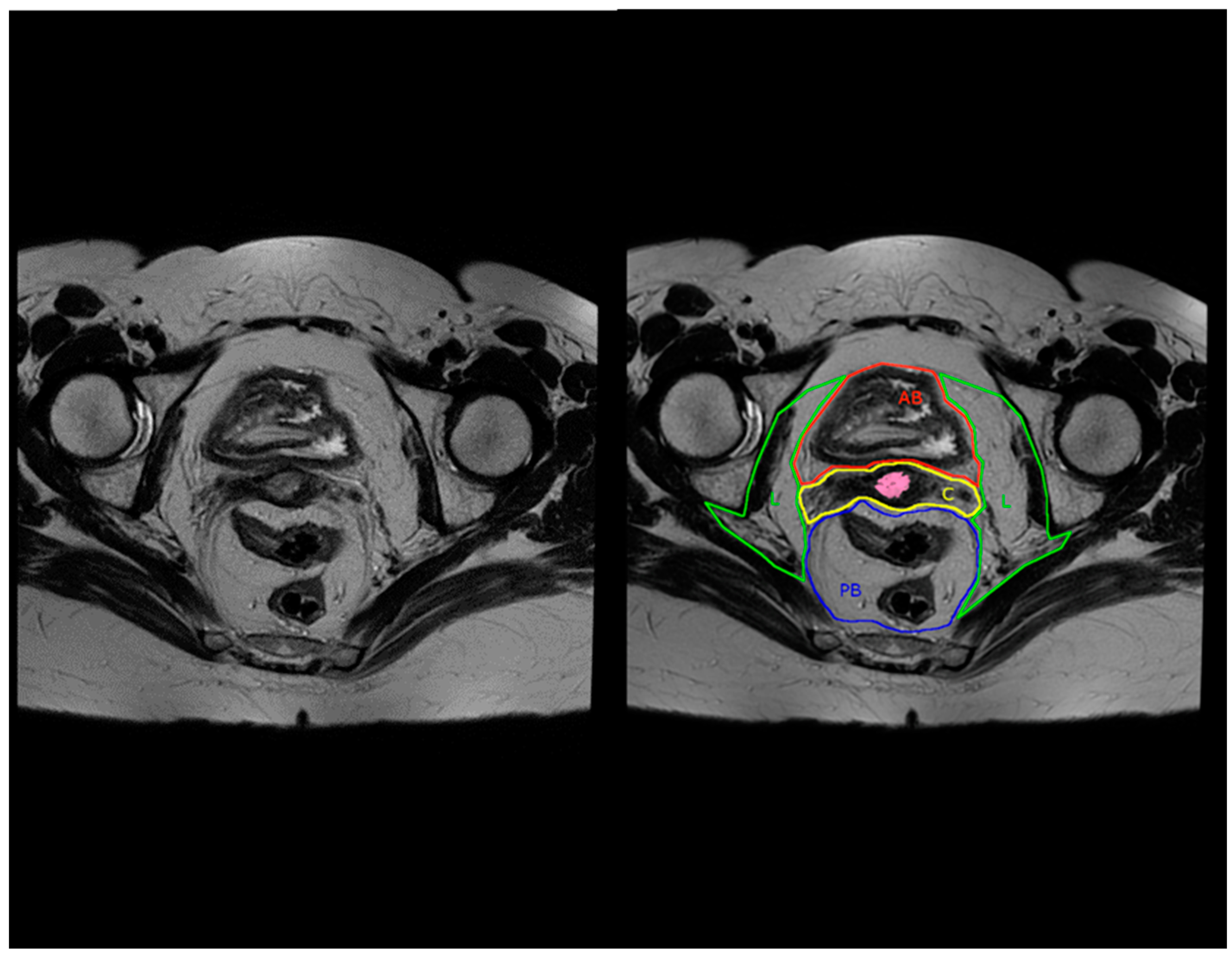
6.2. Compartment-Based Imaging

In order to obtain optimal visualization of the complex anatomy of the pelvis for planning major exenterative surgery, seven pelvic compartments have been described on magnetic resonance imaging (MRI), initially for colorectal cancer [23]. These seven compartments are defined as follows: (1) peritoneal reflection compartment, (2) anterior above peritoneal reflection compartment, (3) anterior below peritoneal reflection compartment, (4) central compartment, (5) posterior compartment, (6) lateral compartments, and (7) inferior compartment. Evaluating the accuracy of compartment-based MRI for detecting local tumor involvement, the sensitivity was more than 93% in all compartments apart from the lateral compartment, where it was 89.3%. The specificity for the posterior and anterior compartments below the peritoneal reflection was 82.2% and 86.4%, respectively, which was lower than the other compartments.
In 2023, in Annals of Surgery, the compartment-based anatomy for colorectal cancer was revisited on the basis of MRI studies of 13 pelvic specimens from body donors, of which 6 females, and the anatomical landmarks for each compartment were described in detail [22]. Given that the original and revisited central compartment description for colorectal cancer contains the rectum and mesorectum as well as the pelvic autonomic nerves, the central compartment for persistent or recurrent cervical cancer should contain instead the uterus and cervix with its parametrium, the vagina with its paracolpium, the proximal round ligaments and the uterine/vaginal vessels.
We have summarized here (Table 2) the other anatomical structures of the different compartments for pelvic exenterations in case of recurrent cervical cancer.
Applying this compartment-based anatomy with MRI in the preoperative evaluation of recurrent cervical cancers for pelvic exenterations may be helpful in standardizing the selection and type of exenteration, especially given the high sensitivity and specificity of this approach, as mentioned above in colorectal cancer. For example, when a recurrent cervical cancer is located in the central compartment and extends towards or crosses the border with the anterior below peritoneal reflection compartment, without any other pelvic disease, then an anterior pelvic exenteration could be considered (example in Figure 3 and Figure 4).

6.3. Positron-Emission Tomography-Computerized Tomography

The preferred imaging modality to evaluate the presence of distant metastases in organs or lymph nodes is positron emission tomography-computed tomography (PET-CT). Chu et al. showed, in 2014, in a systematic review and meta-analysis of 1757 patients, that the sensitivity and specificity of PET-CT in detecting metastases was 90% and 99%, respectively [49]. In 2014, Meads et al. concluded, in a meta-analysis of nine PET-CT studies evaluating the detection and management of recurrent cervical cancer, a sensitivity of 94.8% and a specificity of 86.9% [47].
A more recent study in 2020 on the value of evaluating the presence of para-aortic lymph node metastases in locally advanced cervical cancer by PET-CT versus surgical staging revealed, for PET-CT, a sensitivity and specificity of 23.5% and 93.3%, respectively [50]. The positive predictive value was 47.1% and negative predictive value was 82.8%. A subsequent meta-analysis in 2022 by Thelissen et al. showed, by pooling 12 cohort studies, an upstaging rate of 12% by para-lymph node dissection after a negative PET-CT [51]. When considering only the patients with pelvic nodal metastases on PET-CT without the suspicion of para-aortic lymph node involvement, seven cohorts showed a pooled upstaging rate of 21% by surgical para-aortic staging.

7. Conclusions

In conclusion, robotic-assisted pelvic exenterations have shown to be feasible and safe with reduced operative times, lower blood loss and perioperative as well as postoperative major complications compared to open surgery, while maintaining surgical efficiency. Long-term oncologic outcomes, including survival and recurrence rates, appear comparable between the robotic and conventional open technique, although prospective randomized trials are needed for definitive conclusions. Despite the promising advancements, several challenges remain in the widespread adoption of robotic-assisted pelvic exenterations for cervical cancer, including cost considerations and access to specialized training. MRI and PET-CT are the preferred imaging modalities for evaluating persistent and recurrent cervical cancer to aid decision making when considering pelvic exenteration. The proposed compartment-based anatomy with MRI, with a central compartment containing the uterus and vagina, may be useful in standardizing the selection and type of pelvic exenteration.

Author Contributions

P.V.T. was responsible for the conceptualization of the manuscript, reviewing the literature, writing and preparation of the original draft. P.V.T. and M.-S.W. were responsible for the conceptualization of the imaging section. M.-S.W., S.R., N.C., E.D.C., F.B. and H.A. were responsible for reviewing and editing the manuscript. All authors comply with the requirements for authorship according to ICMJE guidelines. All authors have read and agreed to the published version of the manuscript.

Funding

This research received no external funding.

Institutional Review Board Statement

The study was conducted according to the guidelines of the Declaration of Helsinki. Ethical review and approval were waived for this study, due to the nature of this manuscript being a review of the literature. Images included in the review were obtained retrospectively from the medical file, with previously written informed consent from the individual for anonymous use of images in publications or presentations, in accordance with the local legislation and institutional requirements. Therefore, submission to the local ethics committee was not required.

Conflicts of Interest

Author P.V.T. was employed by the company Intuitive Surgical, Inc. (Sunnyvale, CA, USA) as proctor and for teaching purposes. The remaining authors declare that the research was conducted in the absence of any commercial or financial relationships that could be construed as a potential conflict of interest.

References

  1. Sung, H.; Ferlay, J.; Siegel, R.L.; Laversanne, M.; Soerjomataram, I.; Jemal, A.; Bray, F. Global Cancer Statistics 2020: GLOBOCAN Estimates of Incidence and Mortality Worldwide for 36 Cancers in 185 Countries. CA Cancer J. Clin. 2021, 71, 209–249. [Google Scholar] [CrossRef] [PubMed]
  2. Cibula, D.; Raspollini, M.R.; Planchamp, F.; Centeno, C.; Chargari, C.; Felix, A.; Fischerová, D.; Jahnn-Kuch, D.; Joly, F.; Kohler, C.; et al. ESGO/ESTRO/ESP Guidelines for the management of patients with cervical cancer—Update 2023. Int. J. Gynecol. Cancer 2023, 33, 649–666. [Google Scholar] [CrossRef]
  3. Plante, M.; Kwon, J.S.; Ferguson, S.; Samouëlian, V.; Ferron, G.; Maulard, A.; de Kroon, C.; Van Driel, W.; Tidy, J.; Williamson, K.; et al. Simple versus Radical Hysterectomy in Women with Low-Risk Cervical Cancer. N. Engl. J. Med. 2024, 390, 819–829. [Google Scholar] [CrossRef] [PubMed]
  4. Taliento, C.; Scutiero, G.; Arcieri, M.; Pellecchia, G.; Tius, V.; Bogani, G.; Petrillo, M.; Pavone, M.; Bizzarri, N.; Driul, L.; et al. Simple hysterectomy versus radical hysterectomy in early-stage cervical cancer: A systematic review and meta-analysis. Eur. J. Surg. Oncol. 2024, 50, 108252. [Google Scholar] [CrossRef] [PubMed]
  5. Abu-Rustum, N.R.; Yashar, C.M.; Arend, R.; Barber, E.; Bradley, K.; Brooks, R.; Campos, S.M.; Chino, J.; Chon, H.S.; Crispens, M.A.; et al. NCCN Guidelines® Insights: Cervical Cancer, Version 1.2024. J. Natl. Compr. Cancer Netw. 2023, 21, 1224–1233. [Google Scholar] [CrossRef] [PubMed]
  6. Sardain, H.; Lavoué, V.; Foucher, F.; Levêque, J. Curative pelvic exenteration for recurrent cervical carcinoma in the era of concurrent chemotherapy and radiation therapy. A systematic review. J. Gynecol. Obstet. Biol. Reprod. 2016, 45, 315–329. [Google Scholar] [CrossRef] [PubMed]
  7. Iavazzo, C.; Gkegkes, I.D. Robotic technology for pelvic exenteration in cases of cervical cancer. Int. J. Gynaecol. Obstet. 2014, 125, 15–17. [Google Scholar] [CrossRef]
  8. Schneider, A.; Köhler, C.; Erdemoglu, E. Current developments for pelvic exenteration in gynecologic oncology. Curr. Opin. Obstet. Gynecol. 2009, 21, 4–9. [Google Scholar] [CrossRef] [PubMed]
  9. Marnitz, S.; Köhler, C.; Müller, M.; Behrens, K.; Hasenbein, K.; Schneider, A. Indications for primary and secondary exenterations in patients with cervical cancer. Gynecol. Oncol. 2006, 103, 1023–1030. [Google Scholar] [CrossRef] [PubMed]
  10. Puntambekar, S.; Lawande, A.; Desai, R.; Puntambekar, S.; Joshi, G.A.; Joshi, S.N. Initial experience of robotic anterior pelvic exenteration at a single institute. Int. J. Gynaecol. Obstet. 2014, 126, 41–44. [Google Scholar] [CrossRef]
  11. Matsuo, K.; Matsuzaki, S.; Mandelbaum, R.S.; Kanao, H.; Chang, E.J.; Klar, M.; Roman, L.D.; Wright, J.D. Utilization and perioperative outcome of minimally invasive pelvic exenteration in gynecologic malignancies: A national study in the United States. Gynecol. Oncol. 2021, 161, 39–45. [Google Scholar] [CrossRef] [PubMed]
  12. Pomel, C.; Rouzier, R.; Pocard, M.; Thoury, A.; Sideris, L.; Morice, P.; Duvillard, P.; Bourgain, J.L.; Castaigne, D. Laparoscopic total pelvic exenteration for cervical cancer relapse. Gynecol. Oncol. 2003, 91, 616–618. [Google Scholar] [CrossRef] [PubMed]
  13. Lim, P.C. Robotic assisted total pelvic exenteration: A case report. Gynecol. Oncol. 2009, 115, 310–311. [Google Scholar] [CrossRef]
  14. Zapardiel, I.; Ceccaroni, M.; Minig, L.; Halaska, M.J.; Fujii, S. Avascular spaces in radical hysterectomy. Int. J. Gynecol. Cancer 2023, 33, 285–292. [Google Scholar] [CrossRef]
  15. Bergerot, C.D.; Philip, E.J.; Bergerot, P.G.; Hsu, J.; Dizman, N.; Salgia, M.; Salgia, N.; Vaishampayan, U.; Battle, D.; Loscalzo, M.; et al. Discrepancies between genitourinary cancer patients’ and clinicians’ characterization of the Eastern Cooperative Oncology Group performance status. Cancer 2021, 127, 354–358. [Google Scholar] [CrossRef] [PubMed]
  16. Sardain, H.; Lavoue, V.; Redpath, M.; Bertheuil, N.; Foucher, F.; Levêque, J. Curative pelvic exenteration for recurrent cervical carcinoma in the era of concurrent chemotherapy and radiation therapy. A systematic review. Eur. J. Surg. Oncol. 2015, 41, 975–985. [Google Scholar] [CrossRef] [PubMed]
  17. Cianci, S.; Arcieri, M.; Vizzielli, G.; Martinelli, C.; Granese, R.; La Verde, M.; Fagotti, A.; Fanfani, F.; Scambia, G.; Ercoli, A. Robotic Pelvic Exenteration for Gynecologic Malignancies, Anatomic Landmarks, and Surgical Steps: A Systematic Review. Front. Surg. 2021, 8, 790152. [Google Scholar] [CrossRef] [PubMed]
  18. Peiretti, M.; Zapardiel, I.; Zanagnolo, V.; Landoni, F.; Morrow, C.P.; Maggioni, A. Management of recurrent cervical cancer: A review of the literature. Surg. Oncol. 2012, 21, e59–e66. [Google Scholar] [CrossRef] [PubMed]
  19. Bizzarri, N.; Chiantera, V.; Ercoli, A.; Fagotti, A.; Tortorella, L.; Conte, C.; Cappuccio, S.; Di Donna, M.C.; Gallotta, V.; Scambia, G.; et al. Minimally Invasive Pelvic Exenteration for Gynecologic Malignancies: A Multi-Institutional Case Series and Review of the Literature. J. Minim. Invasive Gynecol. 2019, 26, 1316–1326. [Google Scholar] [CrossRef]
  20. Stanca, M.; Căpîlna, D.M.; Căpîlna, M.E. Long-Term Survival, Prognostic Factors, and Quality of Life of Patients Undergoing Pelvic Exenteration for Cervical Cancer. Cancers 2022, 14, 2346. [Google Scholar] [CrossRef]
  21. Graves, S.; Seagle, B.L.; Strohl, A.E.; Shahabi, S.; Nieves-Neira, W. Survival After Pelvic Exenteration for Cervical Cancer: A National Cancer Database Study. Int. J. Gynecol. Cancer 2017, 27, 390–395. [Google Scholar] [CrossRef]
  22. Stelzner, S.; Heinze, T.; Heimke, M.; Gockel, I.; Kittner, T.; Brown, G.; Mees, S.T.; Wedel, T. Beyond Total Mesorectal Excision: Compartment-based Anatomy of the Pelvis Revisited for Exenterative Pelvic Surgery. Ann. Surg. 2023, 278, e58–e67. [Google Scholar] [CrossRef] [PubMed]
  23. Georgiou, P.A.; Tekkis, P.P.; Constantinides, V.A.; Patel, U.; Goldin, R.D.; Darzi, A.W.; John Nicholls, R.; Brown, G. Diagnostic accuracy and value of magnetic resonance imaging (MRI) in planning exenterative pelvic surgery for advanced colorectal cancer. Eur. J. Cancer 2013, 49, 72–81. [Google Scholar] [CrossRef] [PubMed]
  24. Page, M.J.; McKenzie, J.E.; Bossuyt, P.M.; Boutron, I.; Hoffmann, T.C.; Mulrow, C.D.; Shamseer, L.; Tetzlaff, J.M.; Akl, E.A.; Brennan, S.E.; et al. The PRISMA 2020 statement: An updated guideline for reporting systematic reviews. BMJ 2021, 372, n71. [Google Scholar] [CrossRef] [PubMed]
  25. Lambaudie, E.; Narducci, F.; Leblanc, E.; Bannier, M.; Houvenaeghel, G. Robotically-assisted laparoscopic anterior pelvic exenteration for recurrent cervical cancer: Report of three first cases. Gynecol. Oncol. 2010, 116, 582–583. [Google Scholar] [CrossRef] [PubMed]
  26. Davis, M.A.; Adams, S.; Eun, D.; Lee, D.; Randall, T.C. Robotic-assisted laparoscopic exenteration in recurrent cervical cancer Robotics improved the surgical experience for 2 women with recurrent cervical cancer. Am. J. Obstet. Gynecol. 2010, 202, 663.e1. [Google Scholar] [CrossRef] [PubMed]
  27. Jauffret, C.; Lambaudie, E.; Bannier, M.; Buttarelli, M.; Houvenaeghel, G. Robot-assisted laparoscopy in the management of recurrent pelvic cancer. Gynecol. Obstet. Fertil. 2011, 39, 674–680. [Google Scholar] [CrossRef] [PubMed]
  28. Lawande, A.; Kenawadekar, R.; Desai, R.; Malireddy, C.; Nallapothula, K.; Puntambekar, S.P. Robotic total pelvic exenteration. J. Robot. Surg. 2014, 8, 93–96. [Google Scholar] [CrossRef] [PubMed]
  29. Konstantinidis, I.T.; Chu, W.; Tozzi, F.; Lau, C.; Wakabayashi, M.; Chan, K.; Lee, B. Robotic Total Pelvic Exenteration: Video-Illustrated Technique. Ann. Surg. Oncol. 2017, 24, 3422–3423. [Google Scholar] [CrossRef]
  30. Nguyen Xuan, H.T.; Myriam, D.; Charlotte, N.; Richard, D.; Anne-Sophie, B.; Olivier, T.M.; Arnaud, M.; Fabrice, L. Pelvic exenteration by robotically-assisted laparoscopy: A feasibility series of 6 cases. Gynecol. Oncol. Rep. 2018, 25, 56–59, Erratum in Gynecol. Oncol. Rep. 2021, 35, 100703. [Google Scholar] [CrossRef]
  31. Yang, Q.; Tang, J.; Xiao, L. Disease-free survival after robotic-assisted laparoscopic total pelvic exenteration for recurrent cervical adenocarcinoma: A case report. Medicine 2018, 97, e11611. [Google Scholar] [CrossRef] [PubMed]
  32. Jain, V.; Debnath, S.; Rawal, S. Salvage robotic anterior pelvic exenteration for cervical cancer: Technique and feasibility. J. Robot. Surg. 2021, 15, 945–953. [Google Scholar] [CrossRef] [PubMed]
  33. Dudus, L.; Minciuna, C.; Tudor, S.; Lacatus, M.; Stefan, B.; Vasilescu, C. Robotic or laparoscopic pelvic exenteration for gynecological malignancies: Feasible options to open surgery. J. Gynecol. Oncol. 2024, 35, e12. [Google Scholar] [CrossRef] [PubMed]
  34. Brunschwig, A. Complete excision of pelvic viscera for advanced carcinoma; a one-stage abdominoperineal operation with end colostomy and bilateral ureteral implantation into the colon above the colostomy. Cancer 1948, 1, 177–183. [Google Scholar] [CrossRef]
  35. Berek, J.S.; Howe, C.; Lagasse, L.D.; Hacker, N.F. Pelvic exenteration for recurrent gynecologic malignancy: Survival and morbidity analysis of the 45-year experience at UCLA. Gynecol. Oncol. 2005, 99, 153–159. [Google Scholar] [CrossRef] [PubMed]
  36. Yang, Q.Y.; Tang, J.Y. Application of robot-assisted laparoscopic pelvic exenteration in treating gynecologic malignancies. Chin. Med. J. 2019, 132, 976–979. [Google Scholar] [CrossRef]
  37. Martínez, A.; Filleron, T.; Vitse, L.; Querleu, D.; Mery, E.; Balague, G.; Delannes, M.; Soulie, M.; Pomel, C.; Ferron, G. Laparoscopic pelvic exenteration for gynaecological malignancy: Is there any advantage? Gynecol. Oncol. 2011, 120, 374–379. [Google Scholar] [CrossRef]
  38. Pruthi, R.S.; Stefaniak, H.; Hubbard, J.S.; Wallen, E.M. Robotic anterior pelvic exenteration for bladder cancer in the female: Outcomes and comparisons to their male counterparts. J. Laparoendosc. Adv. Surg. Tech. A 2009, 19, 23–27. [Google Scholar] [CrossRef]
  39. Jäger, L.; Nilsson, P.J.; Rådestad, A.F. Pelvic exenteration for recurrent gynecologic malignancy: A study of 28 consecutive patients at a single institution. Int. J. Gynecol. Cancer 2013, 23, 755–762. [Google Scholar] [CrossRef]
  40. Baiocchi, G.; Guimaraes, G.C.; Rosa Oliveira, R.A.; Kumagai, L.Y.; Faloppa, C.C.; Aguiar, S.; Begnami, M.D.; Soares, F.A.; Lopes, A. Prognostic factors in pelvic exenteration for gynecological malignancies. Eur. J. Surg. Oncol. 2012, 38, 948–954. [Google Scholar] [CrossRef]
  41. Diver, E.J.; Rauh-Hain, J.A.; Del Carmen, M.G. Total pelvic exenteration for gynecologic malignancies. Int. J. Surg. Oncol. 2012, 2012, 693535. [Google Scholar] [CrossRef]
  42. Schmidt, A.M.; Imesch, P.; Fink, D.; Egger, H. Indications and long-term clinical outcomes in 282 patients with pelvic exenteration for advanced or recurrent cervical cancer. Gynecol. Oncol. 2012, 125, 604–609. [Google Scholar] [CrossRef] [PubMed]
  43. Chiantera, V.; Rossi, M.; De Iaco, P.; Koehler, C.; Marnitz, S.; Ferrandina, G.; Legge, F.; Parazzini, F.; Scambia, G.; Schneider, A.; et al. Survival after curative pelvic exenteration for primary or recurrent cervical cancer: A retrospective multicentric study of 167 patients. Int. J. Gynecol. Cancer 2014, 24, 916–922. [Google Scholar] [CrossRef]
  44. Höckel, M.; Horn, L.C.; Einenkel, J. (Laterally) extended endopelvic resection: Surgical treatment of locally advanced and recurrent cancer of the uterine cervix and vagina based on ontogenetic anatomy. Gynecol. Oncol. 2012, 127, 297–302. [Google Scholar] [CrossRef]
  45. Andikyan, V.; Khoury-Collado, F.; Sonoda, Y.; Gerst, S.R.; Alektiar, K.M.; Sandhu, J.S.; Bochner, B.H.; Barakat, R.R.; Boland, P.J.; Chi, D.S. Extended pelvic resections for recurrent or persistent uterine and cervical malignancies: An update on out of the box surgery. Gynecol. Oncol. 2012, 125, 404–408. [Google Scholar] [CrossRef]
  46. Landoni, F.; Zanagnolo, V.; Rosenberg, P.G.; Lopes, A.; Radice, D.; Bocciolone, L.; Aletti, G.; Parma, G.; Colombo, N.; Maggioni, A. Neoadjuvant chemotherapy prior to pelvic exenteration in patients with recurrent cervical cancer: Single institution experience. Gynecol. Oncol. 2013, 130, 69–74. [Google Scholar] [CrossRef] [PubMed]
  47. Meads, C.; Davenport, C.; Małysiak, S.; Kowalska, M.; Zapalska, A.; Guest, P.; Martin-Hirsch, P.; Borowiack, E.; Auguste, P.; Barton, P.; et al. Evaluating PET-CT in the detection and management of recurrent cervical cancer: Systematic reviews of diagnostic accuracy and subjective elicitation. BJOG 2014, 121, 398–407. [Google Scholar] [CrossRef]
  48. Forner, D.M.; Meyer, A.; Lampe, B. Preoperative assessment of complete tumour resection by magnetic resonance imaging in patients undergoing pelvic exenteration. Eur. J. Obstet. Gynecol. Reprod. Biol. 2010, 148, 182–185. [Google Scholar] [CrossRef] [PubMed]
  49. Chu, Y.; Zheng, A.; Wang, F.; Lin, W.; Yang, X.; Han, L.; Chen, Y.; Bai, L. Diagnostic value of 18F-FDG-PET or PET-CT in recurrent cervical cancer: A systematic review and meta-analysis. Nucl. Med. Commun. 2014, 35, 144–150. [Google Scholar] [CrossRef]
  50. De Cuypere, M.; Lovinfosse, P.; Goffin, F.; Gennigens, C.; Rovira, R.; Duch, J.; Fastrez, M.; Gebhart, G.; Squifflet, J.L.; Luyckx, M.; et al. Added value of para-aortic surgical staging compared to 18F-FDG PET/CT on the external beam radiation field for patients with locally advanced cervical cancer: An ONCO-GF study. Eur. J. Surg. Oncol. 2020, 46, 883–887. [Google Scholar] [CrossRef]
  51. Thelissen, A.A.B.; Jürgenliemk-Schulz, I.M.; van der Leij, F.; Peters, M.; Gerestein, C.G.; Zweemer, R.P.; van Rossum, P.S.N. Upstaging by para-aortic lymph node dissection in patients with locally advanced cervical cancer: A systematic review and meta-analysis. Gynecol. Oncol. 2022, 164, 667–674. [Google Scholar] [CrossRef] [PubMed]
Figure 1. PRISMA flow diagram.
Jcm 13 03673 g001
Figure 2. (AF). Ileal conduit formation (Bricker’s derivation) during robotic-assisted anterior pelvic exenteration (APE): (A) clipping left ureter, (B) mobilizing left ureter under meso-sigmoid colon, (C) joining two ileal loops with a stapler, (D) the closure of the ileal pouch with an Endo GIA 60 mm stapler, (E) uretero-ileal anastomosis with the ureter stent in place, (F) the urostoma of the ileal conduit.
Jcm 13 03673 g002
Figure 3. Conventional (left) and compartment-based (right) magnetic resonance imaging, axial T2-weighted image. AB: anterior below peritoneal reflection compartment, C: central compartment, L: lateral compartment, PB: posterior below peritoneal reflection compartment. Cervical tumor is highlighted in pink.
Jcm 13 03673 g003
Figure 4. Conventional (left) and compartment-based (right) magnetic resonance imaging, sagittal T2-weighted image. AA: anterior above peritoneal reflection compartment, PR: peritoneal reflection compartment, AB: anterior below peritoneal reflection compartment, C: central compartment, PB: posterior below peritoneal reflection compartment, I: inferior compartment. Cervical tumor is highlighted in pink and extends towards the anterior below peritoneal reflection compartment (AB).
Jcm 13 03673 g004
Table 1. Robotic-assisted pelvic exenterations in residual or recurrent cervical cancer. Overview of the literature.
AuthorYearNumber of PatientsType of ExenterationTotal Operative Time (mins)EBL (mL)Early Postop ComplicationsLate Postop ComplicationsHospital Stay (Days-Range)Median Follow-Up (Months-Range)Outcome
Lim [13]20091TPE375375noneNS10NSNS
Lambaudi et al. [25]20103APE480400noneFistula, UTI, ureteral stenosis, perineal abscess302–12DFS = 9 m
Davis et al. [26]20102APE540550NSNS8NSRecurrence after 8 and 23 m
Jauffret et al. [27]20112APE480300noneWound dehiscence, fistula, sepsis, urosepsis, prerenal failure, obstructive renal failure6 (3–24)23DFS = 5 m and 8 m, OS = 22 m and 23 m
Lawande et al. [28]20141TPE240300nonenone112NS
Puntambekar et al. [10]201410APE180110nonenone511Disease free: 8 patients
Konstantinidis et al. [29]20171TPE641400NSNSNSNSNS
Nguyen Xuan et al. [30]20185APE (n = 2) PPE (n = 2) TPE (n = 1)390–480NS4 UTI, 1 PE, 1 sepsisWound infection, stenosis ileal anastomosis, renal failure, pulmonary embolism11.5NS3 recurrences after 7 m, 1 died after 10 m
Yang et al. [31]20181TPE700300noneFistula3717no recurrence
Bizzarri et al. [19]201911APE500235none27.3% of cases915DFS = 11 m
Jain et al. [32]202114APE30513536% of cases: urosepsis, anastomosis leak, ileus, fistula, intestinal obstruction28.6% of cases: colon perforation, UTI, large bowel obstruction, bleeding, ureteral stricture6.517.5 (10–68)5 deaths, 12 m DFS = 68.2%, 12 m OS: 77.1%
Dudus et al. [33]202412APE (n = 6) TPE (n = 6)360
440
350Morbidity: 16.6%, urinoma, wound infectionPyelonephritis, hydronephrosis, thrombophlebitis, renal failure, bowel obstruction, iliac artery fistula18 (6–38)2450% alive, DFS = 12 m, OS = 20 m
APE: anterior pelvic exenteration; PPE: posterior pelvic exenteration; TPE: total pelvic exenteration; mins: minutes; NS: not specified; UTI: urinary tract infection; DFS: disease-free survival; OS: overall survival; PE: pulmonary embolism; m: months.
Table 2. Compartment-based magnetic resonance imaging (MRI): anatomical structures of the seven compartments relevant to pelvic exenterations for residual or recurrent cervical cancer.
MRI Compartment NameAnatomical Structures
Peritoneal reflection compartment (PR)Peritoneum (covering Douglas and vesico-uterine space
Anterior above peritoneal reflection compartment (AA)Abdominal cavity with intestines, mesenteric and omental fat,
Retroperitoneal space with proximal two thirds of the ureter, ovarian vessels, genitofemoral nerve, iliopsoas muscle
Anterior below peritoneal reflection compartment (AB)Bladder, urethra, Retzius space, vesico-ureteral junction, superior/inferior vesical vessels, ureter and bladder nerve branches from inferior hypogastric plexus, vesico-uterine ligament
Central compartment (C)Uterine corpus, uterine cervix, proximal round ligaments, parametrium, vagina, paracolpium, uterine and vaginal vessels
Posterior below peritoneal reflection compartment (PB)Rectum, mesorectum, splanchnic branches from the superior/inferior hypogastric plexus and hypogastric nerves, rectosigmoid junction
Lateral compartment (L)Parietal pelvic fascia, inferior hypogastric nerve in meso-ureter, distal third of ureter, iliac vessels, iliac and obturator lymph nodes, sacral nerve plexus, obturator nerve, internal obturator muscle, piriformis muscle, lumbosacral trunk, ovaries, fallopian tubes
Inferior compartment (I)Anorectal junction, pelvic floor muscles, levator ani muscle, transverse perineal muscle (urogenital diaphragm), perineal body, pudendal vessels and nerves
Disclaimer/Publisher’s Note: The statements, opinions and data contained in all publications are solely those of the individual author(s) and contributor(s) and not of MDPI and/or the editor(s). MDPI and/or the editor(s) disclaim responsibility for any injury to people or property resulting from any ideas, methods, instructions or products referred to in the content.

