Management of Cardiovascular Complications in Antiphospholipid Syndrome: A Narrative Review with a Focus on Older Adults

 ,
,  , , , , ,
  ,
,  and
and 
Abstract
1. Introduction
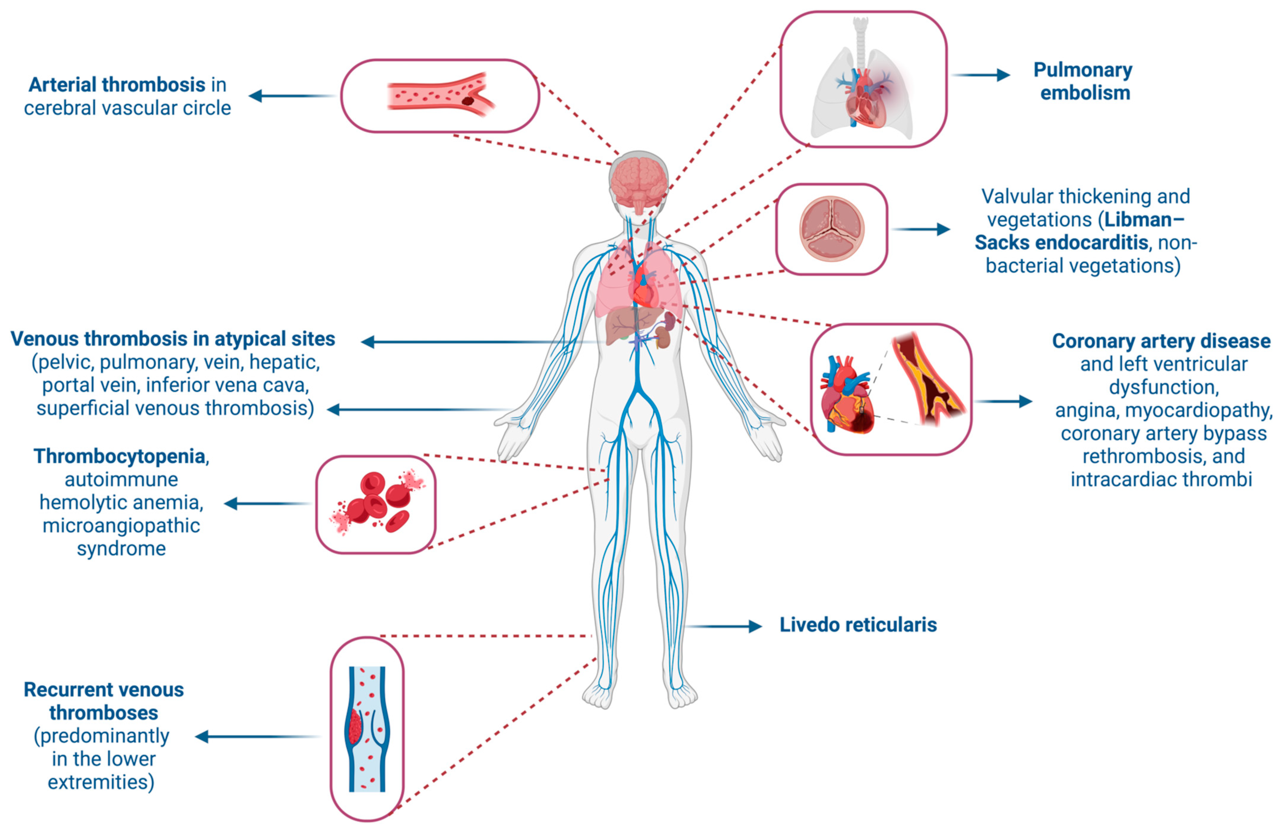
2. Cardiovascular Complications of Antiphospholipid Syndrome: Why Does It Matter?
3. Pathophysiology of Antiphospholipid Syndrome with a Focus on the Cardiovascular System
4. Diagnosis of Antiphospholipid Syndrome with Cardiovascular Manifestations
5. Management Strategies for Antiphospholipid Syndrome with Cardiovascular Complications
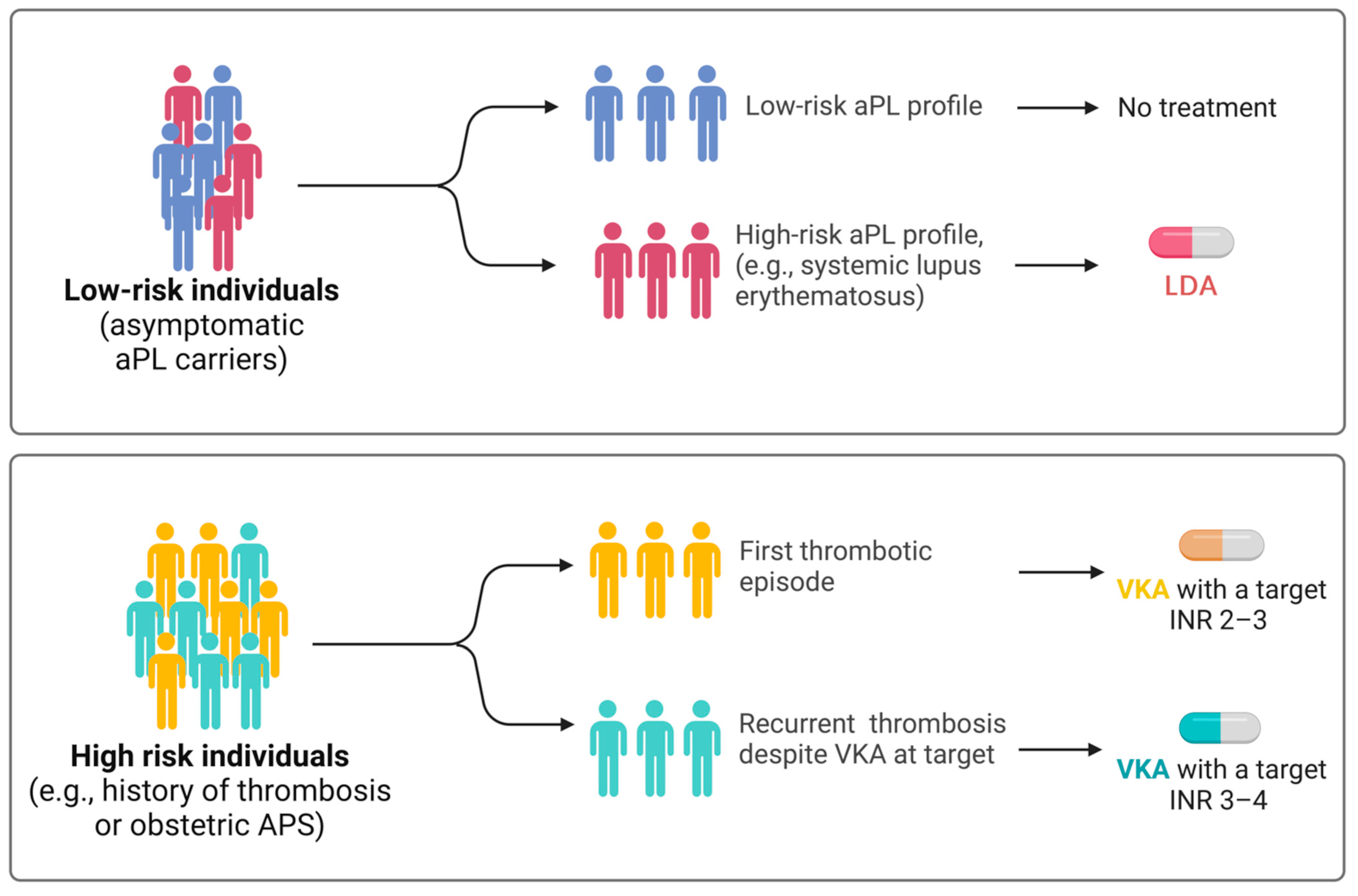
5.1. Current Recommendations for the Management of Antiphospholipid Syndrome
5.2. Role of Anticoagulant Therapy and Related Challenges
5.3. Specific Considerations for Managing Cardiovascular Complications
6. Cardiovascular Implications of Antiphospholipid Syndrome in Older Adults
6.1. Clinical Manifestations of Antiphospholipid Syndrome in Older Adults
6.2. Special Considerations for the Treatment of Antiphospholipid Syndrome in Older Adults
7. Future Perspectives in Antiphospholipid Syndrome Research and Management
7.1. Emerging Biomarkers
7.2. Potential New Treatments and Their Implication in Cardiovascular Complications
8. Conclusions
Author Contributions
Funding
Conflicts of Interest
References
- Miyakis, S.; Lockshin, M.D.; Atsumi, T.; Branch, D.W.; Brey, R.L.; Cervera, R.; Derksen, R.H.; De Groot, P.G.; Koike, T.; Meroni, P.L.; et al. International consensus statement on an update of the classification criteria for definite antiphospholipid syndrome (APS). J. Thromb. Haemost. 2006, 4, 295–306. [Google Scholar] [CrossRef] [PubMed]
- Knight, J.S.; Kanthi, Y. Mechanisms of immunothrombosis and vasculopathy in antiphospholipid syndrome. Semin. Immunopathol. 2022, 44, 347–362. [Google Scholar] [CrossRef] [PubMed]
- Dabit, J.Y.; Valenzuela-Almada, M.O.; Vallejo-Ramos, S.; Duarte-Garcia, A. Epidemiology of Antiphospholipid Syndrome in the General Population. Curr. Rheumatol. Rep. 2022, 23, 8510. [Google Scholar] [CrossRef]
- Cervera, R.; Serrano, R.; Pons-Estel, G.J.; Ceberio-Hualde, L.; Shoenfeld, Y.; de Ramon, E.; Buonaiuto, V.; Jacobsen, S.; Zeher, M.M.; Tarr, T.; et al. Morbidity and mortality in the antiphospholipid syndrome during a 10-year period: A multicentre prospective study of 1000 patients. Ann. Rheum. Dis. 2015, 74, 1011–1018. [Google Scholar] [CrossRef] [PubMed]
- Espinosa, G.; Bucciarelli, S.; Asherson, R.A.; Cervera, R. Morbidity and mortality in the catastrophic antiphospholipid syndrome: Pathophysiology, causes of death, and prognostic factors. Semin. Thromb. Hemost. 2008, 34, 290–294. [Google Scholar] [CrossRef]
- Barbhaiya, M.; Zuily, S.; Naden, R.; Hendry, A.; Manneville, F.; Amigo, M.C.; Amoura, Z.; Andrade, D.; Andreoli, L.; Artim-Esen, B.; et al. The 2023 ACR/EULAR Antiphospholipid Syndrome Classification Criteria. Arthritis Rheumatol. 2023, 75, 1687–1702. [Google Scholar] [CrossRef] [PubMed]
- Sayar, Z.; Moll, R.; Isenberg, D.; Cohen, H. Thrombotic antiphospholipid syndrome: A practical guide to diagnosis and management. Thromb. Res. 2021, 198, 213–221. [Google Scholar] [CrossRef] [PubMed]
- Matyja-Bednarczyk, A.; Swadzba, J.; Iwaniec, T.; Sanak, M.; Dziedzina, S.; Cmiel, A.; Musial, J. Risk factors for arterial thrombosis in antiphospholipid syndrome. Thromb. Res. 2014, 133, 173–176. [Google Scholar] [CrossRef] [PubMed]
- Cervera, R.; Piette, J.C.; Font, J.; Khamashta, M.A.; Shoenfeld, Y.; Camps, M.T.; Jacobsen, S.; Lakos, G.; Tincani, A.; Kontopoulou-Griva, I.; et al. Antiphospholipid syndrome: Clinical and immunologic manifestations and patterns of disease expression in a cohort of 1,000 patients. Arthritis Rheum. 2002, 46, 1019–1027. [Google Scholar] [CrossRef]
- Bitsadze, V.; Yakubova, F.; Khizroeva, J.; Lazarchuk, A.; Salnikova, P.; Vorobev, A.; Tretyakova, M.; Degtyareva, N.; Grigoreva, K.; Gashimova, N.; et al. Catastrophic Antiphospholipid Syndrome. Int. J. Mol. Sci. 2024, 25, 668. [Google Scholar] [CrossRef]
- Farzaneh-Far, A.; Roman, M.J.; Lockshin, M.D.; Devereux, R.B.; Paget, S.A.; Crow, M.K.; Davis, A.; Sammaritano, L.; Levine, D.M.; Salmon, J.E. Relationship of antiphospholipid antibodies to cardiovascular manifestations of systemic lupus erythematosus. Arthritis Rheum. 2006, 54, 3918–3925. [Google Scholar] [CrossRef] [PubMed]
- Zuily, S.; Huttin, O.; Mohamed, S.; Marie, P.Y.; Selton-Suty, C.; Wahl, D. Valvular heart disease in antiphospholipid syndrome. Curr. Rheumatol. Rep. 2013, 15, 320. [Google Scholar] [CrossRef] [PubMed]
- Pons, I.; Louro, J.; Sitges, M.; Vidal, B.; Cervera, R.; Espinosa, G. Heart Valve Involvement in Patients with Antiphospholipid Syndrome: A Long-Term Follow-Up Study of a Single Centre. J. Clin. Med. 2023, 12, 2996. [Google Scholar] [CrossRef] [PubMed]
- Colli, A.; Mestres, C.A.; Espinosa, G.; Plasin, M.A.; Pomar, J.L.; Font, J.; Cervera, R. Heart valve surgery in patients with the antiphospholipid syndrome: Analysis of a series of nine cases. Eur. J. Cardiothorac. Surg. 2010, 37, 154–158. [Google Scholar] [CrossRef] [PubMed]
- Berkun, Y.; Elami, A.; Meir, K.; Mevorach, D.; Naparstek, Y. Increased morbidity and mortality in patients with antiphospholipid syndrome undergoing valve replacement surgery. J. Thorac. Cardiovasc. Surg. 2004, 127, 414–420. [Google Scholar] [CrossRef] [PubMed]
- Gorki, H.; Malinovski, V.; Stanbridge, R.D. The antiphospholipid syndrome and heart valve surgery. Eur. J. Cardiothorac. Surg. 2008, 33, 168–181. [Google Scholar] [CrossRef] [PubMed]
- Denas, G.; Jose, S.P.; Bracco, A.; Zoppellaro, G.; Pengo, V. Antiphospholipid syndrome and the heart: A case series and literature review. Autoimmun. Rev. 2015, 14, 214–222. [Google Scholar] [CrossRef] [PubMed]
- Nevras, V.; Milaras, N.; Katsioulis, C.; Sotiriou, Z.; Tsalamandris, S.; Gkounti, G.; Skevos, S. Acute Coronary Syndromes in Antiphospholipid Syndrome-above Suspicion: A Systematic Review. Curr. Probl. Cardiol. 2023, 48, 101503. [Google Scholar] [CrossRef] [PubMed]
- Zuily, S.; Regnault, V.; Selton-Suty, C.; Eschwege, V.; Bruntz, J.F.; Bode-Dotto, E.; De Maistre, E.; Dotto, P.; Perret-Guillaume, C.; Lecompte, T.; et al. Increased risk for heart valve disease associated with antiphospholipid antibodies in patients with systemic lupus erythematosus: Meta-analysis of echocardiographic studies. Circulation 2011, 124, 215–224. [Google Scholar] [CrossRef]
- Chighizola, C.B.; Andreoli, L.; de Jesus, G.R.; Banzato, A.; Pons-Estel, G.J.; Erkan, D.; Aps, A. The association between antiphospholipid antibodies and pregnancy morbidity, stroke, myocardial infarction, and deep vein thrombosis: A critical review of the literature. Lupus 2015, 24, 980–984. [Google Scholar] [CrossRef]
- Blank, M.; Cohen, J.; Toder, V.; Shoenfeld, Y. Induction of anti-phospholipid syndrome in naive mice with mouse lupus monoclonal and human polyclonal anti-cardiolipin antibodies. Proc. Natl. Acad. Sci. USA 1991, 88, 3069–3073. [Google Scholar] [CrossRef] [PubMed]
- Pierangeli, S.S.; Barker, J.H.; Stikovac, D.; Ackerman, D.; Anderson, G.; Barquinero, J.; Acland, R.; Harris, E.N. Effect of human IgG antiphospholipid antibodies on an in vivo thrombosis model in mice. Thromb. Haemost. 1994, 71, 670–674. [Google Scholar] [CrossRef]
- Cervera, R.; Bucciarelli, S.; Plasin, M.A.; Gomez-Puerta, J.A.; Plaza, J.; Pons-Estel, G.; Shoenfeld, Y.; Ingelmo, M.; Espinos, G.; Catastrophic Antiphospholipid Syndrome Registry Project, G. Catastrophic antiphospholipid syndrome (CAPS): Descriptive analysis of a series of 280 patients from the “CAPS Registry”. J. Autoimmun. 2009, 32, 240–245. [Google Scholar] [CrossRef] [PubMed]
- Ulrich, V.; Gelber, S.E.; Vukelic, M.; Sacharidou, A.; Herz, J.; Urbanus, R.T.; de Groot, P.G.; Natale, D.R.; Harihara, A.; Redecha, P.; et al. ApoE Receptor 2 Mediation of Trophoblast Dysfunction and Pregnancy Complications Induced by Antiphospholipid Antibodies in Mice. Arthritis Rheumatol. 2016, 68, 730–739. [Google Scholar] [CrossRef] [PubMed]
- Urbanus, R.T.; Pennings, M.T.; Derksen, R.H.; de Groot, P.G. Platelet activation by dimeric beta2-glycoprotein I requires signaling via both glycoprotein Ibalpha and apolipoprotein E receptor 2′. J. Thromb. Haemost. 2008, 6, 1405–1412. [Google Scholar] [CrossRef] [PubMed]
- Romay-Penabad, Z.; Aguilar-Valenzuela, R.; Urbanus, R.T.; Derksen, R.H.; Pennings, M.T.; Papalardo, E.; Shilagard, T.; Vargas, G.; Hwang, Y.; de Groot, P.G.; et al. Apolipoprotein E receptor 2 is involved in the thrombotic complications in a murine model of the antiphospholipid syndrome. Blood 2011, 117, 1408–1414. [Google Scholar] [CrossRef] [PubMed]
- Vega-Ostertag, M.; Casper, K.; Swerlick, R.; Ferrara, D.; Harris, E.N.; Pierangeli, S.S. Involvement of p38 MAPK in the up-regulation of tissue factor on endothelial cells by antiphospholipid antibodies. Arthritis Rheum. 2005, 52, 1545–1554. [Google Scholar] [CrossRef] [PubMed]
- Martini, F.; Farsi, A.; Gori, A.M.; Boddi, M.; Fedi, S.; Domeneghetti, M.P.; Passaleva, A.; Prisco, D.; Abbate, R. Antiphospholipid antibodies (aPL) increase the potential monocyte procoagulant activity in patients with systemic lupus erythematosus. Lupus 1996, 5, 206–211. [Google Scholar] [CrossRef] [PubMed]
- Davis, W.D.; Brey, R.L. Antiphospholipid antibodies and complement activation in patients with cerebral ischemia. Clin. Exp. Rheumatol. 1992, 10, 455–460. [Google Scholar]
- Carrera-Marin, A.; Romay-Penabad, Z.; Papalardo, E.; Reyes-Maldonado, E.; Garcia-Latorre, E.; Vargas, G.; Shilagard, T.; Pierangeli, S. C6 knock-out mice are protected from thrombophilia mediated by antiphospholipid antibodies. Lupus 2012, 21, 1497–1505. [Google Scholar] [CrossRef]
- Salmon, J.E.; Girardi, G. Antiphospholipid antibodies and pregnancy loss: A disorder of inflammation. J. Reprod. Immunol. 2008, 77, 51–56. [Google Scholar] [CrossRef] [PubMed]
- Cugno, M.; Cabibbe, M.; Galli, M.; Meroni, P.L.; Caccia, S.; Russo, R.; Bottasso, B.; Mannucci, P.M. Antibodies to tissue-type plasminogen activator (tPA) in patients with antiphospholipid syndrome: Evidence of interaction between the antibodies and the catalytic domain of tPA in 2 patients. Blood 2004, 103, 2121–2126. [Google Scholar] [CrossRef]
- Hojnik, M.; George, J.; Ziporen, L.; Shoenfeld, Y. Heart valve involvement (Libman-Sacks endocarditis) in the antiphospholipid syndrome. Circulation 1996, 93, 1579–1587. [Google Scholar] [CrossRef] [PubMed]
- Jara, L.J.; Medina, G.; Vera-Lastra, O.; Amigo, M.C. Accelerated atherosclerosis, immune response and autoimmune rheumatic diseases. Autoimmun. Rev. 2006, 5, 195–201. [Google Scholar] [CrossRef] [PubMed]
- Matsuura, E.; Kobayashi, K.; Inoue, K.; Lopez, L.R.; Shoenfeld, Y. Oxidized LDL/beta2-glycoprotein I complexes: New aspects in atherosclerosis. Lupus 2005, 14, 736–741. [Google Scholar] [CrossRef] [PubMed]
- Pons-Estel, G.J.; Andreoli, L.; Scanzi, F.; Cervera, R.; Tincani, A. The antiphospholipid syndrome in patients with systemic lupus erythematosus. J. Autoimmun. 2017, 76, 10–20. [Google Scholar] [CrossRef]
- Ciliberti, G.; Bergamaschi, L.; Paolisso, P.; Zilio, F.; Pizzi, C. Ten questions about myocardial infarction with non-obstructed coronary arteries. G. Ital. Cardiol. 2024, 25, 229–238. [Google Scholar] [CrossRef] [PubMed]
- Gandhi, H.; Ahmed, N.; Spevack, D.M. Prevalence of myocardial infarction with non-obstructive coronary arteries (MINOCA) amongst acute coronary syndrome in patients with antiphospholipid syndrome. Int. J. Cardiol. Heart Vasc. 2019, 22, 148–149. [Google Scholar] [CrossRef] [PubMed]
- Stepien, K.; Nowak, K.; Wypasek, E.; Zalewski, J.; Undas, A. High prevalence of inherited thrombophilia and antiphospholipid syndrome in myocardial infarction with non-obstructive coronary arteries: Comparison with cryptogenic stroke. Int. J. Cardiol. 2019, 290, 1–6. [Google Scholar] [CrossRef]
- Svenungsson, E.; Spaak, J.; Strandberg, K.; Wallen, H.N.; Agewall, S.; Brolin, E.B.; Collste, O.; Daniel, M.; Ekenback, C.; Frick, M.; et al. Antiphospholipid antibodies in patients with myocardial infarction with and without obstructive coronary arteries. J. Intern. Med. 2022, 291, 327–337. [Google Scholar] [CrossRef]
- Ramjas, V.; Jain, A.; Lee, R.D.M.; Fioni, F.; Tawfik, N.; Sandhu, O.; Hamid, P. Unraveling the Association Between Myocardial Infarction of Nonobstructive Coronary Arteries and Antiphospholipid Syndrome. Cureus 2021, 13, e17002. [Google Scholar] [CrossRef]
- Kruchinova, S. Prevalence of thrombophilia and antiphospholipid syndrome in myocardial infarction with non-obstructive coronary arteries and cryptogenic stroke. Eur. Heart J. 2020, 41. [Google Scholar] [CrossRef]
- Coletto, L.A.; Gerosa, M.; Valentini, M.; Cimaz, R.; Caporali, R.; Meroni, P.L.; Chighizola, C.B. Myocardial involvement in anti-phospholipid syndrome: Beyond acute myocardial infarction. Autoimmun. Rev. 2022, 21, 102990. [Google Scholar] [CrossRef]
- Yeo, J.; Shin, N.; Ahn, K.J.; Seo, M.; Jang, A.Y.; Kim, M.; Chung, W.J. Pulmonary arterial hypertension due to antiphospholipid syndrome initially mimicking chronic thromboembolic pulmonary hypertension. Clin. Hypertens. 2022, 28, 10. [Google Scholar] [CrossRef] [PubMed]
- Pengo, V.; Testa, S.; Martinelli, I.; Ghirarduzzi, A.; Legnani, C.; Gresele, P.; Passamonti, S.M.; Bison, E.; Denas, G.; Jose, S.P.; et al. Incidence of a first thromboembolic event in carriers of isolated lupus anticoagulant. Thromb. Res. 2015, 135, 46–49. [Google Scholar] [CrossRef] [PubMed]
- Mustonen, P.; Lehtonen, K.V.; Javela, K.; Puurunen, M. Persistent antiphospholipid antibody (aPL) in asymptomatic carriers as a risk factor for future thrombotic events: A nationwide prospective study. Lupus 2014, 23, 1468–1476. [Google Scholar] [CrossRef]
- Djokovic, A.; Stojanovich, L.; Kontic, M.; Stanisavljevic, N.; Radovanovic, S.; Marisavljevic, D. Association between cardiac manifestations and antiphospholipid antibody type and level in a cohort of Serbian patients with primary and secondary antiphospholipid syndrome. Isr. Med. Assoc. J. 2014, 16, 162–167. [Google Scholar]
- Martinuzzo, M.E.; Pombo, G.; Forastiero, R.R.; Cerrato, G.S.; Colorio, C.C.; Carreras, L.O. Lupus anticoagulant, high levels of anticardiolipin, and anti-beta2-glycoprotein I antibodies are associated with chronic thromboembolic pulmonary hypertension. J. Rheumatol. 1998, 25, 1313–1319. [Google Scholar]
- Kolitz, T.; Shiber, S.; Sharabi, I.; Winder, A.; Zandman-Goddard, G. Cardiac Manifestations of Antiphospholipid Syndrome with Focus on Its Primary Form. Front. Immunol. 2019, 10, 941. [Google Scholar] [CrossRef]
- Tektonidou, M.G.; Laskari, K.; Panagiotakos, D.B.; Moutsopoulos, H.M. Risk factors for thrombosis and primary thrombosis prevention in patients with systemic lupus erythematosus with or without antiphospholipid antibodies. Arthritis Rheum. 2009, 61, 29–36. [Google Scholar] [CrossRef]
- Somers, E.; Magder, L.S.; Petri, M. Antiphospholipid antibodies and incidence of venous thrombosis in a cohort of patients with systemic lupus erythematosus. J. Rheumatol. 2002, 29, 2531–2536. [Google Scholar] [PubMed]
- Arnaud, L.; Mathian, A.; Devilliers, H.; Ruffatti, A.; Tektonidou, M.; Forastiero, R.; Pengo, V.; Lambert, M.; Lefevre, G.; Martinez-Zamora, M.A.; et al. Patient-level analysis of five international cohorts further confirms the efficacy of aspirin for the primary prevention of thrombosis in patients with antiphospholipid antibodies. Autoimmun. Rev. 2015, 14, 192–200. [Google Scholar] [CrossRef] [PubMed]
- Rosove, M.H.; Brewer, P.M. Antiphospholipid thrombosis: Clinical course after the first thrombotic event in 70 patients. Ann. Intern. Med. 1992, 117, 303–308. [Google Scholar] [CrossRef] [PubMed]
- Cohen, H.; Hunt, B.J.; Efthymiou, M.; Arachchillage, D.R.; Mackie, I.J.; Clawson, S.; Sylvestre, Y.; Machin, S.J.; Bertolaccini, M.L.; Ruiz-Castellano, M.; et al. Rivaroxaban versus warfarin to treat patients with thrombotic antiphospholipid syndrome, with or without systemic lupus erythematosus (RAPS): A randomised, controlled, open-label, phase 2/3, non-inferiority trial. Lancet Haematol. 2016, 3, e426–e436. [Google Scholar] [CrossRef] [PubMed]
- Dufrost, V.; Risse, J.; Zuily, S.; Wahl, D. Direct Oral Anticoagulants Use in Antiphospholipid Syndrome: Are These Drugs an Effective and Safe Alternative to Warfarin? A Systematic Review of the Literature. Curr. Rheumatol. Rep. 2016, 18, 74. [Google Scholar] [CrossRef] [PubMed]
- Ordi-Ros, J.; Sáez-Comet, L.; Pérez-Conesa, M.; Vidal, X.; Riera-Mestre, A.; Castro-Salomó, A.; Cuquet-Pedragosa, J.; Ortiz-Santamaria, V.; Mauri-Plana, M.; Solé, C.; et al. Rivaroxaban Versus Vitamin K Antagonist in Antiphospholipid Syndrome: A Randomized Noninferiority Trial. Ann. Intern. Med. 2019, 171, 685–694. [Google Scholar] [CrossRef] [PubMed]
- Pastori, D.; Menichelli, D.; Cammisotto, V.; Pignatelli, P. Use of Direct Oral Anticoagulants in Patients with Antiphospholipid Syndrome: A Systematic Review and Comparison of the International Guidelines. Front. Cardiovasc. Med. 2021, 8, 715878. [Google Scholar] [CrossRef] [PubMed]
- Wu, X.; Cao, S.; Yu, B.; He, T. Comparing the efficacy and safety of direct oral anticoagulants versus Vitamin K antagonists in patients with antiphospholipid syndrome: A systematic review and meta-analysis. Blood Coagul. Fibrinolysis 2022, 33, 389–401. [Google Scholar] [CrossRef] [PubMed]
- Bakow, B.R.; Yanek, L.; Crowther, M.A.; Chaturvedi, S. Low recurrent thrombosis rates in single positive antiphospholipid syndrome regardless of type of anticoagulation. Thromb. Res. 2024, 237, 88–93. [Google Scholar] [CrossRef] [PubMed]
- Hu, D.; Emara, O.; Ramos, M.; Aller, C.B. Anticoagulation in a patient with antiphospholipid syndrome and a mechanical heart valve: A case study. Blood Coagul. Fibrinolysis 2018, 29, 472–475. [Google Scholar] [CrossRef]
- Endara, S.A.; Davalos, G.A.; Fierro, C.H.; Ullauri, V.E.; Molina, G.A. Antiphospholipid syndrome and valvular heart disease, a complex scenario of thrombotic events, a case report. J. Cardiothorac. Surg. 2020, 15, 275. [Google Scholar] [CrossRef] [PubMed]
- Vahanian, A.; Beyersdorf, F.; Praz, F.; Milojevic, M.; Baldus, S.; Bauersachs, J.; Capodanno, D.; Conradi, L.; De Bonis, M.; De Paulis, R.; et al. 2021 ESC/EACTS Guidelines for the management of valvular heart disease. Eur. Heart J. 2022, 43, 561–632. [Google Scholar] [CrossRef] [PubMed]
- Mittal, P.; Quattrocchi, G.; Tohidi-Esfahani, I.; Sayar, Z.; Chandratheva, A.; Cohen, H. Antiphospholipid syndrome, antiphospholipid antibodies, and stroke. Int. J. Stroke 2023, 18, 383–391. [Google Scholar] [CrossRef] [PubMed]
- Ruiz-Irastorza, G.; Hunt, B.J.; Khamashta, M.A. A systematic review of secondary thromboprophylaxis in patients with antiphospholipid antibodies. Arthritis Rheum. 2007, 57, 1487–1495. [Google Scholar] [CrossRef] [PubMed]
- Zavaleta, N.E.; Montes, R.M.; Soto, M.E.; Vanzzini, N.A.; Amigo, M.C. Primary antiphospholipid syndrome: A 5-year transesophageal echocardiographic followup study. J. Rheumatol. 2004, 31, 2402–2407. [Google Scholar] [PubMed]
- Sciascia, S.; Khamashta, M.A.; D’Cruz, D.P. Targeted therapy in antiphospholipid syndrome. Curr. Opin. Rheumatol. 2014, 26, 269–275. [Google Scholar] [CrossRef] [PubMed]
- Bakshi, J.; Stevens, R. Rituximab therapy for recurrent thromboembolic disease in antiphospholipid syndrome. Lupus 2013, 22, 865–867. [Google Scholar] [CrossRef] [PubMed]
- Hussain, H.; Tarantino, M.D.; Chaturvedi, S.; McCrae, K.R.; Roberts, J.C. Eculizumab for refractory thrombosis in antiphospholipid syndrome. Blood Adv. 2022, 6, 1271–1277. [Google Scholar] [CrossRef] [PubMed]
- Piette, J.C.; Cacoub, P. Antiphospholipid syndrome in the elderly: Caution. Circulation 1998, 97, 2195–2196. [Google Scholar] [CrossRef]
- Masson, C.; Nguyen, T.T.A.; Dufrost, V.; Audrain, M.; Hémont, C.; Agard, C.; Artifoni, M.; Connault, J.; Fouassier, M.; Hamidou, M.; et al. Antiphospholipid syndrome in patients over 65 years: A comparative study of clinical and biological features and thrombotic relapses. Lupus 2022, 31, 1816–1823. [Google Scholar] [CrossRef]
- Grimaud, F.; Yelnik, C.; Pineton de Chambrun, M.; Amoura, Z.; Arnaud, L.; Costedoat Chalumeau, N.; Hachulla, E.; Lambert, M.; Roriz, M.; Sibilia, J.; et al. Clinical and immunological features of antiphospholipid syndrome in the elderly: A retrospective national multicentre study. Rheumatology 2019, 58, 1006–1010. [Google Scholar] [CrossRef] [PubMed]
- Duarte-García, A.; Pham, M.M.; Crowson, C.S.; Amin, S.; Moder, K.G.; Pruthi, R.K.; Warrington, K.J.; Matteson, E.L. The Epidemiology of Antiphospholipid Syndrome: A Population-Based Study. Arthritis Rheumatol. 2019, 71, 1545–1552. [Google Scholar] [CrossRef] [PubMed]
- Hwang, J.J.; Shin, S.H.; Kim, Y.J.; Oh, Y.M.; Lee, S.D.; Kim, Y.H.; Choi, C.W.; Lee, J.S. Epidemiology of Antiphospholipid Syndrome in Korea: A Nationwide Population-based Study. J. Korean Med. Sci. 2020, 35, e35. [Google Scholar] [CrossRef] [PubMed]
- Quéméneur, T.; Lambert, M.; Hachulla, E.; Dubucquoi, S.; Caron, C.; Fauchais, A.L.; Devulder, B.; Hatron, P.Y. Significance of persistent antiphospholipid antibodies in the elderly. J. Rheumatol. 2006, 33, 1559–1562. [Google Scholar] [PubMed]
- Pengo, V.; Ruffatti, A.; Legnani, C.; Testa, S.; Fierro, T.; Marongiu, F.; De Micheli, V.; Gresele, P.; Tonello, M.; Ghirarduzzi, A.; et al. Incidence of a first thromboembolic event in asymptomatic carriers of high-risk antiphospholipid antibody profile: A multicenter prospective study. Blood 2011, 118, 4714–4718. [Google Scholar] [CrossRef] [PubMed]
- Anderson, F.A., Jr.; Spencer, F.A. Risk factors for venous thromboembolism. Circulation 2003, 107, I-9–I-16. [Google Scholar] [CrossRef] [PubMed]
- Bountola, S.; Perifanis, V.; Kazantzi, A.; Keramidioti, C.; Hassapopoulou-Matami, E.; Hatzitolios, A.; Girtovitis, F. PS1585 Elderly with Antiphospholipid Syndrome Present with Distinct Clinical Profile: A Retrospective Single Center Study. HemaSphere 2019, 3, 730–731. [Google Scholar] [CrossRef]
- Tanne, D.; D’Olhaberriague, L.; Trivedi, A.M.; Salowich-Palm, L.; Schultz, L.R.; Levine, S.R. Anticardiolipin antibodies and mortality in patients with ischemic stroke: A prospective follow-up study. Neuroepidemiology 2002, 21, 93–99. [Google Scholar] [CrossRef]
- Piette, J.C. 1996 diagnostic and classification criteria for the antiphospholipid/cofactors syndrome: A mission impossible? Lupus 1996, 5, 354–363. [Google Scholar] [CrossRef]
- de Godoy, J.M.; de Godoy, M.R.; Cipulo, J.P.; Tognola, V.A. Vascular dementia and anticardiolipin antibodies. Med. Sci. Monit. 2005, 11, Cr430–Gr433. [Google Scholar]
- Finazzi, G.; Brancaccio, V.; Moia, M.; Ciaverella, N.; Mazzucconi, M.G.; Schinco, P.C.; Ruggeri, M.; Pogliani, E.M.; Gamba, G.; Rossi, E.; et al. Natural history and risk factors for thrombosis in 360 patients with antiphospholipid antibodies: A four-year prospective study from the Italian Registry. Am. J. Med. 1996, 100, 530–536. [Google Scholar] [CrossRef] [PubMed]
- Manoussakis, M.N.; Tzioufas, A.G.; Silis, M.P.; Pange, P.J.; Goudevenos, J.; Moutsopoulos, H.M. High prevalence of anti-cardiolipin and other autoantibodies in a healthy elderly population. Clin. Exp. Immunol. 1987, 69, 557–565. [Google Scholar]
- Richaud-Patin, Y.; Cabiedes, J.; Jakez-Ocampo, J.; Vidaller, A.; Llorente, L. High prevalence of protein-dependent and protein-independent antiphospholipid and other autoantibodies in healthy elders. Thromb. Res. 2000, 99, 129–133. [Google Scholar] [CrossRef] [PubMed]
- Galli, M.; Luciani, D.; Bertolini, G.; Barbui, T. Lupus anticoagulants are stronger risk factors for thrombosis than anticardiolipin antibodies in the antiphospholipid syndrome: A systematic review of the literature. Blood 2003, 101, 1827–1832. [Google Scholar] [CrossRef]
- Guerin, J.; Feighery, C.; Sim, R.B.; Jackson, J. Antibodies to beta2-glycoprotein I--a specific marker for the antiphospholipid syndrome. Clin. Exp. Immunol. 1997, 109, 304–309. [Google Scholar] [CrossRef] [PubMed]
- Krause, I.; Cohen, J.; Blank, M.; Bakimer, R.; Cartman, A.; Hohmann, A.; Valesini, G.; Asherson, R.A.; Khamashta, M.A.; Hughes, G.R.; et al. Distribution of two common idiotypes of anticardiolipin antibodies in sera of patients with primary antiphospholipid syndrome, systemic lupus erythematosus and monoclonal gammopathies. Lupus 1992, 1, 91–96. [Google Scholar] [CrossRef]
- Piette, J.C.; Cacoub, P.; Wechsler, B. Renal manifestations of the antiphospholipid syndrome. Semin. Arthritis Rheum. 1994, 23, 357–366. [Google Scholar] [CrossRef]
- Quintarelli, C.; Ferro, D.; Valesini, G.; Basili, S.; Tassone, G.; Violi, F. Prevalence of lupus anticoagulant in patients with cirrhosis: Relationship with beta-2-glycoprotein I plasma levels. J. Hepatol. 1994, 21, 1086–1091. [Google Scholar] [CrossRef] [PubMed]
- Chakravarty, K.; Pountain, G.; Merry, P.; Byron, M.; Hazleman, B.; Scott, D.G. A longitudinal study of anticardiolipin antibody in polymyalgia rheumatica and giant cell arteritis. J. Rheumatol. 1995, 22, 1694–1697. [Google Scholar]
- Ramos-Casals, M.; García-Carrasco, M.; Brito, M.P.; López-Soto, A.; Font, J. Autoimmunity and geriatrics: Clinical significance of autoimmune manifestations in the elderly. Lupus 2003, 12, 341–355. [Google Scholar] [CrossRef]
- Thakkar, J.P.; McCarthy, B.J.; Villano, J.L. Age-specific cancer incidence rates increase through the oldest age groups. Am. J. Med. Sci. 2014, 348, 65–70. [Google Scholar] [CrossRef] [PubMed]
- Bessis, D.; Sotto, A.; Viard, J.P.; Bérard, M.; Ciurana, A.J.; Boffa, M.C. Trousseau’s syndrome with nonbacterial thrombotic endocarditis: Pathogenic role of antiphospholipid syndrome. Am. J. Med. 1995, 98, 511–513. [Google Scholar] [CrossRef] [PubMed]
- Tincani, A.; Taraborelli, M.; Cattaneo, R. Antiphospholipid antibodies and malignancies. Autoimmun. Rev. 2010, 9, 200–202. [Google Scholar] [CrossRef] [PubMed]
- Schved, J.F.; Dupuy-Fons, C.; Biron, C.; Quére, I.; Janbon, C. A prospective epidemiological study on the occurrence of antiphospholipid antibody: The Montpellier Antiphospholipid (MAP) Study. Haemostasis 1994, 24, 175–182. [Google Scholar] [CrossRef] [PubMed]
- McNeil, J.J.; Wolfe, R.; Woods, R.L.; Tonkin, A.M.; Donnan, G.A.; Nelson, M.R.; Reid, C.M.; Lockery, J.E.; Kirpach, B.; Storey, E.; et al. Effect of Aspirin on Cardiovascular Events and Bleeding in the Healthy Elderly. N. Engl. J. Med. 2018, 379, 1509–1518. [Google Scholar] [CrossRef] [PubMed]
- Cervera, R.; Khamashta, M.A.; Shoenfeld, Y.; Camps, M.T.; Jacobsen, S.; Kiss, E.; Zeher, M.M.; Tincani, A.; Kontopoulou-Griva, I.; Galeazzi, M.; et al. Morbidity and mortality in the antiphospholipid syndrome during a 5-year period: A multicentre prospective study of 1000 patients. Ann. Rheum. Dis. 2009, 68, 1428–1432. [Google Scholar] [CrossRef]
- Palareti, G.; Leali, N.; Coccheri, S.; Poggi, M.; Manotti, C.; D’Angelo, A.; Pengo, V.; Erba, N.; Moia, M.; Ciavarella, N.; et al. Bleeding complications of oral anticoagulant treatment: An inception-cohort, prospective collaborative study (ISCOAT). Italian Study on Complications of Oral Anticoagulant Therapy. Lancet 1996, 348, 423–428. [Google Scholar] [CrossRef]
- Torn, M.; Bollen, W.L.; van der Meer, F.J.; van der Wall, E.E.; Rosendaal, F.R. Risks of oral anticoagulant therapy with increasing age. Arch. Intern. Med. 2005, 165, 1527–1532. [Google Scholar] [CrossRef] [PubMed]
- Tumian, N.R.; Hunt, B.J. Clinical Management of Thrombotic Antiphospholipid Syndrome. J. Clin. Med. 2022, 11, 735. [Google Scholar] [CrossRef]
- de Laat, B.; Pengo, V.; Pabinger, I.; Musial, J.; Voskuyl, A.E.; Bultink, I.E.; Ruffatti, A.; Rozman, B.; Kveder, T.; de Moerloose, P.; et al. The association between circulating antibodies against domain I of beta2-glycoprotein I and thrombosis: An international multicenter study. J. Thromb. Haemost. 2009, 7, 1767–1773. [Google Scholar] [CrossRef]
- Sciascia, S.; Radin, M.; Cecchi, I.; Rubini, E.; Scotta, A.; Rolla, R.; Montaruli, B.; Pergolini, P.; Mengozzi, G.; Muccini, E.; et al. Reliability of Lupus Anticoagulant and Anti-phosphatidylserine/prothrombin Autoantibodies in Antiphospholipid Syndrome: A Multicenter Study. Front. Immunol. 2019, 10, 376. [Google Scholar] [CrossRef] [PubMed]
- Müller-Calleja, N.; Hollerbach, A.; Royce, J.; Ritter, S.; Pedrosa, D.; Madhusudhan, T.; Teifel, S.; Meineck, M.; Häuser, F.; Canisius, A.; et al. Lipid presentation by the protein C receptor links coagulation with autoimmunity. Science 2021, 371, eabc0956. [Google Scholar] [CrossRef]
- Tanimura, K.; Jin, H.; Suenaga, T.; Morikami, S.; Arase, N.; Kishida, K.; Hirayasu, K.; Kohyama, M.; Ebina, Y.; Yasuda, S.; et al. β2-Glycoprotein I/HLA class II complexes are novel autoantigens in antiphospholipid syndrome. Blood 2015, 125, 2835–2844. [Google Scholar] [CrossRef] [PubMed]
- Zuo, Y.; Yalavarthi, S.; Gockman, K.; Madison, J.A.; Gudjonsson, J.E.; Kahlenberg, J.M.; Joseph McCune, W.; Bockenstedt, P.L.; Karp, D.R.; Knight, J.S. Anti-Neutrophil Extracellular Trap Antibodies and Impaired Neutrophil Extracellular Trap Degradation in Antiphospholipid Syndrome. Arthritis Rheumatol. 2020, 72, 2130–2135. [Google Scholar] [CrossRef]
- Vandevelde, A.; Chayoua, W.; de Laat, B.; Moore, G.W.; Musiał, J.; Zuily, S.; Wahl, D.; Devreese, K.M.J. Added value of antiphosphatidylserine/prothrombin antibodies in the workup of thrombotic antiphospholipid syndrome: Communication from the ISTH SSC Subcommittee on Lupus Anticoagulant/Antiphospholipid Antibodies. J. Thromb. Haemost. 2022, 20, 2136–2150. [Google Scholar] [CrossRef] [PubMed]
- Zuo, Y.; Shi, H.; Li, C.; Knight, J.S. Antiphospholipid syndrome: A clinical perspective. Chin. Med. J. 2020, 133, 929–940. [Google Scholar] [CrossRef] [PubMed]
- Teruel, R.; Pérez-Sánchez, C.; Corral, J.; Herranz, M.T.; Pérez-Andreu, V.; Saiz, E.; García-Barberá, N.; Martínez-Martínez, I.; Roldán, V.; Vicente, V.; et al. Identification of miRNAs as potential modulators of tissue factor expression in patients with systemic lupus erythematosus and antiphospholipid syndrome. J. Thromb. Haemost. 2011, 9, 1985–1992. [Google Scholar] [CrossRef] [PubMed]
- Wu, M.; Barnard, J.; Kundu, S.; McCrae, K.R. A novel pathway of cellular activation mediated by antiphospholipid antibody-induced extracellular vesicles. J. Thromb. Haemost. 2015, 13, 1928–1940. [Google Scholar] [CrossRef]
- Gysler, S.M.; Mulla, M.J.; Guerra, M.; Brosens, J.J.; Salmon, J.E.; Chamley, L.W.; Abrahams, V.M. Antiphospholipid antibody-induced miR-146a-3p drives trophoblast interleukin-8 secretion through activation of Toll-like receptor 8. Mol. Hum. Reprod. 2016, 22, 465–474. [Google Scholar] [CrossRef]
- Garcia, D.; Erkan, D. Diagnosis and Management of the Antiphospholipid Syndrome. N. Engl. J. Med. 2018, 378, 2010–2021. [Google Scholar] [CrossRef]
- Uthman, I.; Noureldine, M.H.A.; Ruiz-Irastorza, G.; Khamashta, M. Management of antiphospholipid syndrome. Ann. Rheum. Dis. 2019, 78, 155–161. [Google Scholar] [CrossRef] [PubMed]
- Erkan, D. Expert Perspective: Management of Microvascular and Catastrophic Antiphospholipid Syndrome. Arthritis Rheumatol. 2021, 73, 1780–1790. [Google Scholar] [CrossRef] [PubMed]
- Legault, K.; Schunemann, H.; Hillis, C.; Yeung, C.; Akl, E.A.; Carrier, M.; Cervera, R.; Crowther, M.; Dentali, F.; Erkan, D.; et al. McMaster RARE-Bestpractices clinical practice guideline on diagnosis and management of the catastrophic antiphospholipid syndrome. J. Thromb. Haemost. 2018, 16, 1656–1664. [Google Scholar] [CrossRef] [PubMed]
- Hisada, R.; Kato, M.; Sugawara, E.; Kanda, M.; Fujieda, Y.; Oku, K.; Bohgaki, T.; Amengual, O.; Horita, T.; Yasuda, S.; et al. Circulating plasmablasts contribute to antiphospholipid antibody production, associated with type I interferon upregulation. J. Thromb. Haemost. 2019, 17, 1134–1143. [Google Scholar] [CrossRef] [PubMed]
- Cole, S.; Walsh, A.; Yin, X.; Wechalekar, M.D.; Smith, M.D.; Proudman, S.M.; Veale, D.J.; Fearon, U.; Pitzalis, C.; Humby, F.; et al. Integrative analysis reveals CD38 as a therapeutic target for plasma cell-rich pre-disease and established rheumatoid arthritis and systemic lupus erythematosus. Arthritis Res. Ther. 2018, 20, 85. [Google Scholar] [CrossRef] [PubMed]
- Tommy Gambles, M.; Li, J.; Christopher Radford, D.; Sborov, D.; Shami, P.; Yang, J.; Kopeček, J. Simultaneous crosslinking of CD20 and CD38 receptors by drug-free macromolecular therapeutics enhances B cell apoptosis in vitro and in vivo. J. Control Release 2022, 350, 584–599. [Google Scholar] [CrossRef] [PubMed]
- Reddy, V.; Klein, C.; Isenberg, D.A.; Glennie, M.J.; Cambridge, G.; Cragg, M.S.; Leandro, M.J. Obinutuzumab induces superior B-cell cytotoxicity to rituximab in rheumatoid arthritis and systemic lupus erythematosus patient samples. Rheumatology 2017, 56, 1227–1237. [Google Scholar] [CrossRef]
- Reddy, V.R.; Pepper, R.J.; Shah, K.; Cambridge, G.; Henderson, S.R.; Klein, C.; Kell, L.; Taylor, S.J.; Isenberg, D.A.; Cragg, M.S.; et al. Disparity in peripheral and renal B-cell depletion with rituximab in systemic lupus erythematosus: An opportunity for obinutuzumab? Rheumatology 2022, 61, 2894–2904. [Google Scholar] [CrossRef] [PubMed]
- Yoshizuka, R.; Hasegawa, H.; Kamiya, M.; Umezawa, N.; Yasuda, S. Refractory antiphospholipid antibody syndrome-induced thrombocytopaenia successfully treated with belimumab. Lupus 2022, 31, 624–627. [Google Scholar] [CrossRef]
- Shapira, I.; Andrade, D.; Allen, S.L.; Salmon, J.E. Brief report: Induction of sustained remission in recurrent catastrophic antiphospholipid syndrome via inhibition of terminal complement with eculizumab. Arthritis Rheum. 2012, 64, 2719–2723. [Google Scholar] [CrossRef]
- Canaud, G.; Kamar, N.; Anglicheau, D.; Esposito, L.; Rabant, M.; Noël, L.H.; Guilbeau-Frugier, C.; Sberro-Soussan, R.; Del Bello, A.; Martinez, F.; et al. Eculizumab improves posttransplant thrombotic microangiopathy due to antiphospholipid syndrome recurrence but fails to prevent chronic vascular changes. Am. J. Transplant. 2013, 13, 2179–2185. [Google Scholar] [CrossRef] [PubMed]
- Ramos-Casals, M.; Brito-Zerón, P.; Soto, M.J.; Cuadrado, M.J.; Khamashta, M.A. Autoimmune diseases induced by TNF-targeted therapies. Best. Pract. Res. Clin. Rheumatol. 2008, 22, 847–861. [Google Scholar] [CrossRef] [PubMed]
- Cheemalavagu, S.; McCoy, S.S.; Knight, J.S. Digital ischaemia secondary to adalimumab-induced antiphospholipid syndrome. BMJ Case Rep. 2020, 13, e232907. [Google Scholar] [CrossRef] [PubMed]

| Category | Sydney Criteria (2006) [1] | EULAR Recommendations (2023) [6] |
|---|---|---|
| Entry criteria | No. | ≥1 clinical criterion and ≥1 laboratory criterion. |
| Diagnosis | ≥1 clinical criterion and ≥1 laboratory criterion | Weighted criteria, ≥3 points from clinical domain and ≥3 points for laboratory domain. |
| Clinical Criteria | Two clinical criteria | Six clinical domains: |
| (1) Vascular thrombosis (arterial, venous or small vessel thrombosis). (2) Pregnancy morbidity (one or more unexplained deaths of a morphologically normal fetus at the 10th week of gestation or beyond, or three or more unexplained consecutive spontaneous abortions before the 10th week). | (1) VTE: with (1 point) or without (3 points) high-risk profile. (2) AT: with (2 points) or without (4 points) high-risk profile. (3) Microvascular involvement: suspected (2 points) or established (5 points). (4) Pregnancy morbidity: three or more consecutive prefetal (<10 weeks) and/or early (10–16 weeks) fetal deaths, or ≥1 fetal death (16–34 weeks) alone (1 point); preeclampsia with severe features or placental insufficiency with severe features (<34 weeks) with or without fetal death (3 points); preeclampsia with severe features and placental insufficiency with severe features (<34 weeks) with or without fetal death (4 points). (5) Cardiac valve: thickening (2 points) or vegetations (4 points). (6) Thrombocytopenia (platelets < 100 × 109/L, 2 points). | |
| Laboratory Criteria | (1) LAC: present in plasma, on two or more occasions at least 12 weeks apart. (2) aCL: IgG and/or IgM present in plasma with medium or high titers (>40 units or >99th percentile) on two or more occasions at least 12 weeks apart. (3) β2-GPI-Ab: IgG and/or IgM present in plasma with titers >99th percentile on two or more occasions at least 12 weeks apart. | (1) LAC: one-time positivity (1 point) or persistent positivity (≥2 positive results, at least 12 weeks apart, 5 points); (2) β2-GPI-Ab/aCL: moderate */high ** β2-GPI-Ab and/or aCL IgM positivity (1 point); moderate * β2-GPI-Ab and/or aCL IgG positivity (4 points); high ** β2-GPI-Ab and/or aCL IgG positivity (5 points); high ** β2-GPI-Ab and aCL positivity (7 points). |
Disclaimer/Publisher’s Note: The statements, opinions and data contained in all publications are solely those of the individual author(s) and contributor(s) and not of MDPI and/or the editor(s). MDPI and/or the editor(s) disclaim responsibility for any injury to people or property resulting from any ideas, methods, instructions or products referred to in the content. |
© 2024 by the authors. Licensee MDPI, Basel, Switzerland. This article is an open access article distributed under the terms and conditions of the Creative Commons Attribution (CC BY) license (https://creativecommons.org/licenses/by/4.0/).
Share and Cite
Bernardi, M.; Spadafora, L.; Andaloro, S.; Piscitelli, A.; Fornaci, G.; Intonti, C.; Fratta, A.E.; Hsu, C.-E.; Kaziròd-Wolski, K.; Metsovitis, T.; et al. Management of Cardiovascular Complications in Antiphospholipid Syndrome: A Narrative Review with a Focus on Older Adults. J. Clin. Med. 2024, 13, 3064. https://doi.org/10.3390/jcm13113064
Bernardi M, Spadafora L, Andaloro S, Piscitelli A, Fornaci G, Intonti C, Fratta AE, Hsu C-E, Kaziròd-Wolski K, Metsovitis T, et al. Management of Cardiovascular Complications in Antiphospholipid Syndrome: A Narrative Review with a Focus on Older Adults. Journal of Clinical Medicine. 2024; 13(11):3064. https://doi.org/10.3390/jcm13113064
Chicago/Turabian StyleBernardi, Marco, Luigi Spadafora, Silvia Andaloro, Alessandra Piscitelli, Giovanni Fornaci, Chiara Intonti, Alberto Emanuele Fratta, Chieh-En Hsu, Karol Kaziròd-Wolski, Theodora Metsovitis, and et al. 2024. "Management of Cardiovascular Complications in Antiphospholipid Syndrome: A Narrative Review with a Focus on Older Adults" Journal of Clinical Medicine 13, no. 11: 3064. https://doi.org/10.3390/jcm13113064
APA StyleBernardi, M., Spadafora, L., Andaloro, S., Piscitelli, A., Fornaci, G., Intonti, C., Fratta, A. E., Hsu, C.-E., Kaziròd-Wolski, K., Metsovitis, T., Biondi-Zoccai, G., Sabouret, P., Marzetti, E., & Cacciatore, S. (2024). Management of Cardiovascular Complications in Antiphospholipid Syndrome: A Narrative Review with a Focus on Older Adults. Journal of Clinical Medicine, 13(11), 3064. https://doi.org/10.3390/jcm13113064