
Hidden Comorbidities in Asthma: A Perspective for a Personalized Approach
 , , , ,
  and
and 
Abstract
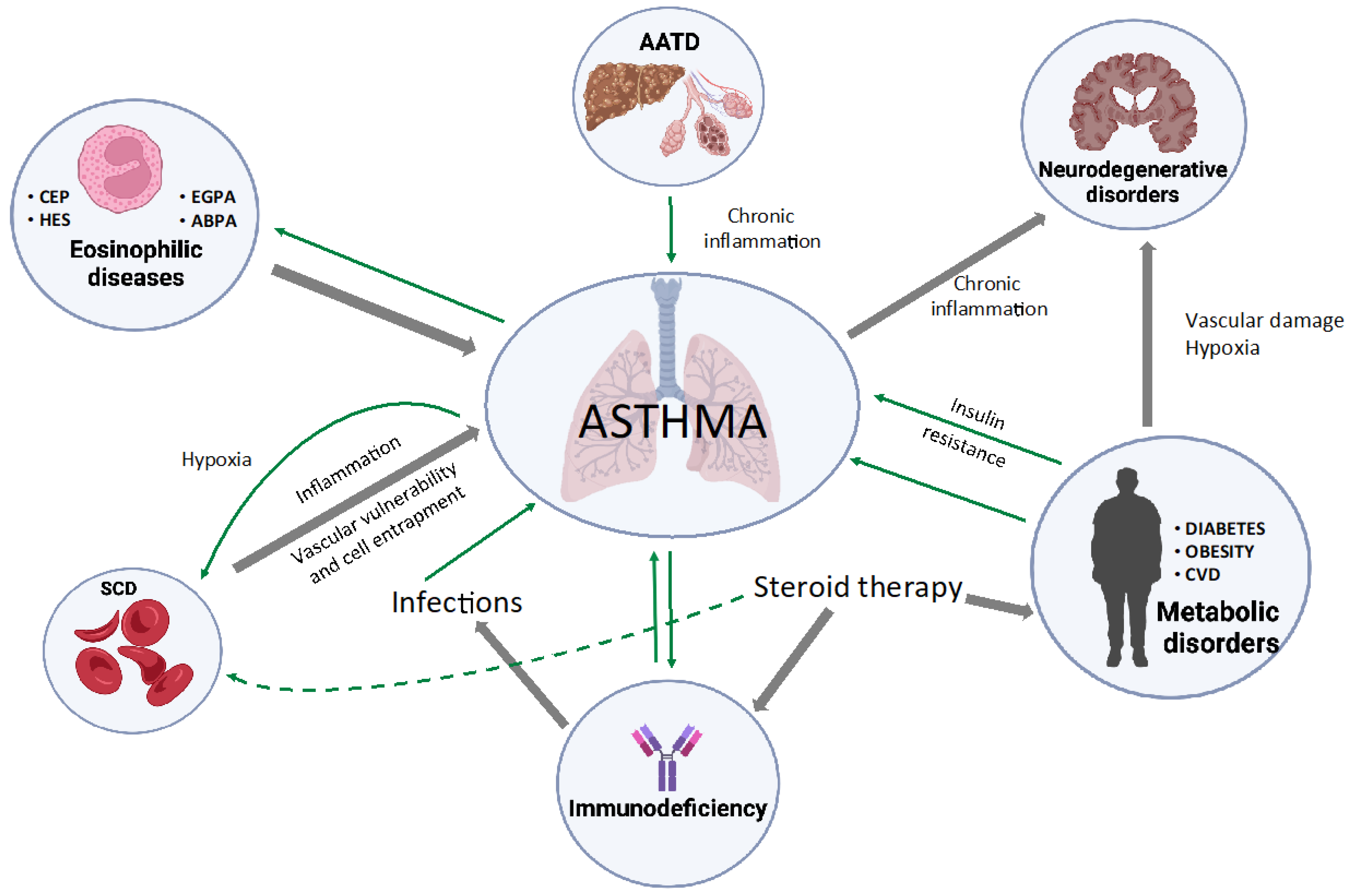
1. Introduction
2. Asthma in Eosinophilic Diseases
3. Alpha1-Antitrypsin Deficiency

| Asthma | AATD | |
|---|---|---|
| Prevalence | 3–9% [42] | 2–3% [33,38] |
| Onset | Variable, childhood > adulthood [42,43] | 40–53 ys, Adulthood > childhood [38] |
| Family history | Allergic diseases (atopic eczema, allergic rhinitis, asthma, food allergies) [42] | Asthma, emphysema, liver cirrhosis, vasculitis [40,41] |
| Lung function test (PFTs) | Airway hyper-responsiveness, reversible obstruction [42] | Fixed or nonreversible obstruction [40,41] |
| Concomitant allergies | 70% [42] | 22% [43] |
| Diagnosis | Clinical history, physical examination, PFTs, demonstration of reversible obstruction [42,43] | Genetic testing, and phenotype testing [38,43] |
| Therapy | ICS, LABA, LAMA, montelukast, Biologics [42,43] | As asthma + AAT augmentation therapy [38,43] |
4. Immunodeficiencies
5. Sickle Cell Disease
6. Neurodegenerative Diseases
7. Metabolic Syndrome and Obesity
8. Cardiovascular Disease
9. Conclusions
Author Contributions
Funding
Institutional Review Board Statement
Informed Consent Statement
Data Availability Statement
Conflicts of Interest
References
- The Global Initiative for Asthma. 2022. Available online: https://ginasthma.org/reports/ (accessed on 28 December 2022).
- Cardet, J.C.; Bulkhi, A.A.; Lockey, R.F. Nonrespiratory Comorbidities in Asthma. J. Allergy Clin. Immunol. Pract. 2021, 9, 3887–3897. [Google Scholar] [CrossRef] [PubMed]
- Caminati, M.; Senna, G.; Stefanizzi, G.; Bellamoli, R.; Longhi, S.; Chieco-Bianchi, F.; Guarnieri, G.; Tognella, S.; Olivieri, M.; Micheletto, C.; et al. Drop-out rate among patients treated with omalizumab for severe asthma: Literature review and real-life experience. BMC Pulm. Med. 2016, 16, 128. [Google Scholar] [CrossRef] [PubMed]
- Eger, K.; Kroes, J.A.; Brinke, A.T.; Bel, E.H. Long-Term Therapy Response to Anti–IL-5 Biologics in Severe Asthma—A Real-Life Evaluation. J. Allergy Clin. Immunol. Pract. 2021, 9, 1194–1200. [Google Scholar] [CrossRef] [PubMed]
- Menzies-Gow, A.N.; McBrien, C.; Unni, B.; Porsbjerg, C.M.; Al-Ahmad, M.; Ambrose, C.S.; Assing, K.D.; von Bülow, A.; Busby, J.; Cosio, B.G.; et al. Real World Biologic Use and Switch Patterns in Severe Asthma: Data from the International Severe Asthma Registry and the US CHRONICLE Study. J. Asthma Allergy 2022, 15, 63–78. [Google Scholar] [CrossRef] [PubMed]
- Olivieri, B.; Tinazzi, E.; Caminati, M.; Lunardi, C. Biologics for the Treatment of Allergic Conditions: Eosinophil Disorders. Immunol. Allergy Clin. North Am. 2020, 40, 649–665. [Google Scholar] [CrossRef]
- Rose, D.M.; Hrncir, D.E. Primary eosinophilic lung diseases. Allergy Asthma Proc. 2013, 34, 19–25. [Google Scholar] [CrossRef]
- Jennette, J.C.; Falk, R.J.; Bacon, P.A.; Basu, N.; Cid, M.C.; Ferrario, F.; Flores-Suarez, L.F.; Gross, W.L.; Guillevin, L.; Hagen, E.C.; et al. 2012 Revised International Chapel Hill Consensus Conference Nomenclature of Vasculitides. Arthritis Rheum. 2013, 17, 603–606. [Google Scholar] [CrossRef]
- Villanueva, K.L.V.; Espinoza, L.R. Eosinophilic Vasculitis. Curr. Rheumatol. Rep. 2020, 22, 5. [Google Scholar] [CrossRef]
- Khoury, P.; Grayson, P.C.; Klion, A.D. Eosinophils in vasculitis: Characteristics and roles in pathogenesis. Nat. Rev. Rheumatol. 2014, 10, 474–483. [Google Scholar] [CrossRef]
- Masi, A.T.; Hunder, G.G.; Lie, J.T.; Michel, B.A.; Bloch, D.A.; Arend, W.P.; Do, L.H.C.; Edworthy, S.M.; Fauci, A.S.; Leavitt, R.Y.; et al. The American College of Rheumatology 1990 criteria for the classification of churg-strauss syndrome (allergic granulomatosis and angiitis). Arthritis Rheum. 2010, 33, 1094–1100. [Google Scholar] [CrossRef]
- White, J.; Dubey, S. Eosinophilic granulomatosis with polyangiitis: A review. Autoimmun. Rev. 2022, 22, 103219. [Google Scholar] [CrossRef]
- Agarwal, R.; Sehgal, I.S.; Dhooria, S.; Aggarwal, A.N. Developments in the diagnosis and treatment of allergic bronchopulmonary aspergillosis. Expert Rev. Respir. Med. 2016, 10, 1317–1334. [Google Scholar] [CrossRef]
- Patel, G.; Greenberger, P.A. Allergic bronchopulmonary aspergillosis. Allergy Asthma Proc. 2019, 40, 421–424. [Google Scholar] [CrossRef]
- Suzuki, Y.; Suda, T. Eosinophilic pneumonia: A review of the previous literature, causes, diagnosis, and management. Allergol. Int. 2019, 68, 413–419. [Google Scholar] [CrossRef]
- Ogbogu, P.U.; Bochner, B.S.; Butterfield, J.H.; Gleich, G.J.; Huss-Marp, J.; Kahn, J.E.; Leiferman, K.M.; Nutman, T.B.; Pfab, F.; Ring, J.; et al. Hypereosinophilic syndrome: A multicenter, retrospective analysis of clinical characteristics and response to therapy. J. Allergy Clin. Immunol. 2009, 124, 1319–1325.e3. [Google Scholar] [CrossRef]
- Dulohery, M.M.; Patel, R.R.; Schneider, F.; Ryu, J.H. Lung involvement in hypereosinophilic syndromes. Respir. Med. 2011, 105, 114–121. [Google Scholar] [CrossRef]
- Shomali, W.; Gotlib, J. World Health Organization-defined eosinophilic disorders: 2019 update on diagnosis, risk stratification, and management. Am. J. Hematol. 2019, 94, 1149–1167. [Google Scholar] [CrossRef]
- Piggott, L.M.; Gill, C.M.; Kent, B.D. Differential diagnosis of pulmonary eosinophilia. In Eosinophilic Lung Diseases; European Respiratory Society: Sheffield, UK, 2022; pp. 19–36. [Google Scholar]
- Rytilä, P.; Metso, T.; Heikkinen, K.; Saarelainen, P.; Helenius, I.; Haahtela, T. Airway inflammation in patients with symptoms suggesting asthma but with normal lung function. Eur. Respir. J. 2000, 16, 824–830. [Google Scholar] [CrossRef]
- Park, S.-W.; Lee, Y.M.; Jang, A.S.; Lee, J.H.; Hwangbo, Y.; Kim, D.J.; Park, C.-S. Development of Chronic Airway Obstruction in Patients with Eosinophilic Bronchitis: A Prospective Follow-Up Study. Chest 2004, 125, 1998–2004. [Google Scholar] [CrossRef]
- Poletti, V. Eosinophilic bronchiolitis: Is it a new syndrome? Eur. Respir. J. 2013, 41, 1012–1013. [Google Scholar] [CrossRef]
- Silverman, E.K.; Sandhaus, R.A. Alpha1-Antitrypsin Deficiency. New Engl. J. Med. 2009, 360, 2749–2757. [Google Scholar] [CrossRef] [PubMed]
- American Thoracic Society; European Respiratory. American Thoracic Society/European Respiratory Society Statement: Standards for the diagnosis and management of individuals with alpha-1 antitrypsin deficiency. Am. J. Respir. Crit. Care Med. 2003, 168, 818–900. [Google Scholar] [CrossRef] [PubMed]
- Veith, M.; Tüffers, J.; Peychev, E.; Klemmer, A.; Kotke, V.; Janciauskiene, S.; Wilhelm, S.; Bals, R.; Koczulla, A.R.; Vogelmeier, C.F.; et al. The Distribution of Alpha-1 Antitrypsin Genotypes Between Patients with COPD/Emphysema, Asthma and Bronchiectasis. Int. J. Chronic Obstr. Pulm. Dis. 2020, 15, 2827–2836. [Google Scholar] [CrossRef] [PubMed]
- Hutchison, D. α1-Antitrypsin deficiency in Europe: Geographical distribution of Pi types S and Z. Respir. Med. 1998, 92, 367–377. [Google Scholar] [CrossRef] [PubMed]
- Sveger, T. Liver Disease in Alpha1-Antitrypsin Deficiency Detected by Screening of 200,000 Infants. N. Engl. J. Med. 1976, 294, 1316–1321. [Google Scholar] [CrossRef]
- Greulich, T.; Nell, C.; Hohmann, D.; Grebe, M.; Janciauskiene, S.; Koczulla, A.R.; Vogelmeier, C.F. The prevalence of diagnosed α1-antitrypsin deficiency and its comorbidities: Results from a large population-based database. Eur. Respir. J. 2017, 49, 1600154. [Google Scholar] [CrossRef]
- Siri, D.; Farah, H.; Hogarth, D.K. Distinguishing alpha1-antitrypsin deficiency from asthma. Ann. Allergy, Asthma Immunol. 2013, 111, 458–464. [Google Scholar] [CrossRef]
- Campos, M.A.; Alazemi, S.; Zhang, G.; Wanner, A.; Sandhaus, R.A. Effects of a Disease Management Program in Individuals with Alpha-1 Antitrypsin Deficiency. COPD J. Chronic Obstr. Pulm. Dis. 2009, 6, 31–40. [Google Scholar] [CrossRef]
- Eden, E.; Mitchell, D.; Mehlman, B.; Khouli, H.; Nejat, M.; Grieco, M.H.; Turino, G.M. Atopy, Asthma, and Emphysema in Patients with Severe α -1-Antitrypysin Deficiency. Am. J. Respir. Crit. Care Med. 1997, 156, 68–74. [Google Scholar] [CrossRef]
- Eden, E.; Holbrook, J.T.; Brantly, M.L.; Turino, G.M.; Wise, R.A. Prevalence of Alpha-1 Antitrypsin Deficiency in Poorly Controlled Asthma—Results from the ALA-ACRC Low-Dose Theophylline Trial. J. Asthma 2007, 44, 605–608. [Google Scholar] [CrossRef]
- Vianello, A.; Caminati, M.; Senna, G.; Arcolaci, A.; Chieco-Bianchi, F.; Ferrarotti, I.; Guarnieri, G.; Molena, B.; Crisafulli, E. Effect of α1 antitrypsin deficiency on lung volume decline in severe asthmatic patients undergoing biologic therapy. J. Allergy Clin. Immunol. Pract. 2021, 9, 1414–1416. [Google Scholar] [CrossRef]
- Suárez-Lorenzo, I.; De Castro, F.R.; Cruz-Niesvaara, D.; Herrera-Ramos, E.; Rodríguez-Gallego, C.; Carrillo-Diaz, T. Alpha 1 antitrypsin distribution in an allergic asthmatic population sensitized to house dust mites. Clin. Transl. Allergy 2018, 8, 44. [Google Scholar] [CrossRef]
- Van Veen, I.H.; Brinke, A.T.; van der Linden, A.; Rabe, K.; Bel, E. Deficient alpha-1-antitrypsin phenotypes and persistent airflow limitation in severe asthma. Respir. Med. 2006, 100, 1534–1539. [Google Scholar] [CrossRef]
- Needham, M. 1-Antitrypsin deficiency 3: Clinical manifestations and natural history. Thorax 2004, 59, 441–445. [Google Scholar] [CrossRef]
- Eden, E. Asthma and COPD in Alpha-1 Antitrypsin Deficiency. Evidence for the Dutch Hypothesis. COPD J. Chronic Obstr. Pulm. Dis. 2010, 7, 366–374. [Google Scholar] [CrossRef]
- Cazzola, M.; Stolz, D.; Rogliani, P.; Matera, M.G. α1-Antitrypsin deficiency and chronic respiratory disorders. Eur. Respir. Rev. 2020, 29, 190073. [Google Scholar] [CrossRef]
- Vignola, A.M.; Bonanno, A.; Mirabella, A.; Riccobono, L.; Mirabella, F.; Profita, M.; Bellia, V.; Bousquet, J.; Bonsignore, G. Increased Levels of Elastase and α1-Antitrypsin in Sputum of Asthmatic Patients. Am. J. Respir. Crit. Care Med. 1998, 157, 505–511. [Google Scholar] [CrossRef]
- Craig, T.J. Suspecting and Testing for Alpha-1 Antitrypsin Deficiency—An Allergist’s and/or Immunologist’s Perspective. J. Allergy Clin. Immunol. Pract. 2015, 3, 506–511. [Google Scholar] [CrossRef]
- Dirksen, A.; Piitulainen, E.; Parr, D.G.; Deng, C.; Wencker, M.; Shaker, S.B.; Stockley, R.A. Exploring the role of CT densitometry: A randomised study of augmentation therapy in 1-antitrypsin deficiency. Eur. Respir. J. 2009, 33, 1345–1353. [Google Scholar] [CrossRef]
- GINA ASTHMA. Available online: www.ginasthma.org (accessed on 1 December 2022).
- Eden, E.; Strange, C.; Holladay, B.; Xie, L. Asthma and allergy in alpha-1 antitrypsin deficiency. Respir. Med. 2006, 100, 1384–1391. [Google Scholar] [CrossRef]
- Tiotiu, A.; Salvator, H.; Jaussaud, R.; Jankowski, R.; Couderc, L.-J.; Catherinot, E.; Devillier, P. Efficacy of immunoglobulin replacement therapy and azithromycin in severe asthma with antibody deficiency. Allergol. Int. 2020, 69, 215–222. [Google Scholar] [CrossRef] [PubMed]
- Urm, S.-H.; Yun, H.D.; Fenta, Y.A.; Yoo, K.H.; Abraham, R.S.; Hagan, J.; Juhn, Y.J. Asthma and Risk of Selective IgA Deficiency or Common Variable Immunodeficiency: A Population-Based Case-Control Study. Mayo Clin. Proc. 2013, 88, 813–821. [Google Scholar] [CrossRef] [PubMed]
- Berger, M.; Geng, B.; Cameron, D.W.; Murphy, L.M.; Schulman, E.S. Primary immune deficiency diseases as unrecognized causes of chronic respiratory disease. Respir. Med. 2017, 132, 181–188. [Google Scholar] [CrossRef] [PubMed]
- Fardet, L.; Petersen, I.; Nazareth, I. Common Infections in Patients Prescribed Systemic Glucocorticoids in Primary Care: A Population-Based Cohort Study. PLoS Med. 2016, 13, e1002024. [Google Scholar] [CrossRef] [PubMed]
- Lee, S.-H.; Ban, G.-Y.; Kim, S.-C.; Chung, C.-G.; Lee, H.-Y.; Lee, J.-H.; Park, H.-S. Association between primary immunodeficiency and asthma exacerbation in adult asthmatics. Korean J. Intern. Med. 2020, 35, 449–456. [Google Scholar] [CrossRef]
- Kim, J.-H.; Ye, Y.-M.; Ban, G.-Y.; Shin, Y.-S.; Lee, H.Y.; Nam, Y.-H.; Lee, S.-K.; Cho, Y.S.; Jang, S.-H.; Jung, K.-S.; et al. Effects of Immunoglobulin Replacement on Asthma Exacerbation in Adult Asthmatics with IgG Subclass Deficiency. Allergy, Asthma Immunol. Res. 2017, 9, 526–533. [Google Scholar] [CrossRef]
- Dupin, C.; Marchand-Adam, S.; Favelle, O.; Costes, R.; Gatault, P.; Diot, P.; Grammatico-Guillon, L.; Guilleminault, L. Asthma and Hypogammaglobulinemia: An Asthma Phenotype with Low Type 2 Inflammation. J. Clin. Immunol. 2016, 36, 810–817. [Google Scholar] [CrossRef]
- Kishiyama, J.; Valacer, D.; Rundlesb, C.; Sperber, K.; Richmond, G.; Abramson, S.; Glovsky, M.; Stiehm, R.; Stocks, J.; Rosenberg, L.; et al. A Multicenter, Randomized, Double-Blind, Placebo-Controlled Trial of High-Dose Intravenous Immunoglobulin for Oral Corticosteroid-Dependent Asthma. Clin. Immunol. 1999, 91, 126–133. [Google Scholar] [CrossRef]
- De Franceschi, L.; Brugnara, C.; Rouyer-Fessard, P.; Jouault, H.; Beuzard, Y. Formation of dense erythrocytes in SAD mice exposed to chronic hypoxia: Evaluation of different therapeutic regimens and of a combination of oral clotrimazole and magnesium therapies. Blood 1999, 94, 4307–4313. [Google Scholar] [CrossRef]
- De Franceschi, L.; Bertoldi, M.; Matte, A.; Franco, S.S.; Pantaleo, A.; Ferru, E.; Turrini, F. Oxidative Stress andβ-Thalassemic Erythroid Cells behind the Molecular Defect. Oxidative Med. Cell. Longev. 2013, 2013, 985210. [Google Scholar] [CrossRef]
- Kalish, B.T.; Matte, A.; Andolfo, I.; Iolascon, A.; Weinberg, O.; Ghigo, A.; Cimino, J.; Siciliano, A.; Hirsch, E.; Federti, E.; et al. Dietary ω-3 fatty acids protect against vasculopathy in a transgenic mouse model of sickle cell disease. Haematologica 2015, 100, 870–880. [Google Scholar] [CrossRef]
- Matte, A.; Recchiuti, A.; Federti, E.; Koehl, B.; Mintz, T.; El Nemer, W.; Tharaux, P.-L.; Brousse, V.; Andolfo, I.; Lamolinara, A.; et al. Resolution of sickle cell disease–associated inflammation and tissue damage with 17R-resolvin D1. Blood 2019, 133, 252–265. [Google Scholar] [CrossRef]
- Matte, A.; Cappellini, M.D.; Iolascon, A.; Enrica, F.; De Franceschi, L. Emerging drugs in randomized controlled trials for sickle cell disease: Are we on the brink of a new era in research and treatment? Expert Opin. Investig. Drugs 2020, 29, 23–31. [Google Scholar] [CrossRef]
- Franceschi, L.; Cappellini, M.; Olivieri, O. Thrombosis and Sickle Cell Disease. Semin. Thromb. Hemost. 2011, 37, 226–236. [Google Scholar] [CrossRef]
- Platt, O.S. The Acute Chest Syndrome of Sickle Cell Disease. New Engl. J. Med. 2000, 342, 1904–1907. [Google Scholar] [CrossRef]
- Kato, G.J.; Piel, F.B.; Reid, C.D.; Gaston, M.H.; Ohene-Frempong, K.; Krishnamurti, L.; Smith, W.R.; Panepinto, J.A.; Weatherall, D.J.; Costa, F.F.; et al. Sickle cell disease. Nat. Rev. Dis. Prim. 2018, 4, 18010. [Google Scholar] [CrossRef]
- Mehari, A.; Klings, E.S. Chronic Pulmonary Complications of Sickle Cell Disease. Chest 2016, 149, 1313–1324. [Google Scholar] [CrossRef]
- Samarasinghe, A.E.; Rosch, J.W. Convergence of Inflammatory Pathways in Allergic Asthma and Sickle Cell Disease. Front. Immunol. 2020, 10, 3058. [Google Scholar] [CrossRef]
- Cohen, R.T.; Klings, E.S.; Strunk, R.C. Sickle cell disease: Wheeze or asthma? Asthma Res. Pract. 2015, 1, 14. [Google Scholar] [CrossRef]
- Arigliani, M.; Gupta, A. Management of chronic respiratory complications in children and adolescents with sickle cell disease. Eur. Respir. Rev. 2020, 29, 200054. [Google Scholar] [CrossRef]
- Strunk, R.C.; Cohen, R.T.; Cooper, B.P.; Rodeghier, M.; Kirkham, F.J.; Warner, J.O.; Stocks, J.; Kirkby, J.; Roberts, I.; Rosen, C.L.; et al. Wheezing Symptoms and Parental Asthma Are Associated with a Physician Diagnosis of Asthma in Children with Sickle Cell Anemia. J. Pediatr. 2014, 164, 821–826.e1. [Google Scholar] [CrossRef] [PubMed]
- Boyd, J.H.; Macklin, E.; Strunk, R.C.; DeBaun, M.R. Asthma is associated with acute chest syndrome and pain in children with sickle cell anemia. Blood 2006, 108, 2923–2927. [Google Scholar] [CrossRef] [PubMed]
- Vichinsky, E.P.; A Styles, L.; Colangelo, L.H.; Wright, E.C.; Castro, O.; Nickerson, B. Acute chest syndrome in sickle cell disease: Clinical presentation and course: Cooperative Study of Sickle Cell Disease. Blood 1997, 89, 1787–1792. [Google Scholar] [CrossRef] [PubMed]
- Vichinsky, E.P.; Neumayr, L.D.; Earles, A.N.; Williams, R.; Lennette, E.T.; Dean, D.; Nickerson, B.; Orringer, E.; McKie, V.; Bellevue, R.; et al. Causes and Outcomes of the Acute Chest Syndrome in Sickle Cell Disease. N. Engl. J. Med. 2000, 342, 1855–1865. [Google Scholar] [CrossRef]
- Cohen, R.T.; Madadi, A.; Blinder, M.A.; DeBaun, M.R.; Strunk, R.C.; Field, J.J. Recurrent, severe wheezing is associated with morbidity and mortality in adults with sickle cell disease. Am. J. Hematol. 2011, 86, 756–761. [Google Scholar] [CrossRef]
- Glassberg, J.A.; Chow, A.; Wisnivesky, J.; Hoffman, R.; DeBaun, M.R.; Richardson, L.D. Wheezing and asthma are independent risk factors for increased sickle cell disease morbidity. Br. J. Haematol. 2012, 159, 472–479. [Google Scholar] [CrossRef]
- Cohen, R.; Klings, E.S. Commentary: Heterogeneity of respiratory disease in children and young adults with sickle cell disease. Thorax 2018, 73, 503–503. [Google Scholar] [CrossRef]
- Bokov, P.; El Jurdi, H.; Denjoy, I.; Peiffer, C.; Medjahdi, N.; Holvoet, L.; Benkerrou, M.; Delclaux, C. Salbutamol Worsens the Autonomic Nervous System Dysfunction of Children with Sickle Cell Disease. Front. Physiol. 2020, 11, 31. [Google Scholar] [CrossRef]
- Green, C.E.; Turner, A.M. The role of the endothelium in asthma and chronic obstructive pulmonary disease (COPD). Respir. Res. 2017, 18, 20. [Google Scholar] [CrossRef]
- Brandow, A.M.; Carroll, C.P.; Creary, S.; Edwards-Elliott, R.; Glassberg, J.; Hurley, R.W.; Kutlar, A.; Seisa, M.; Stinson, J.; Strouse, J.J.; et al. American Society of Hematology 2020 guidelines for sickle cell disease: Management of acute and chronic pain. Blood Adv. 2020, 4, 2656–2701. [Google Scholar] [CrossRef]
- Pervaiz, A.; El-Baba, F.; Dhillon, K.; Daoud, A.; Soubani, A. Pulmonary Complications of Sickle Cell Disease: A Narrative Clinical Review. Adv. Respir. Med. 2021, 89, 173–187. [Google Scholar] [CrossRef]
- Ng, T.-P.; Chiam, P.-C.; Kua, E.-H. Mental disorders and asthma in the elderly: A population-based study. Int. J. Geriatr. Psychiatry 2006, 22, 668–674. [Google Scholar] [CrossRef]
- Eriksson, U.K.; Gatz, M.; Dickman, P.W.; Fratiglioni, L.; Pedersen, N.L. Asthma, Eczema, Rhinitis and the Risk for Dementia. Dement. Geriatr. Cogn. Disord. 2008, 25, 148–156. [Google Scholar] [CrossRef]
- Rusanen, M.; Ngandu, T.; Laatikainen, T.; Tuomilehto, J.; Soininen, H.; Kivipelto, M. Chronic Obstructive Pulmonary Disease and Asthma and the Risk of Mild Cognitive Impairment and Dementia: A Population Based CAIDE Study. Curr. Alzheimer Res. 2013, 10, 549–555. [Google Scholar] [CrossRef]
- Chen, M.-H.; Li, C.-T.; Tsai, C.-F.; Lin, W.-C.; Chang, W.-H.; Chen, T.-J.; Pan, T.-L.; Su, T.-P.; Bai, Y.-M. Risk of Dementia Among Patients with Asthma: A Nationwide Longitudinal Study. J. Am. Med. Dir. Assoc. 2014, 15, 763–767. [Google Scholar] [CrossRef]
- Lutsey, P.L.; Chen, N.; Mirabelli, M.C.; Lakshminarayan, K.; Knopman, D.S.; Vossel, K.A.; Gottesman, R.F.; Mosley, T.H.; Alonso, A. Impaired Lung Function, Lung Disease, and Risk of Incident Dementia. Am. J. Respir. Crit. Care Med. 2019, 199, 1385–1396. [Google Scholar] [CrossRef]
- Peng, Y.-H.; Wu, B.-R.; Su, C.-H.; Liao, W.-C.; Muo, C.-H.; Hsia, T.-C.; Kao, C.-H. Adult asthma increases dementia risk: A nationwide cohort study. J. Epidemiol. Community Health 2014, 69, 123–128. [Google Scholar] [CrossRef]
- Caldera-Alvarado, G.; Khan, D.A.; DeFina, L.F.; Pieper, A.; Brown, E.S. Relationship between asthma and cognition: The Cooper Center Longitudinal Study. Allergy 2013, 68, 545–548. [Google Scholar] [CrossRef]
- Irani, F.; Barbone, J.M.; Beausoleil, J.; Gerald, L. Is asthma associated with cognitive impairments? A meta-analytic review. J. Clin. Exp. Neuropsychol. 2017, 39, 965–978. [Google Scholar] [CrossRef]
- Peters, M.C.; McGrath, K.W.; A Hawkins, G.; Hastie, A.T.; Levy, B.D.; Israel, E.; Phillips, B.R.; Mauger, D.T.; A Comhair, S.; Erzurum, S.C.; et al. Plasma interleukin-6 concentrations, metabolic dysfunction, and asthma severity: A cross-sectional analysis of two cohorts. Lancet Respir. Med. 2016, 4, 574–584. [Google Scholar] [CrossRef]
- Tuleta, I.; Skowasch, D.; Aurich, F.; Eckstein, N.; Schueler, R.; Pizarro, C.; Schahab, N.; Nickenig, G.; Schaefer, C.; Pingel, S. Asthma is associated with atherosclerotic artery changes. PLoS ONE 2017, 12, e0186820. [Google Scholar] [CrossRef] [PubMed]
- Tattersall, M.C.; Evans, M.D.; Korcarz, C.E.; Mitchell, C.; Anderson, E.; DaSilva, D.F.; Salazar, L.P.; Gern, J.E.; Jackson, D.J.; Stein, J.H. Asthma is associated with carotid arterial injury in children: The Childhood Origins of Asthma (COAST) Cohort. PLoS ONE 2018, 13, e0204708. [Google Scholar] [CrossRef] [PubMed]
- Rosenkranz, M.A.; Dean, D.C.; Bendlin, B.B.; Jarjour, N.N.; Esnault, S.; Zetterberg, H.; Heslegrave, A.; Evans, M.D.; Davidson, R.J.; Busse, W.W. Neuroimaging and biomarker evidence of neurodegeneration in asthma. J. Allergy Clin. Immunol. 2022, 149, 589–598.e6. [Google Scholar] [CrossRef] [PubMed]
- Rosenkranz, M.A.; Busse, W.W.; Sheridan, J.F.; Crisafi, G.M.; Davidson, R.J. Are There Neurophenotypes for Asthma? Functional Brain Imaging of the Interaction between Emotion and Inflammation in Asthma. PLoS ONE 2012, 7, e40921. [Google Scholar] [CrossRef]
- Kroll, J.L.; Steele, A.M.; Pinkham, A.E.; Choi, C.; Khan, D.A.; Patel, S.V.; Chen, J.R.; Aslan, S.; Brown, E.S.; Ritz, T. Hippocampal metabolites in asthma and their implications for cognitive function. NeuroImage Clin. 2018, 19, 213–221. [Google Scholar] [CrossRef]
- Brown, E.S.; Woolston, D.J.; Frol, A.B. Amygdala Volume in Patients Receiving Chronic Corticosteroid Therapy. Biol. Psychiatry 2008, 63, 705–709. [Google Scholar] [CrossRef]
- Marschallinger, J.; Altendorfer, B.; Rockenstein, E.; Holztrattner, M.; Garnweidner-Raith, J.; Pillichshammer, N.; Leister, I.; Hutter-Paier, B.; Strempfl, K.; Unger, M.S.; et al. The Leukotriene Receptor Antagonist Montelukast Reduces Alpha-Synuclein Load and Restores Memory in an Animal Model of Dementia with Lewy Bodies. Neurotherapeutics 2020, 17, 1061–1074. [Google Scholar] [CrossRef]
- Karamzad, N.; Izadi, N.; Sanaie, S.; Ahmadian, E.; Eftekhari, A.; Sullman, M.J.; Safiri, S. Asthma and metabolic syndrome: A comprehensive systematic review and meta-analysis of observational studies. J. Cardiovasc. Thorac. Res. 2020, 12, 120–128. [Google Scholar] [CrossRef]
- De Boer, G.M.; Tramper-Stranders, G.A.; Houweling, L.; van Zelst, C.M.; Pouw, N.; Verhoeven, G.T.; Klerk, B.M.B.-D.; Veen, J.C.I.; van Rossum, E.F.; Hendriks, R.W.; et al. Adult but not childhood onset asthma is associated with the metabolic syndrome, independent from body mass index. Respir. Med. 2021, 188, 106603. [Google Scholar] [CrossRef]
- Brumpton, B.M.; Camargo, C.A.; Romundstad, P.R.; Langhammer, A.; Chen, Y.; Mai, X.-M. Metabolic syndrome and incidence of asthma in adults: The HUNT study. Eur. Respir. J. 2013, 42, 1495–1502. [Google Scholar] [CrossRef]
- Wu, T.D.; Brigham, E.P.; Keet, C.A.; Brown, T.T.; Hansel, N.N.; McCormack, M.C. Association Between Prediabetes/Diabetes and Asthma Exacerbations in a Claims-Based Obese Asthma Cohort. J. Allergy Clin. Immunol. Pract. 2019, 7, 1868–1873.e5. [Google Scholar] [CrossRef]
- Yang, G.; Han, Y.-Y.; Forno, E.; Yan, Q.; Rosser, F.; Chen, W.; Celedón, J.C. Glycated Hemoglobin A1c, Lung Function, and Hospitalizations Among Adults with Asthma. J. Allergy Clin. Immunol. Pract. 2020, 8, 3409–3415.e1. [Google Scholar] [CrossRef]
- Thuesen, B.H.; Husemoen, L.L.N.; Hersoug, L.-G.; Pisinger, C.; Linneberg, A. Insulin resistance as a predictor of incident asthma-like symptoms in adults. Clin. Exp. Allergy 2009, 39, 700–707. [Google Scholar] [CrossRef]
- Wu, T.D. Diabetes, insulin resistance, and asthma: A review of potential links. Curr. Opin. Pulm. Med. 2021, 27, 29–36. [Google Scholar] [CrossRef]
- Christiansen, S.C.; Schatz, M.; Yang, S.-J.; Ngor, E.; Chen, W.; Zuraw, B. Hypertension and Asthma: A Comorbid Relationship. J. Allergy Clin. Immunol. Pract. 2016, 4, 76–81. [Google Scholar] [CrossRef]
- Madhur, M.S.; Lob, H.E.; McCann, L.A.; Iwakura, Y.; Blinder, Y.; Guzik, T.J.; Harrison, D.G. Interleukin 17 Promotes Angiotensin II–Induced Hypertension and Vascular Dysfunction. Hypertension 2010, 55, 500–507. [Google Scholar] [CrossRef]
- Chesné, J.; Braza, F.; Chadeuf, G.; Mahay, G.; Cheminant, M.-A.; Loy, J.; Brouard, S.; Sauzeau, V.; Loirand, G.; Magnan, A. Prime role of IL-17A in neutrophilia and airway smooth muscle contraction in a house dust mite–induced allergic asthma model. J. Allergy Clin. Immunol. 2015, 135, 1643–1645.e5. [Google Scholar] [CrossRef]
- Irvin, C.; Zafar, I.; Good, J.; Rollins, D.; Christianson, C.; Gorska, M.M.; Martin, R.J.; Alam, R. Increased frequency of dual-positive TH2/TH17 cells in bronchoalveolar lavage fluid characterizes a population of patients with severe asthma. J. Allergy Clin. Immunol. 2014, 134, 1175–1186.e7. [Google Scholar] [CrossRef]
- Myou, S.; Fujimura, M.; Kamio, Y.; Kita, T.; Watanabe, K.; Ishiura, Y.; Hashimoto, T.; Nakao, S. Effect of candesartan, a type 1 angiotensin II receptor antagonist, on bronchial hyper-responsiveness to methacholine in patients with bronchial asthma. Br. J. Clin. Pharmacol. 2002, 54, 622–626. [Google Scholar] [CrossRef]
- Kankaanranta, H.; Kauppi, P.; Tuomisto, L.E.; Ilmarinen, P. Emerging Comorbidities in Adult Asthma: Risks, Clinical Associations, and Mechanisms. Mediat. Inflamm. 2016, 2016, 3690628. [Google Scholar] [CrossRef]
- Enright, P.L.; Ward, B.J.; Tracy, R.P.; Lasser, E.C. Asthma and Its Association with Cardiovascular Disease in the Elderly. J. Asthma 1996, 33, 45–53. [Google Scholar] [CrossRef] [PubMed]
- Picado, C.; Deulofeu, R.; Casals, E.; Lleonart, R.; Agustí, M.; Quintó, L.; Mullol, J. Lipid and protein metabolism in asthma. Effects of diet and corticosteroid therapy. Allergy 1999, 54, 569–575. [Google Scholar] [CrossRef] [PubMed]
- Su, X.; Ren, Y.; Li, M.; Zhao, X.; Kong, L.; Kang, J. Association between lipid profile and the prevalence of asthma: A meta-analysis and systemic review. Curr. Med. Res. Opin. 2018, 34, 423–433. [Google Scholar] [CrossRef] [PubMed]
- Van Zelst, C.M.; de Boer, G.M.; Türk, Y.; van Huisstede, A.; Veen, J.C.C.M.I.; Birnie, E.; de Klerk, B.M.B.; Stranders, G.A.T.; Braunstahl, G.-J. Association between elevated serum triglycerides and asthma in patients with obesity: An explorative study. Allergy Asthma Proc. 2021, 42, e71–e76. [Google Scholar] [CrossRef]
- Alabed, M.; Elemam, N.M.; Ramakrishnan, R.K.; Sharif-Askari, N.S.; Kashour, T.; Hamid, Q.; Halwani, R. Therapeutic effect of statins on airway remodeling during asthma. Expert Rev. Respir. Med. 2022, 16, 17–24. [Google Scholar] [CrossRef]
- Serafino-Agrusa, L. Asthma and metabolic syndrome: Current knowledge and future perspectives. World J. Clin. Cases 2015, 3, 285–292. [Google Scholar] [CrossRef]
- Kim, J.-H.; Wee, J.-H.; Choi, H.G.; Park, J.-Y.; Hwang, Y.I.; Jang, S.H.; Jung, K.-S. Association Between Statin Medication and Asthma/Asthma Exacerbation in a National Health Screening Cohort. J. Allergy Clin. Immunol. Pract. 2021, 9, 2783–2791. [Google Scholar] [CrossRef]
- Peters, U.; Dixon, A.E.; Forno, E. Obesity and asthma. J. Allergy Clin. Immunol. 2018, 141, 1169–1179. [Google Scholar] [CrossRef]
- Dixon, A.E.; Poynter, M.E. Mechanisms of Asthma in Obesity. Pleiotropic Aspects of Obesity Produce Distinct Asthma Phenotypes. Am. J. Respir. Cell Mol. Biol. 2016, 54, 601–608. [Google Scholar] [CrossRef]
- Miethe, S.; Karsonova, A.; Karaulov, A.; Renz, H. Obesity and asthma. J. Allergy Clin. Immunol. 2020, 146, 685–693. [Google Scholar] [CrossRef]
- Bassi, M.; Singh, S. Impact of Obesity on Response to Biologic Therapies in Patients with Inflammatory Bowel Diseases. Biodrugs 2022, 36, 197–203. [Google Scholar] [CrossRef]
- Menzies-Gow, A.; Corren, J.; Bourdin, A.; Chupp, G.; Israel, E.; Wechsler, M.E.; Brightling, C.E.; Griffiths, J.M.; Hellqvist, A.; Bowen, K.; et al. Tezepelumab in Adults and Adolescents with Severe, Uncontrolled Asthma. N. Engl. J. Med. 2021, 384, 1800–1809. [Google Scholar] [CrossRef]
- Ross, R. Atherosclerosis as an inflammatory disease. N. Engl. J. Med. 1999, 340, 115–126. [Google Scholar] [CrossRef]
- Hansson, G.K. Inflammation, Atherosclerosis, and Coronary Artery Disease. N. Engl. J. Med. 2005, 352, 1685–1695. [Google Scholar] [CrossRef]
- Croce, K.; Libby, P. Inflammation in Atherosclerosis. Arter. Thromb. Vasc. Biol. 2012, 32, 2045–2051. [Google Scholar] [CrossRef]
- Ridker, P.M.; Everett, B.M.; Thuren, T.; MacFadyen, J.G.; Chang, W.H.; Ballantyne, C.; Fonseca, F.; Nicolau, J.; Koenig, W.; Anker, S.D.; et al. Antiinflammatory Therapy with Canakinumab for Atherosclerotic Disease. N. Engl. J. Med. 2017, 377, 1119–1131. [Google Scholar] [CrossRef]
- Shi, G.-P.; Bot, I.; Kovanen, P.T. Mast cells in human and experimental cardiometabolic diseases. Nat. Rev. Cardiol. 2015, 12, 643–658. [Google Scholar] [CrossRef]
- Niccoli, G.; Montone, R.A.; Sabato, V.; Crea, F. Role of Allergic Inflammatory Cells in Coronary Artery Disease. Circulation 2018, 138, 1736–1748. [Google Scholar] [CrossRef]
- Sun, J.; Sukhova, G.K.; Wolters, P.J.; Yang, M.; Kitamoto, S.; Libby, P.; A Macfarlane, L.; Clair, J.M.-S.; Shi, G.-P. Mast cells promote atherosclerosis by releasing proinflammatory cytokines. Nat. Med. 2007, 13, 719–724. [Google Scholar] [CrossRef]
- Sun, J.; Sukhova, G.K.; Yang, M.; Wolters, P.J.; MacFarlane, L.A.; Libby, P.; Sun, C.; Zhang, Y.; Liu, J.; Ennis, T.L.; et al. Mast cells modulate the pathogenesis of elastase-induced abdominal aortic aneurysms in mice. J. Clin. Investig. 2007, 117, 3359–3368. [Google Scholar] [CrossRef]
- Wang, J.; Sjöberg, S.; Tia, V.; Secco, B.; Chen, H.; Yang, M.; Sukhova, G.K.; Shi, G.-P. Pharmaceutical stabilization of mast cells attenuates experimental atherogenesis in low-density lipoprotein receptor-deficient mice. Atherosclerosis 2013, 229, 304–309. [Google Scholar] [CrossRef] [PubMed]
- Furukawa, H.; Wada, K.; Tada, Y.; Kuwabara, A.; Sato, H.; Ai, J.; Lawton, M.T.; Hashimoto, T. Mast Cell Promotes the Development of Intracranial Aneurysm Rupture. Stroke 2020, 51, 3332–3339. [Google Scholar] [CrossRef] [PubMed]
- Marx, C.; Novotny, J.; Salbeck, D.; Zellner, K.R.; Nicolai, L.; Pekayvaz, K.; Kilani, B.; Stockhausen, S.; Bürgener, N.; Kupka, D.; et al. Eosinophil-platelet interactions promote atherosclerosis and stabilize thrombosis with eosinophil extracellular traps. Blood 2019, 134, 1859–1872. [Google Scholar] [CrossRef] [PubMed]
- Gudbjartsson, D.; Bjornsdottir, U.S.; Halapi, E.; Helgadottir, A.; Sulem, P.; Jonsdottir, G.M.; Thorleifsson, G.; Helgadottir, H.; Steinthorsdottir, V.; Stefansson, H.; et al. Sequence variants affecting eosinophil numbers associate with asthma and myocardial infarction. Nat. Genet. 2009, 41, 342–347. [Google Scholar] [CrossRef]
- Madjid, M.; Awan, I.; Willerson, J.T.; Casscells, S.W. Leukocyte count and coronary heart disease: Implications for risk assessment. J. Am. Coll. Cardiol. 2004, 44, 1945–1956. [Google Scholar] [CrossRef]
- Madjid, M.; Fatemi, O. Components of the complete blood count as risk predictors for coronary heart disease: In-depth review and update. Tex. Hear. Inst. J. 2013, 40, 17–29. [Google Scholar]
- Pongdee, T.; Manemann, S.M.; Decker, P.A.; Larson, N.B.; Moon, S.; Killian, J.M.; Liu, H.; Kita, H.; Bielinski, S.J. Rethinking blood eosinophil counts: Epidemiology, associated chronic diseases, and increased risks of cardiovascular disease. J. Allergy Clin. Immunol. Glob. 2022, 1, 233–240. [Google Scholar] [CrossRef]
- Niccoli, G.; Calvieri, C.; Flego, D.; Scalone, G.; Imaeva, A.; Sabato, V.; Schiavino, D.; Liuzzo, G.; Crea, F. Allergic Inflammation Is Associated with Coronary Instability and a Worse Clinical Outcome After Acute Myocardial Infarction. Circ. Cardiovasc. Interv. 2015, 8, e002554. [Google Scholar] [CrossRef]
- Pizzolo, F.; Castagna, A.; Olivieri, O.; Girelli, D.; Friso, S.; Stefanoni, F.; Udali, S.; Munerotto, V.; Baroni, M.; Cetera, V.; et al. Basophil Blood Cell Count Is Associated with Enhanced Factor II Plasma Coagulant Activity and Increased Risk of Mortality in Patients with Stable Coronary Artery Disease: Not Only Neutrophils as Prognostic Marker in Ischemic Heart Disease. J. Am. Hear. Assoc. 2021, 10, e018243. [Google Scholar] [CrossRef]
- Morshed, M.; Hlushchuk, R.; Simon, D.; Walls, A.F.; Obata-Ninomiya, K.; Karasuyama, H.; Djonov, V.; Eggel, A.; Kaufmann, T.; Simon, H.-U.; et al. NADPH Oxidase–Independent Formation of Extracellular DNA Traps by Basophils. J. Immunol. 2014, 192, 5314–5323. [Google Scholar] [CrossRef]
- Moreno-Sanchez, D.; Hernandez-Ruiz, L.; Ruiz, F.A.; Docampo, R. Polyphosphate Is a Novel Pro-inflammatory Regulator of Mast Cells and Is Located in Acidocalcisomes. J. Biol. Chem. 2012, 287, 28435–28444. [Google Scholar] [CrossRef]
- Smith, S.A.; Morrissey, J.H. Polyphosphate: A new player in the field of hemostasis. Curr. Opin. Hematol. 2014, 21, 388–394. [Google Scholar] [CrossRef]
- Onufrak, S.; Abramson, J.; Vaccarino, V. Adult-onset asthma is associated with increased carotid atherosclerosis among women in the atherosclerosis risk in communities (ARIC) study. Atherosclerosis 2007, 195, 129–137. [Google Scholar] [CrossRef]
- Pacholczak-Madej, R.; Kuszmiersz, P.; Iwaniec, T.; Zaręba, L.; Zarychta, J.; Walocha, J.; Dropiński, J.; Bazan-Socha, S. Endothelial Dysfunction and Pentraxin-3 in Clinically Stable Adult Asthma Patients. J. Investig. Allergol. Clin. Immunol. 2021, 31, 417–425. [Google Scholar] [CrossRef]
- Bazan-Socha, S.; Mastalerz, L.; Cybulska, A.; Zareba, L.; Kremers, R.; Zabczyk, M.; Pulka, G.; Iwaniec, T.; Hemker, C.; Undas, A. Asthma is associated with enhanced thrombin formation and impaired fibrinolysis. Clin. Exp. Allergy 2016, 46, 932–944. [Google Scholar] [CrossRef]
- Rhee, T.-M.; Choi, E.-K.; Han, K.-D.; Lee, S.-R.; Oh, S. Impact of the Combinations of Allergic Diseases on Myocardial Infarction and Mortality. J. Allergy Clin. Immunol. Pract. 2021, 9, 872–880.e4. [Google Scholar] [CrossRef]
- Tattersall, M.C.; Guo, M.; Korcarz, C.E.; Gepner, A.D.; Kaufman, J.D.; Liu, K.J.; Barr, R.G.; Donohue, K.M.; McClelland, R.L.; Delaney, J.A.; et al. Asthma Predicts Cardiovascular Disease Events: The multi-ethnic study of atherosclerosis. Arter. Thromb. Vasc. Biol. 2015, 35, 1520–1525. [Google Scholar] [CrossRef]
- Strand, L.B.; Tsai, M.K.; Wen, C.P.; Chang, S.-S.; Brumpton, B.M. Is having asthma associated with an increased risk of dying from cardiovascular disease? A prospective cohort study of 446 346 Taiwanese adults. BMJ Open 2018, 8, e019992. [Google Scholar] [CrossRef]
- Wee, J.H.; Park, M.W.; Min, C.; Byun, S.H.; Park, B.; Choi, H.G. Association between asthma and cardiovascular disease. Eur. J. Clin. Investig. 2021, 51, e13396. [Google Scholar] [CrossRef]
- Zhang, B.; Li, Z.-F.; An, Z.-Y.; Zhang, L.; Wang, J.-Y.; Hao, M.-D.; Jin, Y.-J.; Li, D.; Song, A.-J.; Ren, Q.; et al. Association Between Asthma and All-Cause Mortality and Cardiovascular Disease Morbidity and Mortality: A Meta-Analysis of Cohort Studies. Front. Cardiovasc. Med. 2022, 9, 861798. [Google Scholar] [CrossRef]
- Gao, S.; Wang, C.; Li, W.; Shu, S.; Zhou, J.; Yuan, Z.; Wang, L. Allergic asthma aggravated atherosclerosis increases cholesterol biosynthesis and foam cell formation in apolipoprotein E-deficient mice. Biochem. Biophys. Res. Commun. 2019, 519, 861–867. [Google Scholar] [CrossRef]
- Gunstone, R.F. Right heart pressures in bronchial asthma. Thorax 1971, 26, 39–45. [Google Scholar] [CrossRef] [PubMed][Green Version]
- Karasu, B.B.; Aydıncak, H.T. Right ventricular-pulmonary arterial uncoupling in mild-to-moderate asthma. J. Asthma 2022, 12, 543–552. [Google Scholar] [CrossRef] [PubMed]
- Harkness, L.M.; Kanabar, V.; Sharma, H.S.; Westergren-Thorsson, G.; Larsson-Callerfelt, A.-K. Pulmonary vascular changes in asthma and COPD. Pulm. Pharmacol. Ther. 2014, 29, 144–155. [Google Scholar] [CrossRef] [PubMed]
- Shedeed, S.A. Right Ventricular Function in Children with Bronchial Asthma: A Tissue Doppler Echocardiographic Study. Pediatr. Cardiol. 2010, 31, 1008–1015. [Google Scholar] [CrossRef]
- Ozde, C.; Dogru, M.; Ozde, Ş.; Kayapinar, O.; Kaya, A.; Korkmaz, A. Subclinical right ventricular dysfunction in intermittent and persistent mildly asthmatic children on tissue Doppler echocardiography and serum NT -pro BNP: Observational study. Pediatr. Int. 2018, 60, 1024–1032. [Google Scholar] [CrossRef]
- Suissa, S.; Assimes, T.; Brassard, P.; Ernst, P. Inhaled corticosteroid use in asthma and the prevention of myocardial infarction. Am. J. Med. 2003, 115, 377–381. [Google Scholar] [CrossRef]

Disclaimer/Publisher’s Note: The statements, opinions and data contained in all publications are solely those of the individual author(s) and contributor(s) and not of MDPI and/or the editor(s). MDPI and/or the editor(s) disclaim responsibility for any injury to people or property resulting from any ideas, methods, instructions or products referred to in the content. |
© 2023 by the authors. Licensee MDPI, Basel, Switzerland. This article is an open access article distributed under the terms and conditions of the Creative Commons Attribution (CC BY) license (https://creativecommons.org/licenses/by/4.0/).
Share and Cite
Maule, M.; Olivieri, B.; Guarnieri, G.; De Franceschi, L.; Martinelli, N.; Vaia, R.; Argentino, G.; Vianello, A.; Senna, G.; Caminati, M. Hidden Comorbidities in Asthma: A Perspective for a Personalized Approach. J. Clin. Med. 2023, 12, 2294. https://doi.org/10.3390/jcm12062294
Maule M, Olivieri B, Guarnieri G, De Franceschi L, Martinelli N, Vaia R, Argentino G, Vianello A, Senna G, Caminati M. Hidden Comorbidities in Asthma: A Perspective for a Personalized Approach. Journal of Clinical Medicine. 2023; 12(6):2294. https://doi.org/10.3390/jcm12062294
Chicago/Turabian StyleMaule, Matteo, Bianca Olivieri, Gabriella Guarnieri, Lucia De Franceschi, Nicola Martinelli, Rachele Vaia, Giuseppe Argentino, Andrea Vianello, Gianenrico Senna, and Marco Caminati. 2023. "Hidden Comorbidities in Asthma: A Perspective for a Personalized Approach" Journal of Clinical Medicine 12, no. 6: 2294. https://doi.org/10.3390/jcm12062294
APA StyleMaule, M., Olivieri, B., Guarnieri, G., De Franceschi, L., Martinelli, N., Vaia, R., Argentino, G., Vianello, A., Senna, G., & Caminati, M. (2023). Hidden Comorbidities in Asthma: A Perspective for a Personalized Approach. Journal of Clinical Medicine, 12(6), 2294. https://doi.org/10.3390/jcm12062294