Article

Preimplantation Genetic Testing (PGT) and Prenatal Diagnosis of Schaaf-Yang Syndrome: A Report of Three Families and a Research on Genotype–Phenotype Correlations

1
Obstetrics and Gynecology Hospital, Institute of Reproduction and Development, Fudan University, Shanghai 200030, China
2
Shanghai Key Laboratory of Embryo Original Diseases, Shanghai 200030, China
3
International Peace Maternity and Child Health Hospital, School of Medicine, Shanghai Jiao Tong University, Shanghai 200030, China
4
Research Units of Embryo Original Diseases, Chinese Academy of Medical Sciences (No. 2019RU056), Shanghai 200030, China
*
Authors to whom correspondence should be addressed.
J. Clin. Med. 2023, 12(4), 1688; https://doi.org/10.3390/jcm12041688
Submission received: 28 December 2022 / Revised: 10 February 2023 / Accepted: 17 February 2023 / Published: 20 February 2023
(This article belongs to the Section Reproductive Medicine & Andrology)

Abstract

:
Schaaf-Yang Syndrome (SYS) is a genetic disorder caused by truncating pathogenic variants in the paternal allele of the maternally imprinted, paternally expressed gene MAGEL2 and is characterized by genital hypoplasia, neonatal hypotonia, developmental delay, intellectual disability, autism spectrum disorder (ASD), and other features. In this study, eleven SYS patients from three families were enrolled and comprehensive clinical features were gathered regarding each family. Whole-exome sequencing (WES) was performed for the definitive molecular diagnosis of the disease. Identified variants were validated using Sanger sequencing. Three couples underwent PGT for monogenic diseases (PGT-M) and/or a prenatal diagnosis. Haplotype analysis was performed to deduce the embryo’s genotype by using the short tandem repeats (STRs) identified in each sample. The prenatal diagnosis results showed that the fetus in each case did not carry pathogenic variants, and all the babies of the three families were born at full term and were healthy. We also performed a review of SYS cases. In addition to the 11 patients in our study, a total of 127 SYS patients were included in 11 papers. We summarized all variant sites and clinical symptoms thus far, and conducted a genotype–phenotype correlation analysis. Our results also indicated that the variation in phenotypic severity may depend on the specific location of the truncating variant, suggestive of a genotype–phenotype association.

1. Introduction

A group of imprinted genes necessary for typical mammalian neurodevelopment can be found on chromosome 15q11-q13 [1]. Prader–Willi syndrome (PWS, OMIM #176270) is caused by paternal deletions of this area [2]. Truncating variants in MAGEL2, one of the paternally expressed genes in this region, were shown to induce Schaaf-Yang syndrome (SYS, OMIM #615547). The first four patients with truncating viariants in the paternal copy of MAGEL2 were described by Schaaf et al. in 2013 [3]. These patients were referred to as having a “PW-like syndrome” because several of their phenotypic traits, such as hypogonadism, neonatal hypotonia, feeding issues, developmental delay, and intellectual disability, mirrored those of patients with PWS [3]. Their distinctive phenotypic profile can be recognized more accurately as there are more individuals with pathogenic truncating variants of MAGEL2. Patients with SYS had higher rates of autism spectrum disorder (ASD) than those with PWS [4,5]. Truncating variants in MAGEL2 cause severe arthrogryposis with respiratory distress that results in neonatal mortality. Moreover, despite a variety of widespread clinical signs, including endocrine disorders such as hypopituitarism, SYS individuals exhibit a broad phenotypic spectrum [3,4,6]. MAGEL2 variants were recently discovered in patients with Chitayat–Hall syndrome (CHS), broadening the clinical breadth of MAGEL2-related illnesses. However, CHS and SYS seem to have the most common clinical features and genetic causes, so many researchers regard them as the same syndrome [7,8].
MAGEL2 is a single-exon, highly GC-rich gene that encodes a protein involved in endosomal protein recycling and is a member of the MAGE family of ubiquitin ligase regulators [9]. As a matrilineal-imprinted gene (the maternal chromosome is methylated), MAGEL2 is only expressed by unmethylated paternal alleles, and the individual is only impacted when the variation occurs on the paternal allele [3]. Variants in the maternal-imprinted allele can cause the symptoms to skip several generations in contrast to autosomal dominant inheritance. However, for male individuals carrying a deleterious MAGEL2 variant, 50% of their offspring will be clinically affected [4]. Even though the majority of SYS patients are able to survive the illness, fatal cases cannot be disregarded. Neurodevelopmental delay with unexplained dyspnea is clinically extremely hazardous, leading to death in the neonatal period of SYS patients [9,10]. As SYS does not comply with Mendelian inheritance laws and the underlying pathological mechanisms and genotype–phenotype correlations remain to be elucidated, clinical suspicion of SYS in the differential diagnosis in pediatric patients with hypotonia and developmental delay/intellectual disability is not high because of the physicians’ unfamiliarity [11].
Preimplantation genetic testing for monogenic disorders (PGT-M) and prenatal diagnosis are essential for families with SYS-affected members to prevent the transmission of the variant [12,13,14]. PGT-M is a component of the in vitro fertilization (IVF) process that genetically profiles oocytes or embryos before implantation [15,16,17]. It is generally accessible for any monogenic condition when the causative variant is known [18,19,20]. Couples who decide to use PGT-M for IVF have two major challenges: identifying the disease-causing variation and performing an appropriate biopsy and subsequent genetic analysis that prevents any harm to embryo viability [21].
Here, we report on eleven patients with SYS from three Chinese families, along with the variant spectrum and phenotype profile of SYS. In addition, we performed PGT and/or a prenatal diagnosis in three couples from families who suffered from SYS and successfully prevented the transmission of the truncating variants in MAGEL2. Moreover, we performed a review of SYS-related literature, summarized all variant sites and clinical symptoms of SYS thus far, and conducted a genotype–phenotype correlation analysis.

2. Materials and Methods

2.1. Study Subjects

Three families with SYS were recruited from the International Peace Maternity and Child Health Hospital in this study. The study was authorized by the International Peace Maternity and Child Health Hospital, and all participants provided informed consent in accordance with ethical norms.

2.2. DNA Extraction and Variant Detection

These methods have been described in detail in our previous research [21].

2.3. In Vitro Fertilization (IVF)

Controlled ovarian hyperstimulation (COH) was conducted with human chorionic gonadotropin (hCG). Gonadotropin-releasing hormone (GnRH) antagonists constitute a multiple-dose flexible regimen for the prevention of ovarian hyperstimulation syndrome (OHSS). Intracytoplasmic sperm injection (ICSI) was performed on metaphase II (MII) oocytes, and the resultant embryos were cultured to the blastocyst stage for biopsy. The biopsied blastocysts were cryopreserved for further embryo transfer cycles utilizing the vitrification procedure with individual tubes containing single blastocysts.

2.4. Identity Testing and Haplotype Analysis

According to the instructions, short tandem repeats (STRs) were utilized for identity testing and detecting probable maternal contamination with an identification detection kit (R1004T; GENESKY, Shanghai, China). The general sample processing kit for gene sequencing (Yikon Genomics, Shanghai, China) was used to perform whole-genome amplification on each embryo biopsy sample according to the manufacturer’s instructions. Using a 1.5 Mb custom probe encompassing 350 kb upstream to 350 kb downstream of the MAGEL2 gene, multiple displacement amplification (MDA) products and gDNA libraries were created and collected. Haplotype analysis was performed to confirm the carrier status in the family and the embryonic inheritance based on informative STRs that cosegregate with the detected variant and are found in the same region in both parents but are homozygous in one and heterozygous in the other.

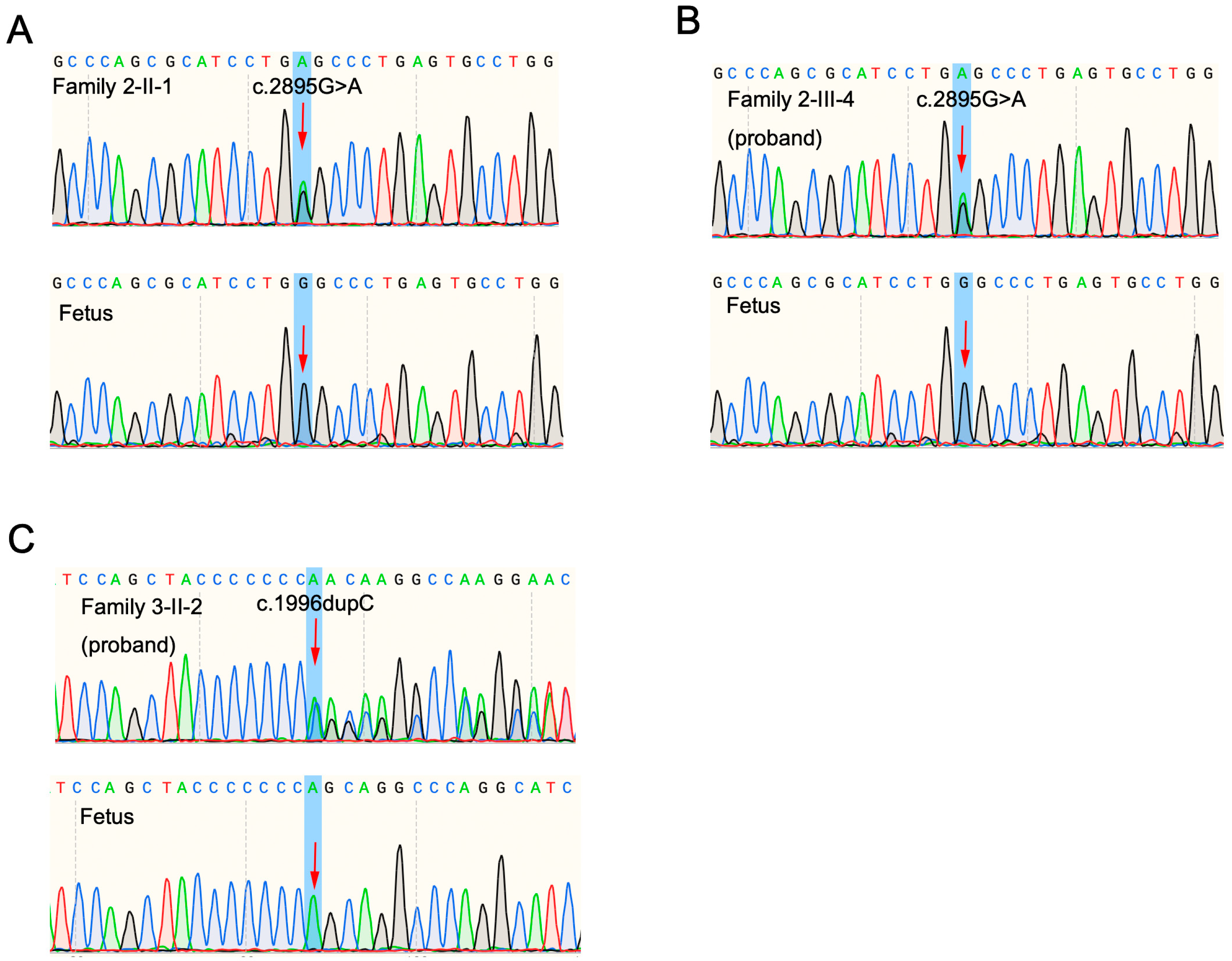
2.5. Prenatal Diagnosis

Pregnancy was verified by a blood hCG level of 25 U/L 14 days after transplantation and determined by ultrasound of the fetal sac with heart rhythm in the uterine cavity 30~40 days after transplantation. In the second trimester, a prenatal diagnosis was carried out via amniocentesis. Under the direction of ultrasound, 20 microliters of amniotic fluid were taken. After the genomic DNA was collected as previously mentioned, the genotypes of the fetuses were verified by Sanger sequencing.

2.6. Literature Review and Genotype–Clinical Phenotype Correlation Analysis

The PubMed, EMBASE, and Web of Science databases were used to conduct literature searches. The following terms were used in the search: “Schaaf-Yang syndrome”, “Prader-Willi and Schaaf-Yang syndromes”, “MAGEL2”, “MAGEL2-related disorders”, “Chitayat-Hall syndrome”, and “Chitayat-Hall and Schaaf-Yang syndromes”. Published cases of SYS caused by MAGEL2 gene variants, as well as information on the current patient are included in our literature review. The associations between genotype and clinical phenotype were examined after collecting the clinical symptoms, laboratory test results, imaging, and gene variant status of the patients. Two patient groups were examined to determine the relationship between genotype and phenotype: patients with the c.1996dupC (p.Gln666Profs*47) variant (n = 51) and patients with any other variant (n = 67).

2.7. Statistical Analysis

For statistical analysis, SPSS 26.0 was used. Continuous variables are presented as mean (standard error), and categorical variables are described as n/N(percentages). Differences between groups of continuous variables were tested using a parametric two-tailed t test or Mann–Whitney U test when the normality assumption was not met. Differences between the two groups regarding categorical variables were evaluated using Fisher’s exact test or the chi-square test when the expected count was ≥5. p values less than 0.05 were considered statistically significant.

3. Results

3.1. History of Three Families

Figure 1 illustrates the pedigrees of the three families with SYS included in our study. Table 1 shows the specific MAGEL2 variant detected in the probands of these three families. The clinical characteristics of the eleven patients with SYS from the three families are summarized in Table A1.

3.1.1. Family 1

The couple (II-1 and II-2) in Family 1 had five children presenting with hypotonia and abnormal mandibular development. The male children presented with gonadal dysplasia and contractures. All five children were born with hypoxia and an inability to breathe spontaneously. After rescue treatment, they showed irregular breathing, cyanosis, poor response, and a weak cry. They were admitted to the neonatal intensive care unit for respiratory and feeding support. Four children (III-1/2/4/5) died of respiratory failure shortly after birth (7–17 days). Family 1-III-3 died about 110 days after birth, and doctors suspected it might be caused by sleep apnea.
Whole-exome sequencing (WES) revealed NM019066.4(MAGEL2): c.1996dupC (p. Gln666Profs*47) heterozygous variant of the proband (III-3) and III-5. Variants were inherited from the unaffected father (II-1) as a heterozygous variant (c.1996dupC).

3.1.2. Family 2

The proband in Family 2 (III-4) was an 11-year-old boy, diagnosed with cerebral palsy a week after birth, and complaining of feeding difficulties in the neonatal period, intellectual disability, obesity, and bulimia. A physical examination showed a micropenis. Both the older brother (III-3) and younger brother (III-5) of the proband were diagnosed with neonatal asphyxia and atelectasis after birth and died at 7–14 days of age due to respiratory failure. The parents (II-4 and II-5) were phenotypically normal. The uncle of the proband (II-1) had fathered two sons (III-1 and III-2), both of whom died of atelectasis after birth.
WES revealed the presence of NM019066(MAGEL2):c.2895G>A (p.W965*) heterozygous variant in the proband (III-4). The same heterozygous variant was detected in the proband’s father (II-4), uncle (II-1), and grandmother (I-2).

3.1.3. Family 3

The proband (II-1) in Family 3 had pneumonia, sepsis, encephalopathy, and intracranial hemorrhage in the neonatal period. The physical examination was notable for the presence of laryngeal chondromalacia, increased muscle tone in the upper extremities, and stiffness of the middle and fourth finger joints of both hands, which could not be fully extended. Laboratory tests suggested elevated thyroid-stimulating hormone levels. Echocardiography suggested an atrial septal defect and patent foramen ovale. The proband died of apnea caused by pneumonia 32 days after birth.
Trio-whole-exome sequencing (trio-WES) revealed the presence of NM_019066.5(MAGEL2):c.1996dupC de novo heterozygous variants in the proband (II-1).

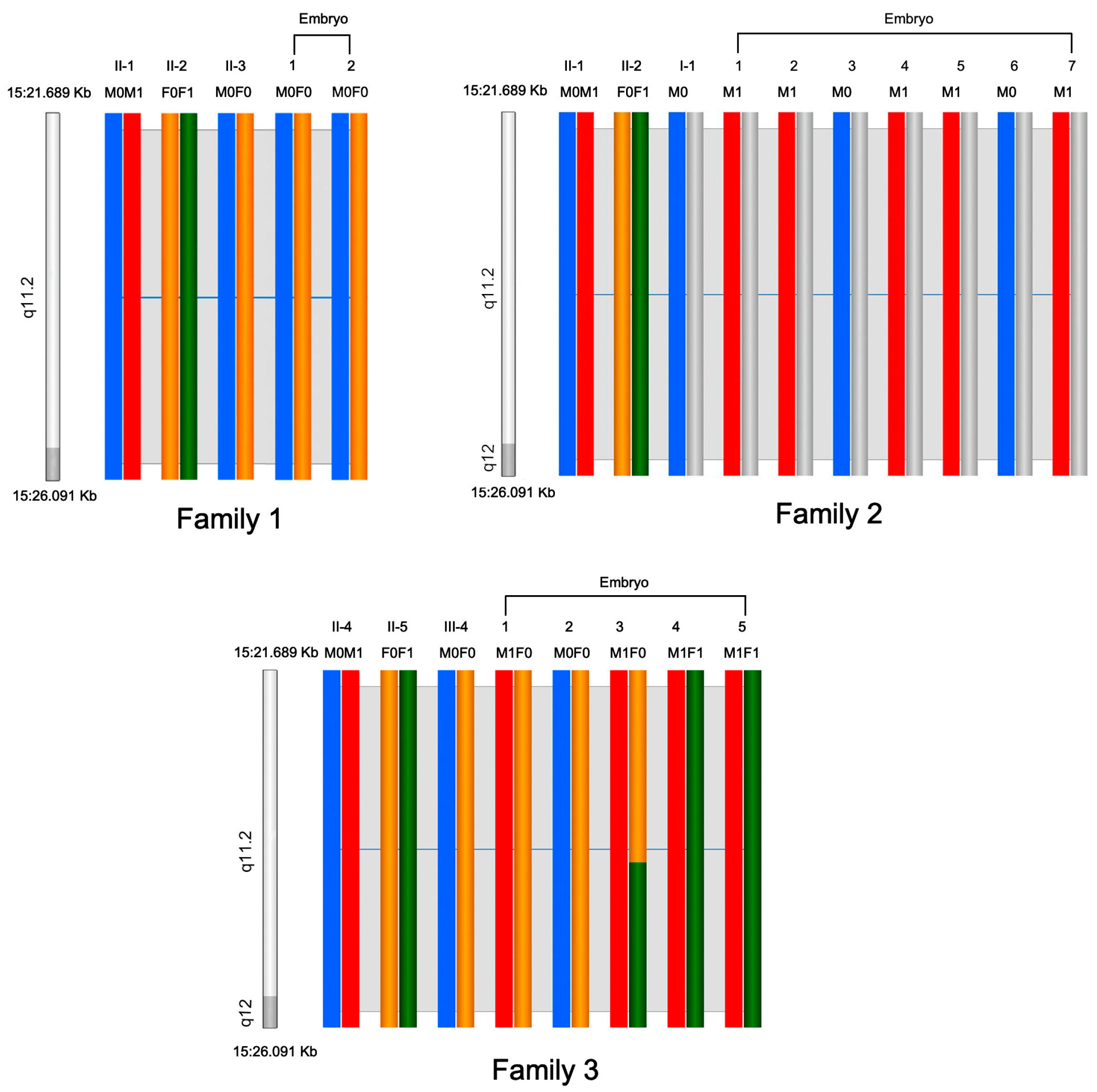
3.2. Preimplantation Genetic Testing and Prenatal Diagnosis

Three couples in particular conceived via in vitro fertilization following PGT, a process whereby a 5-day-old embryo is examined in the lab to see if it possesses a specific disease-causing variation. Four PGT cycles were completed by the three couples (Family 1-II-2, Family 2-II-2, Family 2-II-5). The 14 blastocysts underwent trophectoderm biopsies and PGT-M/PGT-A (PGT for aneuploidies). The genotypes of 14 embryos were all successfully determined using haplotype analysis (Figure 2). Nine (64.2%) of the fourteen embryos were found not to harbor any MAGEL2 variants by means of PGT-M, whereas 7 of the 14 embryos were found to be euploid by means of PGT-A (Table 2). Two embryos (Embryo 2 of Family 2-II-2, Embryo 4 of Family 2-II-5) found to be both negative for variant and euploid were used for transfer. Family 1 had only two available embryos, and both carried the disease-causing variant. The couple (Family 1-II-2) terminated PGT and embryo implantation.
The other couple (Family 3-I-2) elected to undergo a prenatal diagnosis on the fetus after the mother had conceived naturally. The results and outcomes of the prenatal diagnosis in the three families are summarized in Table 3. The prenatal diagnosis results showed that the fetus did not carry variants (Figure A1). All babies of the three families were born at full term and were healthy.

3.3. Literature Review

After excluding duplicate cases, 11 papers were found from the literature review in which these syndromes were discussed (Figure 3). Together with the 11 patients in our study, data from a total of 127 patients with SYS were pooled and were available for analysis. We numbered patients according to the literature sources: 1–91 [8], 92–95 [23], 96 [24], 97–102 [6], 103–104 [9], 105–108 [11], 109 [25], 110 [26], 111 [27], 112–115 [28], 116 [29] and 117–127 (New).

3.4. MAGEL2 Gene Variant Spectrum

Figure 4 shows the MAGEL2 gene variant spectrum in the included 127 SYS patients. We confirmed the most common variant as c.1996dupC. Most of the variants were present in the second half of the gene after c.1580, where most cases were found in nucleotides c.1990–c.1996, a region that represents a variant hotspot. The c.1996dupC variant represents the most prevalent variant with the largest number of individuals.

3.5. Clinical Phenotype Profile

Table 4 summarizes the clinical characteristics and related examinations of the included 127 SYS patients. The proportions of male and female patients were roughly the same.
It is worth noting that in terms of prenatal symptoms, a fetus with SYS rarely has characteristic imaging features. A small proportion of pregnant women experience decreased fetal movements and polyhydramnios. Patients with SYS in the perinatal period are more symptomatic, and those who die in the neonatal period comprise the highest proportion of all SYS deaths. Hypotonia (103/114, 90%) and poor suck (93/104, 89%), as well as feeding issues (98/116, 84%), are the most common symptoms in the perinatal period. In addition, approximately 70% (71/102) of patients have respiratory distress/defects, which lead to a higher rate of intubation (60/96, 63%) and mechanical ventilator use (63/104, 61%). The majority (112/128, 88%) of SYS patients have characteristic contractures of the fingers, and approximately half of the patients (28/54, 52%) have small hand features. In addition, scoliosis (38/94, 40%) and kyphosis (12/69, 17%) are also present in SYS patients.
Data are shown as n/N (%). Many patients with SYS have digestive symptoms, such as gastroesophageal reflux (40/84, 48%) and chronic constipation (49/83, 59%). On the assessment of the endocrine system, the most prominent symptom of SYS patients is short stature (29/44, 66%) due to growth hormone deficiency. Hypogonadism (44/103, 43%) is also present. In addition, hyperphagia (17/65, 26%) and excessive weight gain (23/90, 26%) are present after six years of age in a small percentage of patients.
Developmental delay (106/110, 96%) is present in the vast majority of patients with SYS. Approximately 67% (37/55) of the patients are diagnosed with autistic spectrum disorder. A small number of patients also have other psychiatric disorders, including attention deficit disorder, obsessive compulsive disorder, and some self-injurious behaviors.
Sleep apnea was present in 66 of 94 SYS patients (70%). Abnormal sleep cycles were present in a small proportion of SYS patients. Regarding the imaging findings, echocardiography abnormalities and brain MRI abnormalities were present in 18 of 31 patients. Hypopigmentation (4/32, 13%) was present in a small percentage of patients. Eye anomalies were present in 28 of 53 patients (53%).

3.6. MAGEL2 Genotype–Clinical Phenotype Correlation Analysis

In this study, we also contrasted the phenotypes in two groups of patients based on the sites of their MAGEL2 variants: individuals with any variant other than c.1996dupC (n = 67) and individuals with the c.1996dupC variant (n = 51) (Table 5). Compared with patients with any variant other than c.1996dupC, respiratory distress/defects and intubation were more common symptoms in patients with the c.1996dupCvariant (81 vs. 56%, p value: 0.008; 73 vs. 47%, p value: 0.011). Among individuals with the c.1996dupC variant, 32 of 44 individuals (73%) used a mechanical ventilator compared to 22 of 47 individuals (47%) without the c.1996dupc variant (p value: 0.012). We also found a substantial enrichment of four symptoms in patients who harbored 1996dupC variants in comparison with patients who harbored any other variant (poor suck in infancy [p value: 0.009]; dysphagia [p value: 0.005]; use of nasogastric tube [p value: 0.00004]; use of G tube [p value: 0.005]). In the patients with the c.1996dupC variant, contractures were more common than in those with any variant other than c.1996dupC (98 vs. 82%, p value: 0.038). There was also a difference in the endocrine profile between the two groups, and endocrine symptoms seemed to be milder in patients with the c.1996dupC variant. Among those with a c.1996dupC variant, 5 of 40 individuals (13%) displayed excessive weight gain, while 18 of 49 individuals (37%) without a c.1996dupC variant were similarly affected (p value: 0.009). Those with MAGEL2 variants other than c.1996dupC variant were more likely to have hyperphagia; there were 13 of 37 (35%) in the former case compared to 4 of 34 (12%) in the case of variants of c.1996dupC (p value: 0.021). In addition, there was a significant relationship between the number of symptoms and the presence of the 1996dupC variant when compared to other variants (mean number of symptoms in patients with c.1996DupC = 13.6, SE = 0.57; mean number of symptoms in patients without c.1996dupC = 11.5, SE = 0.54; p value: 0.012).

4. Discussion

The eleven patients from three families in our case series all harbored MAGEL2 variants and had overlapping clinical presentations, including multiple congenital anomalies, contractures, respiratory stress, and micropenises in male infants. Most patients died in the neonatal period from respiratory failure or neonatal pneumonia. In childhood, surviving patients presented with developmental delay, obesity, and bulimia. Of note is that congenital heart disease (atrial septal defect and patent foramen ovale) was present in the affected child (Family 3-II-1) with the c.1996dupC variant, which has been less commonly reported in related variants and only mentioned in one case report [9]. Since the disease does not follow Mendelian inheritance laws and the patients did not undergo genetic screening and diagnosis in time, both Family 1 and Family 2 in our study had a history of recurrent birth defects and neonatal deaths. As SYS is a rare autosomal dominant and matrilineal-imprinted neurodevelopmental illness, when the mutant gene is inherited from the father, the likelihood of the disease emerging in the progeny of an affected individual is estimated to be approximately 50%. De novo variants, which account for 50% of cases, have a 2–3% likelihood of recurrence [30]. It is consequently critical in such circumstances to be able to provide PGT and a prenatal diagnosis to prevent the recurrence of SYS in the affected families. In families that underwent PGT-M, informative STRs were distributed from upstream to downstream of the MAGEL2 gene, ensuring that any recombination would be identified. We determined each embryo successfully. Family 1-II-2 had only two available embryos, and both carried the disease-causing variant, so PGT and embryo implantation were terminated. We successfully performed PGT and/or a prenatal diagnosis in another three couples and prevented the transmission of truncating variants in MAGEL2.
In this paper, we have summarized the data of 127 patients from around the world diagnosed with truncating variants in the MAGEL2 gene to increase clinicians’ understanding and the diagnostic ability of the disease. In terms of prenatal symptoms, ultrasound of the SYS fetus rarely yields characteristic ultrasound images, and only a small number of fetuses may have nonspecific features, such as polyhydramnios or decreased fetal movement. This poses a particular difficulty in the diagnosis of SYS by routine prenatal testing. In the neonatal period, the most prominent and contributing symptom to the highest case fatality rate is respiratory defects, which in turn also leads to an elevated rate of intubation and ventilator use. Respiratory disorders may persist into infancy and adulthood, as sleep apnea is often reported as a problem that must be handled throughout one’s life. Hypotonia is almost universally found (90%) in patients with SYS and contributes to many clinical manifestations of the disease, including feeding difficulties and poor suck in infancy, so a nasogastric tube or G tube is needed. The most prominent and exclusive dysmorphic feature of patients with SYS is contractures (88%), which is also a symptom that distinguishes SYS from PWS [23,31]. Short stature, increased fat mass, and low IGF-1 levels are common in SYS patients, indicating a growth hormone deficiency similar to PWS [32]. Hypogonadism is a consistent finding in both males and females with SYS [4,33]. Furthermore, patients with truncating variants in MAGEL2 have a higher prevalence (67%) of autism spectrum disorder (ASD) than patients with classical PWS [31,34,35]. In general, SYS and PWS have a considerable overlap in symptoms, which further proves that the molecular diagnosis of SYS is useful and necessary. We also summarize the overlap and differences of SYS, PWS, and CHS, with the hope that the clinical and molecular characteristics of this group of disorders will become increasingly clear (Figure A2). In addition, understanding the pathogenesis through PGT can help patients block the transmission of pathogenic MAGEL2 variants and give birth to healthy babies.
MAGEL2 contains a proline-rich domain (PRD), a USP7-binding segment (U7BS), and a MAGE homology domain (MHD) [36,37,38]. We summarized the truncating variants of MAGEL2 that have been reported and found that these variants are located in the PRD most commonly, followed by the MHD and U7BS domains. The c.1996dupC variant represents the most prevalent variant, affecting the largest number of individuals. By comparing clinical characteristics between variant groups, we demonstrated further genotype–phenotype associations found in SYS. Our findings suggest that the position of the truncating variant may affect how severe a syndromic condition is since people with the c.1996dupC variant exhibit a more severe and varied phenotypic profile than people with other truncating variants; this is consistent with previous research conclusions [8,39]. Specifically, joint contractures, respiratory distress/defects, intubation and the use of mechanical ventilators, difficulty sucking in infancy, and the use of a nasogastric tube and G tube all occur at a higher prevalence in people with this duplication. Interestingly, patients with the c.1996dupC variant appear to have milder endocrine symptoms. It is important to emphasize that this conclusion needs to be taken with caution because abnormalities of the endocrine system mostly present in childhood rather than the neonatal period, and some patients with the c.1996dupC variant may not survive into childhood because of a more severe respiratory distress phenotype, thus leading to a bias that patients with the c.1996dupC variant appear to have milder endocrine symptoms. Although the pathomechanism of these variations is unknown, it is worth noting that MAGEL2 is a single exon gene, and variants that result in a premature stop codon are unlikely to generate nonsense-mediated mRNA decay [36,40,41]. Instead, a shortened protein product is anticipated as a result of such variations. Because each truncated protein product would originate from a different site of the variant on MAGEL2, it is possible to hypothesize that the pathogenic effect may vary depending on the location of the variant. Further research will be required to determine the underlying pathomechanics in the varied expressivity of SYS phenotypes.
MAGEL2 is truncatingly mutated in SYS and inactivated in PWS, but it is unclear how the loss of MAGEL2 function contributes to the pathophysiology of these disorders. MAGEL2 proteins regulate protein ubiquitination by interacting with the MHD and variable domains of E3 ubiquitin ligases and deubiquitinases to generate MAGE-RING E3 ligase complexes that serve as multifunctional hubs for the alteration of important substrates in the cell [38,42]. MAGEL2 is widely expressed in the hypothalamus and plays a vital role in a critical biological process that recycles membrane proteins from endosomes via the retromer sorting process [36]. Variants in MAGEL2 impair its capacity to promote the retromer-dependent recycling of proteins from endosomes back to the trans-Golgi network, to enhance leptin receptor expression on cell surfaces and to modulate the ubiquitination and stability of the circadian rhythm protein CRY1 [43,44,45]. Mice with a targeted Magel2 deletion recapitulated key elements of SYS, further suggesting that MAGEL2 plays an important role in the etiology of these disorders. Neonatal Magel2 null mice fail to thrive, have a modest increase in embryonic mortality, and demonstrate growth retardation in early life, which is followed by weight gain after weaning, and increased obesity with disturbed metabolic and endocrine homeostasis [46,47]. The loss of Magel2 in mice leads to the reduction in male and female fertility by extending breeding intervals and early reproductive decline and termination, which is consistent with the symptoms of hypogonadism in patients with SYS [48]. Due to a blunted circadian rhythm, these mice exhibit aberrant eating behavior and general hypoactivity, which are symptoms similar to those reported in SYS patients [49,50]. The MAGEL2 protein was demonstrated to interact with and modify the activity of the major elements of the circadian clock at the cellular level, providing more evidence that MAGEL2 plays an important role in controlling the circadian rhythm [45,51].

5. Conclusions

In conclusion, we identified eleven patients harboring pathogenic truncating MAGEL2 variants from three Chinese families with SYS, so the MAGEL2 variant spectrum was enriched. Meanwhile, a prenatal diagnosis and/or targeted NGS-based PGT-M was performed to prevent the transmission of the pathogenic variants of MAGEL2 in their families. Furthermore, this study also summarized the clinical symptoms of patients with SYS to date and analyzed genotype–clinical phenotype correlations, which helps to inform families about the range of symptoms associated with SYS and may help further elucidate SYS pathogenesis, providing benefits for clinical diagnosis and management of the disease.

Author Contributions

Conceptualization, N.X., C.X. and S.C.; methodology, W.S.; software, X.C.; validation, X.Z. and W.S.; resources, W.S.; writing—original draft preparation, N.X.; writing—review and editing, C.X.; visualization, N.X.; supervision, H.H. All authors have read and agreed to the published version of the manuscript.

Funding

This work was supported by the National Key R&D Program of China (2021YFC2701002, 2022YFC2703700 and 2022YFC2703702), the National Natural Science Foundation of China (Nos. 81971344, 82171677, 82192864, 81901495 and 82088102), the Shanghai Municipal Commission of Science and Technology Program (No. 22S31901500, No. 21Y21901002, No. 15411966700), Shanghai Municipal Health Commission (202140110, GW-10.1-XK07), Shanghai Municipal Commission of Health and family planning (202140110, 20215Y0216), CAMS Innovation Fund for Medical Sciences (2019-I2M-5-064), Collaborative Innovation Program of Shanghai Municipal Health Commission (2020CXJQ01), Clinical Research Plan of SHDC (SHDC2020CR1008A), Shanghai Clinical Research Center for Gynecological Diseases (22MC1940200), Shanghai Urogenital System Diseases Research Center (2022ZZ01012) and Shanghai Frontiers Science Research Base of Reproduction and Development.

Institutional Review Board Statement

This study was approved by the institutional review boards of the International Peace Maternity and Child Health Hospital (GKLW2019-52) and the Obstetrics and Gynecology Hospital of Fudan University (2020-178).

Informed Consent Statement

Informed consent was obtained from all subjects involved in the study. Written informed consent has been obtained from the patient(s) to publish this paper.

Data Availability Statement

The data presented in this study are available upon request from the corresponding author.

Acknowledgments

The authors would like to thank the family members for participating in this study.

Conflicts of Interest

The authors declare no conflict of interest.

Appendix A

Table A1. Clinical characteristics of the eleven SYS patients in three families.
PatientSexAgeVariant LocationRespiratory DistressHypotoniaHypogonadismOther Clinical Phenotypes
Family 1-III-1M7 dc.1996dupCNeonatal anoxia++Camptodactyly
Family 1-III-2M15 dc.1996dupCNeonatal anoxia++Camptodactyly, patent foramen ovale
Family 1-III-3F110 dc.1996dupCRecurrent apnea, respiratory failure+Camptodactyly, simian line
Family 1-III-4M17 dc.1996dupCNeonatal anoxia++Camptodactyly, simian line, small mandible, high palate arch, small tongue, low ear position
Family 1-III-5F9 dc.1996dupCNeonatal anoxiaCamptodactyly, high palate arch, low ear position, bilateral polydactyly
Family 2-III-4M11 yc.2895G>ANeonatal anoxia+Feeding difficulties, intellectual disability, obesity, bulimia
Family 2-III-1/2/3/5M1–14 dc.2895G>ANeonatal anoxia, atelectasis++-
Family 3-III-4F32 dc.1996dupCNeonatal pneumoniaNeonatal sepsis, neonatal encephalopathy, intracranial hemorrhage, camptodactyly, thyroid-stimulating hormone, atrial septal defect, and patent foramen ovale
M, male; F, female; d, days; y, years.
Figure A1. Prenatal genetic diagnosis after PGT. Sanger sequencing of the fetal samplings from amniotic fluid indicated normal genotypes in the three families. (A) Family 2-II-2. (B) Family 2-II-5. (C) Family 3-I-2.
Jcm 12 01688 g0a1
Figure A2. Venn diagram comparing the clinical presentations of Schaaf-Yang syndrome (SYS), Prader–Willi syndrome (PWS) and Chitayat–Hall syndrome (CHS). a, disorder defined by truncating variants of the MAGEL2 gene; b, disorder defined by gene deletions in the 15q11-q13 chromosomal region.
Jcm 12 01688 g0a2

References

  1. Cassidy, S.B.; Schwartz, S.; Miller, J.L.; Driscoll, D.J. Prader-Willi syndrome. Genet. Med. 2012, 14, 10–26. [Google Scholar] [CrossRef] [PubMed] [Green Version]
  2. Chao, Y.; Gao, L.; Wang, X.; Cai, Y.; Shu, Y.; Zou, X.; Qin, Y.; Hu, C.; Dai, Y.; Zhu, M.; et al. Dysregulated adipose tissue expansion and impaired adipogenesis in Prader-Willi syndrome children before obesity-onset. Metabolism 2022, 136, 155295. [Google Scholar] [CrossRef]
  3. Schaaf, C.P.; Gonzalez-Garay, M.L.; Xia, F.; Potocki, L.; Gripp, K.W.; Zhang, B.; Peters, B.A.; McElwain, M.A.; Drmanac, R.; Beaudet, A.L.; et al. Truncating mutations of MAGEL2 cause Prader-Willi phenotypes and autism. Nat. Genet. 2013, 45, 1405–1408. [Google Scholar] [CrossRef] [Green Version]
  4. Fountain, M.D.; Aten, E.; Cho, M.T.; Juusola, J.; Walkiewicz, M.A.; Ray, J.W.; Xia, F.; Yang, Y.; Graham, B.H.; Bacino, C.A.; et al. The phenotypic spectrum of Schaaf-Yang syndrome: 18 new affected individuals from 14 families. Genet. Med. 2017, 19, 45–52. [Google Scholar] [CrossRef] [PubMed] [Green Version]
  5. Mejlachowicz, D.; Nolent, F.; Maluenda, J.; Ranjatoelina-Randrianaivo, H.; Giuliano, F.; Gut, I.; Sternberg, D.; Laquerrière, A.; Melki, J. Truncating Mutations of MAGEL2, a Gene within the Prader-Willi Locus, Are Responsible for Severe Arthrogryposis. Am. J. Hum. Genet. 2015, 97, 616–620. [Google Scholar] [CrossRef] [Green Version]
  6. Negishi, Y.; Ieda, D.; Hori, I.; Nozaki, Y.; Yamagata, T.; Komaki, H.; Tohyama, J.; Nagasaki, K.; Tada, H.; Saitoh, S. Schaaf-Yang syndrome shows a Prader-Willi syndrome-like phenotype during infancy. Orphanet J. Rare Dis. 2019, 14, 277. [Google Scholar] [CrossRef] [PubMed] [Green Version]
  7. Jobling, R.; Stavropoulos, D.J.; Marshall, C.R.; Cytrynbaum, C.; Axford, M.M.; Londero, V.; Moalem, S.; Orr, J.; Rossignol, F.; Lopes, F.D.; et al. Chitayat-Hall and Schaaf-Yang syndromes: A common aetiology: Expanding the phenotype of MAGEL2-related disorders. J. Med. Genet. 2018, 55, 316–321. [Google Scholar] [CrossRef] [Green Version]
  8. Patak, J.; Gilfert, J.; Byler, M.; Neerukonda, V.; Thiffault, I.; Cross, L.; Amudhavalli, S.; Pacio-Miguez, M.; Palomares-Bralo, M.; Garcia-Minaur, S.; et al. MAGEL2-related disorders: A study and case series. Clin. Genet. 2019, 96, 493–505. [Google Scholar] [CrossRef]
  9. Chen, X.; Ma, X.; Zou, C. Phenotypic spectrum and genetic analysis in the fatal cases of Schaaf-Yang syndrome: Two case reports and literature review. Medicine 2020, 99, e20574. [Google Scholar] [CrossRef]
  10. Tong, W.; Wang, Y.; Lu, Y.; Ye, T.; Song, C.; Xu, Y.; Li, M.; Ding, J.; Duan, Y.; Zhang, L.; et al. Whole-exome Sequencing Helps the Diagnosis and Treatment in Children with Neurodevelopmental Delay Accompanied Unexplained Dyspnea. Sci. Rep. 2018, 8, 5214. [Google Scholar] [CrossRef] [Green Version]
  11. Ahn, H.; Seo, G.H.; Oh, A.; Lee, Y.; Keum, C.; Heo, S.H.; Kim, T.; Choi, J.; Kim, G.H.; Ko, T.S.; et al. Diagnosis of Schaaf-Yang syndrome in Korean children with developmental delay and hypotonia. Medicine 2020, 99, e23864. [Google Scholar] [CrossRef]
  12. Carlson, L.M.; Vora, N.L. Prenatal Diagnosis: Screening and Diagnostic Tools. Obstet. Gynecol. Clin. N. Am. 2017, 44, 245–256. [Google Scholar] [CrossRef] [Green Version]
  13. Dokras, A.; Barlow, D. Preimplantation genetic testing for monogenic diseases-bench to bedside reflections. Fertil. Steril. 2022, 118, 849–851. [Google Scholar] [CrossRef] [PubMed]
  14. Vermeesch, J.R.; Voet, T.; Devriendt, K. Prenatal and pre-implantation genetic diagnosis. Nat. Rev. Genet. 2016, 17, 643–656. [Google Scholar] [CrossRef]
  15. Besser, A.G.; Blakemore, J.K.; Grifo, J.A.; Mounts, E.L. Transfer of embryos with positive results following preimplantation genetic testing for monogenic disorders (PGT-M): Experience of two high-volume fertility clinics. J. Assist. Reprod. Genet. 2019, 36, 1949–1955. [Google Scholar] [CrossRef]
  16. Gibbons, W.E. Preimplantation diagnosis for genetic susceptibility. N. Engl. J. Med. 2006, 355, 2048. [Google Scholar] [CrossRef] [PubMed]
  17. Beaudet, A.L. Preimplantation genetic screens. Science 2015, 349, 1423. [Google Scholar] [CrossRef] [PubMed] [Green Version]
  18. Dolan, S.M.; Goldwaser, T.H.; Jindal, S.K. Preimplantation Genetic Diagnosis for Mendelian Conditions. JAMA 2017, 318, 859–860. [Google Scholar] [CrossRef] [PubMed]
  19. Treff, N.R.; Fedick, A.; Tao, X.; Devkota, B.; Taylor, D.; Scott, R.T., Jr. Evaluation of targeted next-generation sequencing-based preimplantation genetic diagnosis of monogenic disease. Fertil. Steril. 2013, 99, 1377–1384.e1376. [Google Scholar] [CrossRef] [PubMed]
  20. Lee, I.; Alur-Gupta, S.; Gallop, R.; Dokras, A. Utilization of preimplantation genetic testing for monogenic disorders. Fertil. Steril. 2020, 114, 854–860. [Google Scholar] [CrossRef]
  21. Chen, S.; Fei, H.; Zhang, J.; Chen, Y.; Huang, H.; Lu, D.; Xu, C. Classification and Interpretation for 11 FBN1 Variants Responsible for Marfan Syndrome and Pre-implantation Genetic Testing (PGT) for Two Families Successfully Blocked Transmission of the Pathogenic Mutations. Front. Mol. Biosci. 2021, 8, 749842. [Google Scholar] [CrossRef]
  22. Richards, S.; Aziz, N.; Bale, S.; Bick, D.; Das, S.; Gastier-Foster, J.; Grody, W.W.; Hegde, M.; Lyon, E.; Spector, E.; et al. Standards and guidelines for the interpretation of sequence variants: A joint consensus recommendation of the American College of Medical Genetics and Genomics and the Association for Molecular Pathology. Genet. Med. 2015, 17, 405–424. [Google Scholar] [CrossRef] [Green Version]
  23. Marbach, F.; Elgizouli, M.; Rech, M.; Beygo, J.; Erger, F.; Velmans, C.; Stumpel, C.; Stegmann, A.P.A.; Beck-Wödl, S.; Gillessen-Kaesbach, G.; et al. The adult phenotype of Schaaf-Yang syndrome. Orphanet J. Rare Dis. 2020, 15, 294. [Google Scholar] [CrossRef] [PubMed]
  24. Halloun, R.; Habib, C.; Ekhilevitch, N.; Weiss, R.; Tiosano, D.; Cohen, M. Expanding the spectrum of endocrinopathies identified in Schaaf-Yang syndrome—A case report and review of the literature. Eur. J. Med. Genet. 2021, 64, 104252. [Google Scholar] [CrossRef]
  25. Kleinendorst, L.; Pi Castán, G.; Caro-Llopis, A.; Boon, E.M.J.; van Haelst, M.M. The role of obesity in the fatal outcome of Schaaf-Yang syndrome: Early onset morbid obesity in a patient with a MAGEL2 mutation. Am. J. Med. Genet. Part A 2018, 176, 2456–2459. [Google Scholar] [CrossRef] [PubMed]
  26. Mizuno, S.; Yokoyama, K.; Yokoyama, A.; Nukata, T.; Ikeda, Y.; Hara, S. Longitudinal analysis of electroencephalography pattern changes in an infant with Schaaf-Yang syndrome and a novel mutation in melanoma antigen L2 (MAGEL2). Mol. Genet. Genomic. Med. 2022, 10, e1932. [Google Scholar] [CrossRef]
  27. Duan, Y.; Liu, L.; Zhang, X.; Jiang, X.; Xu, J.; Guan, Q. Phenotypic spectrum and mechanism analysis of Schaff Yang syndrome: A case report on new mutation of MAGEL2 gene. Medicine 2021, 100, e26309. [Google Scholar] [CrossRef] [PubMed]
  28. Gregory, L.C.; Shah, P.; Sanner, J.R.F.; Arancibia, M.; Hurst, J.; Jones, W.D.; Spoudeas, H.; Le Quesne Stabej, P.; Williams, H.J.; Ocaka, L.A.; et al. Mutations in MAGEL2 and L1CAM Are Associated with Congenital Hypopituitarism and Arthrogryposis. J. Clin. Endocrinol. Metab. 2019, 104, 5737–5750. [Google Scholar] [CrossRef] [PubMed]
  29. Guo, W.; Nie, Y.; Yan, Z.; Zhu, X.; Wang, Y.; Guan, S.; Kuo, Y.; Zhang, W.; Zhi, X.; Wei, Y.; et al. Genetic testing and PGD for unexplained recurrent fetal malformations with MAGEL2 gene mutation. Sci. China Life Sci. 2019, 62, 886–894. [Google Scholar] [CrossRef] [PubMed]
  30. Nunes, S.; Xavier, M.; Lourenço, C.; Melo, M.; Godinho, C. Schaaf-Yang Syndrome: A Real Challenge for Prenatal Diagnosis. Cureus 2021, 13, e20414. [Google Scholar] [CrossRef]
  31. Fountain, M.D.; Schaaf, C.P. Prader-Willi Syndrome and Schaaf-Yang Syndrome: Neurodevelopmental Diseases Intersecting at the MAGEL2 Gene. Diseases 2016, 4, 2. [Google Scholar] [CrossRef] [Green Version]
  32. McCarthy, J.M.; McCann-Crosby, B.M.; Rech, M.E.; Yin, J.; Chen, C.A.; Ali, M.A.; Nguyen, H.N.; Miller, J.L.; Schaaf, C.P. Hormonal, metabolic and skeletal phenotype of Schaaf-Yang syndrome: A comparison to Prader-Willi syndrome. J. Med. Genet. 2018, 55, 307–315. [Google Scholar] [CrossRef]
  33. Hidalgo-Santos, A.D.; del Carmen DeMingo-Alemany, M.; Moreno-Macián, F.; Roselló, M.; Orellana, C.; Martínez, F.; Caro-Llopis, A.; León-Cariñena, S.; Tomás-Vila, M. A Novel Mutation of MAGEL2 in a Patient with Schaaf-Yang Syndrome and Hypopituitarism. Int. J. Endocrinol. Metab. 2018, 16, e67329. [Google Scholar] [CrossRef] [Green Version]
  34. Althammer, F.; Muscatelli, F.; Grinevich, V.; Schaaf, C.P. Oxytocin-based therapies for treatment of Prader-Willi and Schaaf-Yang syndromes: Evidence, disappointments, and future research strategies. Transl. Psychiatry 2022, 12, 318. [Google Scholar] [CrossRef]
  35. Crutcher, E.; Pal, R.; Naini, F.; Zhang, P.; Laugsch, M.; Kim, J.; Bajic, A.; Schaaf, C.P. mTOR and autophagy pathways are dysregulated in murine and human models of Schaaf-Yang syndrome. Sci. Rep. 2019, 9, 15935. [Google Scholar] [CrossRef] [PubMed] [Green Version]
  36. Tacer, K.F.; Potts, P.R. Cellular and disease functions of the Prader-Willi Syndrome gene MAGEL2. Biochem. J. 2017, 474, 2177–2190. [Google Scholar] [CrossRef] [PubMed]
  37. Basak, S.; Basak, A. Proteins and proteases of Prader-Willi syndrome: A comprehensive review and perspectives. Biosci. Rep. 2022, 42, BSR20220610. [Google Scholar] [CrossRef] [PubMed]
  38. Sanderson, M.R.; Fahlman, R.P.; Wevrick, R. The N-terminal domain of the Schaaf-Yang syndrome protein MAGEL2 likely has a role in RNA metabolism. J. Biol. Chem. 2021, 297, 100959. [Google Scholar] [CrossRef]
  39. McCarthy, J.; Lupo, P.J.; Kovar, E.; Rech, M.; Bostwick, B.; Scott, D.; Kraft, K.; Roscioli, T.; Charrow, J.; Schrier Vergano, S.A.; et al. Schaaf-Yang syndrome overview: Report of 78 individuals. Am. J. Med. Genet. Part A 2018, 176, 2564–2574. [Google Scholar] [CrossRef] [Green Version]
  40. Matuszewska, K.E.; Badura-Stronka, M.; Śmigiel, R.; Cabała, M.; Biernacka, A.; Kosinska, J.; Rydzanicz, M.; Winczewska-Wiktor, A.; Sasiadek, M.; Latos-Bieleńska, A.; et al. Phenotype of two Polish patients with Schaaf-Yang syndrome confirmed by identifying mutation in MAGEL2 gene. Clin. Dysmorphol. 2018, 27, 49–52. [Google Scholar] [CrossRef]
  41. Ieda, D.; Negishi, Y.; Miyamoto, T.; Johmura, Y.; Kumamoto, N.; Kato, K.; Miyoshi, I.; Nakanishi, M.; Ugawa, S.; Oishi, H.; et al. Two mouse models carrying truncating mutations in MAGEL2 show distinct phenotypes. PLoS ONE 2020, 15, e0237814. [Google Scholar] [CrossRef]
  42. Kozakova, L.; Vondrova, L.; Stejskal, K.; Charalabous, P.; Kolesar, P.; Lehmann, A.R.; Uldrijan, S.; Sanderson, C.M.; Zdrahal, Z.; Palecek, J.J. The melanoma-associated antigen 1 (MAGEA1) protein stimulates the E3 ubiquitin-ligase activity of TRIM31 within a TRIM31-MAGEA1-NSE4 complex. Cell Cycle 2015, 14, 920–930. [Google Scholar] [CrossRef] [Green Version]
  43. Wijesuriya, T.M.; De Ceuninck, L.; Masschaele, D.; Sanderson, M.R.; Carias, K.V.; Tavernier, J.; Wevrick, R. The Prader-Willi syndrome proteins MAGEL2 and necdin regulate leptin receptor cell surface abundance through ubiquitination pathways. Hum. Mol. Genet. 2017, 26, 4215–4230. [Google Scholar] [CrossRef] [Green Version]
  44. Hao, Y.H.; Fountain, M.D., Jr.; Fon Tacer, K.; Xia, F.; Bi, W.; Kang, S.H.; Patel, A.; Rosenfeld, J.A.; Le Caignec, C.; Isidor, B.; et al. USP7 Acts as a Molecular Rheostat to Promote WASH-Dependent Endosomal Protein Recycling and Is Mutated in a Human Neurodevelopmental Disorder. Mol. Cell 2015, 59, 956–969. [Google Scholar] [CrossRef] [Green Version]
  45. Carias, K.V.; Zoeteman, M.; Seewald, A.; Sanderson, M.R.; Bischof, J.M.; Wevrick, R. A MAGEL2-deubiquitinase complex modulates the ubiquitination of circadian rhythm protein CRY1. PLoS ONE 2020, 15, e0230874. [Google Scholar] [CrossRef] [Green Version]
  46. Tennese, A.A.; Wevrick, R. Impaired hypothalamic regulation of endocrine function and delayed counterregulatory response to hypoglycemia in MAGEL2-null mice. Endocrinology 2011, 152, 967–978. [Google Scholar] [CrossRef] [Green Version]
  47. Bischof, J.M.; Stewart, C.L.; Wevrick, R. Inactivation of the mouse MAGEL2 gene results in growth abnormalities similar to Prader-Willi syndrome. Hum. Mol. Genet. 2007, 16, 2713–2719. [Google Scholar] [CrossRef] [PubMed] [Green Version]
  48. Mercer, R.E.; Wevrick, R. Loss of MAGEL2, a candidate gene for features of Prader-Willi syndrome, impairs reproductive function in mice. PLoS ONE 2009, 4, e4291. [Google Scholar] [CrossRef] [PubMed] [Green Version]
  49. Kozlov, S.V.; Bogenpohl, J.W.; Howell, M.P.; Wevrick, R.; Panda, S.; Hogenesch, J.B.; Muglia, L.J.; Van Gelder, R.N.; Herzog, E.D.; Stewart, C.L. The imprinted gene MAGEL2 regulates normal circadian output. Nat. Genet. 2007, 39, 1266–1272. [Google Scholar] [CrossRef] [PubMed]
  50. Fountain, M.D.; Tao, H.; Chen, C.A.; Yin, J.; Schaaf, C.P. MAGEL2 knockout mice manifest altered social phenotypes and a deficit in preference for social novelty. Genes Brain Behav. 2017, 16, 592–600. [Google Scholar] [CrossRef] [Green Version]
  51. Devos, J.; Weselake, S.V.; Wevrick, R. MAGEL2, a Prader-Willi syndrome candidate gene, modulates the activities of circadian rhythm proteins in cultured cells. J. Circadian Rhythm. 2011, 9, 12. [Google Scholar] [CrossRef] [PubMed]
Figure 1. Pedigrees of three families with SYS. The arrows indicate the proband of each family. Squares represent males; circles represent females; solid symbols indicate affected patients; open symbols indicate unaffected subjects; a slash through the symbol means deceased; the circle with the letter D in the middle represents the donor egg.
Jcm 12 01688 g001
Figure 2. The haplotype of the MAGEL2 gene in embryos from three families. M0 means male disease-causing chromosome; M1 means male normal chromosome; F0 and F1 mean female normal chromosome.
Jcm 12 01688 g002
Figure 3. Flow diagram of the literature inclusion.
Jcm 12 01688 g003
Figure 4. MAGEL2 gene variant spectrum in all the included literature. (A) Summary of the location of variants in all genes in MAGEL2. The highest peak represented the c.1996 region, which is predicted to contain a “variant hotspot”. (B) MAGEL2-specific variant types.
Jcm 12 01688 g004
Table 1. MAGEL2 variant detected in probands of three families.
FamilyTranscript IDDNA_VariantAmino Acid ChangesClassification of Variants #Inheritance
1NM_019066.4c.1996dupCp.(Gln666Profs*47)pathogenicPat
2NM_019066.4c.2895G>Ap.(Trp965*)pathogenicPat
3NM_019066.5c.1996dupCp.(Gln666Profs*47)pathogenicDe novo
# The interpretation and classification of variants were based on ACMG guidelines [22]; * Termination of protein translation; pat, paternal; mat, maternal.
Table 2. Summary of PGT-M/PGT-A results from three families.
Sample No.EmbryoPGT-M ResultPGT-A ResultOutcome
HaplotypeSanger Sequencing
Family 1-II-2Embryo 1M0c.1996dupC−2, −15, −12
Embryo 2M0c.1996dupCN
Family 2-II-2Embryo 1M1/N46, XN
Embryo 2M1/N46, XNTransfer
Embryo 3M0/c.2895G>A46, XN
Embryo 4M1/N46, XN
Embryo 5M1/N46, XN
Embryo 6M0/c.2895G>A46, XN
Embryo 7M1/N46, XN
Family 2-II-5Embryo 1M1/N47, XN, +22
Embryo 2M0/c.2895G>A46, XN
Embryo 3M1/N46, XN, −14q21.2qter
Embryo 4M1/N46, XNTransfer
Embryo 5M1/N46, XN
M0: male disease-causing chromosome; M1, male normal chromosome.
Table 3. Summary of the results of prenatal diagnosis in three families.
Sample No.Gestation, WeeksFetus ResultOutcome
Family 2-II-216 WFamily 2-III-3NBorn healthy
Family 2-II-517 + 4 WFamily 2-III-7NBorn healthy
Family 3-I-215 WFamily 3-II-2NBorn healthy
Table 4. Patients’ symptom summary.
Patients and Source PublicationTotals d
(n = 127)
1–9192–959697–102103–104105–108109110111112–115116New
Sex (M/F)N/A4/40/15/10/23/10/10/11/01/31/08/344/47
Prenatal symptomsHistory of polyhydramniosN/AN/AN/AN/A0/21/40/10/10/11/31/10/10N/A
Decreased fetal movement8/232/80/1N/AN/A0/41/10/10/10/40/10/711/51 (22%)
Perinatal historyTemperature instability40/60N/AN/A4/6N/AN/A0/11/10/10/4N/A2/747/80 (59%)
Respiratory distress/defects49/70N/A1/10/62/23/41/11/10/12/41/111/1171/102 (70%)
Intubation38/64N/A1/11/62/22/41/11/10/12/41/111/1160/96 (63%)
Mechanical ventilator36/644/81/11/62/23/41/11/10/12/41/111/1163/104 (61%)
Tracheostomy13/58N/AN/A0/60/20/40/11/10/12/40/10/1016/88 (18%)
Hypotonia72/786/71/16/62/23/41/11/10/11/11/19/11103/114 (90%)
Feeding issues71/807/71/14/6N/A1/41/11/10/11/41/110/1098/116 (84%)
Poor suck in infancy65/726/7N/A6/62/22/41/11/10/1N/AN/A10/1093/104 (89%)
Dysphagia49/62N/AN/A1/62/22/41/11/10/1N/AN/A6/662/83 (75%)
Use of nasogastric (NG) tube45/604/50/11/61/22/41/11/10/1N/AN/A9/1164/92 (70%)
Use of G tube30/58N/A1/1N/A0/21/40/10/10/1N/AN/A0/1132/79 (41%)
Dysmorphic featuresScoliosis29/545/80/10/61/20/40/10/10/13/40/10/1138/94 (40%)
Kyphosis12/37N/A0/10/60/20/40/10/10/10/40/10/1112/69 (17%)
Contractures 77/906/71/16/62/23/41/1N/A0/14/41/111/11112/128 (88%)
Small hands15/24N/AN/A6/62/21/40/10/11/11/4N/A2/1128/54 (52%)
Small feet12/24N/AN/AN/A0/21/40/10/11/11/4N/A2/1117/48 (35%)
GastrointestinalReflux/GERD36/66N/AN/AN/A1/21/40/10/10/12/4N/A0/540/84 (48%)
Chronic constipation40/606/8N/AN/AN/A0/40/10/1N/A3/4N/A0/549/83 (59%)
Endocrine assessmentExcessive weight gain15/672/5N/A3/5N/A0/31/10/11/1N/AN/A1/723/90 (26%)
Hyperphagia14/56N/AN/AN/AN/AN/A1/10/11/1N/AN/A1/617/65 (26%)
Hypogonadism29/743/7N/A4/6N/A1/2N/A1/11/11/41/13/744/103 (43%)
Hypopituitarism3/9N/A1/13/3N/AN/AN/A1/11/13/4N/AN/AN/A
Growth hormone deficiency3/82/81/13/3N/AN/AN/A1/11/14/4N/AN/AN/A
Short stature13/27N/A1/15/5N/A3/4N/A1/11/14/4N/A1/129/44 (66%)
Increased fatty tissue4/5N/A0/1N/A0/20/41/10/11/10/4N/A1/7N/A
Cortical blindness1/2N/AN/AN/AN/AN/AN/AN/AN/AN/AN/AN/AN/A
NeurodevelopmentDevelopmental delay79/838/81/16/6N/A4/41/11/11/14/4N/A1/1106/110 (96%)
Attention deficit disorderN/A2/5N/AN/AN/AN/AN/AN/AN/AN/AN/AN/AN/A
Obsessive compulsive disorderN/A2/5N/AN/AN/AN/AN/AN/AN/AN/AN/AN/AN/A
ASD diagnosis29/394/7N/A1/1N/A2/4N/AN/AN/A1/4N/AN/A37/55 (67%)
Seizures25/73N/A1/12/61/21/40/10/10/10/40/10/530/99 (30%)
Self-injurious behavior9/164/8N/A4/6 aN/AN/AN/AN/A0/11/4N/A0/118/36 (50%)
Intellectual disabilityN/A5/5N/A5/6N/A4/41/1N/A1/1N/AN/A1/1N/A
Sleep problemsAbnormal sleep cycle bN/A8/8N/A5/6 cN/AN/AN/A0/10/1N/AN/A0/1N/A
Sleep apnea43/585/71/11/5N/A2/41/11/10/12/41/19/1166/94 (70%)
Assistant
examination
Echocardiography FindingsN/AN/AN/AN/A2/2N/AN/A0/10/11/41/12/2N/A
Brain MRI findings6/11N/A0/14/60/12/4N/A1/1N/A2/4N/A3/318/31 (58%)
OthersHypopigmentationN/AN/A0/14/60/20/40/10/10/10/40/10/114/32 (13%)
Hirsutism1/6N/AN/AN/AN/A0/40/1N/A0/1N/AN/A0/2N/A
Eye abnormalities18/25N/A0/15/61/20/40/10/10/14/40/10/728/53 (53%)
a Skin picking. b A sleeping routine that is different from the normal sleep cycle (e.g., sleeping from 2 pm to 1 am). A “reverse” sleep cycle would be the most extreme form. c Infantile lethargy, weak cry. d To avoid ascertainment bias, symptoms that have been assessed or reported in less than 25% of all patients (n = 127) are not applicable.
Table 5. Phenotype comparison between patients with any variant other than c.1996dupC and patients with the c.1996dupC variant.
Patients with Any Variant Other Than c.1996dupC (n = 67)Patients with the c.1996dupC Variant (n = 51)p-Value
Prenatal symptomsHistory of polyhydramnios2/11 (18%)1/12 (8%)0.590
Decreased fetal movement3/16 (19%)0/12 (0%)0.112
Perinatal historyTemperature instability23/46 (50%)24/43 (56%)0.583
Respiratory distress/defects29/52 (56%)35/43 (81%)0.008 *
Intubation26/55 (47%)32/44 (73%)0.0111 *
Mechanical ventilator22/47 (47%)32/44 (73%)0.012 *
Tracheostomy6/45 (13%)9/52 (17%)0.589
Hypotonia57/63 (90%)45/47 (96%)0.426
Feeding issues55/65 (85%)45/50 (90%)0.395
Poor suck in infancy49/57 (86%)45/45 (100%)0.009 *
Dysphagia28/44 (64%)36/40 (90%)0.005 *
Use of nasogastric (NG) tube24/51 (47%)39/45 (87%)0.00004 *
Use of G tube11/46 (24%)21/39 (54%)0.005 *
Dysmorphic featuresScoliosis17/51 (33%)19/38 (50%)0.113
Kyphosis5/38 (13%)7/35 (20%)0.431
Contractures 54/66 (82%)51/52 (98%)0.038 *
Small hands7/17 (41%)7/14 (50%)0.623
Small feet2/12 (17%)4/13 (31%)0.645
GastrointestinalReflux/GERD15/40 (38%)25/43 (58%)0.351
Chronic constipation23/44 (52%)27/42 (64%)0.259
Endocrine assessmentExcessive weight gain18/49 (37%)5/40 (13%)0.009 *
Hyperphagia13/37 (35%)4/34 (12%)0.021 *
Hypogonadism25/53 (47%)18/44 (41%)0.537
Hypopituitarism7/7 (100%)3/4 (75%)0.364
Growth hormone deficiency22/43 (51%)17/30 (57%)0.643
Short stature30/48 (63%)24/37 (65%)0.822
Increased fatty tissue3/9 (33%)0/13 (0%)0.025 *
Cortical blindness0/1 (0%)N/A (/)/
NeurodevelopmentDevelopmental delay45/53 (85%)33/34 (97%)0.069
Attention deficit disorder2/6 (33%)N/A (/)/
Obsessive compulsive disorder2/6 (33%)1/1 (100%)0.429
ASD diagnosis
Seizures
24/34 (71%)14/21 (67%)0.760
Seizures17/53 (32%)12/43 (28%)0.658
Self-injurious behavior7/16 (44%)2/5 (40%)1.000
Intellectual disability15/15 (100%)3/3 (100%)1.000
Sleep problemsAbnormal sleep cycle11/15 (73%)2/2 (100%)1.000
Sleep apnea36/52 (69%)27/40 (68%)0.859
Assistant
examination
Echocardiography Findings2/4 (50%)4/7 (57%)1.000
Brain MRI findings6/11 (55%)6/9 (67%)0.670
OthersHypopigmentation4/19 (21%)0/14 (0%)0.119
Hirsutism0/6 (0%)0/3 (0%)1.000
Eye abnormalities5/14 (36%)5/14 (36%)1.000
Total number of symptoms11.5 (0.54)13.6 (0.57)0.012 *
Statistically significant p values were marked in bold. Data are shown as n/N (%) or mean (SE); * p < 0.05.
Disclaimer/Publisher’s Note: The statements, opinions and data contained in all publications are solely those of the individual author(s) and contributor(s) and not of MDPI and/or the editor(s). MDPI and/or the editor(s) disclaim responsibility for any injury to people or property resulting from any ideas, methods, instructions or products referred to in the content.

