Article

Greater Tuberosity Fractures after RTSA: A Matched Group Analysis

1
Department of Orthopaedic Surgery, Balgrist University Hospital, University of Zurich, 8008 Zurich, Switzerland
2
Department of Orthopaedic Surgery, Royal Melbourne Hospital, Parkville, VIC 3050, Australia
3
Department of Biomedical Engineering, University of Melbourne, Parkville, VIC 3010, Australia
*
Author to whom correspondence should be addressed.
J. Clin. Med. 2023, 12(3), 1153; https://doi.org/10.3390/jcm12031153
Submission received: 29 November 2022 / Revised: 12 January 2023 / Accepted: 31 January 2023 / Published: 1 February 2023

Abstract

:
Periprosthetic fractures, such as acromial and spine fractures, are known complications following implantation of reverse shoulder arthroplasty (RTSA). The entity of greater tuberosity fractures (GTF) has rarely been studied in the literature. The purpose of this study was to analyze the outcome of postoperative greater tuberosity fractures after RTSA compared to a matched control group. The main findings of this study are that a GTF after RTSA is associated with worse clinical outcome scores (mean absolute CS 50 ± 19 (p = 0.032); SSV 63% ± 26 (p = 0.022); mean force 1 kg ± 2 kg (p = 0.044)) compared with the control group (mean absolute CS 62 ± 21; SSV 77% ± 29; mean force 2 kg ± 2 kg). In terms of postoperative range of motion, the fracture group was significantly worse in terms of external rotation (17° ± 19° vs. 30° ± 19° (p = 0.029)). Internal rotation, flexion, as well as abduction of the shoulder appear to be unaffected (internal rotation GTF 4 ± 2, control group 5 ± 3 (p = 0.138); flexion GTF 102° ± 28°, control group 114° ± 27° (p = 0.160); abduction GTF 109° ± 42°, control group 120° ± 39° (p = 0.317)).

1. Introduction

Since the first case series published by Grammont et al. [1] in 1987, there has been a consistently increasing use of reverse total shoulder arthroplasties (RTSA) worldwide. Due to good clinical outcomes for a variety of indications, such as massive rotator cuff tear, osteoarthritis, fractures, tumors, and for revision surgery [2,3,4,5], RTSA is increasingly replacing the anatomical shoulder prosthesis [6,7]. However, with more frequent use, more complications are being identified. Instability, infections, material loosening, and fractures of the surrounding osseous structures are among the most common complications [7,8,9,10,11]. Isolated periprosthetic fractures, mostly of the acromion, account for approximately 1–5% of all complications and are related to a poorer outcome and higher revision rate [8,12,13,14,15]. The underlying risk factors and pathomechanism of periprosthetic fractures are still incompletely understood. Female sex, osteoporosis, and impaired bone quality in general were reported to have a negative influence [14,16]. Furthermore, previous surgery and increased deltoid length are associated with a higher rate of acromial fractures [14]. The effect of RTSA positioning and design on postoperative fractures has been mainly studied for acromion and scapula fractures and remains controversial [14,17,18,19].
Greater tuberosity fractures (GTF) as a complication after RTSA are rarely mentioned in the literature [13,20]. In fracture arthroplasty for proximal humeral fractures, postoperative union of the greater tuberosity (GT) seems to have an important biomechanical effect. As several studies have shown, correct healing of the GT is associated with better clinical long-term outcomes [21,22,23]. This concerns especially the external rotation function after RTSA [23].
Hochreiter et al. [24] recently published that less preoperative to postoperative distalization of the GT is associated with poor functional internal rotation [24]. These findings suggest that the position of the GT relative to the RTSA does influence the postoperative shoulder function.
Therefore, we hypothesize that RTSA with postoperative GTF is associated with poorer clinical results.
The aim of this study was to compare and report the clinical outcome in patients with and without a fracture of the GT after RTSA.

2. Materials and Methods

2.1. Patients

A prospectively enrolled RTSA database of 1360 RTSA implantations between September 2005 and December 2019 was reviewed retrospectively for a GTF. The inclusion criteria were (1) an isolated postoperative greater tuberosity fracture after RTSA, regardless of the indication for RTSA, (2) agreement to be included in the study, (3) age ≥18 years, and (4) a minimum clinical and radiological follow-up period of 2 years after RTSA implantation. The exclusion criteria were (1) a preoperative fracture of the greater tuberosity and (2) further postoperative fractures. The degree of dislocation was specified for each GTF. Displacement was defined as a displacement of the greater tuberosity ≥1 cm, slight displacement if displacement was >3 mm but <1 cm, and no displacement with ≤3 mm displacement on standard radiographs [25].
The GTF group was pair matched in a 2:1 manner to a control group with regards to sex, age at surgery, indication for RTSA, length of follow-up, and body mass index.

2.2. Surgical Technique

According to the indication, patients were treated with different RTSA designs. The indication for RTSA varied from cuff tear arthropathy, osteoarthritis, proximal humeral fracture, instability, or conversion surgery from failed anatomic prosthesis. Seven of the 17 stems were cemented (41%), the remaining stems were implanted in press-fit technique. All surgeries were performed through a deltopectoral approach by different senior shoulder surgeons as previously described [26]. The subscapularis tendon was detached if needed and sutured/reinserted if possible.
Postoperatively, all patients had the same standard follow-up treatment.

2.3. Clinical and Radiological Evaluation

All patients were evaluated postoperatively at 6 weeks, 4.5 months, 1 and/or 2 years and then every 2–4 years. At each consultation, functional outcomes were determined with the absolute Constant–Murley score (aCS) and relative Constant–Murley score (rCS), including pain assessment [27], subjective shoulder value (SSV) [28] and range-of-motion (ROM) assessment.
The definitions for minimal clinically important differences (MCIDs) are based on studies by Torrens [29] and Simovitch [30] et al. The MCID cutoff for active flexion is 12°, abduction 7°, and 3° for internal and external rotation. The anchor-based MCID for the Constant score is 5.7 points.
Radiologic follow-up consisted of routine radiographs in 3 standardized planes and, if necessary, additional imaging with computed tomography (CT) or magnetic resonance imaging (MRI). The GTFs were diagnosed by an orthopedic resident, a consultant with a specialization in shoulder surgery, and/or a musculoskeletal radiologist.

2.4. Statistical Analysis and Data Collection

Study data were collected and edited using REDCap (Research Electronic Data Capture) tools provided by Balgrist University Hospital [31,32]. Preoperative and postoperative scores were compared using the Wilcoxon rank sum test. Comparison of 2 groups were performed using the Mann–Whitney-U test. The χ2 test was used for categorical variables. A p-value of less than 0.05 was considered significant. Owing to the given population of patients with GTFs, no power analysis was carried out. Statistical analysis was performed using SPSS software (version 27.0; IBM, Armonk, NY, USA).

3. Results

3.1. Incidence and Indication for RTSA

We identified 19 (1.4%) of 1360 shoulders with a postoperative GTF at a mean of 24 months (±38 months). Two patients (10.5%) had a follow-up <2 years and were excluded. Basic demographic and arthroplasty-related data are summarized in Table 1.
The GTFs occurred immediately in the first postoperative X-ray (day 0) up to 9 years postoperatively (Figure 1). The day 0 fractures were not detected intraoperatively. Otherwise, they would have been treated during the initial surgery. Further, 16 patients (94%) had no trauma causing the GTF. One patient had a GTF 125 days after RTSA after a fall with unclear trauma mechanism. The atraumatic GTFs were accidentally noticed in a follow-up examination or during work-up of postoperative functional limitations or pain. Thus, we were not able to fully reconstruct the fracture mechanism or exact fracture timing. The fracture time is equated with the radiological diagnosis time.
Six GTFs (35%) were nondisplaced, five patients (30%) had a slightly displaced GTF, and six (35%) patients had a dislocation of the greater tuberosity.
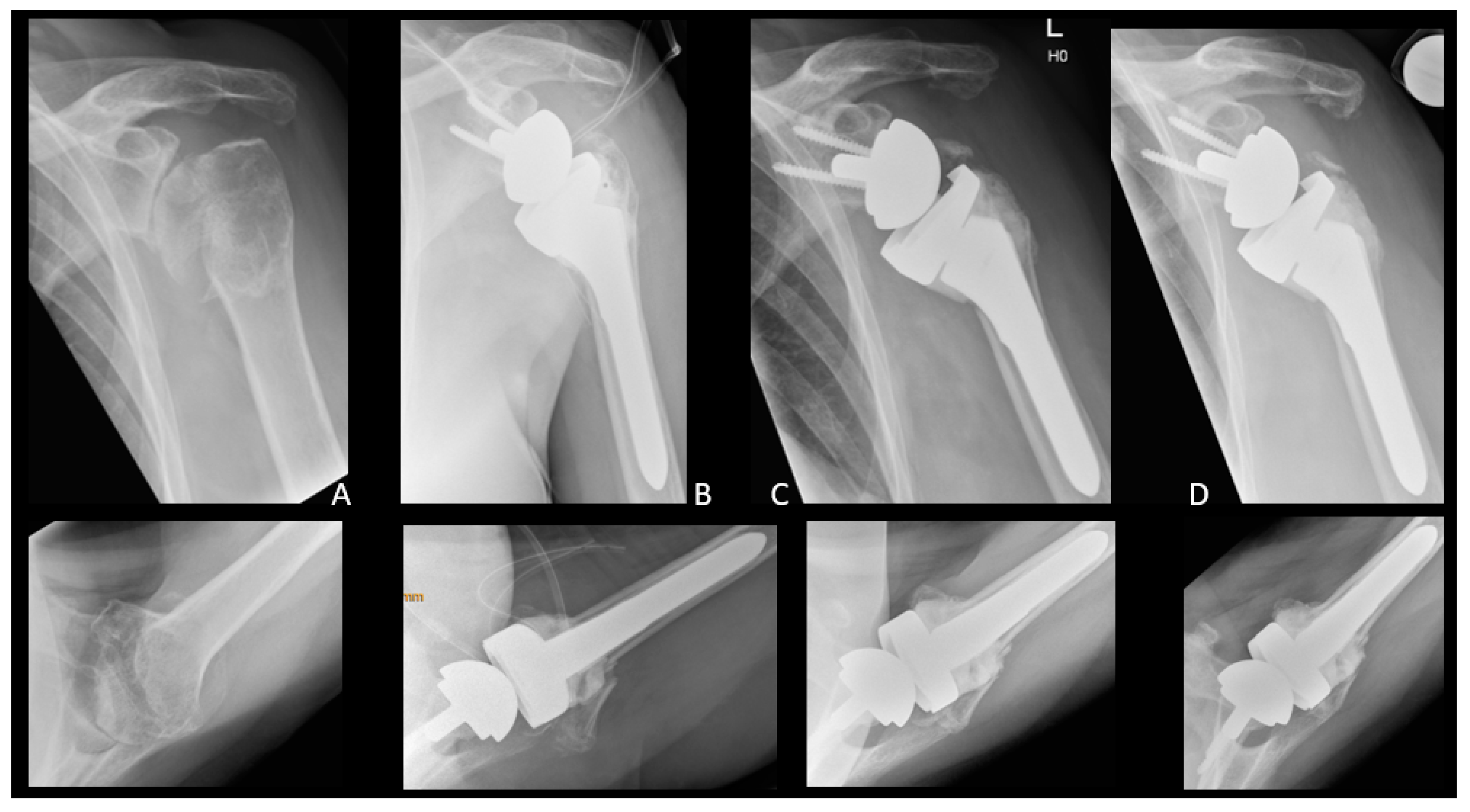
Most GTFs were treated conservatively (12 out of 17, 70%). Conservative treatment of postoperative GTF included immobilization in a brace for 4–6 weeks with initial passive mobilization and consecutive active mobilization. Five patients (five out of 17, 30%) were treated surgically by means of suture cerclage with FiberWire (Arthrex, Naples, FL, USA) (Figure 2).
Most GTFs (15 out of 17, 88%) did not further displace, regardless of surgical or conservative treatment. More than half of GTFs (nine out of 17, 53%) showed radiological consolidation at follow-up. One patient’s surgically treated GTF did not heal and was further dislocated. After revision surgery with a second FiberWire refixation, sufficient healing of the GT could be achieved. One patient with severe external rotation malfunction and a GTF nonunion was treated with resection of the GT and a latissimus dorsi tendon transfer.
Patient data according to GTFs are summarized in Table 2.

3.2. Clinical Outcome

Clinical outcome measurements with a minimum follow-up period of two years following RTSA implantation were available for 17 patients (100%) in GTF group and 34 patients (100%) in the control group. Outcome data at two-year follow up were compared in each case.
CSs improved in both groups after RTSA (absolute CS: GTF preoperative 30° ± 17°, postoperative 50° ± 19°, p ≤ 0.001; control group preoperative 32° ± 13°, postoperative 62° ± 21°, p ≤ 0.001; relative CS: GTF preoperative 36° ± 20°, postoperative 62° ± 21°, p ≤ 0.001; control group preoperative 40° ± 15°, postoperative 75° ± 22°, p ≤ 0.001). Preoperative CSs were comparable in both groups (absolute CS p = 0.508, relative CS p = 0.241). However, the control group achieved a significantly higher postoperative CS compared to the GTF group (absolute CS 62° ± 21° vs. 50° ± 19°, p = 0.032; relative CS 75° ± 20° vs. 62° ± 21°, p = 0.015).
The same result applies to the SSV, which improved in both groups as a result of the surgical treatment (GTF preoperative 26% ± 16%, postoperative 63% ± 26%, p ≤ 0.001; control group preoperative 36% ± 17%, postoperative 77% ± 29%, p ≤ 0.001). Postoperative SSV was significantly higher in the control group compared to the GTF group (p = 0.022).
Pain, measured by the CS pain points, was comparable preoperatively (GTF 8 ± 4, control 7 ± 4, p = 0.367) and improved in both groups after RTSA (p < 0.001) with no significant difference (GTF 12 ± 4, control 14 ± 2, p = 0.271).
Mean flexion was comparable preoperatively (GTF 67° ± 42°, control 80° ± 37°, p = 0.146) and improved in both groups after RTSA (p < 0.001) with no significant difference (GTF 102° ± 28°, control 114° ± 27°, p = 0.160).
Analog to flexion, shoulder abduction was comparable preoperatively (GTF 54° ± 31°, control 69° ± 35°, p = 0.076) and improved in both groups after RTSA (p < 0.001) with no significant difference (GTF 109° ± 42°, control 120° ± 39°, p = 0.317).
Mean external rotation was preoperatively comparable in both groups (GTF 32° ± 26°, control 28° ± 21°, p = 0.822). Mean external rotation did not significantly change in the control group after RTSA (preoperative 28° ± 21°, postoperative 30° ± 19°, p = 0.770), whereas the GTF group had a significant worsening in external rotation after RTSA (preoperative 32° ± 26°, postoperative 17° ± 19°, p = 0.010). The postoperative difference in external rotation was significant between both groups (p = 0.029).
The internal rotation ability was preoperatively comparable in both groups (GTF 4 ± 2, control 4 ± 3, p = 0.965) and could not be positively influenced by the prosthesis in both groups (GTF p = 0.627, control p = 0.137).
The same results appear for preoperative mean force in both groups (GTF 1 kg ± 1 kg, control 0 kg ± 1 kg, p = 0.155). The control group showed significantly more force after RTSA (2 kg ± 2 kg, p ≤ 0.001), whereas the GTF group did not improve (1 kg ± 2 kg, p = 0.441). The difference in postoperative mean force between both groups was statistically significant (p = 0.004).
The detailed outcome analysis is provided in Table 3.

4. Discussion

The most important finding of our study is that patients with a postoperative GTF had worse external rotation after RTSA compared to a matched control group.
The difference in external rotation may have a clinical impact, as the postoperative SSV and Constant scores were significantly higher in the control group compared to the GTF group. Additionally, according to Simovitch et al. [30] a difference of 3° ± 2° in external rotation may be clinically relevant. These findings are not unexpected, as it has been shown previously that healing of the GT is relevant to achieve a clinical outcome and the external rotation ability after fracture RTSA [21,22,23,33,34,35,36,37,38]. Ohl et al. [36] concluded in a multicenter study with 420 patients that anatomic healing of the GT in fracture RTSA improved objective and subjective outcome. Excision of the GT is associated with worse functional outcome and increased risk of postoperative instability [36]. Gallinet et al. [22] compared two cohorts of fracture RTSAs with and without GT-fixation retrospectively. They concluded that anatomic fixation of the GT led to better active range of motion, especially in external rotation with an improvement of +35°. These results show the importance of an intact and correct positioned GT for external rotation, as this can only be provided by functioning infraspinatus and teres minor muscles.
In our cohort, there was a non-significant difference in postoperative flexion and abduction. It seems that these results are not consistent with other studies showing a significant difference in elevation after resection or malunion of the GT [36,37,38,39]. Gallinet et al. [37] reported a significant mean difference of 14° in forward elevation comparing overall population (115° ± 29°) to a group, where the GT was resected (101° ± 25°). Gunst et al. [39] showed a difference of 19° in a comparable study (p = 0.04). Ohl et al. [36] had a mean difference of 27° between anatomical healing of the GT (128° ± 28°) and resection (101° ± 25°). We reported an overall difference in flexion and abduction of approximately 10°. Despite the lack of statistical significance in our comparably small study group with postoperative GTF, we agree that active elevation may be affected negatively by a GTF.
The overall clinical outcomes in this series of RTSA are comparable with reported data from previous studies [38,40,41,42]. The GTF group showed significant pain relief and improvement in all functional outcome parameters, except for active external and internal rotation. As expected, the comparative matching group was also unable to show any improvement in internal rotation after RTSA. These results are in agreement with the data of Hochreiter et al. [24], who pointed out factors influencing frequently observed limited internal rotation after RTSA.
Most of the GTF patients were treated conservatively, regardless of the grade of dislocation. Further, 50% of conservative treated patients achieved consolidation of the GTF during follow up. It remains unclear whether surgical treatment of GTFs can achieve a significant improvement of GTF healing and external rotation function, especially because the fractures occurred at a wide timeline. It is conceivable that, after a certain time, the muscles responsible for external rotation cannot be sufficiently activated despite refixation of the GT. Successful anatomical healing of the GT after surgical treatment is difficult to achieve. Gallinet et al. [22] and Sebastià-Forceda et al. [43] reported only 64% of anatomic healing utilizing suture cerclage fixation as described by Boileau et al. [44]. Our cohort showed similar results with 60% of GTF healing after surgical treatment.
The exact incidence of a GTF as a complication after RTSA remains unclear. In our database, an incidence of 1.4% was observed. If GTFs occur unnoticed in the postoperative follow-up or do not cause any complaints, these fractures might frequently remain undetected and are, therefore, potentially underreported.
The specific cause and risk factors for GTFs were not investigated in this study. However, all fractures except one occurred without a trauma history. Therefore, we hypothesize that these fractures either develop from an intraoperative fissure during the implantation of the stem or due to stress shielding in elderly patient with poor bone quality. Especially female patients are at risk of sustaining a GTF, similar to postoperative fractures of the acromion and scapular spine, with 76% (13 out of 17) females in our series.
The main limitations of the study are the inconsistent patient cohort with GTF and the retrospective study design. Due to different indications, different RTSA glenoid types and revision systems were chosen and implanted by different shoulder surgeons. However, the implanted stem design was the same (ZimmerBiomet Anatomical Inverse/Reverse onlay design) in all patients. We further tried to minimalize the bias by the study design with a 2:1 matching.
At the follow-up appointments, GTF was assessed on plain radiographs and computed tomography scans were not performed routinely. Non-displaced fractures may have been underreported.
Despite these limitations, the findings of this study add a valuable aspect to scientific knowledge in RTSA complications.

5. Conclusions

In this series of primary and secondary RTSA, the rare complication of a greater tuberosity fracture is significantly associated with an impaired external rotation, less abduction strength, and inferior clinical outcome scores compared to a matched control group.

Author Contributions

Conceptualization, P.B., K.W. and F.S.; methodology, P.B. and P.K.; formal analysis, P.K.; data curation, P.K.; writing—original draft preparation, F.S.; writing—review and editing, F.S., P.B., L.E., P.K. and K.W.; visualization, F.S.; supervision, P.B. and K.W. All authors have read and agreed to the published version of the manuscript.

Funding

This research received no external funding.

Institutional Review Board Statement

Not applicable.

Informed Consent Statement

Not applicable.

Data Availability Statement

Data will be provided by the corresponding author upon request.

Conflicts of Interest

The authors declare no conflict of interest. The funders had no role in the design of the study; in the collection, analyses, or interpretation of data; in the writing of the manuscript; or in the decision to publish the results. All involved authors are registered doctors.

References

  1. Grammont, P. Etude et réalisation d’une nouvelle prothèse d’épaule. Rheumatologie 1987, 39, 27–38. [Google Scholar]
  2. Hao, K.A.; Wright, T.W.; Schoch, B.S.; Wright, J.O.; Dean, E.W.; Struk, A.M.; King, J.J. Rate of improvement in shoulder strength after anatomic and reverse total shoulder arthroplasty. JSES Int. 2021, 6, 247–252. [Google Scholar] [CrossRef] [PubMed]
  3. Haupt, S.; Weber, S.; Frima, H.; Hutter, R.; Grehn, H.; Sommer, C. Proximal humeral fracture-dislocation: Outcome analysis in osteosynthesis and arthroplasties. Eur. J. Orthop. Surg. Traumatol. 2022, 1–10. [Google Scholar] [CrossRef] [PubMed]
  4. Karademir, G.; Tunalı, O.; Erşen, A.; Akpınar, S.; Atalar, A.C. Reverse total shoulder arthroplasty for failed treatment of proximal humerus fractures. reconstruction 2021, 6, 8. [Google Scholar] [CrossRef]
  5. Ferlauto, H.R.; Wickman, J.R.; Lazarides, A.L.; Hendren, S.; Visgauss, J.D.; Brigman, B.E.; Anakwenze, O.A.; Klifto, C.S.; Eward, W.C. Reverse total shoulder arthroplasty for oncologic reconstruction of the proximal humerus: A systemat-ic review. J. Shoulder Elb. Surg. 2021, 30, e647–e658. [Google Scholar] [CrossRef] [PubMed]
  6. Westermann, R.W.; Pugely, A.J.; Martin, C.T.; Gao, Y.; Wolf, B.R.; Hettrich, C.M. Reverse Shoulder Arthroplasty in the United States: A Comparison of National Volume, Patient Demographics, Complications, and Surgical Indications. Iowa Orthop. J. 2015, 35, 1–7. [Google Scholar]
  7. Schairer, W.W.; Nwachukwu, B.U.; Lyman, S.; Craig, E.V.; Gulotta, L.V. National utilization of reverse total shoulder arthroplasty in the United States. J. Shoulder Elb. Surg. 2015, 24, 91–97. [Google Scholar] [CrossRef]
  8. Farshad, M.; Gerber, C. Reverse total shoulder arthroplasty—From the most to the least common complication. Int. Orthop. 2010, 34, 1075–1082. [Google Scholar] [CrossRef]
  9. Shah, S.S.; Gaal, B.T.; Roche, A.M.; Namdari, S.; Grawe, B.M.; Lawler, M.; Dalton, S.; King, J.J.; Helmkamp, J.; Garrigues, G.E.; et al. The modern reverse shoulder arthroplasty and an updated systematic review for each complication: Part I. JSES Int. 2020, 4, 929–943. [Google Scholar] [CrossRef]
  10. Bohsali, K.I.; Wirth, M.A.; Rockwood, C.A., Jr. Complications of total shoulder arthroplasty. JBJS 2006, 88, 2279–2292. [Google Scholar]
  11. Boileau, P. Complications and revision of reverse total shoulder arthroplasty. Orthop. Traumatol. Surg. Res. 2016, 102, S33–S43. [Google Scholar] [CrossRef] [PubMed]
  12. Molé, D.; Favard, L. Excentered scapulohumeral osteoarthritis. Revue de Chirurgie Orthopedique et Reparatrice de l’ap-pareil Moteur 2007, 93 (Suppl. 6), 37–94. [Google Scholar] [CrossRef] [PubMed]
  13. Zumstein, M.A.; Pinedo, M.; Old, J.; Boileau, P. Problems, complications, reoperations, and revisions in reverse total shoulder arthroplasty: A sys-tematic review. J. Shoulder Elb. Surg. 2011, 20, 146–157. [Google Scholar] [CrossRef] [PubMed]
  14. Cho, C.-H.; Rhee, Y.G.; Yoo, J.C.; Ji, J.H.; Kim, D.-S.; Kim, Y.-S.; Rhee, S.-M.; Kim, D.-H. Incidence and risk factors of acromial fracture following reverse total shoulder arthroplasty. J. Shoulder Elb. Surg. 2020, 30, 57–64. [Google Scholar] [CrossRef]
  15. Kriechling, P.; Zaleski, M.; Loucas, R.; Loucas, M.; Fleischmann, M.; Wieser, K. Complications and further surgery after reverse total shoulder arthroplasty: Report of 854 primary cases. Bone Joint J. 2022, 104, 401–407. [Google Scholar] [CrossRef]
  16. Moverman, M.A.; Menendez, M.E.; Mahendraraj, K.A.; Polisetty, T.; Jawa, A.; Levy, J.C. Patient risk factors for acromial stress fractures after reverse shoulder arthroplasty: A multicenter study. J. Shoulder Elb. Surg. 2020, 30, 1619–1625. [Google Scholar] [CrossRef]
  17. Mahendraraj, K.A.; Abboud, J.; Armstrong, A.; Austin, L.; Brolin, T.; Entezari, V.; Friedman, L.; Garrigues, G.E.; Grawe, B.; Gulotta, L.; et al. Predictors of acromial and scapular stress fracture after reverse shoulder arthroplasty: A study by the ASES Complications of RSA Multicenter Research Group. J. Shoulder Elb. Surg. 2021, 30, 2296–2305. [Google Scholar] [CrossRef]
  18. Werthel, J.-D.; Schoch, B.S.; van Veen, S.C.; Elhassan, B.T.; An, K.-N.; Cofield, R.H.; Sperling, J.W. Acromial Fractures in Reverse Shoulder Arthroplasty: A Clinical and Radiographic Analysis. J. Shoulder Elb. Arthroplast. 2018, 2, 2471549218777628. [Google Scholar] [CrossRef]
  19. Otto, R.J.; Virani, N.A.; Levy, J.C.; Nigro, P.T.; Cuff, D.J.; Frankle, M.A. Scapular fractures after reverse shoulder arthroplasty: Evaluation of risk factors and the reliability of a proposed classification. J. Shoulder Elb. Surg. 2013, 22, 1514–1521. [Google Scholar] [CrossRef]
  20. LaCoste, K.L.; Arguello, A.M.; Ponce, B.A. Physical Therapy-Induced Fracture After Reverse Shoulder Arthroplasty: A Case Report. JBJS Case Connect 2021, 11, e20. [Google Scholar] [CrossRef]
  21. Ernstbrunner, L.; Rahm, S.; Suter, A.; Imam, M.A.; Catanzaro, S.; Grubhofer, F.; Gerber, C. Salvage reverse total shoulder arthroplasty for failed operative treatment of proximal humeral fractures in patients younger than 60 years: Long-term results. J. Shoulder Elb. Surg. 2019, 29, 561–570. [Google Scholar] [CrossRef] [PubMed]
  22. Gallinet, D.; Adam, A.; Gasse, N.; Rochet, S.; Obert, L. Improvement in shoulder rotation in complex shoulder fractures treated by reverse shoulder arthro-plasty. J. Shoulder Elb. Surg. 2013, 22, 38–44. [Google Scholar] [CrossRef] [PubMed]
  23. Simovitch, R.W.; Roche, C.P.; Jones, R.B.; Routman, H.D.; Marczuk, Y.; Wright, T.W.; Zuckerman, J. Effect of Tuberosity Healing on Clinical Outcomes in Elderly Patients Treated With a Reverse Shoulder Arthroplasty for 3- and 4-Part Proximal Humerus Fractures. J. Orthop. Trauma 2019, 33, e39–e45. [Google Scholar] [CrossRef] [PubMed]
  24. Hochreiter, B.; Hasler, A.; Hasler, J.; Kriechling, P.; Borbas, P.; Gerber, C. Factors influencing functional internal rotation after reverse total shoulder arthroplasty. JSES Int. 2021, 5, 679–687. [Google Scholar] [CrossRef]
  25. Nyffeler, R.W.; Seidel, A.; Werlen, S.; Bergmann, M. Radiological and biomechanical assessment of displaced greater tuberosity fractures. Int. Orthop. 2018, 43, 1479–1486. [Google Scholar] [CrossRef]
  26. Kriechling, P.; Loucas, R.; Loucas, M.; Künzler, T.; Gerber, C.; Wieser, K. Primary reverse total shoulder arthroplasty in patients older than 80 years: Clinical and radiologic outcome measures. J. Shoulder Elb. Surg. 2021, 30, 877–883. [Google Scholar] [CrossRef]
  27. Constant, C.R.; Murley, A.H. A clinical method of functional assessment of the shoulder. Clin. Orthop. Relat. Res. 1987, 160–164. [Google Scholar] [CrossRef]
  28. Gilbart, M.K.; Gerber, C. Comparison of the subjective shoulder value and the Constant score. J. Shoulder Elb. Surg. 2007, 16, 717–721. [Google Scholar] [CrossRef]
  29. Torrens, C.; Guirro, P.; Santana, F. The minimal clinically important difference for function and strength in patients undergoing reverse shoulder arthroplasty. J. Shoulder Elb. Surg. 2015, 25, 262–268. [Google Scholar] [CrossRef]
  30. Simovitch, R.; Flurin, P.-H.; Wright, T.; Zuckerman, J.D.; Roche, C.P. Quantifying success after total shoulder arthroplasty: The minimal clinically important difference. J. Shoulder Elb. Surg. 2018, 27, 298–305. [Google Scholar] [CrossRef]
  31. Harris, P.A.; Taylor, R.; Minor, B.L.; Elliott, V.; Fernandez, M.; O’Neal, L.; McLeod, L.; Delacqua, G.; Delacqua, F.; Kirby, J.; et al. The REDCap consortium: Building an international community of software platform partners. J. Biomed. Inform. 2019, 95, 103208. [Google Scholar] [CrossRef] [PubMed]
  32. Harris, P.A.; Taylor, R.; Thielke, R.; Payne, J.; Gonzalez, N.; Conde, J.G. Research electronic data capture (REDCap)—A metadata-driven methodology and workflow process for providing translational research informatics support. J. Biomed. Inform. 2009, 42, 377–381. [Google Scholar] [CrossRef] [PubMed]
  33. Cazeneuve, J.-F.; Cristofari, D.-J. Grammont reversed prosthesis for acute complex fracture of the proximal humerus in an elderly population with 5 to 12 years follow-up. Orthop. Traumatol. Surg. Res. 2014, 100, 93–97. [Google Scholar] [CrossRef] [Green Version]
  34. Grubhofer, F.; Wieser, K.; Meyer, D.C.; Catanzaro, S.; Beeler, S.; Riede, U.; Gerber, C. Reverse total shoulder arthroplasty for acute head-splitting, 3- and 4-part fractures of the proximal humerus in the elderly. J. Shoulder Elbow Surg. 2016, 25, 1690–1698. [Google Scholar] [CrossRef]
  35. Grubhofer, F.; Wieser, K.; Meyer, D.C.; Catanzaro, S.; Schürholz, K.; Gerber, C. Reverse total shoulder arthroplasty for failed open reduction and internal fixation of fractures of the proximal humerus. J. Shoulder Elb. Surg. 2017, 26, 92–100. [Google Scholar] [CrossRef] [PubMed]
  36. Ohl, X.; Bonnevialle, N.; Gallinet, D.; Ramdane, N.; Valenti, P.; Decroocq, L.; Boileau, P. How the greater tuberosity affects clinical outcomes after reverse shoulder arthroplasty for proximal humeral fractures. J. Shoulder Elb. Surg. 2018, 27, 2139–2144. [Google Scholar] [CrossRef] [PubMed]
  37. Gallinet, D.; Cazeneuve, J.-F.; Boyer, E.; Menu, G.; Obert, L.; Ohl, X.; Bonnevialle, N.; Valenti, P.; Boileau, P. Reverse shoulder arthroplasty for recent proximal humerus fractures: Outcomes in 422 cases. Orthop. Traumatol. Surg. Res. 2019, 105, 805–811. [Google Scholar] [CrossRef]
  38. Boileau, P.; Alta, T.D.; Decroocq, L.; Sirveaux, F.; Clavert, P.; Favard, L.; Chelli, M. Reverse shoulder arthroplasty for acute fractures in the elderly: Is it worth reattaching the tuberosities? J. Shoulder Elb. Surg. 2019, 28, 437–444. [Google Scholar] [CrossRef]
  39. Gunst, S.; Louboutin, L.; Swan, J.; Lustig, S.; Servien, E.; Nove-Josserand, L. Does healing of both greater and lesser tuberosities improve functional outcome after reverse shoulder arthroplasty for fracture? A retrospective study of twenty-eight cases with a computed tomography scan at a minimum of one-year follow-up. Int. Orthop. 2021, 45, 681–687. [Google Scholar] [CrossRef]
  40. Ek, E.T.; Neukom, L.; Catanzaro, S.; Gerber, C. Reverse total shoulder arthroplasty for massive irreparable rotator cuff tears in patients younger than 65 years old: Results after five to fifteen years. J. Shoulder Elb. Surg. 2013, 22, 1199–1208. [Google Scholar] [CrossRef]
  41. Hasler, A.; Fornaciari, P.; Jungwirth-Weinberger, A.; Jentzsch, T.; Wieser, K.; Gerber, C. Reverse shoulder arthroplasty in the treatment of glenohumeral instability. J. Shoulder Elb. Surg. 2019, 28, 1587–1594. [Google Scholar] [CrossRef] [PubMed]
  42. Hasler, A.; Kriechling, P.; Passaplan, C.; Wieser, K. Inadvertent, intraoperative, non- to minimally displaced periprosthetic humeral shaft fractures in RTSA do not affect the clinical and radiographic short-term outcome. Arch. Orthop. Trauma. Surg. 2021, 1–7. [Google Scholar] [CrossRef] [PubMed]
  43. Sebastiá-Forcada, E.; Cebrián-Gómez, R.; Lizaur-Utrilla, A.; Gil-Guillén, V. Reverse shoulder arthroplasty versus hemiarthroplasty for acute proximal humeral fractures. A blinded, randomized, controlled, prospective study. J. Shoulder Elb. Surg. 2014, 23, 1419–1426. [Google Scholar] [CrossRef] [PubMed]
  44. Boileau, P.; Krishnan, S.; Tinsi, L.; Walch, G.; Coste, J.; Molé, D. Tuberosity malposition and migration: Reasons for poor outcomes after hemiarthroplasty for displaced fractures of the proximal humerus. J. Shoulder Elb. Surg. 2002, 11, 401–412. [Google Scholar] [CrossRef]
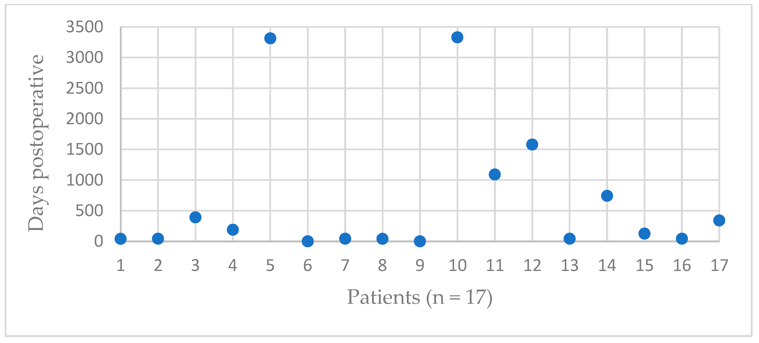
Figure 1. Occurrence of GTP depending on the time.
Jcm 12 01153 g001
Figure 2. Surgical treatment of a postoperative GTF with FiberWire suture cerclage. Left shoulder, X-rays in a.p. and axial views. (A) Preoperative arthritis after humeral head fracture. (B) Intraoperative control of RTSA. (C) Greater Tuberosity fracture. (D) Greater Tuberosity after surgical suture cerclage, unconsolidated, but with no further dislocation.
Jcm 12 01153 g002
Table 1. Basic demographic and arthroplasty-related data. Mann-Whitney-U testing (*) and Chi square testing (#) were used to compare the groups. Abbreviations: ASA, American Society of Anesthesiologists; CTA, cuff tear arthropathy; MRCT, massive rotator cuff tear; SD, standard deviation.
CharacteristicFractureControl p-Value
Patients, n1734
Mean age, years (SD)69 (9)69 (10)0.984 *
Male, n (%)4 (24)6 (17)0.618 #
Right side, n (%)13 (76)17 (50)0.070 #
ASA grade, n (%) 0.077 *
I01
II925
III88
Indication 1.000 #
CTA12
MRCT without osteoarthritis12
MRCT with osteoarthritis12
Osteoarthritis48
Instability12
Fracture36
Arthroplasty revision612
Mean BMI, kg/m2 (SD)29 (7)28 (6)0.704 *
Mean FU, moths (SD)72 (37)84 (35)0.263 *
Table 2. GTF-related data.
PatientOccurrence of GTF [Days after RTSA] TraumaDislocationTreatmentFurther Dislocation of GTFRadiological Consolidation of GTF
141NoSlightlyOperativeNoYes
243NoNoOperativeYesYes, after 2nd refixation
3391NoSlightlyConservativeNoNo
4188NoNoConservativeNoNo
53313NoYesConservativeNoPartial
60NoYesConservativeNoNo
743NoNoConservativeNoYes
841NoYesConservativeNoYes
90NoYesOperativeYesNo
103329NoSlightlyConservativeNoYes
111091NoNoConservativeNoYes
121580NoNoOperative NoYes
1343NoSlightlyOperativeNoNo
14743NoYesConservativeNoNo
15125YesYesConservativeNoNo
1643NoNoConservativeNoNo
17340NoSlightlyConservativeNoYes
Table 3. Clinical outcome comparing greater tuberosity fracture following RTSA with the control group. Wilcoxon-Ranksum testing 1 and Mann-Whitney-U testing 2 were used to compare the groups. Abbreviations: CS, Constant Score; ER, External rotation; FUP, Follow-up; IR, internal rotation; kg, kilograms; SD, Standard deviation; SSV, Subjective Shoulder Value.
FractureControlp-Value 2
Number 1734
Mean CS absolute, points (SD)Preop30 (17)32 (13)0.508
Postop50 (19)62 (21)0.032
p-Value 1<0.001<0.001
Mean CS relative, % (SD)Preop36 (20)40 (15)0.241
Postop62 (21)75 (20)0.015
p-Value 1<0.001<0.001
Mean SSV, % (SD)Preop26 (16)36 (17)0.101
Postop63 (26)77 (29)0.022
p-Value 1<0.001<0.001
Mean CS pain, points (SD)Preop8 (4)7 (4)0.367
Postop12 (4)14 (2)0.271
p-Value 1<0.001<0.001
Mean flexion, ° (SD)Preop67 (42)80 (37)0.146
Postop102 (28)114 (27)0.160
p-Value 1<0.001<0.001
Mean abduction, ° (SD)Preop54 (31)69 (35)0.076
Postop109 (42)120 (39)0.317
p-Value 1<0.001<0.001
Mean ER, ° (SD)Preop32 (26)28 (21)0.822
Postop17 (19)30 (19)0.029
p-Value 10.0100.770
Mean IR, ° (SD)Preop4 (2)4 (3)0.965
Postop4 (2)5 (3)0.138
p-Value 10.6270.137
Mean force, kg (SD)Preop1 (1)0 (1)0.155
Postop1 (2)2 (2)0.044
p-Value 10.441<0.001
Disclaimer/Publisher’s Note: The statements, opinions and data contained in all publications are solely those of the individual author(s) and contributor(s) and not of MDPI and/or the editor(s). MDPI and/or the editor(s) disclaim responsibility for any injury to people or property resulting from any ideas, methods, instructions or products referred to in the content.