
Same Clinical Reality of Spontaneous Rupture of the Common Iliac Artery with Pseudoaneurysm Formation—Comparison of Two Therapeutical Solutions, Endovascular Stent-Graft and Open Surgical Correction, for Two Cases and Review of the Literature

 ,
,  ,
,  ,
,  , ,
, , 
Abstract
1. Introduction
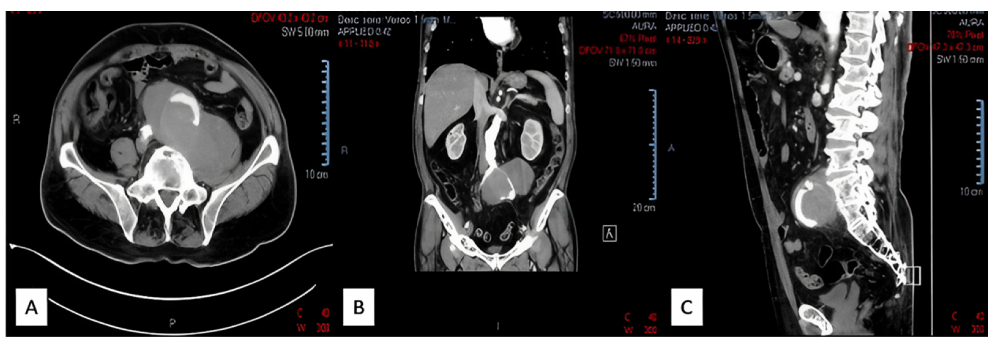
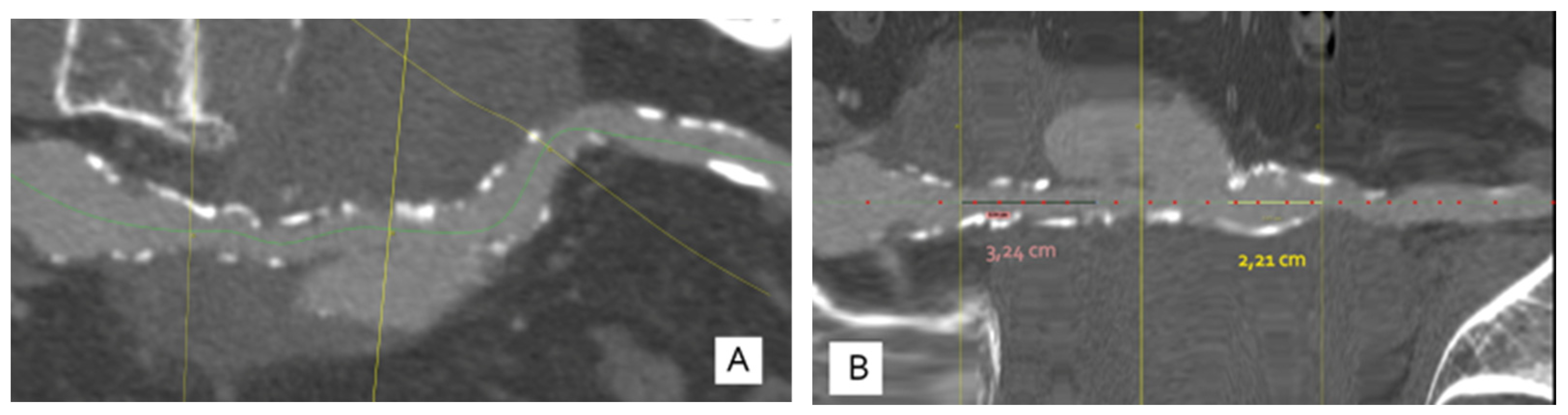
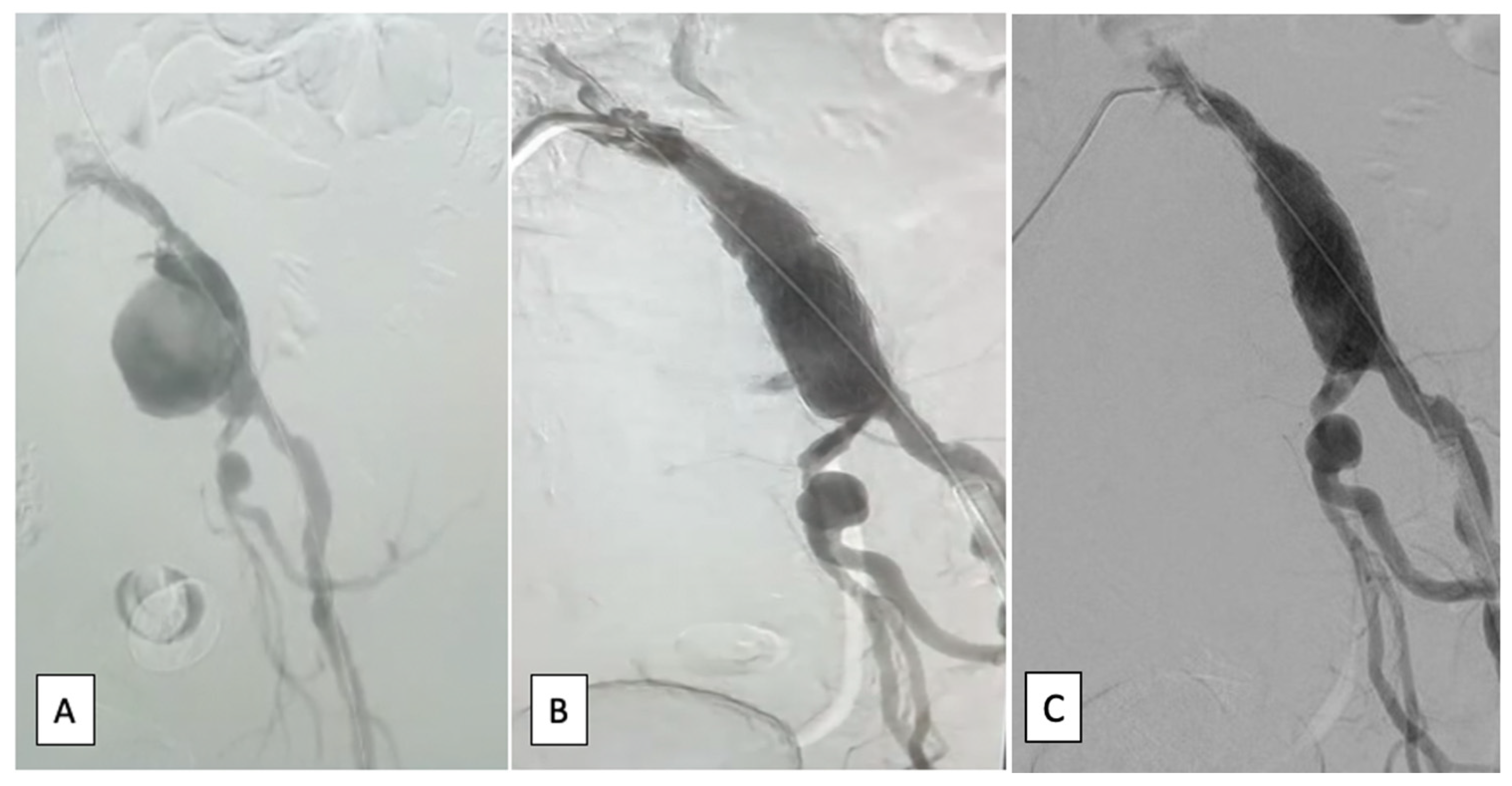
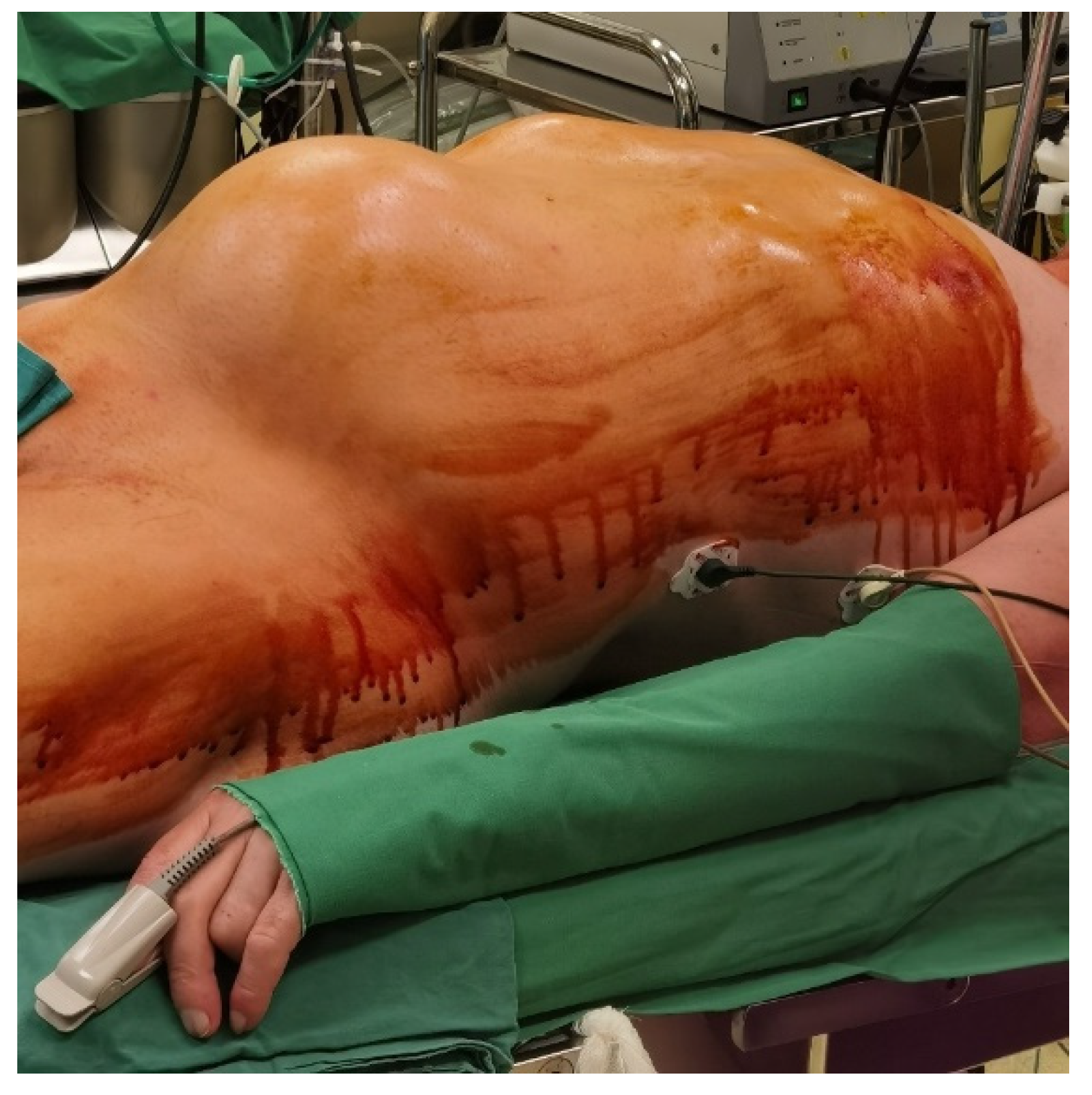
2. Case Reports
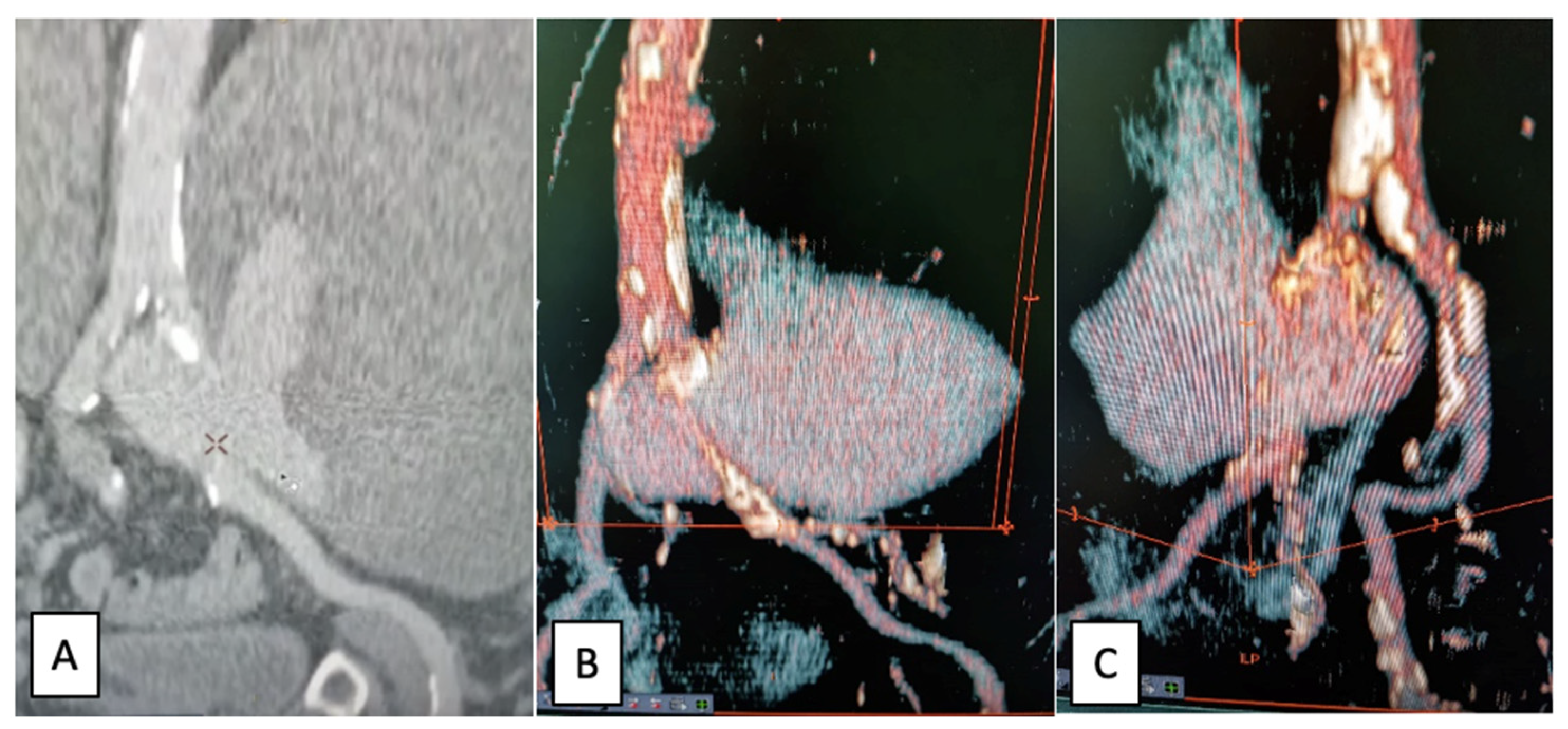
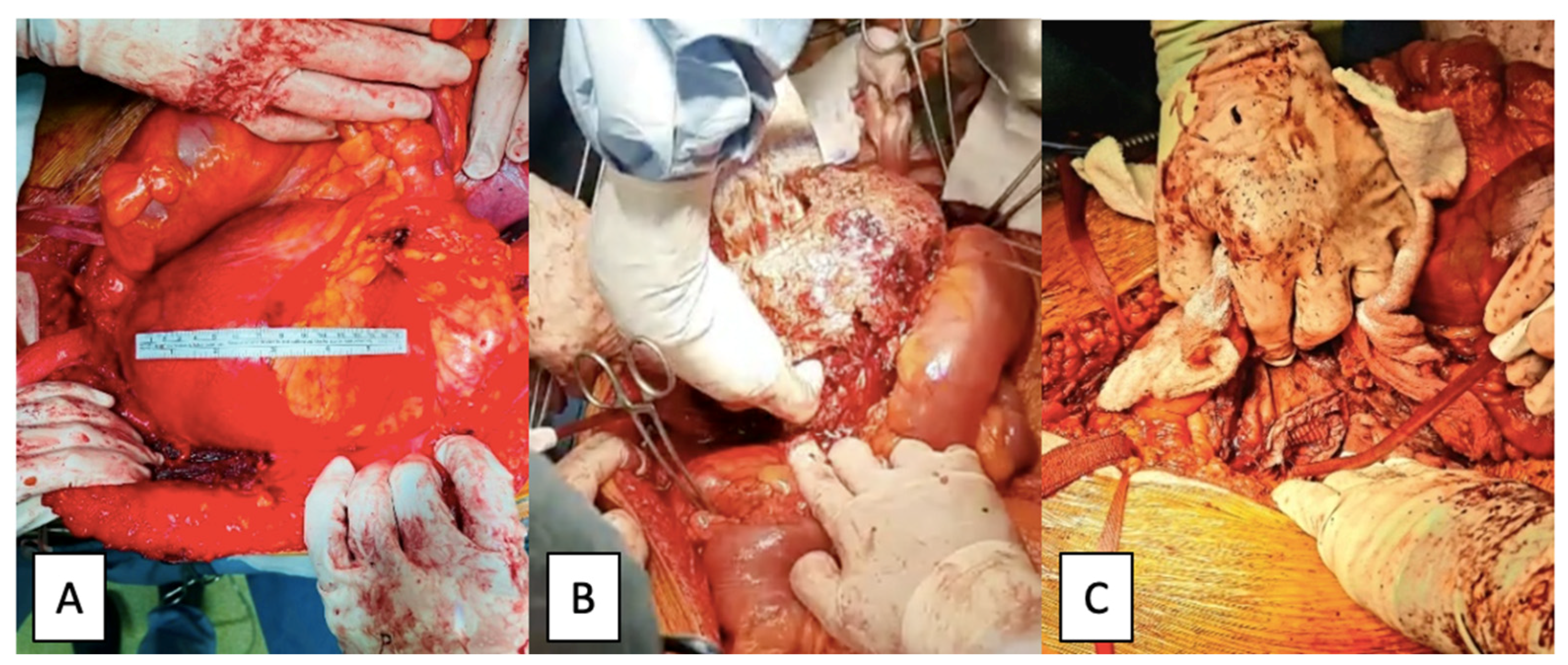
2.1. Case Report I
2.2. Case Report II
3. Discussion
4. Conclusions
Author Contributions
Funding
Institutional Review Board Statement
Informed Consent Statement
Data Availability Statement
Conflicts of Interest
References
- Bigarella, M.P.; Lopes, R.I.; Gentile, G.; Faustino, C.B.; Gamba, L.d.C.; Lima, G.B.B.; Melo, H.A.H.; Mulatti, G.C. Bilateral external iliac artery pseudoaneurysms causing urinary obstruction and acute renal failure. CVIR Endovasc. 2022, 5, 22. [Google Scholar] [CrossRef]
- Pitton, M.B.; Dappa, E.; Jungmann, F.; Kloeckner, R.; Schotten, S.; Wirth, G.M.; Mittler, J.; Lang, H.; Mildenberger, P.; Kreitner, K.-F.; et al. Visceral artery aneurysms: Incidence, management, and outcome analysis in a tertiary care center over one decade. Eur. Radiol. 2015, 25, 2004–2014. [Google Scholar] [CrossRef]
- Hussain, A.S.; Aziz, A. Giant External Iliac Artery Aneurysm. Ann. Vasc. Surg. 2019, 58, 386.e1–386.e3. [Google Scholar] [CrossRef]
- Moldovan, H.; Bulescu, C.; Sibisan, A.-M.; Tiganasu, R.; Cacoveanu, C.; Nica, C.; Rachieru, A.; Gheorghita, D.; Zaharia, O.; Balanescu, S.; et al. A Large Ascending Aorta Thrombus in a Patient with Acute Myocardial Infarction—Case Report. Medicina 2021, 57, 1176. [Google Scholar] [CrossRef]
- Bai, H.; Sun, P.; Wei, S.; Xie, B.; Li, M.; Xu, Y.; Wang, W.; Liu, Y.; Zhang, L.; Wu, H.; et al. A novel intramural TGF β 1 hydrogel delivery method to decrease murine abdominal aortic aneurysm and rat aortic pseudoaneurysm formation and progression. Biomed. Pharmacother. 2021, 137, 111296. [Google Scholar] [CrossRef]
- Rocha, R.V.; Lindsay, T.F.; Friedrich, J.O.; Shan, S.; Sinha, S.; Yanagawa, B.; Al-Omran, M.; Forbes, T.L.; Ouzounian, M. Systematic review of contemporary outcomes of endovascular and open thoracoabdominal aortic aneurysm repair. J. Vasc. Surg. 2020, 71, 1396–1412.e12. [Google Scholar] [CrossRef]
- Prabhakar, A.; Kumar, A.; Gupta, V.; Khandelwal, N.; Ahuja, C.K.; Singhal, M.; Vyas, S.; Panda, N.K.; Vaidhya, P.C. Endovascular management of internal carotid artery pseudoaneurysms: A single-centre experience of 20 patients. Neurol. India 2018, 66, 1067–1074. [Google Scholar] [CrossRef]
- Osmán, I.; Barrero, R.; León, E.; Medina-Lopez, R.; Torrubia, F. Mycotic pseudoaneurysm following a kidney transplant: A case report and review of the literature. Pediatr. Transplant. 2009, 13, 615–619. [Google Scholar] [CrossRef]
- Peng, Q.; Zhang, W. Rupture of multiple pseudoaneurysms as a rare complication of common iliac artery balloon occlusion in a patient with placenta accreta: A case report and review of literature. Medicine 2018, 97, e9896. [Google Scholar] [CrossRef]
- Moulakakis, K.G.; Alexiou, V.G.; Sfyroeras, G.S.; Kakisis, J.; Lazaris, A.; Vasdekis, S.N.; Brountzos, E.N.; Geroulakos, G.; Information, R. Endovascular management of infected iliofemoral pseudoaneurysms—A systematic review. Vasa 2017, 46, 5–9. [Google Scholar] [CrossRef]
- Corvino, A.; Catalano, O.; De Magistris, G.; Corvino, F.; Giurazza, F.; Raffaella, N.; Vallone, G. Usefulness of doppler techniques in the diagnosis of peripheral iatrogenic pseudoaneurysms secondary to minimally invasive interventional and surgical procedures: Imaging findings and diagnostic performance study. J. Ultrasound 2020, 23, 563–573. [Google Scholar] [CrossRef]
- Sousa, J.; Costa, D.; Mansilha, A. Visceral artery aneurysms: Review on indications and current treatment strategies. Int. Angiol. 2019, 38, 381–394. [Google Scholar] [CrossRef]
- Costache, V.S.; Meekel, J.P.; Costache, A.; Melnic, T.; Solomon, C.; Chitic, A.M.; Bucurenciu, C.; Moldovan, H.; Antoniac, I.; Candea, G.; et al. Geometric Analysis of Type B Aortic Dissections Shows Aortic Remodeling After Intervention Using Multilayer Stents. Materials 2020, 13, 2274. [Google Scholar] [CrossRef] [PubMed]
- Caño-Velasco, J.; Polanco-Pujol, L.; González-García, J.; Herranz-Amo, F.; Hernández-Fernández, C. Renal artery infectious (mycotic) pseudoaneurysms in renal transplantation recipients. Actas Urológicas Españolas 2021, 45, 335–344. [Google Scholar] [CrossRef]
- Xue, J.; Yao, Y.; Liu, L. Treatment of tuberculous aortic pseudoaneurysm associated with vertebral tuberculosis: A case series and a literature review. Medicine 2018, 97, e0382. [Google Scholar] [CrossRef]
- Qiu, C.; Liu, Z.; Huang, L.; Guo, L.; Lu, W.; Zhang, H.; He, Y.; Tian, L.; Li, D.; Wang, X.; et al. Covered Stents for Treatment of Visceral Artery Aneurysms: A Multicenter Study. J. Vasc. Interv. Radiol. 2022, 33, 640–647. [Google Scholar] [CrossRef]
- Buzatu, M.; Geantă, V.; Ştefănoiu, R.; Petrescu, M.-I.; Antoniac, I.; Iacob, G.; Niculescu, F.; Ghica, S.-I.; Moldovan, H. Investigations into Ti-15Mo-W Alloys Developed for Medical Applications. Materials 2019, 12, 147. [Google Scholar] [CrossRef]
- Miculescu, F.; Bojin, D.; Ciocan, L.T.; Antoniac, I.V.; Miculescu, M.; Miculescu, N. Experimental researches on biomaterial-tissue interface interactions. J. Optoelectron. Adv. Mater. 2007, 9, 3303–3306. [Google Scholar]
- Duta, L.; Ristoscu, C.; Stan, G.; Husanu, M.; Besleaga, C.; Chifiriuc, M.; Lazar, V.; Bleotu, C.; Miculescu, F.; Mihailescu, N.; et al. New bio-active, antimicrobial and adherent coatings of nanostructured carbon double-reinforced with silver and silicon by Matrix-Assisted Pulsed Laser Evaporation for medical applications. Appl. Surf. Sci. 2018, 441, 871–883. [Google Scholar] [CrossRef]
- Quan, P.H.; Antoniac, I.; Miculescu, F.; Antoniac, A.; Păltânea, V.M.; Robu, A.; Bița, A.-I.; Miculescu, M.; Saceleanu, A.; Bodog, A.D.; et al. Fluoride Treatment and In Vitro Corrosion Behavior of Mg-Nd-Y-Zn-Zr Alloys Type. Materials 2022, 15, 566. [Google Scholar] [CrossRef]
- Zabicki, B.; Limphaibool, N.; Holstad, M.J.V.; Juszkat, R. Endovascular management of pancreatitis-related pseudoaneurysms: A review of techniques. PLoS ONE 2018, 13, e0191998. [Google Scholar] [CrossRef] [PubMed]
- Sarioglu, O.; Capar, A.; Belet, U. Interventional treatment options in pseudoaneurysms: Different techniques in different localizations. Pol. J. Radiol. 2019, 84, 319–327. [Google Scholar] [CrossRef] [PubMed]
- Iliuta, L. Impact of Severe Pulmonary Hypertension on Outcomes Late After Aortic Valve Replacement for Aortic Stenosis Compared with Aortic Regurgitation. Cardiology 2014, 128, 177. [Google Scholar]
- Loffroy, R.; Chevallier, O.; Gehin, S.; Midulla, M.; Berthod, P.-E.; Galland, C.; Briche, P.; Duperron, C.; Majbri, N.; Mousson, C.; et al. Endovascular management of arterial injuries after blunt or iatrogenic renal trauma. Quant. Imaging Med. Surg. 2017, 7, 434–442. [Google Scholar] [CrossRef]
- Wang, C.; Regar, E.; Lachat, M.; Von Segesser, L.K.; Maisano, F.; Ferrari, E. Endovascular treatment of non-dissected ascending aorta disease: A systematic review. Eur. J. Cardio-Thoracic Surg. 2018, 53, 317–324. [Google Scholar] [CrossRef]
- Henry, J.C.; Franz, R.W. Pseudoaneurysms of the Peripheral Arteries. Int. J. Angiol. 2019, 28, 020–024. [Google Scholar] [CrossRef]
- Baptista-Sincos, A.P.W.; Simplício, A.B.; Sincos, I.R.; Leaderman, A.; Neto, F.S.; Moraes, A.; Aun, R. Flow-diverting Stent in the Treatment of Cervical Carotid Dissection and Pseudoaneurysm: Review of Literature and Case Report. Ann. Vasc. Surg. 2018, 46, 372–379. [Google Scholar] [CrossRef]
- Glaser, J.D.; Kalapatapu, V.R. Endovascular Therapy of Vascular Trauma-Current Options and Review of the Literature. Vasc. Endovasc. Surg. 2019, 53, 477–487. [Google Scholar] [CrossRef]
- Kasulke, R.J.; Clifford, A.; Nichols, W.K.; Silver, D. Isolated atherosclerotic aneurysms of the internal iliac arteries: Report of two cases and review of literature. Arch. Surg. 1982, 117, 73–77. [Google Scholar] [CrossRef]
- Sueyoshi, E.; Sakamoto, I.; Nakashima, K.; Minami, K.; Hayashi, K. Visceral and Peripheral Arterial Pseudoaneurysms. Am. J. Roentgenol. 2005, 185, 741–749. [Google Scholar] [CrossRef]
- Taif, S.; Alrawi, A.; Al-Kalbani, J. Iliac artery pseudoaneurysm presenting as a paravertebral collection: A potentially fatal mimic. BMJ Case Rep. 2014, 2014, bcr2013203428. [Google Scholar] [CrossRef] [PubMed]
- Ramakrishnan, P.; Hote, M.P.; Sreedhar, N.; Kumar, S.; Malik, V.; Choudhary, S.K. Internal iliac artery pseudoaneurysm: A rare presentation with foot drop and sciatica. Indian J. Thorac. Cardiovasc. Surg. 2019, 35, 222–225. [Google Scholar] [CrossRef]
- Wong, C.J.; Kraus, E.E. An Unusual Case of Acute Foot Drop Caused by a Pseudoaneurysm. Case Rep. Med. 2011, 2011, 1–3. [Google Scholar] [CrossRef]
- Chen, Y.; Gilman, M.D.; Humphrey, K.L.; Salazar, G.M.; Sharma, A.; Muniappan, A.; Shepard, J.-A.O.; Wu, C.C. Pulmonary Artery Pseudoaneurysms: Clinical Features and CT Findings. Am. J. Roentgenol. 2017, 208, 84–91. [Google Scholar] [CrossRef] [PubMed]
- Marshall, M.M.; Muiesan, P.; Srinivasan, P.; Kane, P.A.; Rela, M.; Heaton, N.D.; Karani, J.B.; Sidhu, P. Hepatic Artery Pseudoaneurysms Following Liver Transplantation: Incidence, Presenting Features and Management. Clin. Radiol. 2001, 56, 579–587. [Google Scholar] [CrossRef]
- Lugmayr, H.; Hartl, P.; Schwarz, C.; Zisch, R. Stent implantation in solitary aneurysm of the common iliac artery. Dtsch. Med. Wochenschr. 1993, 118, 499–502. [Google Scholar] [CrossRef] [PubMed]
- Marin, M.L.; Veith, F.J.; Panetta, T.F.; Cynamon, J.; Sanchez, L.A.; Schwartz, M.L.; Lyon, R.T.; Bakal, C.W.; Suggs, W.D. Transluminally placed endovascular stented graft repair for arterial trauma. J. Vasc. Surg. 1994, 20, 466–473. [Google Scholar] [CrossRef]
- O’Brien, C.J.; Rankin, R.N. Percutaneous Management of Large-Neck Pseudoaneurysms with Arterial Stent Placement and Coil Embolization. J. Vasc. Interv. Radiol. 1994, 5, 443–445. [Google Scholar] [CrossRef]
- Fakhro, A.; Shah, N.; Barnes, T. Endovascular Repair of a Common Iliac Pseudoaneurysm and Aortic Ectasia in a Patient with Horseshoe Kidney and Pancreatitis: A Case Report. Ann. Med. Surg. 2013, 2, 65–67. [Google Scholar] [CrossRef]
- Chaer, R.A.; Barbato, J.E.; Lin, S.C.; Zenati, M.; Kent, K.C.; McKinsey, J.F. Isolated iliac artery aneurysms: A contemporary comparison of endovascular and open repair. J. Vasc. Surg. 2008, 47, 708–713.e1. [Google Scholar] [CrossRef]
- Pitoulias, G.A.; Donas, K.P.; Schulte, S.; Horsch, S.; Papadimitriou, D.K. Isolated iliac artery aneurysms: Endovascular versus open elective repair. J. Vasc. Surg. 2007, 46, 648–654. [Google Scholar] [CrossRef] [PubMed]
- Hsu, J.-S.; Wu, I.-H.; Liu, K.-L. A Common Disease with an Unusual Complication of Acute Abdomen. Gastroenterology 2012, 142, e16–e18. [Google Scholar] [CrossRef]
- Chandler, B.T.; Ryer, E.J.; Keyser, B.M.; Elmore, J.R. A hybrid approach to appendicitis with right external iliac artery pseudo aneurysm: A case report. Int. J. Surg. Case Rep. 2017, 33, 99–101. [Google Scholar] [CrossRef] [PubMed]
- Pitcher, G.; Shuja, F.; Bacharach, J.M. A Mycotic Common Iliac Artery Pseudoaneurysm of Indeterminate Etiology. Ann. Vasc. Surg. 2020, 67, 567.e5–567.e8. [Google Scholar] [CrossRef]
- Doleman, B.; Kaushal, S.; Patel, A.; Kirk, J.; Quarmby, J. Rupture of the Left External Iliac Artery and Right Groin Pseudoaneurysm Formation following Angioplasty in a Patient with Neurofibromatosis Type 1 and Undiagnosed Bilateral Phaeochromocytoma. Case Rep. Radiol. 2013, 2013, 1–5. [Google Scholar] [CrossRef] [PubMed]
- de Oliveira Leite, T.F.; Pazinato, L.V.; Nunes, T.F.; da Motta Leal Filho, J.M. Endovascular management of giant common iliac artery pseudoaneurysm after complications in simultaneous pancreas-kidney transplant: A case report. J. Med. Case Rep. 2021, 15, 329. [Google Scholar] [CrossRef]
- Borges, L.; Oliveira, N.; Dias, E.; Cássio, I. Iliac artery pseudoaneurysm: A rare complication following allograft nephrectomy. BMJ Case Rep. 2014, 2014, bcr2013202596. [Google Scholar] [CrossRef]
- Wanhainen, A.; Verzini, F.; Van Herzeele, I.; Allaire, E.; Bown, M.; Cohnert, T.; Dick, F.; van Herwaarden, J.; Karkos, C.; Koelemay, M.; et al. Editor’s Choice—European Society for Vascular Surgery (ESVS) 2019 Clinical Practice Guidelines on the Management of Abdominal Aorto-iliac Artery Aneurysms. Eur. J. Vasc. Endovasc. Surg. 2019, 57, 8–93. [Google Scholar] [CrossRef] [PubMed]
- Melas, N.; Saratzis, A.; Dixon, H.; Saratzis, N.; Lazaridis, J.; Perdikides, T.; Kiskinis, D. Isolated Common Iliac Artery Aneurysms:A Revised Classification to Assist Endovascular Repair. J. Endovasc. Ther. 2011, 18, 697–715. [Google Scholar] [CrossRef]


Disclaimer/Publisher’s Note: The statements, opinions and data contained in all publications are solely those of the individual author(s) and contributor(s) and not of MDPI and/or the editor(s). MDPI and/or the editor(s) disclaim responsibility for any injury to people or property resulting from any ideas, methods, instructions or products referred to in the content. |
© 2023 by the authors. Licensee MDPI, Basel, Switzerland. This article is an open access article distributed under the terms and conditions of the Creative Commons Attribution (CC BY) license (https://creativecommons.org/licenses/by/4.0/).
Share and Cite
Moldovan, H.; Tiganasu, R.; Câlmâc, L.; Voica, C.; Broască, M.; Diaconu, C.; Ichim, V.; Cacoveanu, M.; Mirea, L.; Nica, C.; et al. Same Clinical Reality of Spontaneous Rupture of the Common Iliac Artery with Pseudoaneurysm Formation—Comparison of Two Therapeutical Solutions, Endovascular Stent-Graft and Open Surgical Correction, for Two Cases and Review of the Literature. J. Clin. Med. 2023, 12, 713. https://doi.org/10.3390/jcm12020713
Moldovan H, Tiganasu R, Câlmâc L, Voica C, Broască M, Diaconu C, Ichim V, Cacoveanu M, Mirea L, Nica C, et al. Same Clinical Reality of Spontaneous Rupture of the Common Iliac Artery with Pseudoaneurysm Formation—Comparison of Two Therapeutical Solutions, Endovascular Stent-Graft and Open Surgical Correction, for Two Cases and Review of the Literature. Journal of Clinical Medicine. 2023; 12(2):713. https://doi.org/10.3390/jcm12020713
Chicago/Turabian StyleMoldovan, Horațiu, Robert Tiganasu, Lucian Câlmâc, Cristian Voica, Marian Broască, Camelia Diaconu, Vlad Ichim, Mihai Cacoveanu, Liliana Mirea, Claudia Nica, and et al. 2023. "Same Clinical Reality of Spontaneous Rupture of the Common Iliac Artery with Pseudoaneurysm Formation—Comparison of Two Therapeutical Solutions, Endovascular Stent-Graft and Open Surgical Correction, for Two Cases and Review of the Literature" Journal of Clinical Medicine 12, no. 2: 713. https://doi.org/10.3390/jcm12020713
APA StyleMoldovan, H., Tiganasu, R., Câlmâc, L., Voica, C., Broască, M., Diaconu, C., Ichim, V., Cacoveanu, M., Mirea, L., Nica, C., Minoiu, C., Dobra, I., Gheorghiță, D., Dorobanțu, L., Molnar, A., & Iliuță, L. (2023). Same Clinical Reality of Spontaneous Rupture of the Common Iliac Artery with Pseudoaneurysm Formation—Comparison of Two Therapeutical Solutions, Endovascular Stent-Graft and Open Surgical Correction, for Two Cases and Review of the Literature. Journal of Clinical Medicine, 12(2), 713. https://doi.org/10.3390/jcm12020713