Systematic Review

Systematic Literature Review Shows Gaps in Data on Global Prevalence and Birth Prevalence of Sickle Cell Disease and Sickle Cell Trait: Call for Action to Scale Up and Harmonize Data Collection

1
Clinic of Pediatric Hematology Oncology, Department of Child and Maternal Health, Azienda Ospedaliera, University of Padova, 35122 Padua, Italy
2
Novo Nordisk Pharma AG, 8058 Zürich, Switzerland
3
Novo Nordisk A/S, 2860 Søborg, Denmark
*
Author to whom correspondence should be addressed.
J. Clin. Med. 2023, 12(17), 5538; https://doi.org/10.3390/jcm12175538
Submission received: 17 July 2023 / Revised: 18 August 2023 / Accepted: 22 August 2023 / Published: 25 August 2023
(This article belongs to the Section Hematology)

Abstract

:
Sickle cell disease (SCD) is an inherited monogenic disorder with high prevalence throughout sub-Saharan Africa, the Mediterranean basin, the Middle East, and India. Sources of SCD epidemiology remain scarce and fragmented. A systematic literature review (SLR) to identify peer-reviewed studies on SCD epidemiology was performed, with a search of bibliographic databases and key conference proceedings from 1 January 2010 to 25 March 2022 (congress abstracts after 2018). The SLR followed PRISMA (Preferred Reporting Items for Systematic Reviews and Meta-Analyses) guidelines. Meta-analyses, using a binomial normal random-effects model, were performed to estimate global and regional prevalence and birth prevalence. Of 1770 journal articles and 468 abstracts screened, 115 publications met the inclusion criteria. Prevalence was highest in Africa (~800/100,000), followed by the Middle East (~200/100,000) and India (~100/100,000), in contrast to ~30/100,000 in Europe. Birth prevalence was highest in Africa (~1000/100,000) and lowest in North America (~50/100,000) and Europe (~30/100,000). This SLR confirmed that sub-Saharan and North-East Africa, India, the Middle East, and the Caribbean islands are global SCD hotspots. Publications including mortality data were sparse, and no conclusions could be drawn about mortality. The identified data were limited due to gaps in the published literature for large parts of the world population; the inconsistent reporting of SCD genotypes, diagnostic criteria, and settings; and a sparsity of peer-reviewed publications from countries with assumed high prevalence. This SLR demonstrated a lack of systematic knowledge and a need to provide uniform data collection on SCD prevalence and mortality.

1. Introduction

Sickle cell disease (SCD) is one of the most common severe monogenic disorders in the world [1,2]. SCD is a hereditary chronic hemolytic anemia resulting from mutations in the subunit β-chain gene of hemoglobin (Hb) that cause stiff, sickle-shaped red blood cells that are involved in a complex series of adhesive events among blood cells, leading to small blood vessel obstruction, which reduces the oxygen supply to body tissues. Sickle-shaped red blood cells are prone to hemolysis, entailing at least three separate mechanisms involved in SCD pathophysiology: endothelial damage with vaso-occlusion, vasculopathy, and anemia [1,2]. Patients with SCD suffer from repeated infections, tissue hypoxia, acute and chronic pain, and multi-organ damage [1,3,4].
The acute and chronic complications of SCD, particularly vaso-occlusive crises, require frequent emergency room visits and hospitalizations [1,3]. Eventually, cumulative organ damage reduces survival compared with the general population [5]. The introduction of newborn screening programs and preventative measures have contributed to the improved life expectancy of patients with SCD, but the mortality rate in low-income regions is still high in children ≤3 years of age [6].
There are various genotypes that cause SCD, and the genotypes generally result in different severities of symptoms. Homozygosity for the sickle cell mutation in the Hb gene (HbS), known as sickle cell anemia or HbSS, is the most common and severe type of SCD [3,7]. Other types of SCD involve one HbS gene plus other mutations of the Hb gene, which lead to other altered forms of hemoglobin such as HbC [1,7]. HbSC is a common compound genotype that generally leads to a different phenotype, with fewer critical events but more retinopathy than HbSS [1]. HbS/β+/0-thalassemia, which is compound heterozygosity for HbS and β0 or β+ (the latter two leading to the impaired production of Hb β-chains), occurs in approximately 10% of people with SCD [3,7]. The HbS/β+ genotype generally results in a milder phenotype, whereas the HbS/β0 genotype leads to a severe form of SCD comparable to HbSS [3,7,8]. There is also a range of rarer genotypes, including regional variants, such as HbSDPunjab, HbSE, HbSOArab, and HbS Lepore [1,7,9]. People with one HbS gene and one normal gene (HbAS or sickle cell trait, SCT) rarely have symptoms or complications, although some evidence exists of possible progressive organ damage in some individuals with HbAS [1,7].
SCD is highly prevalent throughout large areas of sub-Saharan Africa, the Mediterranean basin, the Middle East, and India, which is possibly related to the high level of protection that SCT provides against severe malaria [2]. Studies suggest that approximately 300,000 infants worldwide are born every year with SCD [2]. A study with the objectives of estimating and mapping the global distribution of neonates with heterozygous (HbAS) and homozygous (HbSS) genotypes found that frequencies of HbS (>10%) were almost exclusively encountered in sub-Saharan Africa, with additional relatively high frequencies in India [10]. In 2008, the World Health Organization estimated that the global prevalence of SCD was 2.28 cases per 1000 people [11]. A systematic literature review (SLR) of 67 papers published between January 1980 and July 2017 reported a global birth prevalence of SCD homogenous genotypes (HbSS) in children under 5 years age of 112/100,000 live births, with a birth prevalence in Africa of 1125/100,000 compared with 43/100,000 in Europe [12].
With the increased global population movement, the prevalence and disease burden of SCD are changing in various regions. However, studies estimating the current prevalence of SCD and the mortality associated with it are scarce; fragmented; and dependent on sources that are difficult to compare (e.g., administrative databases, registries, screening programs, and clinical trials), limiting our understanding of the number of contemporary patients living with SCD across specific geographies worldwide and the impact that this disease has on their life expectancy. It is important to obtain sound data on prevalence to understand the health economic burden and complications of SCD in patients with various genotypes, as it will ensure the correct assessment of healthcare needs, assist policymakers with evidence-based decision making on how to allocate funding and resources, and determine adequate prevention and screening strategies.
The objective of this study was to perform an SLR of recent publications, from 2010 to 2022, on the prevalence, birth prevalence, and mortality associated with SCD globally and regionally, and to identify any knowledge gaps.

2. Materials and Methods

This SLR was performed in accordance with guidelines from the National Institute for Health and Clinical Excellence (NICE) using methodologies from the Cochrane Collaboration [13,14] and Preferred Reporting Items for Systematic Reviews and Meta-Analysis (PRISMA) guidance [15,16], and the study was registered with PROSPERO (ID 321574). The results are reported according to the 2020 PRISMA statement [17].
This SLR drew on data that are readily available in the public domain; hence, it did not require ethical review and approval.

2.1. Search Strategy

A comprehensive literature search of bibliographic databases and proceedings from key conferences was performed to identify peer-reviewed studies reporting on SCD epidemiology from 1 January 2010 to 25 March 2022. Congress publications published before 2018 were excluded, as it was assumed they would have been published subsequently as full-text articles. The bibliographic databases searched were: Excerpta Medica Database (Embase), MEDLINE, MEDLINE Epub Ahead of Print, BIOSIS, and Current Contents. The conference abstract databases searched were: Insightmeme and Embase. The proceedings from key congresses, the European Haematology Association, and the American Society of Hematology were also searched.
The search terms included a combination of subject headings (namely, variations of thesaurus and indexing terms (for example, MESH in MEDLINE)) and ‘free-text’ terms (e.g., synonyms, acronyms and abbreviations, spelling variants, and medical terminology) related to the prevalence or birth prevalence of SCD, common SCD genotypes, and SCT, or its associated impact on mortality. Comprehensive details of the search terms for prevalence, birth prevalence, and mortality and the searches performed can be found in Table S1.
Additional full-text articles and congress publications outside of the search identified as references within references, but relevant for outcomes of interest, could be included. In addition, data from national databases could be included to cover any gaps in peer-reviewed publications.

2.2. Eligibility Criteria for Publications

A Population, Intervention, Comparison, Outcomes, and Study (PICOS) framework was used to formulate the inclusion/exclusion criteria for the SLR (outlined in Table S2). All publications between 1 January 2010 and 25 March 2022 published in English, French, Spanish, or other languages where an English abstract was available, were eligible for inclusion if the population studied included people with any form of SCD (including HbSS, HbSC, HbS/β0-thalassemia, HbS/β+-thalassemia, HbSD, or HbSOArab) or SCT, irrespective of age (including children, adolescents, and adults); race; gender; or setting, and only if the publication had an outcome measure of interest (prevalence of SCD/SCT, birth prevalence of SCD/SCT, mortality related to SCD and its complications, or life expectancy in people with SCD/SCT).
Studies that were not deemed eligible for inclusion were animal studies, in vitro/ex vivo studies, gene/protein expression studies, or modeling studies; single-center studies (unless the center gathered data from a wider geographic region than a single town or community); and studies that reported data from a specific ethnic group within a country. Conference or meeting abstracts, meeting posters, or conference papers published before 2018 were excluded. Reviews and literature reviews; letters, case reports, editorials, comments, or editorial material; news, interviews, lectures, video-audio material, tombstone advertisements, meeting summary errata, corrections, and retracted publications were also excluded. For any studies published in more than one report, the most comprehensive and up-to-date version was used.

2.3. Study Selection

All identified search hits were imported into a reference management program (Endnote X9) for duplication checks. Two independent reviewers screened the titles and abstracts of records identified in the searches using the pre-defined eligibility criteria. Any discrepancies were resolved by consensus and by consulting a third reviewer as needed.
Congress abstracts and abstracts of full-text peer-reviewed publications deemed relevant during the first review step were retrieved, reviewed again, and selected for data extraction based on the eligibility criteria by the two independent reviewers, with a third reviewer to resolve any differences.
After identifying and validating the articles to be included in the review, a full list of included studies was collated. The flow diagram of screening, assessment, and inclusion is outlined in Figure 1.

2.4. Study Quality

Study quality was assessed by two independent reviewers using a version of the Grading of Recommendations, Assessment, Development, and Evaluations (GRADE) framework, adapted to observational studies. In this adaptation, publication bias and dose–response gradient were not evaluated, as publication bias would be difficult to assess in the context of epidemiological studies (e.g., a comparison of published literature versus registered clinical trials), and dose–response gradients do not apply to a non-interventional setting. The risk of bias was assessed in terms of factors that allowed the true estimate to be under-/over-reported. A GRADE assessment was applied to each study, rather than outcome. Where the assessment varied between outcomes, the highest-graded outcome was used to guide the overall study assessment.

2.5. Data Extraction, Reporting, and Synthesis

An extraction dashboard set up in a spreadsheet program was used to capture the data. The variables with sufficient data to be included in this report were: (1) authors, year, and title of publication; (2) year(s) of data collection; (3) greater region (regions defined based on available data in current SLR); (4) country; (5) number of people with SCD; (6) total population at risk; (7) disease classification: genotype, including HbSS, HbSC, sickle β-thalassemia, and regional SCD variants, or SCT; (8) proportion male (%); (9) prevalence data: prevalence (%), number of people with genotype; (10) birth prevalence data: birth prevalence (%); (11) mortality rate: by age category; (12) mean survival: survival curves; (13) cause of mortality; and (14) study quality: very low, low, moderate, or high. Other variables were extracted but were not sufficiently comprehensive to use in further analyses. A complete list of the parameters extracted is given in Table S3.
Data on the prevalence, birth prevalence, and mortality from the included studies were synthesized by either SCD genotype (HbSS, HbSC, sickle HbS/β-thalassemia [HbS/β0-thalassemia, HbS/β+-thalassemia] or undefined if the genotype was not described or if it was mixed) or SCT, as well as by greater region (Africa, Europe, India, Middle East, North America, and South America/the Caribbean). Initially, data were intended to be reported from Asia as a region. However, this was revised to India as a region, as no data were available from other countries in Asia. Studies investigating prevalence in children <1 year of age were considered to report on birth prevalence, while data from older children, adolescents, and adults were included as a prevalence measurement.

2.6. Quantitative Analyses of Prevalence and Birth Prevalence

A summary estimate was determined for the prevalence and birth prevalence globally and regionally (when sufficient data were available). The summary estimates were calculated from a random-effects model that assumed a binomial distribution for the results of each individual study, with the true value drawn from a normal distribution centered on a true regional/global value (assumed to exist). This will be referred to as a binomial normal (BN) model. Inhomogeneity scores (I2) are reported for each summary estimate to quantify the model assumption of a shared regional/global prevalence. The calculations were made using R statistics software [18] with the metafor package [19].
The summary estimates are presented as forest plots for prevalence and birth prevalence for SCD grouped (i.e., across all SCD genotypes) by individual SCD genotypes (when sufficient data were available), and for SCT. The quantitative analyses only included studies concerning groups that represented the general population and had a total study population larger than 1000.

2.7. Quantitative Analyses of Mortality

Due to the heterogeneity in reporting mortality across the identified studies, data on mortality are presented as survival curves. Publications found in the SLR with information on survival rates were identified and the data extracted. When a dataset with all individuals and their censoring or death date was not available, the survival curves presented in the papers were either digitized to extract the values, or survival curves were drawn based on other mortality information (e.g., mortality rate or deaths and at-risk population). For papers reporting survival rates for intervals, the survival curves reflected this by plotting the corresponding drop in the middle of the interval. If only deaths and at-risk population were reported, the survival probability was estimated by the Kaplan–Meier estimator.
In addition to the survival curves for SCD identified through the SLR, the equivalent curve for the general population was acquired from either the Human Mortality Database (HMD) [20] or the Human Life-Table Database (HLD) [21]. Notably, not all SCD populations had a corresponding general population from the same country and years. In these cases, a survival curve for the general population from another country in the same region, or the same country and another year, was used for comparison.

3. Results

Of 1770 journal articles and 468 abstracts screened, 115 publications met the inclusion criteria for the SLR (Figure 1). Of these, 23 included data on SCD prevalence and 23 on SCT prevalence, 50 included data on the birth prevalence of SCD, and 43 on the birth prevalence of SCT. Thirty-three publications included some aspect of data on mortality or life expectancy.

3.1. Qualitative Data Analysis

Key data on prevalence and birth prevalence for SCD/SCT are synthesized in Tables S4–S7 in the Supplementary Materials. Mortality data were extracted from the text, and the identified causes of death among people with SCD are shown in Table S8 in the Supplementary Materials. Data extracted from the text, tables, and figures were used to generate survival curves.

3.2. Quantitative Meta-Analysis of Prevalence and Birth Prevalence

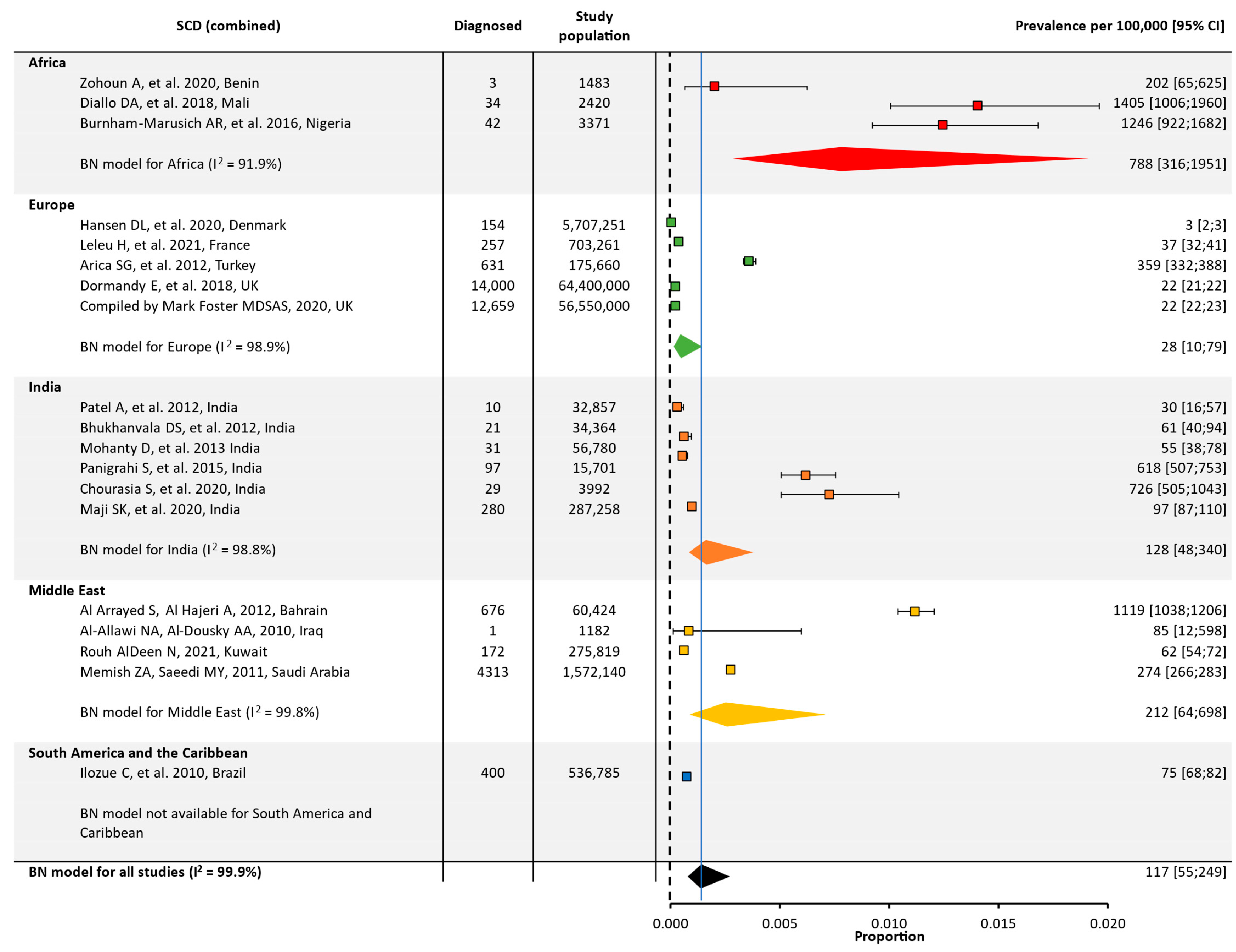
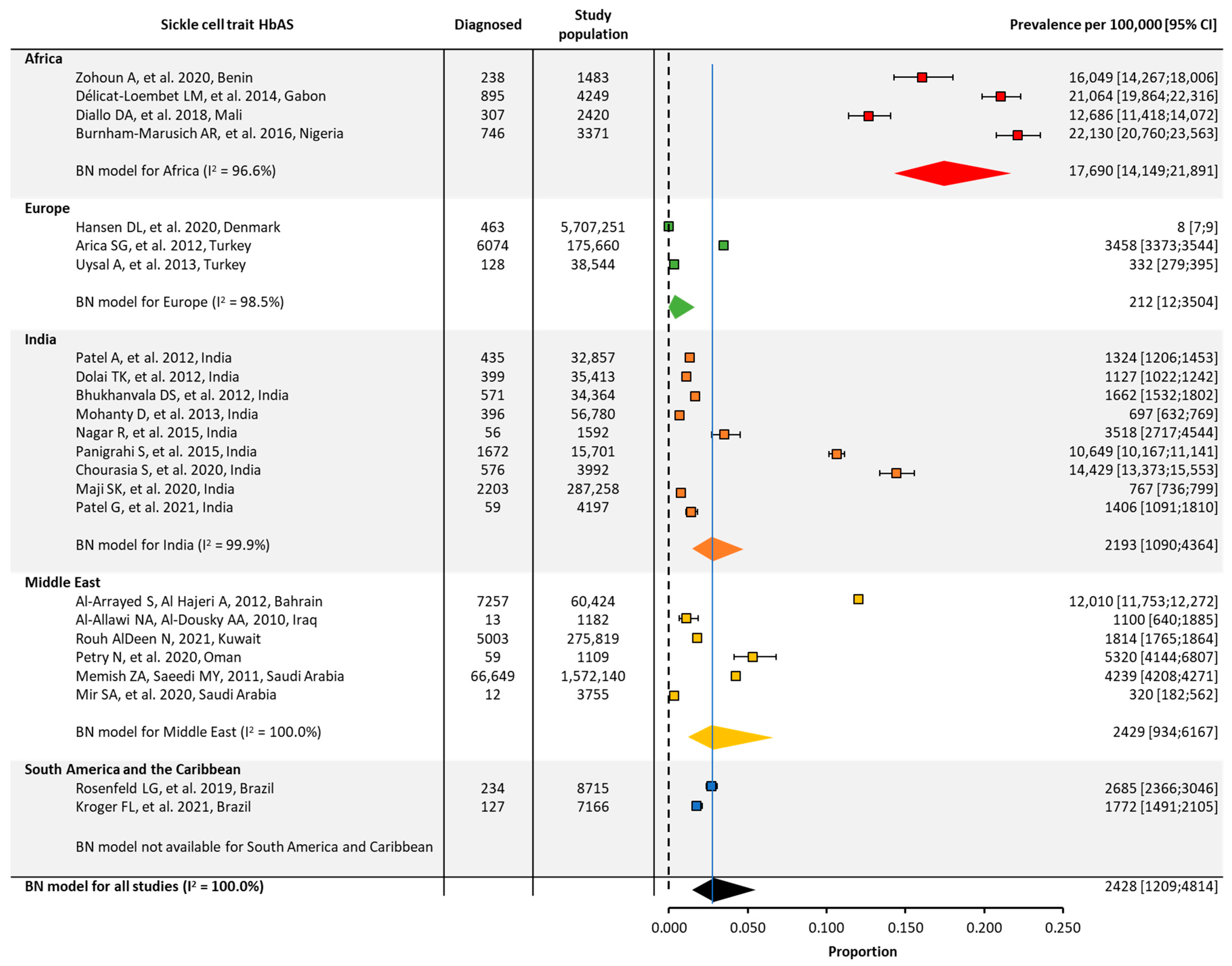
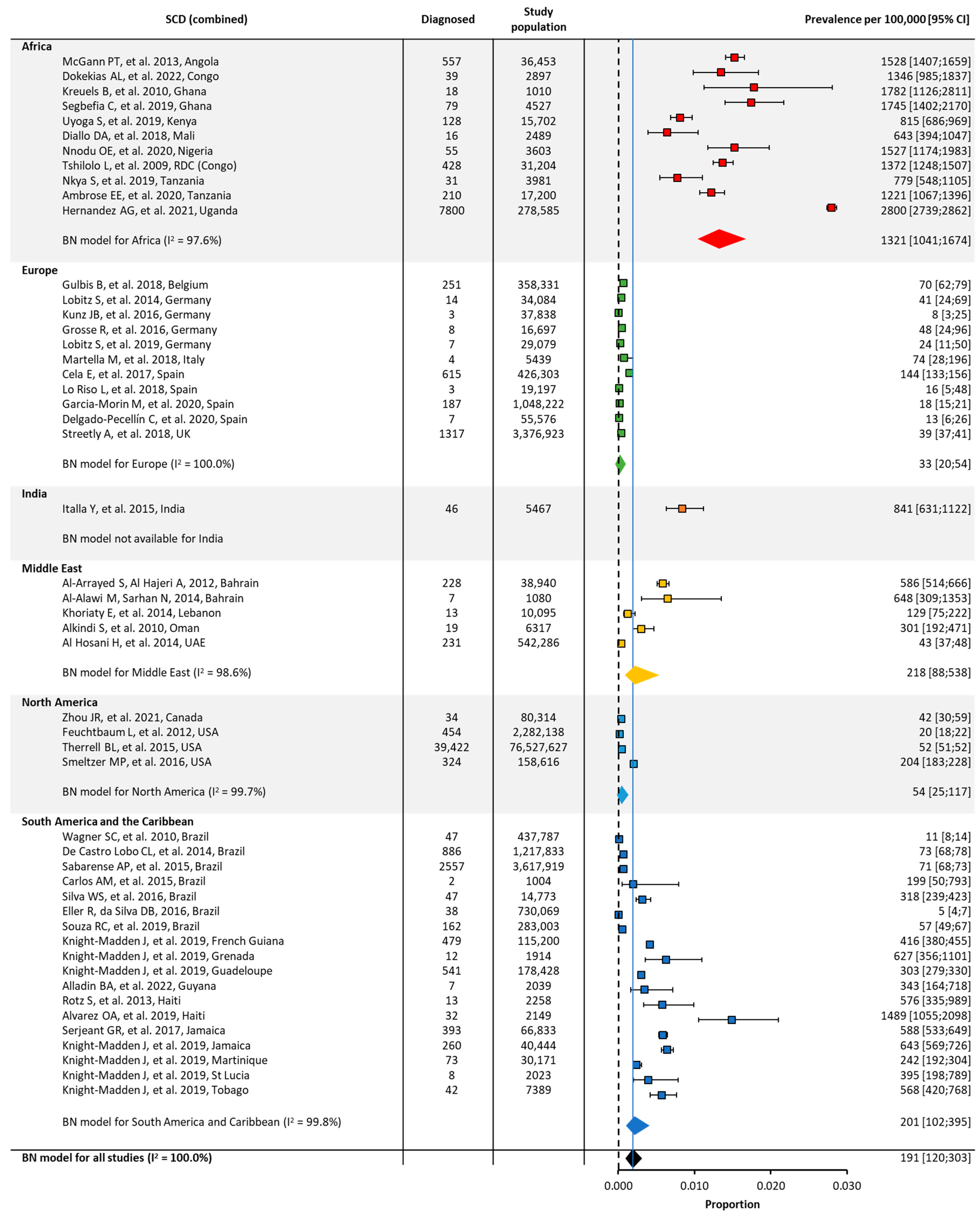
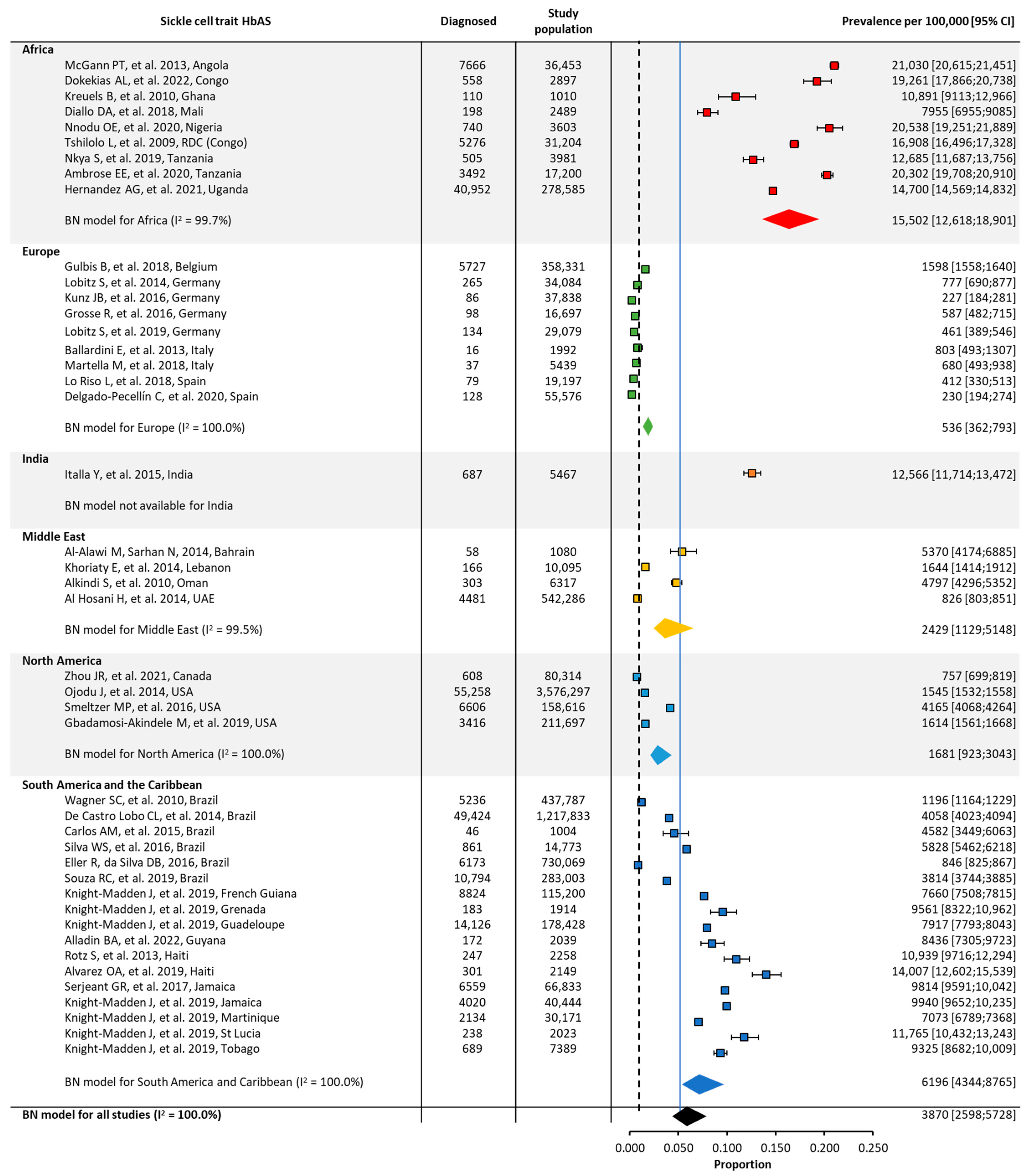
Quantitative analyses on the global and regional prevalence and birth prevalence of SCD and SCT are shown in Figure 2, Figure 3, Figure 4 and Figure 5.
SCD prevalence was highest in Africa (~800/100,000), followed by the Middle East (~200/100,000) and India (~100/100,000). Prevalence was lower in Europe (30/100,000) and could not be calculated for North America or South America/the Caribbean due to a low number of eligible studies (Table 1, Figure 2).
SCT prevalence followed a similar pattern to that of SCD, with the highest prevalence in Africa (~18,000/100,000), followed by the Middle East (~2000/100,000), India (~2000/100,000), and Europe (~200/100,000). The prevalence of SCT could not be calculated for North America or South America and the Caribbean due to the insufficient number of papers from these regions (Table 1, Figure 3).
The birth prevalence of SCD was highest in Africa (~1000/100,000) and lowest in North America (~50/100,000) and Europe (~30/100,000) (Table 1, Figure 4). Similarly, the birth prevalence of SCT was highest in Africa (~16,000/100,000) and lowest in North America (~2000/100,000) and Europe (~500/100,000) (Table 1, Figure 5). The birth prevalence for SCD and SCT for India were not calculated, as only one study included these data. The birth prevalence of SCD reported in this study was ~800/100,000, and the birth prevalence of SCT was ~13,000/100,000 (Table 1, Figure 4 and Figure 5).
As expected, there was considerable heterogeneity (I2) in the overall prevalence or birth prevalence for a given region, due to the low number of identified eligible studies; a wide variation across studies, even within the same country; and differing methods of data collection (e.g., the screening of subpopulations or in specific settings such as military drafts or school students versus database studies) (Figure 3, Figure 4 and Figure 5).
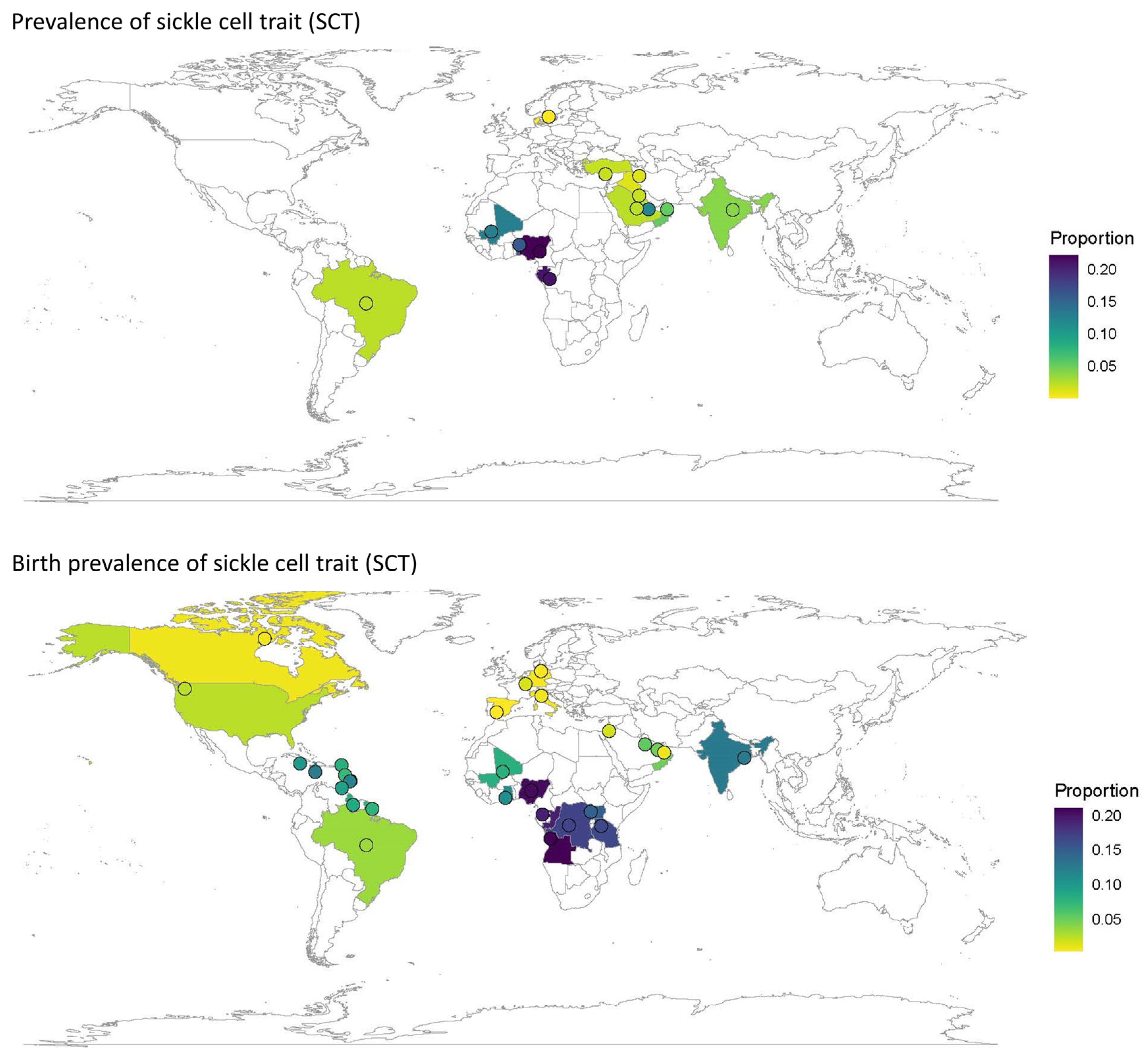
The prevalence and birth prevalence of SCD (Figure 6) and SCT (Figure 7) plotted by country on a world map visually represents the regions in which the data gaps on any type of prevalence exist. There were no large-scale published data on SCD prevalence from North America; however, some birth prevalence data were available, including one large study of case findings from newborn screening across the US. Large-scale studies of prevalence were available for a few countries in Northern Europe, but only smaller studies from key countries in Southern Europe with an expected increasing prevalence due to migration, such as France, Italy, and Spain (Figure 6).
SCD prevalence data from Africa were also sparse (three countries), while SCD birth prevalence was available from eight African countries (Figure 6). SCD prevalence data for South America and the Caribbean were only available from a single study in Brazil. Although SCD birth prevalence data were available from seven studies in Brazil and one study including a number of Caribbean countries, the majority of the data came from Brazilian publications (Figure 4). SCD prevalence was reported for nine studies in India, but only one study reported SCD birth prevalence (Figure 3).
Prevalence and birth prevalence by genotype (HbSS, HbSC, and sickle β-thalassemia), where these data were available, are reported in Figures S1–S6.

3.3. Mortality

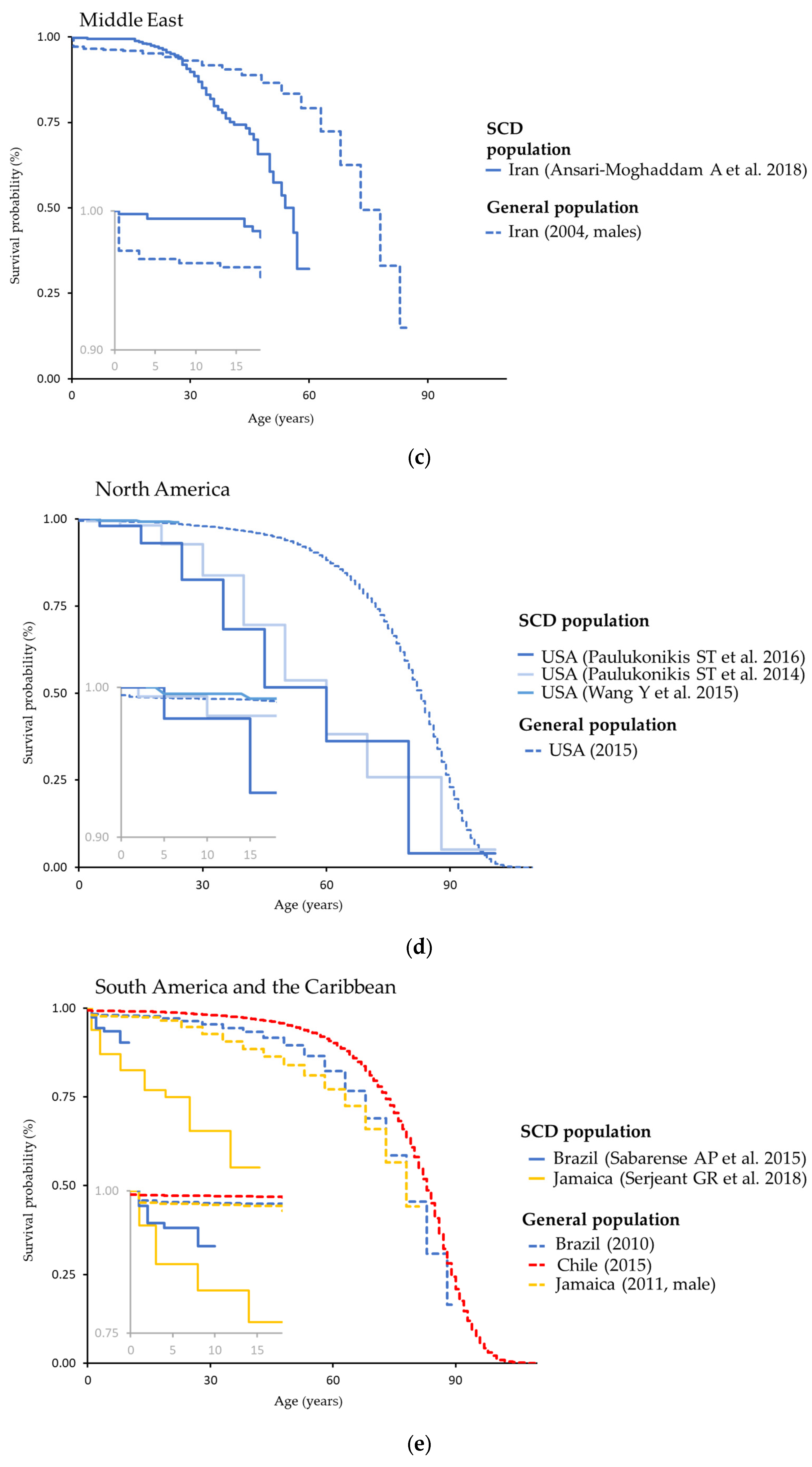
Survival curves were determined for Africa, Europe, the Middle East, North America, and South America and the Caribbean, as presented in Figure 8. Each plot presents survival curves for SCD populations, from the studies referenced, and from the general population for comparison [19,20] for the included countries. Generally, observed survival in the SCD population was lower than for the general population. The difference between the SCD population and overall population tended to be larger for regions with less access to healthcare (e.g., Africa) compared with those that had higher access (e.g., North America or Europe).
In the nine studies where the cause of death was reported, the leading causes of death were aligned with the known complications of SCD and included acute chest syndromes (61/345 [17.7%]), various infections (40/345 [11.6%]), sepsis or septicemia (34/345 [9.9%]), splenic sequestration (28/345 [8.1%]), and stroke (26/345 [7.5%]) (Table S8).

4. Discussion

As expected, SCD prevalence and/or birth prevalence were highest in Africa, followed by the Middle East, India, and the Caribbean. The prevalence data for South America and the Caribbean were limited by the low number of eligible studies in the region, although the existing studies covered a range of Caribbean countries. For SCT, these relationships were preserved for Africa and India, but not for the Middle East. There could be an actual difference in the SCT to SCD ratio across regions due, for example, to differing mortality rates from SCD. However, this apparent difference may also be due to missing data in high-prevalence areas or may reflect different data gathering methods used in the publications; for example, in the Middle East region, a high portion of publications were of premarital screening studies, which were not widely seen in other regions. Having multiple data sources from individual countries of interest within a specific region (i.e., those with significant expected effects on healthcare resources, such as countries in Europe with an expected increase in prevalence due to migration), would be highly beneficial to allow the calculation of an accurate prevalence and absolute number of people living with SCD in any given country. Although both our study and other previous SLRs showed certain trends in terms of key countries and hotspot regions, differences between countries and regions observed here were likely not wholly driven by geography. There are other possible contributing factors such as differences in study protocols and population size. With multiple potential sources of bias affecting the accuracy of the prevalence estimates for each country, reliable absolute numbers could not be calculated for countries of interest with the data currently available.
A previous SLR by Wastnedge et al. [12], which included 67 papers from January 1980 to July 2017 in a meta-analysis, gave a global estimate for the birth prevalence of homozygous SCD of 112/100,000 live births (95% CI 101–123). In Africa, the birth prevalence was 1125/100,000 (95% CI 680–1570) compared with 43/100,000 (95% CI 30–56) in Europe [12]. While the findings of the current study were broadly in line with the results of this meta-analysis, with Africa as an SCD hotspot, the Wastnedge et al. review included prevalence data that covered over three decades [12]. This SLR included publications from 2010 onwards, as it was considered that epidemiological and mortality data from older publications would not be representative of the current situation, considering changes over time in healthcare practices and the prevalence and disease burden of SCD in various regions arising from global population movements [2].
A study by Piel et al. [10] using a database of sickle hemoglobin surveys to estimate and map the global distribution of neonates with SCT (HbAS) or homozygous SCD (HbSS) showed that high frequencies (>10%) of the HbS allele (i.e., HbAS plus HbSS) were almost exclusively encountered in sub-Saharan Africa, with additional relatively high frequencies in India [10]. However, Piel et al. covered only neonate estimates, focusing on HbAS and HbSS, and did not include SCD birth prevalence from other SCD genotypes, meaning their study did not cover the estimation of healthcare needs for all aspects of the disease. While the previous SLRs focused on birth prevalence, this current SLR addressed some of the limitations of these earlier SLRs by investigating both prevalence and updated estimates for birth prevalence. The current SLR also included a wider range of both SCD genotypes and SCT in the search terms; however, data on the rare or regional forms were limited. The individual genotypes were described in the synthesized qualitative overview but were included in the overall SCD groups for the meta-analyses. Furthermore, most publications reporting on the prevalence and birth prevalence of HbS/β-thalassemia unfortunately did not separate the more severe form of HbS/β0-thalassemia and the less severe form of HbS/β+-thalassemia, a granularity that would be important for an accurate understanding of the healthcare needs related to SCD in a specific region.
The current SLR revealed important gaps in the published literature, with missing data on prevalence and/or birth prevalence from countries where SCD is believed to be highly prevalent, including African countries such as Cameroon and Mozambique. In addition, although some identified studies represented large African countries, such as Nigeria, Egypt, and the Democratic Republic of Congo, the data were limited due to the total study populations being in the 10,000s. There were also limited or no data from other regions where prevalence is expected to be on the rise through migration and higher birth rates of the migrant population relative to the native population, such as Europe and North America. Up-to-date information from these regions is important to understand the changing prevalence in these areas in order to ensure early detection, adequate prevention programs, and appropriate healthcare resources.
Countries with a very low prevalence, for example, countries in Eastern Europe, would likely not collect data on SCD, as the disease does not pose a significant clinical issue in these countries. However, in some countries with a low or very heterogeneous prevalence of SCD in the population, screening programs are often limited to at-risk populations, for example, members of specific ethnic groups. For instance, Mueller et al. [106] reported on SCD prevalence in Ghanaian migrants in Germany, where the prevalence of SCT and SCD were 17.4% and 1.2%, respectively, much higher than the estimates reported for the general German population [107]. With the rising prevalence in Germany, a nationwide registry of patients across centers caring for children with SCD has been established to understand specifically the disease burden and patient needs in Germany [108]. Similarly, in India, screening for sickle cell disease is carried out among children in tribal schools to allow the development of genetic counselling and intervention programs to reduce morbidity and mortality [30,35,109,110]. Although these studies may not be representative of the general population and are often not large enough to be included in meta-analyses, in the absence of the screening of the whole population they may provide important insights into the prevalence of SCD among certain subpopulations in the respective regions.
Mortality, life-expectancy, and survival data were limited, as publications reporting these were sparse, and the information was reported in many different ways (e.g., by median length of life, mean length of life, life expectancy, and survival time). The causes of death reported by the identified publications generally aligned with key complications of the SCD pathology, such as thrombosis/sickle cell crises and ensuing tissue/organ damage, as well as an increased susceptibility to infections [3]. As noted in other aspects of data collection in the field of SCD, gaps in the data on the cause of death were prevalent, with ~25% (8/345) of deaths across the nine publications being reported as other/not known.
Furthermore, SCD genotypes, diagnostic criteria, and study settings differed widely between publications that included mortality data. However, for a number of studies, it was possible to present mortality data as survival curves. In general, survival observed in the SCD population was lower than what was reported for the general population. Furthermore, a trend could be observed towards a larger difference for regions with less access to healthcare. This difference was particularly noticeable in Africa (Angola and Kenya), where mortality in infancy was elevated in the first 5 years of life. However, no data were available for older children or adults. Unfortunately, the reasons for differences in the cause of death between people with SCD and the general population were hard to ascertain, as only nine of the 115 identified publications reported causes of death, and only in a few of these cases did the same paper report on mortality and causes of death. The complications of SCD tend to differ in children and adults; the available data did not allow for the further confirmation of causes of death across age groups.
A limitation of a meta-analysis based on the data in this SLR would be that there were few studies in some regions, such as North America. A number of US studies identified in the search were excluded from the analyses as they were from a single center only, which would make them difficult to generalize to the broader population of the country. In addition, there was considerable heterogeneity between countries within regions and even within countries, for example in Brazil and India [111]. Given the large inhomogeneity (I2)—close to 100%—the regional/global results were more difficult to interpret and should be used with caution. The main reason for the large inhomogeneity was the actual differences between populations across regions and/or different groups across countries. In addition, the inhomogeneity could have also been due to differences in sampling methods, imprecise estimations in individual studies, or different subpopulations.
The limitations to this SLR were mainly due to the identified gaps in the published literature, including the inconsistent reporting of the different SCD genotypes, variability in diagnostic criteria, and different study settings. There was a clear deficit of studies from some large regions of the world (e.g., US, Russia, Asian countries apart from India) in the current SLR. The scarce information published over the 12-year study period highlights the need to scale up systematic epidemiological data collection on SCD and SCT by registries, databases, and longitudinal studies using specific and clearly defined parameters to properly estimate the disease burden.
Few countries reported both prevalence and birth prevalence. The studies on birth prevalence (i.e., prevalence in children <1 year of age) were mainly based on newborn screening programs. Newborn screening data have advantages like standardized data collection and are high-quality, providing a good basis for estimating future care needs. However, for a better estimate of care needs in any given population, we would need to combine the birth prevalence data with up-to-date survival data in the SCD population from the equivalent region/country. With the great heterogeneity in the research on mortality identified in the current SLR, it is clear that further research would be needed to determine up-to-date and adequate prevention and screening strategies, targeting the causes of SCD mortality. The Rare Anaemia Disorders European Epidemiological Platform (RADeep, https://www.radeepnetwork.eu/, accessed on 21 August 2023) aims to set up a European registry of patients affected by rare anemia diseases by creating a network of centers and registries and establishing a framework for sharing data. Similarly, the Sickle Pan-African Research Consortium (SPARCO) has started enrolling patients into a centralized hemoglobinopathy database for the collection of longitudinal data [112]. We hope that these initiatives will increase the amount of comprehensive data collection for all genotypes, which this SLR demonstrated to be lacking at the moment. This could be achieved by strengthening uniform and standardized routine data collection.

5. Conclusions

This SLR confirmed earlier studies of areas with a high prevalence of SCD but identified important data gaps. Resources are needed for additional studies across regions to provide uniform data collection on prevalence and mortality, ensuring increased SCD awareness among healthcare professionals and public health policymakers worldwide.

Supplementary Materials

The following supporting information can be downloaded at: https://www.mdpi.com/article/10.3390/jcm12175538/s1, Table S1: Search terms and strategy for bibliographic databases, Table S2: Study eligibility criteria according to PICOS, Table S3: Categories of data extraction captured on data extraction dashboard based on Excel, Table S4: Synthesis of regional prevalence of overall SCD and specific genotypes, Table S5: Synthesis of regional prevalence of SCT (HbAS), Table S6: Synthesis of regional birth prevalence of overall SCD and specific genotypes, Table S7: Synthesis of regional birth prevalence of SCT (HbAS), Table S8: Causes of death among people with sickle cell disease, Figure S1: Global and regional prevalence of homozygous SCD (HbSS), Figure S2: Global and regional prevalence of heterozygous SCD (HbSC), Figure S3: Global and regional prevalence of sickle cell β-thalassemia, Figure S4: Global and regional birth prevalence of homozygous SCD (HbSS), Figure S5: Global and regional birth prevalence of heterozygous SCD (HbSC), Figure S6: Global and regional birth prevalence of sickle cell β-thalassemia. References [113,114,115,116,117,118,119,120,121,122,123,124,125,126,127,128,129,130,131,132,133,134,135,136,137,138,139,140,141,142,143,144,145,146,147,148,149,150,151,152,153,154].

Author Contributions

Design, implementation, and data collection, R.C., M.M. and C.B.; data analysis R.C., I.H., M.M. and C.B.; writing and critical review R.C., I.H., M.M. and C.B. All authors have read and agreed to the published version of the manuscript. All named authors meet the International Committee of Medical Journal Editors (ICMJE) criteria for authorship for this article.

Funding

This systematic literature review was funded by Novo Nordisk A/S.

Institutional Review Board Statement

Not applicable. This SLR drew on data that are readily available in the public domain; hence, it did not require ethical review and approval.

Informed Consent Statement

Not applicable.

Data Availability Statement

Data may be available on reasonable request from the authors.

Acknowledgments

The authors would like to thank Ana Podlasek for her assistance with the Prospero submission for the study and for adapting and performing the bias grading for the identified paper within this study. We also thank Tim Latham and Daniella Pfeifer (AXON Communications) who provided medical writing and editorial assistance for this manuscript (funded by Novo Nordisk A/S).

Conflicts of Interest

R.C. has received consultancy fees from Global Blood Therapeutics; advisory board honoraria from AddMedica, Novartis, Forma Therapeutics, Global Blood Therapeutics, Novo Nordisk A/S, and Vertex; and research funding from Global Blood Therapeutics. I.H., M.M. and C.B. are employees of Novo Nordisk A/S.

References

  1. Kato, G.J.; Piel, F.B.; Reid, C.D.; Gaston, M.H.; Ohene-Frempong, K.; Krishnamurti, L.; Smith, W.R.; Panepinto, J.A.; Weatherall, D.J.; Costa, F.F.; et al. Sickle cell disease. Nat. Rev. Dis. Prim. 2018, 4, 18010. [Google Scholar] [CrossRef] [PubMed]
  2. Piel, F.B.; Steinberg, M.H.; Rees, D.C. Sickle Cell Dis. N. Engl. J. Med. 2017, 376, 1561–1573. [Google Scholar] [CrossRef] [PubMed]
  3. Kanter, J.; Kruse-Jarres, R. Management of sickle cell disease from childhood through adulthood. Blood Rev. 2013, 27, 279–287. [Google Scholar] [CrossRef]
  4. Ranque, B.; Kitenge, R.; Ndiaye, D.D.; Ba, M.D.; Adjoumani, L.; Traore, H.; Coulibaly, C.; Guindo, A.; Boidy, K.; Mbuyi, D.; et al. Estimating the risk of child mortality attributable to sickle cell anaemia in sub-Saharan Africa: A retrospective, multicentre, case-control study. Lancet Haematol. 2022, 9, e208–e216. [Google Scholar] [CrossRef] [PubMed]
  5. Lubeck, D.; Agodoa, I.; Bhakta, N.; Danese, M.; Pappu, K.; Howard, R.; Gleeson, M.; Halperin, M.; Lanzkron, S. Estimated life expectancy and income of patients with sickle cell disease compared with those without sickle cell disease. JAMA Netw. Open 2019, 2, e1915374. [Google Scholar] [CrossRef]
  6. Makani, J.; Ofori-Acquah, S.F.; Nnodu, O.; Wonkam, A.; Ohene-Frempong, K. Sickle cell disease: New opportunities and challenges in Africa. Sci. World J. 2013, 2013, 193252. [Google Scholar] [CrossRef]
  7. Smith, Y. Sickle-Cell Disease Classification. Available online: https://www.news-medical.net/health/Sickle-Cell-Disease-Classification.aspx (accessed on 21 August 2023).
  8. Notarangelo, L.D.; Agostini, A.; Casale, M.; Samperi, P.; Arcioni, F.; Gorello, P.; Perrotta, S.; Masera, N.; Barone, A.; Bertoni, E.; et al. HbS/b+ thalassemia: Really a mild disease? A National survey from the AIEOP Sickle Cell Disease Study Group with genotype-phenotype correlation. Eur. J. Haematol. 2020, 104, 214–222. [Google Scholar] [CrossRef]
  9. Torres, L.D.S.; Okumura, J.V.; Silva, D.G.J.; Bonini-Domingos, C.R. Hemoglobin D-Punjab: Origin, distribution and laboratory diagnosis. Rev. Bras. Hematol. Hemoter. 2015, 37, 120–126. [Google Scholar] [CrossRef]
  10. Piel, F.B.; Patil, A.P.; Howes, R.E.; Nyangiri, O.A.; Gething, P.W.; Dewi, M.; Temperley, W.H.; Williams, T.N.; Weatherall, D.J.; Hay, S.I. Global epidemiology of sickle haemoglobin in neonates: A contemporary geostatistical model-based map and population estimates. Lancet 2013, 381, 142–151. [Google Scholar] [CrossRef]
  11. Modell, B.; Darlison, M. Global epidemiology of haemoglobin disorders and derived service indicators. Bull. World Health Organ. 2008, 86, 480–487. [Google Scholar] [CrossRef]
  12. Wastnedge, E.; Waters, D.; Patel, S.; Morrison, K.; Goh, M.Y.; Adeloye, D.; Rudan, I. The global burden of sickle cell disease in children under five years of age: A systematic review and meta-analysis. J. Glob. Health 2018, 8, 021103. [Google Scholar] [CrossRef] [PubMed]
  13. Cochrane Handbook for Systematic Reviews of Interventions. 2011. Available online: https://training.cochrane.org/handbook (accessed on 21 August 2023).
  14. Higgins, J.P.; Altman, D.G.; Gøtzsche, P.C.; Juni, P.; Moher, D.; Oxman, A.D.; Savovic, J.; Schulz, K.F.; Weeks, L.; Sterne, J.A.; et al. The Cochrane Collaboration’s tool for assessing risk of bias in randomised trials. BMJ 2011, 343, d5928. [Google Scholar] [CrossRef] [PubMed]
  15. Moher, D.; Shamseer, L.; Clarke, M.; Ghersi, D.; Liberati, A.; Petticrew, M.; Shekelle, P.; Stewart, L.A.; Group, P.-P. Preferred reporting items for systematic review and meta-analysis protocols (PRISMA-P) 2015 statement. Syst. Rev. 2015, 4, 1. [Google Scholar] [CrossRef] [PubMed]
  16. Liberati, A.; Altman, D.G.; Tetzlaff, J.; Mulrow, C.; Gotzsche, P.C.; Ioannidis, J.P.; Clarke, M.; Devereaux, P.J.; Kleijnen, J.; Moher, D. The PRISMA statement for reporting systematic reviews and meta-analyses of studies that evaluate health care interventions: Explanation and elaboration. PLoS Med. 2009, 6, e1000100. [Google Scholar] [CrossRef]
  17. Page, M.J.; McKenzie, J.E.; Bossuyt, P.M.; Boutron, I.; Hoffmann, T.C.; Mulrow, C.D.; Shamseer, L.; Tetzlaff, J.M.; Akl, E.A.; Brennan, S.E.; et al. The PRISMA 2020 statement: An updated guideline for reporting systematic reviews. BMJ 2021, 372, n71. [Google Scholar] [CrossRef]
  18. R Core Team. A Language and Environment for Statistical Computing; R Foundation for Statistical Computing: Vienna, Austria, 2021. [Google Scholar]
  19. Viechtbauer, W. Conducting meta-analyses in R with the metafor package. J. Stat. Softw. 2010, 36, 1–48. [Google Scholar] [CrossRef]
  20. Human Mortality Database. Max Planck Institute for Demographic Research (Germany), University of California, Berkeley (USA), and French Institute for Demographic Studies (France). Available online: www.mortality.org (accessed on 21 August 2023).
  21. Human Life-Table Database. 2022. Available online: https://www.lifetable.de/ (accessed on 21 August 2023).
  22. Burnham-Marusich, A.; Ezeanolue, C.; Obiefune, M.; Yang, W.; Osuji, A.; Ogidi, A.; Hunt, A.; Patel, D.; Ezeanolue, E.E. Prevalence Of Sickle Cell Trait And Reliability Of Self-Reported Status Among Expectant Parents In Nigeria: Implications For Targeted Newborn Screening. Public Health Genom. 2016, 19, 298–306. [Google Scholar] [CrossRef]
  23. Diallo, D.A.; Guindo, A.; Toure, B.A.; Sarro, Y.S.; Sima, M.; Tessougue, O.; Baraika, M.A.; Guindo, P.; Traore, M.; Diallo, M.; et al. Targeted newborn screening for sickle-cell anemia: Sickling test (Emmel test) boundaries in the prenatal assessment in West African area. Rev. Epidemiol. Sante Publique 2018, 66, 181–185. [Google Scholar] [CrossRef]
  24. Zohoun, A.; Baglo Agbodande, T.; Zohoun, L.; Anani, L. Prevalence of hemoglobin abnormalities in an apparently healthy population in Benin. Hematol. Transfus. Cell Ther. 2020, 42, 145–149. [Google Scholar] [CrossRef]
  25. Dormandy, E.; James, J.; Inusa, B.; Rees, D. How many people have sickle cell disease in the UK? J. Public Health 2018, 40, e291–e295. [Google Scholar] [CrossRef]
  26. Arıca, S.; Turhan, E.; Özer, C.; Arıca, V.; Şilfeler, D.; Şilfeler, I.; Altun, A. Evaluation of hemoglobinopathy screening results of a six year period in Turkey. Int. J. Collab. Res. Intern. Med. Public Health 2012, 4, 145–151. [Google Scholar]
  27. Leleu, H.; Arlet, J.B.; Habibi, A.; Etienne-Julan, M.; Khellaf, M.; Adjibi, Y.; Pirenne, F.; Pitel, M.; Granghaud, A.; Sinniah, C.; et al. Epidemiology and disease burden of sickle cell disease in France: A descriptive study based on a French nationwide claim database. PLoS ONE 2021, 16, e0253986. [Google Scholar] [CrossRef] [PubMed]
  28. Hansen, D.L.; Glenthoj, A.; Moller, S.; Biemond, B.J.; Andersen, K.; Gaist, D.; Petersen, J.; Frederiksen, H. Prevalence of Congenital Hemolytic Disorders in Denmark, 2000-2016. Clin. Epidemiol. 2020, 12, 485–495. [Google Scholar] [CrossRef] [PubMed]
  29. National Haemoglobinopathy Registry (Compiled by Mark Foster). Annual Data Report; 2019/2020. Available online: https://nhr.mdsas.com/wp-content/uploads/2021/01/2020_12_NHR_AnnualReport201920_Final.pdf (accessed on 21 August 2023).
  30. Chourasia, S.; Kumar, R.; Singh, M.; Vishwakarma, C.; Gupta, A.K.; Shanmugam, R. High Prevalence of Anemia and Inherited Hemoglobin Disorders in Tribal Populations of Madhya Pradesh State, India. Hemoglobin 2020, 44, 391–396. [Google Scholar] [CrossRef]
  31. Maji, S.K.; Dolai, T.K.; Pradhan, S.; Maity, A.; Mandal, S.; Mondal, T.; Manna, S.; Mandal, P.K. Implications of Population Screening for Thalassemias and Hemoglobinopathies in Rural Areas of West Bengal, India: Report of a 10-Year Study of 287,258 Cases. Hemoglobin 2020, 44, 432–437. [Google Scholar] [CrossRef]
  32. Panigrahi, S.; Patra, P.K.; Khodiar, P.K. The screening and morbidity pattern of sickle cell anemia in chhattisgarh. Indian J. Hematol. Blood Transfus. 2015, 31, 104–109. [Google Scholar] [CrossRef] [PubMed]
  33. Mohanty, D.; Colah, R.B.; Gorakshakar, A.C.; Patel, R.Z.; Master, D.C.; Mahanta, J.; Sharma, S.K.; Chaudhari, U.; Ghosh, M.; Das, S.; et al. Prevalence of beta-thalassemia and other haemoglobinopathies in six cities in India: A multicentre study. J. Community Genet. 2013, 4, 33–42. [Google Scholar] [CrossRef]
  34. Bhukhanvala, D.S.; Sorathiya, S.M.; Shah, A.P.; Patel, A.G.; Gupte, S.C. Prevalence and hematological profile of beta-thalassemia and sickle cell anemia in four communities of Surat city. Indian J. Hum. Genet. 2012, 18, 167–171. [Google Scholar] [PubMed]
  35. Patel, A.G.; Shah, A.P.; Sorathiya, S.M.; Gupte, S.C. Hemoglobinopathies in South Gujarat population and incidence of anemia in them. Indian J. Hum. Genet. 2012, 18, 294–298. [Google Scholar]
  36. Memish, Z.A.; Saeedi, M.Y. Six-year outcome of the national premarital screening and genetic counseling program for sickle cell disease and beta-thalassemia in Saudi Arabia. Ann. Saudi Med. 2011, 31, 229–235. [Google Scholar] [CrossRef]
  37. Rouh AlDeen, N.; Osman, A.; Alhabashi, M.; Al Khaldi, R.; Alawadi, H.; Alromh, M.; Alyafai, E.; Akbulut-Jeradi, N. The Prevalence of β-Thalassemia and Other Hemoglobinopathies in Kuwaiti Premarital Screening Program: An 11-Year Experience. J. Pers. Med. 2021, 11, 980. [Google Scholar] [CrossRef] [PubMed]
  38. Al-Allawi, N.; Al-Dousky, A. Frequency of haemoglobinopathies at premarital health screening in Dohuk, Iraq: Implications for a regional prevention programme. East Mediterr. Health J. 2010, 16, 381–385. [Google Scholar] [CrossRef] [PubMed]
  39. Al Arrayed, S.; Al Hajeri, A. Newborn Screening Services in Bahrain between 1985 and 2010. Adv. Hematol. 2012, 2012, 903219. [Google Scholar] [CrossRef]
  40. Ilozue, C.; Cipolotti, R.; Melo, C.A.; Gurgel, R.Q.; Cuevas, L.E. Estimating the post-neonatal prevalence of sickle cell disease in a Brazilian population. Trop. Med. Int. Health 2010, 15, 1125–1131. [Google Scholar] [CrossRef] [PubMed]
  41. Delicat-Loembet, L.M.; Elguero, E.; Arnathau, C.; Durand, P.; Ollomo, B.; Ossari, S.; Mezui-me-ndong, J.; Mbang Mboro, T.; Becquart, P.; Nkoghe, D.; et al. Prevalence of the sickle cell trait in Gabon: A nationwide study. Infect. Genet. Evol. 2014, 25, 52–56. [Google Scholar] [CrossRef]
  42. Uysal, A.; Genc, A.; Tasyurek, N.; Turkyilmaz, B. Prevalence of beta-thalassemia trait and abnormal hemoglobin in premarital screening in the province of Izmir, Turkey. Pediatr. Hematol. Oncol. 2013, 30, 46–50. [Google Scholar] [CrossRef]
  43. Dolai, T.K.; Dutta, S.; Bhattacharyya, M.; Ghosh, M.K. Prevalence of hemoglobinopathies in rural Bengal, India. Hemoglobin 2012, 36, 57–63. [Google Scholar] [CrossRef]
  44. Nagar, R.; Sinha, S.; Raman, R. Haemoglobinopathies in eastern Indian states: A demographic evaluation. J. Community Genet. 2015, 6, 1–8. [Google Scholar] [CrossRef]
  45. Patel, G.M.; Parmar, A.; Zalavadiya, D.; Talati, K. Tackling the Menace of Anemia and Hemoglobinopathies among Young Adults—Conceptualizing University-Level Screening. Indian J. Community Med. 2021, 46, 117–120. [Google Scholar]
  46. Mir, S.A.; Alshehri, B.M.; Alaidarous, M.; Banawas, S.S.; Dukhyil, A.; Alturki, M.K. Prevalence of Hemoglobinopathies (beta-Thalassemia and Sickle Cell Trait) in the Adult Population of Al Majma’ah, Saudi Arabia. Hemoglobin 2020, 44, 47–50. [Google Scholar] [CrossRef]
  47. Petry, N.; Al-Maamary, S.A.; Woodruff, B.A.; Alghannami, S.; Al-Shammakhi, S.M.; Al-Ghammari, I.K.; Tyler, V.; Rohner, F.; Wirth, J.P. National Prevalence of Micronutrient Deficiencies, Anaemia, Genetic Blood Disorders and Over- and Undernutrition in Omani Women of Reproductive Age and Preschool Children. Sultan Qaboos Univ. Med. J. 2020, 20, e151–e164. [Google Scholar] [CrossRef] [PubMed]
  48. Kroger, F.L.; Ernesto, I.C.; Silva, M.S.; Santos, O.F.D.; Medeiros, R.L.; Rodrigues, D.O.W. Hemoglobin S identification in blood donors: A cross section of prevalence. Hematol. Transfus. Cell Ther. 2022, 44, 336–340. [Google Scholar] [CrossRef] [PubMed]
  49. Rosenfeld, L.G.; Bacal, N.S.; Cuder, M.A.M.; Silva, A.G.D.; Machado, I.E.; Pereira, C.A.; Souza, M.F.M.; Malta, D.C. Prevalence of hemoglobinopathies in the Brazilian adult population: National Health Survey 2014–2015. Rev. Bras Epidemiol. 2019, 22, E190007-SUPL. [Google Scholar]
  50. Hernandez, A.G.; Kiyaga, C.; Howard, T.A.; Ssewanyana, I.; Ndeezi, G.; Aceng, J.R.; Ware, R.E. Trends in sickle cell trait and disease screening in the Republic of Uganda, 2014–2019. Trop. Med. Int. Health 2021, 26, 23–32. [Google Scholar] [CrossRef]
  51. Ambrose, E.E.; Smart, L.R.; Charles, M.; Hernandez, A.G.; Latham, T.; Hokororo, A.; Beyanga, M.; Howard, T.A.; Kamugisha, E.; McElhinney, K.E.; et al. Surveillance for sickle cell disease, United Republic of Tanzania. Bull. World Health Organ. 2020, 98, 859–868. [Google Scholar] [CrossRef]
  52. Nkya, S.; Mtei, L.; Soka, D.; Mdai, V.; Mwakale, P.; Mrosso, P.; Mchoropa, I.; Rwezaula, S.; Azayo, M.; Ulenga, N.; et al. Newborn screening for sickle cell disease: An innovative pilot program to improve child survival in Dar es Salaam, Tanzania. Int. Health 2019, 11, 589–595. [Google Scholar] [CrossRef]
  53. Tshilolo, L.; Aissi, L.M.; Lukusa, D.; Kinsiama, C.; Wembonyama, S.; Gulbis, B.; Vertongen, F. Neonatal screening for sickle cell anaemia in the Democratic Republic of the Congo: Experience from a pioneer project on 31 204 newborns. J. Clin. Pathol. 2009, 62, 35–38. [Google Scholar] [CrossRef]
  54. Nnodu, O.E.; Sopekan, A.; Nnebe-Agumadu, U.; Ohiaeri, C.; Adeniran, A.; Shedul, G.; Isa, H.A.; Owolabi, O.; Chianumba, R.I.; Tanko, Y.; et al. Implementing newborn screening for sickle cell disease as part of immunisation programmes in Nigeria: A feasibility study. Lancet Haematol. 2020, 7, e534–e540. [Google Scholar] [CrossRef]
  55. Uyoga, S.; Macharia, A.W.; Mochamah, G.; Ndila, C.M.; Nyutu, G.; Makale, J.; Tendwa, M.; Nyatichi, E.; Ojal, J.; Otiende, M.; et al. The epidemiology of sickle cell disease in children recruited in infancy in Kilifi, Kenya: A prospective cohort study. Lancet Glob. Health 2019, 7, e1458–e1466. [Google Scholar] [CrossRef]
  56. Segbefia, C.; Goka, B.; Welbeck, J.; Amegan-Aho, K.; Dwuma-Badu, D.; Rao, S.; Salifu, N.; Oppong, S.; Odei, E.; Ohene-Frempong, K.; et al. Implementing newborn screening for sickle cell disease in Accra, Ghana: Results and challenges. Pediatr. Blood Cancer 2021, 68, e29068. [Google Scholar] [CrossRef]
  57. Kreuels, B.; Kreuzberg, C.; Kobbe, R.; Ayim-Akonor, M.; Apiah-Thompson, P.; Thompson, B.; Ehmen, C.; Adjei, S.; Langefeld, I.; Adjei, O.; et al. Differing effects of HbS and HbC traits on uncomplicated falciparum malaria, anemia, and child growth. Blood 2010, 115, 4551–4558. [Google Scholar] [CrossRef] [PubMed]
  58. Dokekias, A.E.; Ocko Gokaba, L.T.; Louokdom, J.S.; Ocini, L.N.; Galiba Atipo Tsiba, F.O.; Ondzotto Ibatta, C.I.; Kouandzi, Q.N.; Tamekue, S.T.; Bango, J.C.; Nziengui Mboumba, J.V.; et al. Neonatal Screening for Sickle Cell Disease in Congo. Anemia 2022, 2022, 9970315. [Google Scholar] [CrossRef] [PubMed]
  59. McGann, P.T.; Ferris, M.G.; Ramamurthy, U.; Santos, B.; de Oliveira, V.; Bernardino, L.; Ware, R.E. A prospective newborn screening and treatment program for sickle cell anemia in Luanda, Angola. Am. J. Hematol. 2013, 88, 984–989. [Google Scholar] [CrossRef] [PubMed]
  60. Streetly, A.; Sisodia, R.; Dick, M.; Latinovic, R.; Hounsell, K.; Dormandy, E. Evaluation of newborn sickle cell screening programme in England: 2010-2016. Arch. Dis. Child. 2018, 103, 648–653. [Google Scholar] [CrossRef]
  61. Delgado-Pecellin, C.; Alvarez Rios, I.; Bueno Delgado, M.D.A.; Jimenez Jambrina, M.M.; Quintana Gallego, M.E.; Ruiz Salas, P.; Marcos Luque, I.; Melguizo Madrid, E. Results of the neonatal screening on Western Andalusia after a decade of experience. Rev. Esp. Salud Publica 2020, 94, e202012174. [Google Scholar] [PubMed]
  62. Garcia-Morin, M.; Bardon-Cancho, E.J.; Belendez, C.; Zamarro, R.; Beliz-Mendiola, C.; Gonzalez-Rivera, M.; Vecilla, C.; Llorente-Otones, L.; Perez-Alonso, V.; Roman, S.S.; et al. Fifteen years of newborn sickle cell disease screening in Madrid, Spain: An emerging disease in a European country. Ann. Hematol. 2020, 99, 1465–1474. [Google Scholar] [CrossRef]
  63. Lo Riso, L.; Ortuño Cabrero, A.; Bauza, J.; Garcia-Recio, M.; Rodriguez, B.; Andrade, B.; Sanchez-Raga, J.; Mayol, A.; Pastor, M. Newborn screening for sickle cell disease: Experience in Balearic islands 2 years after of the implementation of the screening program. In Proceedings of the 23rd European Hematology Association Congress, Stockholm, Sweden, 14–17 June 2018. [Google Scholar]
  64. Cela, E.; Bellon, J.M.; de la Cruz, M.; Belendez, C.; Berrueco, R.; Ruiz, A.; Elorza, I.; Diaz de Heredia, C.; Cervera, A.; Valles, G.; et al. National registry of hemoglobinopathies in Spain (REPHem). Pediatr. Blood Cancer 2017, 64, e26322. [Google Scholar] [CrossRef]
  65. Martella, M.; Viola, G.; Azzena, S.; Schiavon, S.; Biondi, A.; Basso, G.; Corti, P.; Colombatti, R.; Masera, N.; Sainati, L. Evaluation of Technical Issues in a Pilot Multicenter Newborn Screening Program for Sickle Cell Disease. Int. J. Neonatal. Screen 2019, 5, 2. [Google Scholar] [CrossRef]
  66. Grosse, R.; Lukacs, Z.; Cobos, P.N.; Oyen, F.; Ehmen, C.; Muntau, B.; Timmann, C.; Noack, B. The Prevalence of Sickle Cell Disease and Its Implication for Newborn Screening in Germany (Hamburg Metropolitan Area). Pediatr. Blood Cancer 2016, 63, 168–170. [Google Scholar] [CrossRef]
  67. Kunz, J.B.; Awad, S.; Happich, M.; Muckenthaler, L.; Lindner, M.; Gramer, G.; Okun, J.G.; Hoffmann, G.F.; Bruckner, T.; Muckenthaler, M.U.; et al. Significant prevalence of sickle cell disease in Southwest Germany: Results from a birth cohort study indicate the necessity for newborn screening. Ann. Hematol. 2016, 95, 397–402. [Google Scholar] [CrossRef]
  68. Lobitz, S.; Frommel, C.; Brose, A.; Klein, J.; Blankenstein, O. Incidence of sickle cell disease in an unselected cohort of neonates born in Berlin, Germany. Eur. J. Hum. Genet. 2014, 22, 1051–1053. [Google Scholar] [CrossRef] [PubMed]
  69. Lobitz, S.; Telfer, P.; Cela, E.; Allaf, B.; Angastiniotis, M.; Backman Johansson, C.; Badens, C.; Bento, C.; Bouva, M.J.; Canatan, D.; et al. Newborn screening for sickle cell disease in Europe: Recommendations from a Pan-European Consensus Conference. Br. J. Haematol. 2018, 183, 648–660. [Google Scholar] [CrossRef] [PubMed]
  70. Gulbis, B.; Le, P.Q.; Ketelslegers, O.; Dresse, M.F.; Adam, A.S.; Cotton, F.; Boemer, F.; Bours, V.; Minon, J.M.; Ferster, A. Neonatal Screening for Sickle Cell Disease in Belgium for More than 20 Years: An Experience for Comprehensive Care Improvement. Int. J. Neonatal. Screen 2018, 4, 37. [Google Scholar] [CrossRef]
  71. Italia, Y.; Krishnamurti, L.; Mehta, V.; Raicha, B.; Italia, K.; Mehta, P.; Ghosh, K.; Colah, R. Feasibility of a newborn screening and follow-up programme for sickle cell disease among South Gujarat (India) tribal populations. J. Med. Screen 2015, 22, 1–7. [Google Scholar] [CrossRef] [PubMed]
  72. Al Hosani, H.; Salah, M.; Osman, H.M.; Farag, H.M.; El-Assiouty, L.; Saade, D.; Hertecant, J. Expanding the comprehensive national neonatal screening programme in the United Arab Emirates from 1995 to 2011. East Mediterr. Health J. 2014, 20, 17–23. [Google Scholar] [CrossRef] [PubMed]
  73. Alkindi, S.; Al Zadjali, S.; Al Madhani, A.; Daar, S.; Al Haddabi, H.; Al Abri, Q.; Gravell, D.; Berbar, T.; Pravin, S.; Pathare, A.; et al. Forecasting hemoglobinopathy burden through neonatal screening in Omani neonates. Hemoglobin 2010, 34, 135–144. [Google Scholar] [CrossRef]
  74. Khoriaty, E.; Halaby, R.; Berro, M.; Sweid, A.; Abbas, H.A.; Inati, A. Incidence of sickle cell disease and other hemoglobin variants in 10,095 Lebanese neonates. PLoS ONE 2014, 9, e105109. [Google Scholar] [CrossRef]
  75. Al-Alawi, M.; Sarhan, N. Prevalence of anemia among nine-month-old infants attending primary care in Bahrain. J. Bahrain Med. Soc. 2014, 25, 29–32. [Google Scholar] [CrossRef]
  76. Smeltzer, M.P.; Nolan, V.G.; Yu, X.; Nottage, K.A.; Davis, B.A.; Yang, Y.; Wang, W.C.; Gurney, J.G.; Hankins, J.S. Birth Prevalence of Sickle Cell Trait and Sickle Cell Disease in Shelby County, TN. Pediatr. Blood Cancer 2016, 63, 1054–1059. [Google Scholar] [CrossRef]
  77. Therrell, B.L., Jr.; Lloyd-Puryear, M.A.; Eckman, J.R.; Mann, M.Y. Newborn screening for sickle cell diseases in the United States: A review of data spanning 2 decades. Semin. Perinatol. 2015, 39, 238–251. [Google Scholar] [CrossRef]
  78. Feuchtbaum, L.; Carter, J.; Dowray, S.; Currier, R.J.; Lorey, F. Birth prevalence of disorders detectable through newborn screening by race/ethnicity. Genet. Med. 2012, 14, 937–945. [Google Scholar] [CrossRef] [PubMed]
  79. Zhou, J.R.; Ridsdale, R.; MacNeil, L.; Lilley, M.; Hoang, S.; Christian, S.; Blumenschein, P.; Wolan, V.; Bruce, A.; Singh, G.; et al. The Alberta Newborn Screening Approach for Sickle Cell Disease: The Advantages of Molecular Testing. Int. J. Neonatal. Screen 2021, 7, 78. [Google Scholar] [CrossRef] [PubMed]
  80. Souza, R.C.; Miranda Neto, P.A.D.; Santos, J.R.N.; Monteiro, S.G.; Goncalves, M.C.; Silva, F.B.; Holanda, R.A.; Santos, J.R.A. Sickle Cell Anaemia Prevalence Among Newborns in the Brazilian Amazon-Savanna Transition Region. Int. J. Environ. Res. Public Health 2019, 16, 1638. [Google Scholar] [CrossRef] [PubMed]
  81. Eller, R.; da Silva, D.B. Evaluation of a neonatal screening program for sickle-cell disease. J. Pediatr. 2016, 92, 409–413. [Google Scholar] [CrossRef]
  82. Silva, W.S.; de Oliveira, R.F.; Ribeiro, S.B.; da Silva, I.B.; de Araujo, E.M.; Baptista, A.F. Screening for Structural Hemoglobin Variants in Bahia, Brazil. Int. J. Environ. Res. Public Health 2016, 13, 225. [Google Scholar] [CrossRef]
  83. Carlos, A.M.; Souza, R.A.; Souza, B.M.; Pereira Gde, A.; Tostes Junior, S.; Martins, P.R.; Moraes-Souza, H. Hemoglobinopathies in newborns in the southern region of the Triangulo Mineiro, Brazil. Cross-sectional study. Sao Paulo Med. J. 2015, 133, 439–444. [Google Scholar] [CrossRef]
  84. Sabarense, A.P.; Lima, G.O.; Silva, L.M.; Viana, M.B. Survival of children with sickle cell disease in the comprehensive newborn screening programme in Minas Gerais, Brazil. Paediatr. Int. Child Health 2015, 35, 329–332. [Google Scholar] [CrossRef]
  85. Lobo, C.L.; Ballas, S.K.; Domingos, A.C.; Moura, P.G.; do Nascimento, E.M.; Cardoso, G.P.; de Carvalho, S.M. Newborn screening program for hemoglobinopathies in Rio de Janeiro, Brazil. Pediatr. Blood Cancer 2014, 61, 34–39. [Google Scholar] [CrossRef]
  86. Wagner, S.C.; de Castro, S.M.; Gonzalez, T.P.; Santin, A.P.; Zaleski, C.F.; Azevedo, L.A.; Dreau, H.; Henderson, S.; Old, J.; Hutz, M.H. Neonatal screening for hemoglobinopathies: Results of a public health system in South Brazil. Genet. Test Mol. Biomark. 2010, 14, 565–569. [Google Scholar] [CrossRef]
  87. Serjeant, G.R.; Serjeant, B.E.; Mason, K.P.; Gardner, R.; Warren, L.; Gibson, F.; Coombs, M. Newborn screening for sickle cell disease in Jamaica: Logistics and experience with umbilical cord samples. J. Community Genet. 2017, 8, 17–22. [Google Scholar] [CrossRef]
  88. Alvarez, O.A.; Hustace, T.; Voltaire, M.; Mantero, A.; Liberus, U.; Saint Fleur, R. Newborn Screening for Sickle Cell Disease Using Point-of-Care Testing in Low-Income Setting. Pediatrics 2019, 144, e20184105. [Google Scholar] [CrossRef] [PubMed]
  89. Rotz, S.; Arty, G.; Dall’Amico, R.; De Zen, L.; Zanolli, F.; Bodas, P. Prevalence of sickle cell disease, hemoglobin S, and hemoglobin C among Haitian newborns. Am. J. Hematol. 2013, 88, 827–828. [Google Scholar] [CrossRef] [PubMed]
  90. Alladin, B.A.; Mohamed-Rambaran, P.; Grey, V.; Hunter, A.; Chakraborty, P.; Henderson, M.; Milburn, J.; Tessier, L. Cross-sectional prospective feasibility study of newborn screening for sickle cell anaemia and congenital hypothyroidism in Guyana. BMJ Open 2022, 12, e046240. [Google Scholar] [CrossRef] [PubMed]
  91. Knight-Madden, J.; Lee, K.; Elana, G.; Elenga, N.; Marcheco-Teruel, B.; Keshi, N.; Etienne-Julan, M.; King, L.; Asnani, M.; Romana, M.; et al. Newborn Screening for Sickle Cell Disease in the Caribbean: An Update of the Present Situation and of the Disease Prevalence. Int. J. Neonatal. Screen 2019, 5, 5. [Google Scholar] [CrossRef] [PubMed]
  92. Ballardini, E.; Tarocco, A.; Marsella, M.; Bernardoni, R.; Carandina, G.; Melandri, C.; Guerra, G.; Patella, A.; Zucchelli, M.; Ferlini, A.; et al. Universal neonatal screening for sickle cell disease and other haemoglobinopathies in Ferrara, Italy. Blood Transfus. 2013, 11, 245–249. [Google Scholar]
  93. Gbadamosi-Akindele, M.; Aurit, S.; Johnson, A.; Nester, A. A State Level Retrospective Analysis of Newborn Screening for Hemoglobinopathies. Blood 2019, 134, 5806. [Google Scholar] [CrossRef]
  94. Ojodu, J.; Hulihan, M.M.; Pope, S.N.; Grant, A.M.; Centers for Disease Control and Prevention. Incidence of sickle cell trait—United States, 2010. MMWR Morb. Mortal Wkly Rep. 2014, 63, 1155–1158. [Google Scholar]
  95. INDEPTH Network. Population and Health in Developing Countries. Volume 1, Population, Health, and Survival at INDEPTH Sites 2002; International Development Research Centre: Ottawa, ON, Canada, 2002.
  96. Brousse, V.; Arnaud, C.; Lesprit, E.; Quinet, B.; Odievre, M.H.; Etienne-Julan, M.; Guillaumat, C.; Elana, G.; Belloy, M.; Garnier, N.; et al. Evaluation of outcomes and quality of care in children with sickle cell disease diagnosed by newborn screening: A real-world nation-wide study in France. J. Clin. Med. 2019, 8, 1594. [Google Scholar] [CrossRef]
  97. Bardón Cancho, E.J.; García-Morín, M.; Belendez, C.; Velasco, P.; Beneitez, D.; Ruiz-Llobet, A.; Berrueco, R.; Argiles, B.; Cervera, A.; Salinas, J.A.; et al. Update of the Spanish registry of haemoglobinopathies in children and adults. Med. Clin. 2020, 155, 95–103. [Google Scholar] [CrossRef]
  98. Rettenbacher, E.; Zaal, J.; Heijboer, H.; van der Plas, E.M.; Hof, M.; Biemond, B.J.; Fijnvandraat, K.; on behalf of the SCORE consortium. Mortality and Causes of Death From Sickle Cell Disease in The Netherlands, 1985–2017. J. Pediatr. Hematol. Oncol. 2021, 43, 258–265. [Google Scholar] [CrossRef]
  99. Ansari-Moghaddam, A.; Adineh, H.A.; Zareban, I.; Mohammadi, M.; Maghsoodlu, M. The survival rate of patients with beta-thalassemia major and intermedia and its trends in recent years in Iran. Epidemiol. Health 2018, 40, e2018048. [Google Scholar] [CrossRef] [PubMed]
  100. Khosravi, A.; Taylor, R.; Naghavi, M.; Lopez, A.D. Mortality in the Islamic Republic of Iran, 1964–2004. Bull. World Health Organ. 2007, 85, 607–614. [Google Scholar] [CrossRef] [PubMed]
  101. Paulukonis, S.T.; Harris, W.T.; Coates, T.D.; Neumayr, L.; Treadwell, M.; Vichinsky, E.; Feuchtbaum, L.B. Population based surveillance in sickle cell disease: Methods, findings and implications from the California registry and surveillance system in hemoglobinopathies project (RuSH). Pediatr. Blood Cancer 2014, 61, 2271–2276. [Google Scholar] [CrossRef] [PubMed]
  102. Wang, Y.; Liu, G.; Caggana, M.; Kennedy, J.; Zimmerman, R.; Oyeku, S.O.; Werner, E.M.; Grant, A.M.; Green, N.S.; Grosse, S.D. Mortality of New York children with sickle cell disease identified through newborn screening. Genet. Med. 2015, 17, 452–459. [Google Scholar] [CrossRef] [PubMed]
  103. Paulukonis, S.T.; Eckman, J.R.; Snyder, A.B.; Hagar, W.; Feuchtbaum, L.B.; Zhou, M.; Grant, A.M.; Hulihan, M.M. Defining sickle cell disease mortality using a population-based surveillance system, 2004 through 2008. Public Health Rep. 2016, 131, 367–375. [Google Scholar] [CrossRef]
  104. Instituto Brasiliero de Geografia e Estatística (IBGE). Tábuas Abreviadas de Mortalidade por Sexo e Idade; Grandes Regiões e Unidades da Federaçao: Brasil, 2010. Available online: www.ibge.gov.br (accessed on 21 August 2023).
  105. Statistical Institute of Jamaica. STATIN: Abridged Life Table 2011. In Demographic Statistics 2014; Statistical Institute of Jamaica: Kingston, Jamaica, 2014; pp. 36–37. [Google Scholar]
  106. Muller, S.A.; Amoah, S.K.; Meese, S.; Spranger, J.; Mockenhaupt, F.P. High prevalence of anaemia among African migrants in Germany persists after exclusion of iron deficiency and erythrocyte polymorphisms. Trop. Med. Int. Health 2015, 20, 1180–1189. [Google Scholar] [CrossRef]
  107. Lobitz, S.; Klein, J.; Brose, A.; Blankenstein, O.; Frömmel, C. Newborn screening by tandem mass spectrometry confirms the high prevalence of sickle cell disease among German newborns. Ann. Hematol. 2019, 98, 47–53. [Google Scholar] [CrossRef]
  108. Kunz, J.B.; Lobitz, S.; Grosse, R.; Oevermann, L.; Hakimeh, D.; Jarisch, A.; Cario, H.; Beier, R.; Schenk, D.; Schneider, D.; et al. Sickle cell disease in Germany: Results from a national registry. Pediatr. Blood Cancer 2020, 67, e28130. [Google Scholar] [CrossRef]
  109. Gunjal, S.; Narlawar, U.; Humne, A.; Chaudhari, V. Prevalence of sickle cell disorder and anaemia in tribal school students from central India. Int. J. Collab. Res. Intern. Med. Public Health 2012, 4, 1321–1329. [Google Scholar]
  110. Oberoi, A.; Kanakia, S. Prevalence of thalassemia syndromes, hemoglobinopathies and mutation analysis in a tribal school in India. In Proceedings of the 11th World Hematology and Oncology Congress & 47th World Congress on Nursing Care, Rome, Italy, 24–25 July 2020. [Google Scholar]
  111. Seide, S.E.; Röver, C.; Friede, T. Likelihood-based random-effects meta-analysis with few studies: Empirical and simulation studies. BMC Med. Res. Methodol. 2019, 19, 16. [Google Scholar] [CrossRef]
  112. Paintsil, V.; Amuzu, E.X.; Nyanor, I.; Asafo-Adjei, E.; Mohammed, A.R.; Yawnumah, S.A.; Oppong-Mensah, Y.G.; Nguah, S.B.; Obeng, P.; Dogbe, E.E.; et al. Establishing a Sickle Cell Disease Registry in Africa: Experience From the Sickle Pan-African Research Consortium, Kumasi-Ghana. Front. Genet. 2022, 13, 802355. [Google Scholar] [CrossRef] [PubMed]
  113. Adam, M.A.; Adam, N.K.; Mohamed, B.A. Prevalence of sickle cell disease and sickle cell trait among children admitted to Al Fashir Teaching Hospital North Darfur State, Sudan. BMC Res. Notes 2019, 12, 659. [Google Scholar] [CrossRef]
  114. Adewoyin, A.; Busari, O.; Aworanti, O. Hemoglobin phenotypes in Nigeria: Data from a national reference laboratory. Am. J. Clin. Pathol. 2019, 152, S115–S116. [Google Scholar] [CrossRef]
  115. Al Arrayed, S. Prevalence of abnormal hemoglobins among students in Bahrain: A ten-year study. Bahrain Med. Bull. 2011, 33. [Google Scholar]
  116. Alayed, N.; Kezouh, A.; Oddy, L.; Abenhaim, H.A. Sickle cell disease and pregnancy outcomes: Population-based study on 8.8 million births. J. Perinat. Med. 2014, 42, 487–492. [Google Scholar] [CrossRef] [PubMed]
  117. Alsaeed, E.S.; Farhat, G.N.; Assiri, A.M.; Memish, Z.; Ahmed, E.M.; Saeedi, M.Y.; Al-Dossary, M.F.; Bashawri, H. Distribution of hemoglobinopathy disorders in Saudi Arabia based on data from the premarital screening and genetic counseling program, 2011–2015. J. Epidemiol. Glob. Health 2018, 7 (Suppl. 1), S41–S47. [Google Scholar] [CrossRef] [PubMed]
  118. Ameen, H.; Abidoye, A.; Alatishe-Muhammad, B.; Aderibigbe, S.; Uthman, M.; Bolarinwa, O.; Saludeen, A.; Musa, O.; Akande, T. Prevalence of heamoglobin genotype screening and awareness of SCD among undergraduate students of Unilorin. J. Med. Biomed. Res. 2016, 14–27. [Google Scholar]
  119. Brízido, H.; Silva, C.; Sousa, M.; Ribeiro, R.; Sousa, J.; Sousa, G. Hemoglobinopathies: A Five Year Revised Experience of Centro Medicina Laboratorial Germano de Sousa. Clin. Chem. Lab. Med. 2018, 56, eA20–eA21. [Google Scholar]
  120. Chunda-Liyoka, C.; Kumar, A.A.; Sambo, P.; Lubinda, F.; Nchimba, L.; Humpton, T.; Okuku, P.; Miyanda, C.; Im, J.; Maguire, K.; et al. Application of a public health strategy to large-scale point-of-care screening for sickle cell disease in rural sub-Saharan Africa. Blood Adv. 2018, 2, 1–3. [Google Scholar] [CrossRef]
  121. Daak, A.A.; Elsamani, E.; Ali, E.H.; Mohamed, F.A.; Abdel-Rahman, M.E.; Elderdery, A.Y.; Talbot, O.; Kraft, P.; Ghebremeskel, K.; Elbashir, M.I.; et al. Sickle cell disease in western Sudan: Genetic epidemiology and predictors of knowledge attitude and practices. Trop. Med. Int. Health 2016, 21, 642–653. [Google Scholar] [CrossRef]
  122. De Assis, E.; Araújo, J.; de Rezende, M.; da Cunha Oliveira, C.; Francisco, P.; de Melo, M. Prevalence of variant hemoglobins and thalassemias in a maroon community in Sergipe, Brazil. Acta Sci. Health Sci. 2015, 37, 211–216. [Google Scholar] [CrossRef]
  123. Desselas, E.; Thuret, I.; Kaguelidou, F.; Benkerrou, M.; de Montalembert, M.; Odievre, M.H.; Lesprit, E.; Rumpler, E.; Fontanet, A.; Pondarre, C.; et al. Mortality in children with sickle cell disease in mainland France from 2000 to 2015. Haematologica 2020, 105, e440–e443. [Google Scholar] [CrossRef] [PubMed]
  124. Eastburg, L.; Peckham, A.; Kawira, E.; Chirangi, B.; Adler, D.; Akungo, B.D.; Smart, L.R.; Ambrose, E.E. Extremely high birth prevalence of sickle cell disease in rural Tanzania. Pediatr. Blood Cancer 2020, 67, e28620. [Google Scholar] [CrossRef] [PubMed]
  125. El Ariss, A.B.; Younes, M.; Matar, J.; Berjaoui, Z. Prevalence of Sickle Cell Trait in the Southern Suburb of Beirut, Lebanon. Mediterr. J. Hematol. Infect. Dis. 2016, 8, e2016015. [Google Scholar] [CrossRef]
  126. Elsayid, M.; Al-Shehri, M.J.; Alkulaibi, Y.A.; Alanazi, A.; Qureshi, S. Frequency distribution of sickle cell anemia, sickle cell trait and sickle/beta-thalassemia among anemic patients in Saudi Arabia. J. Nat. Sci. Biol. Med. 2015, 6, S85–S88. [Google Scholar]
  127. Gibbons, C.; Geoghegan, R.; Conroy, H.; Lippacott, S.; O’Brien, D.; Lynam, P.; Langabeer, L.; Cotter, M.; Smith, O.; McMahon, C. Sickle cell disease: Time for a targeted neonatal screening programme. Ir. Med. J. 2015, 108, 43–45. [Google Scholar]
  128. Gualandro, S.F.; Fonseca, G.H.; Yokomizo, I.K.; Gualandro, D.M.; Suganuma, L.M. Cohort study of adult patients with haemoglobin SC disease: Clinical characteristics and predictors of mortality. Br. J. Haematol. 2015, 171, 631–637. [Google Scholar] [CrossRef]
  129. Hemminki, K.; Li, X.; Forsti, A.; Sundquist, J.; Sundquist, K. Thalassemia and sickle cell anemia in Swedish immigrants: Genetic diseases have become global. SAGE Open Med. 2015, 3, 2050312115613097. [Google Scholar] [CrossRef]
  130. Kjellander, C.; Hernlund, E.; Ivergård, M.; Svedbom, A.; Dibbern, T.; Stenling, A.; Sjöö, F.; Vertuani, S.; Glenthøj, A.; Cherif, H. Sickle Cell Disease in Sweden—Prevalence and Resource Use Estimated through Population-Based National Registers. Blood Rev. 2021, 138, 2040. [Google Scholar] [CrossRef]
  131. Lippi, G.; Mattiuzzi, C. Updated Worldwide Epidemiology of Inherited Erythrocyte Disorders. Acta Haematol. 2020, 143, 196–203. [Google Scholar] [CrossRef]
  132. Madhubala, R.; Gupta, A.; Kumar, M.; Saini, S.; Purohit, A.; Didel, S.; Elhence, P.; Singh, K. Prevalence of Haemoglobinopathies in Western Rajasthan. Indian J. Hematol. Blood Transfus 2020, 36, S82. [Google Scholar]
  133. Marcheco-Teruel, B. Sickle Cell Anemia in Cuba: Prevention and Management, 1982–2018. MEDICC Rev. 2019, 21, 34–38. [Google Scholar] [PubMed]
  134. Moez, P.; Younan, D.N. High prevalence of haemoglobin S in the closed Egyptian community of Siwa Oasis. J. Clin. Pathol. 2016, 69, 632–636. [Google Scholar] [CrossRef] [PubMed]
  135. Nakajima, L.; Dascalu, I.; Marchand, K.; Khaira, A.; Yu, S.; Xu, R. Assessment of the Sickle Cell Disease Screening Program in the Indigenous Tharu Population in Nepal. J. Investig. Med. 2020, 68 (Suppl. 1), A178. [Google Scholar]
  136. Nalbandian, M.; Kaminsky, H.; Baghdasaryan, P.; Keleny, D.; Nalbandyan, K.; Jalonen, T. Epidemiology of Sickle Cell Disease in Grenada: A comparison with Haiti, Jamaica and the United States of America. West Indian Med. J. 2017, 66, 491–496. [Google Scholar]
  137. Okocha, C.; Onubogu, C.U.; Aneke, J.; Onah, C.; Ajuba, I.; Ibeh, N.; Egbuonu, I. Prevalence of sickle cell gene among apparently healthy under-two south-east Nigerian children: What is the role of parental premarital counselling and socio-demographic characteristics? A pilot study. Niger. J. Med. 2016, 25, 176–181. [Google Scholar]
  138. Oktay, G.; Acıpayam, C.; İlhan, G.; Karal, Y.; Sakallı, G.; Yılmazoglu, N.; Basun, S. The results of hemoglobinopathy screening in Hatay, the southern part of Turkey. J. Clin. Anal. Med. 2016, 7, 6–9. [Google Scholar]
  139. Okwi, A.L.; Byarugaba, W.; Ndugwa, C.M.; Parkes, A.; Ocaido, M.; Tumwine, J.K. An up-date on the prevalence of sickle cell trait in Eastern and Western Uganda. BMC Blood Disord. 2010, 10, 5. [Google Scholar] [CrossRef]
  140. Patel, J.; Patel, B.; Gamit, N.; Serjeant, G.R. Screening for the sickle cell gene in Gujarat, India: A village-based model. J. Community Genet. 2013, 4, 43–47. [Google Scholar] [CrossRef]
  141. Pinto, A.C.; Costa, F.F.; Gualandro, S.; Fonseca, P.; Bueno, C.; Cançado, R. Burden of sickle cell disease: A Brazilian societal perspective analysis. Blood 2020, 136, 10–11. [Google Scholar] [CrossRef]
  142. Pinto, V.M.; Graziadei, G.; Voi, V.; Quota, A.; Rigano, P.; Spadola, V.; Fidone, C.; Gianesin, B.; De Franceschi, L.; Forni, G.L. Survival rates and causes of death in elderly patients with sickle cell disease. HemaSphere 2019, 3, 322–323. [Google Scholar] [CrossRef]
  143. Purohit, P.; Dehury, S.; Patel, S.; Patel, D.K. Prevalence of deletional alpha thalassemia and sickle gene in a tribal dominated malaria endemic area of eastern India. ISRN Hematol. 2014, 2014, 745245. [Google Scholar] [PubMed]
  144. Santiago, R.P.; Oliveira, R.M.; Soares, L.F.; Figueiredo, C.V.B.; Silva, D.O.; Hurtado-Guerrero, A.F.; Fiuza, L.M.; Guarda, C.C.; Adorno, E.V.; Barbosa, C.G.; et al. Hemoglobin Variant Profiles among Brazilian Quilombola Communities. Hemoglobin 2017, 41, 83–88. [Google Scholar] [CrossRef] [PubMed]
  145. Shah, A.; Hussain, R.; Fareed, M.; Afzal, M. Gene frequency of sickle cell trait among Muslim populations in a malarial belt of India, i.e., Manipur. Egypt. J. Med. Hum. Genet. 2012, 13, 323–330. [Google Scholar] [CrossRef]
  146. Shrestha, R.M.; Pandit, R.; Yadav, U.K.; Das, R.; Yadav, B.K.; Upreti, H.C. Distribution of Hemoglobinopathy in Nepalese Population. J. Nepal. Health Res. Counc. 2020, 18, 52–58. [Google Scholar] [CrossRef]
  147. Sidhu, H.; Donaldson, M.; Dayan, Z.; Dean, P.; Dhinsa, J.; Marchand, N.; Wang, A.; Zou, V.; Kapoor, V. Follow-up assessment of the sickle cell disease screening program in the indigenous Tharu population of Nepal. J. Investig. Med. 2019, 67, 167. [Google Scholar]
  148. Smart, L.; Ambrose, E.; Charles, M.; Hernandez, A.; Latham, T.; Hokororo, A.; Beyanga, M.; Kamugisha, E.; Tebuka, E.; Howard, T.; et al. Genetic Analysis in the Tanzania Sickle Surveillance Study (TS3): Modifiers of Sickle Cell Disease and Identification of Hemoglobin Variants. Blood 2019, 134, 988. [Google Scholar] [CrossRef]
  149. Suchdev, P.S.; Ruth, L.J.; Earley, M.; Macharia, A.; Williams, T.N. The burden and consequences of inherited blood disorders among young children in western Kenya. Matern. Child Nutr. 2014, 10, 135–144. [Google Scholar] [CrossRef] [PubMed]
  150. Telfer, P.; Agodoa, I.; Fox, K.M.; Burke, L.; Mant, T.; Jurek, M.; Tonda, M.; Lehrer-Graiwer, J. Impact of voxelotor (GBT440) on unconjugated bilirubin and jaundice in sickle cell disease. Hematol. Rep. 2018, 10, 7643. [Google Scholar] [CrossRef]
  151. Teli, A.B.; Deori, R.; Saikia, S.P.; Pathak, K.; Panyang, R.; Rajkakati, R. Beta-Thalassaemia and its Co-existence with Haemoglobin E and Haemoglobin S in Upper Assam Region of North Eastern India: A Hospital Based Study. J. Clin. Diagn. Res. 2016, 10, GC01–GC04. [Google Scholar]
  152. Theodoridou, S.; Prapas, N.; Balassopoulou, A.; Boutou, E.; Vyzantiadis, T.A.; Adamidou, D.; Delaki, E.E.; Yfanti, E.; Economou, M.; Teli, A.; et al. Efficacy of the National Thalassaemia and Sickle Cell Disease Prevention Programme in Northern Greece: 15-Year Experience, Practice and Policy Gaps for Natives and Migrants. Hemoglobin 2018, 42, 257–263. [Google Scholar] [CrossRef] [PubMed]
  153. Umoh, A.V.; Abah, G.M.; Ekanem, T.I.; Essien, E.M. Haemoglobin genotypes: A prevalence study and implications for reproductive health in Uyo, Nigeria. Niger. J. Med. 2010, 19, 36–41. [Google Scholar] [CrossRef] [PubMed]
  154. Vovor, A.; Feteke, L.; Kueviakoe, I.M.; Kpatarou, L.; Mawussi, K.; Magnang, H.; Segbena, A.Y. Blood typing profile of a school-aged population of a North Togo township. Hemoglobin 2014, 38, 316–318. [Google Scholar] [CrossRef] [PubMed]
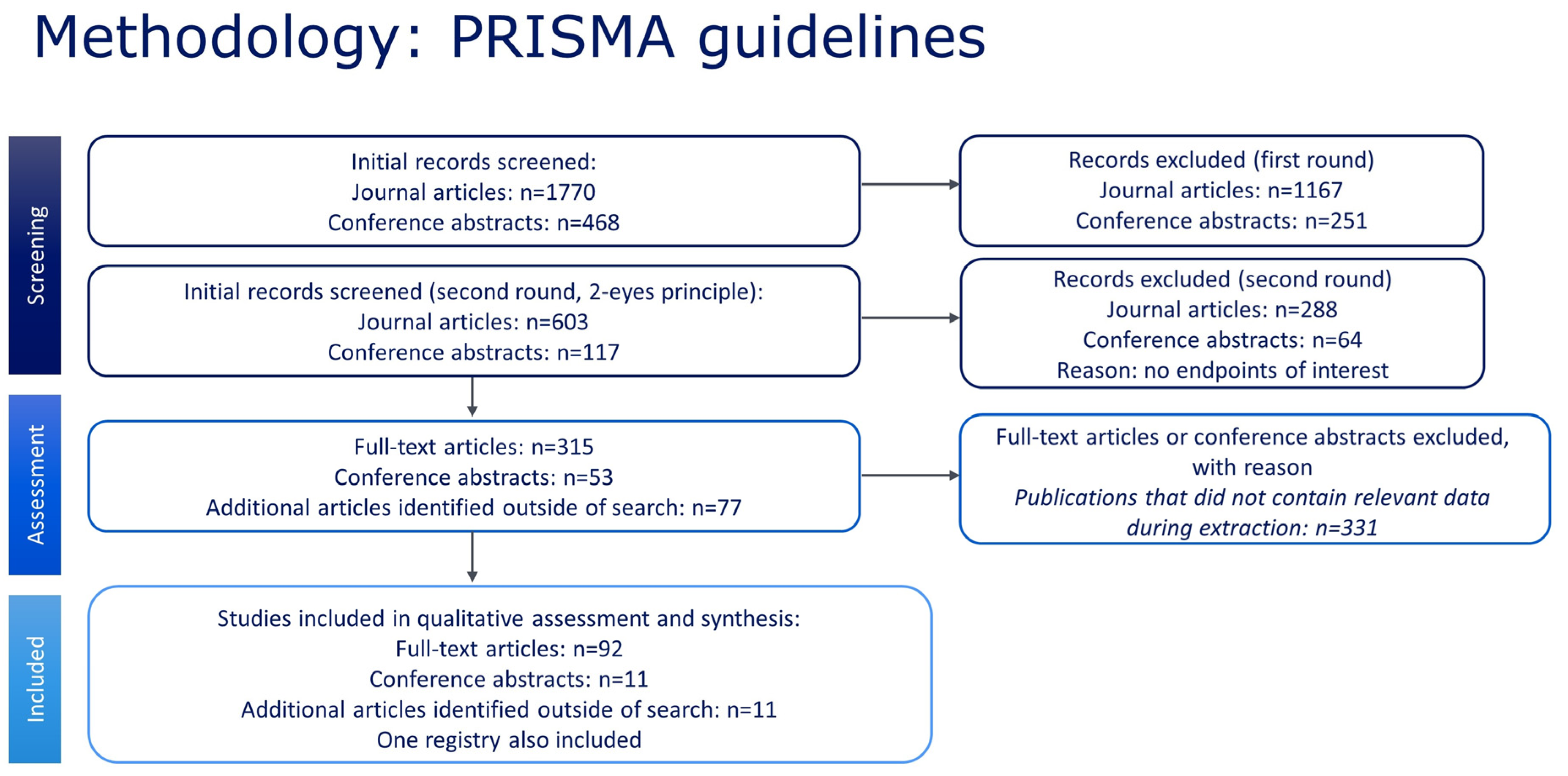
Figure 1. PRISMA diagram for the search strategy and selected studies.
Jcm 12 05538 g001
Figure 2. Global and regional prevalence a of SCD in Africa [22,23,24], Europe [25,26,27,28,29], India [30,31,32,33,34,35], the Middle East [36,37,38,39], and South America and the Caribbean [40]. a Within each region the prevalence was estimated using a binomial normal model, which assumed a binomial distribution for the individual studies with a mean value drawn from a normal distribution for a regional/global value. The prevalence for each reference was determined from the log odds. A summary estimate was determined for each region with >2 studies. North America had insufficient data to determine the prevalence of SCD. I2 describes the percentage of variation across studies that was due to heterogeneity rather than chance, scored from 0 to 100%, where 100% is maximum heterogeneity. BN, binomial normal; CI, confidence interval; SCD, sickle cell disease.
Jcm 12 05538 g002
Figure 3. Global and regional prevalence a of sickle cell trait in Africa [22,23,24,41], Europe [26,28,42], India [30,31,32,33,34,35,43,44,45], the Middle East [36,37,38,39,46,47], and South America and the Caribbean [48,49]. a Within each region the prevalence was estimated using a binomial normal model, which assumed a binomial distribution for the individual studies with a mean value drawn from a normal distribution for a regional/global value. The prevalence for each reference was determined from the log odds. A summary estimate was determined for each region with >2 studies. North America had insufficient data to determine the prevalence of SCD. I2 describes the percentage of variation across studies that was due to heterogeneity rather than chance, scored from 0 to 100%, where 100% is maximum heterogeneity. BN, binomial normal; CI, confidence interval; SCD, sickle cell disease.
Jcm 12 05538 g003
Figure 4. Global and regional birth prevalence a of SCD in Africa [23,50,51,52,53,54,55,56,57,58,59], Europe [60,61,62,63,64,65,66,67,68,69,70], India [71], the Middle East [39,72,73,74,75], North America [76,77,78,79], and South America and Caribbean [80,81,82,83,84,85,86,87,88,89,90,91]. a Within each region, the birth prevalence was estimated using a binomial normal model, which assumed a binomial distribution for the individual studies with a mean value drawn from a normal distribution for a regional/global value. The prevalence for each reference was determined from the log odds. A summary estimate was determined for each region with >2 studies. I2 describes the percentage of variation across studies that was due to heterogeneity rather than chance, scored from 0 to 100%, where 100% is maximum heterogeneity. BN, binomial normal; CI, confidence interval; SCD, sickle cell disease.
Jcm 12 05538 g004
Figure 5. Global and regional birth prevalence a of sickle cell trait in Africa [23,50,51,52,53,54,57,58,59], Europe [61,63,65,66,67,68,69,70,92], India [71], the Middle East [72,73,74,75], North America [76,79,93,94], and South America and Caribbean [80,81,82,83,85,86,87,88,89,90,91]. a Within each region the birth prevalence was estimated using a binomial normal model, which assumed a binomial distribution for the individual studies with a mean value drawn from a normal distribution for a regional/global value. The prevalence for each reference was determined from the log odds. A summary estimate was determined for each region with >2 studies. I2 describes the percentage of variation across studies that was due to heterogeneity rather than chance, scored from 0 to 100%, where 100% is maximum heterogeneity. BN, binomial normal; CI, confidence interval; SCD, sickle cell disease.
Jcm 12 05538 g005
Figure 6. Worldwide prevalence a and birth prevalence of SCD. a Each country with results for the specific genotype and prevalence measure are colored according to the reported prevalence (or average if multiple results were available for the given country). Dots are placed at the center of each country (and therefore do not indicate a specific location within the country) and may have been marginally moved in a random direction in order to be able to see multiple dots if the countries are close together. SCD, sickle cell disease.
Jcm 12 05538 g006
Figure 7. Worldwide prevalence and birth prevalence of sickle cell trait. Each country with results for the specific genotype and prevalence measure are colored according to the reported prevalence (or average if multiple results were available for the given country). Dots are placed at the center of each country (and therefore do not indicate a specific location within the country) and may have been marginally moved in a random direction in order to be able to see multiple dots if the countries are close together.
Jcm 12 05538 g007
Figure 8. Survival curves (a,b) for all ages and for the first 15 years of life (inset) for people with SCD and matched general populations in: (a) Africa, (b) Europe, (c) the Middle East, (d) North America, and (e) South America and the Caribbean. (a) Mortality curves for SCD populations from Angola 2013 [59]; Kenya 2019 [55]; and for the general population: Tanzania—Dar Es Salaam 1994–1999 [21,95], Tanzania—Morogoro 1994–1999 [21,95], Gambia 1991–1995 [21,95], and Mozambique. (b) Mortality curves for SCD populations from France 2019 [96]; Spain 2020 [97]; the Netherlands 2021 [98]; and for the general population: France 2015 [20], Spain 2015 [20], and the Netherlands 2015 [20]. (c) Mortality curves for SCD populations from Iran 2018 [99] and for the general population: Iran 2004 [100]. (d) Mortality curves for SCD populations from USA 2014 [101]; USA 2015 [102]; USA 2016 [103]; and for the general population: USA 2015 [20]. (e) Mortality curves for SCD populations from Brazil 2015; Jamaica 2018; and for the general population: Brazil 2010 [21,104], Jamaica 2011 [21,105], and Chile 2015 [20]. (a) Steps occur at each death (if available) or end of age intervals (for age-binned data). (b) Mortality data for the general population were obtained from matched sources for the same country as and similar year(s) to the SCD study. If datasets that matched geographical locations and years were unavailable, data from neighboring countries covering the same geographical area or close in time to the SCD study were used. SCD, sickle cell disease.
Jcm 12 05538 g008aJcm 12 05538 g008b
Table 1. Quantitative analysis of SCD and SCT for prevalence and birth prevalence.
Prevalence
SCDSCT
RegionNo. of studiesTotal studied populationPrevalence per 100,000 [95% CI]No. of studiesTotal studied populationPrevalence per 100,000 [95% CI]
Global19130,420,748117 [55; 249]248,335,4422427 [1209; 4814]
Africa37274788 [316; 1951]411,52317,690 [14,149; 21,891]
Europe6126,641,17228 [10; 79]35,921,455212 [12; 3503]
India6430,952128 [48; 340]9472,1542193 [1090; 4364]
Middle East41,909,565212 [64; 698]61,914,4292429 [934; 6167]
North America--NA--NA
South America/the Caribbean1536,785NA215,881NA
Birth prevalence
SCDSCT
RegionNo. of studiesTotal studied populationPrevalence per 100,000 [95% CI]No. of studiesTotal studied populationPrevalence per 100,000 [95% CI]
Global4492,209,456191 [120; 303]448,661,1413870 [2597; 5728]
Africa11397,6511321 [1041; 1674]9377,42215,502 [12,618; 18,901]
Europe115,407,68933 [20; 54]9558,233535 [362; 792]
India15467NA15467NA
Middle East5598,718218 [88; 538]4559,7782429 [1129; 5148]
North America479,048,69554 [25;117]44,026,9241681 [923; 3043]
South America/the Caribbean126,751,236201 [102; 395]113,133,3176196 [4344; 8765]
Within each region the prevalence was estimated using a binomial normal model, which assumed a binomial distribution for the individual studies with a mean value drawn from a normal distribution for a regional/global value. A summary estimate was determined for each region with >2 studies. CI, confidence interval; NA, not available; SCD, sickle cell disease; SCT, sickle cell trait.
Disclaimer/Publisher’s Note: The statements, opinions and data contained in all publications are solely those of the individual author(s) and contributor(s) and not of MDPI and/or the editor(s). MDPI and/or the editor(s) disclaim responsibility for any injury to people or property resulting from any ideas, methods, instructions or products referred to in the content.