Article

Association between Lipid Levels, Anti-SARS-CoV-2 Spike Antibodies and COVID-19 Mortality: A Prospective Cohort Study

1
Central Medical Laboratories, 6800 Feldkirch, Austria
2
Medical-Scientific Faculty, Private University of the Principality of Liechtenstein, 9495 Triesen, Liechtenstein
3
VIVIT Institute, Academic Teaching Hospital Feldkirch, 6800 Feldkirch, Austria
4
Department of Internal Medicine, Academic Teaching Hospital Feldkirch, 6800 Feldkirch, Austria
5
Drexel University College of Medicine, Philadelphia, PA 19129, USA
*
Author to whom correspondence should be addressed.
J. Clin. Med. 2023, 12(15), 5068; https://doi.org/10.3390/jcm12155068
Submission received: 7 July 2023 / Revised: 21 July 2023 / Accepted: 30 July 2023 / Published: 1 August 2023
(This article belongs to the Section Infectious Diseases)

Abstract

:
Background: Recent studies suggest that both lipid levels and anti-severe-acute-respiratory-syndrome-coronavirus-2 (SARS-CoV-2) antibody levels are associated with outcome in coronavirus disease 2019 (COVID-19). While both parameters have separately been implicated in the neutralization and clearance of pathogens during severe infections, it is currently unclear whether the interplay of these parameters affects outcome in COVID-19. We therefore aimed to determine whether there was a relationship between lipoproteins, anti-SARS-CoV-2 antibodies, and COVID-19 mortality. Methods: In this prospective, multicenter cohort study, we recruited 1152 hospitalized patients with COVID-19 from five hospitals. Total cholesterol (TC), LDL-C, HDL-C, triglycerides, and anti-SARS-CoV-2 spike antibodies were measured on hospital admission. The investigated endpoint was in-hospital mortality. Results: LDL-C, HDL-C, and TC were significantly lower in non-survivors than in survivors (mg/dL, 95%CI; 56.1, 50.4–61.8 vs. 72.6, 70.2–75.0, p < 0.001; 34.2, 31.7–36.8 vs. 38.1, 37.2–39.1, p = 0.025; 139.3, 130.9–147.7 vs. 157.4, 54.1–160.6, p = 0.002). Mortality risk increased progressively with lower levels of LDL-C, HDL-C, and TC (aOR 1.73, 1.30–2.31, p < 0.001; 1.44, 1.10–1.88, p = 0.008; 1.49, 1.14–1.94, p < 0.001). Mortality rates varied between 2.1% for high levels of both LDL-C and anti-SARS-CoV-2 antibodies and 16.3% for low levels of LDL-C and anti-SARS-CoV-2 antibodies (aOR 9.14, 95%CI 3.17–26.34, p < 0.001). Accordingly, for total cholesterol and anti-SARS-CoV-2 antibodies, mortality rates varied between 2.1% and 15.0% (aOR 8.01, 95%CI 2.77–23.18, p < 0.001). Conclusion: The combination of serum lipid levels and anti-SARS-CoV-2 antibodies is strongly associated with in-hospital mortality of patients with COVID-19. Patients with low levels of LDL-C and total cholesterol combined with low levels of anti-SARS-CoV-2 antibodies exhibited the highest mortality rates.

1. Introduction

Previous studies have suggested that plasma lipoproteins are vital for the removal of pathogen-associated lipids and toxins in severe infections such as sepsis [1,2,3,4]. Although the pathophysiological role of lipoproteins in COVID-19 is still incompletely understood, experimental studies show that the lipid biogenesis pathways of the host may affect virus replication in several ways [5]. Viral internalization into a cell requires the attachment and fusion of viral membrane with plasma membrane by way of endocytosis [6,7]. Lipids can act as direct receptors or as entry co-factors at both the cell surface and the endosomes [8,9]. In experimental studies, enhanced transport of cholesterol from the cell membrane to the endoplasmatic reticulum by the enzyme cholesterol-25-hydroxylase inhibits SARS-CoV-2 entry into a cell by depriving it of accessible cholesterol at the plasma membrane. Conversely, this entry inhibition can be removed by adding soluble cholesterol [10]. Lipids further play an important role in the formation and function of the viral replication complex [11,12]. Accordingly, in animal models, intracellular cholesterol was found to enhance SARS-CoV2 infectivity [13,14].
In clinical studies, COVID-19 is associated with increased rates of dyslipidaemia, which may persist up to one year after infection [15,16]. Pre-existing dyslipidaemia and atherosclerosis have been linked to increased severity and mortality of COVID-19 [17,18]. In addition, several studies describe an association between plasma lipid levels and outcome in patients infected with SARS-CoV-2, suggesting that plasma lipids may be involved in the immune response to COVID-19 [19,20,21,22].
Accordingly, antibodies are known to play a crucial role in the neutralization of pathogens. With regard to COVID-19, neutralizing antibodies have been correlated with lower infection rates and protection against severe courses of COVID-19 [23,24,25,26]. Positive results have been reported following the administration of neutralizing monoclonal antibodies [27,28,29]. In addition, previous studies have demonstrated a protective effect of booster vaccinations against severe illness and hospitalization due to COVID-19 [30,31,32,33,34], and high anti-SARS-CoV-2 spike antibody levels on hospital admission have been shown to be associated with lower in-hospital mortality [35].
Taken together, these points suggest that both lipoproteins and antibodies are of high importance in the immune response against SARS-CoV-2. However, it is currently unclear whether the interplay between lipoproteins and antibodies affects outcome in SARS-CoV-2 infections. We therefore evaluated whether there was a relationship between anti-SARS-CoV-2 spike antibody, lipoprotein levels and COVID-19 mortality.

2. Methods

2.1. Study Design and Participants

The design of this study has previously been reported elsewhere [35]. In brief, we conducted a prospective, multicenter cohort study in which we enrolled patients from five Austrian hospitals who were hospitalized between 1 August 2021 and 10 April 2022.
Patients were considered eligible for recruitment if they tested positive for SARS-CoV-2 with a polymerase chain reaction (PCR)-based assay and a blood sample was drawn on hospital admission. Patients were excluded from the study if no blood sample was collected, if the residual sample material was insufficient, if they had been previously hospitalized during the study period, or if their hospital stay had not concluded by the end of the study.
Sample size calculation for logistic regression with a binary covariate, Wald’s method, yielded a minimum sample size of 465 participants (alpha 0.05, two-sided, power 0.8).

2.2. Variables

The investigated endpoint was in-hospital mortality from any cause.
Diabetes status was defined based on previous diagnosis according to ICD-10 codes or HbA1c levels above 6.4%, as obtained from patient files. Patients with a BMI of 30 or above were classified as obese.
Known risk factors for severe courses and mortality in COVID-19 were chosen as predefined covariates. It has been well documented that obesity and an underlying condition of diabetes predispose to a higher mortality risk through a combination of inflammation, hypercoagulation, and mechanical obstruction [36]. Age, due to the accumulation of underlying conditions and frailty, is another known risk factor for severe course and mortality in COVID-19 [37]. Statin therapy was chosen as an additional covariate because of its effects on lipid levels.
Laboratory parameters that were used as covariates were anti-SARS-CoV-2 antibodies, total cholesterol, LDL-C, HDL-C, and triglyceride levels.

2.3. Data Sources and Measurements

Total cholesterol, LDL-C, HDL-C, and triglycerides were measured on hospital admission using the respective Roche assays on Roche Cobas 6000 or Cobas 8000 systems. Lipid measurements were conducted with the Roche CHOL2 assay for cholesterol, the HDL-C4 assay for HDL-C, the LDL-C3 assay for LDL-C, and the TRIGL assay for triglycerides. The Roche LDL-C3 assay allows direct measurement of LDL-C utilizing a chromogenic enzymatic method. Anti-SARS-CoV-2 spike antibodies were measured using the Elecsys Anti-SARS-CoV-2 S assay.
Clinical data obtained from patient files included age, gender, body mass index (BMI), vaccination status, type of vaccine, COVID-19 variant, PCR-derived cycle threshold values, reason for hospitalization, main diagnosis, symptoms at admission such as coughing, fever, loss of taste and/or smell, oxygen requirement (none/without endotracheal intubation/with endotracheal intubation), duration of hospital stay, and, if applicable, duration in intensive care unit (ICU) as well as in-hospital survival.

2.4. Statistical Methods

All statistical analyses were calculated using the IBM Statistical Package for the Social Sciences (SPSS), version 28 (IBM, Armonk, NY, USA). Baseline statistical characteristics, including frequencies, percentages, means, medians, standard deviations, and interquartile ranges were determined with the functiondescriptive statistics. The 95% confidence intervals for means were calculated with one-sample t tests. Statistical significance was determined by Mann–Whitney U, chi-square test, or log-rank test, as appropriate. As is customary, two-sided p values of <0.05 were considered statistically significant.
In order to quantify the respective risk associated with lower lipid levels, we built logistic regression models. Odds ratios with a 95% confidence interval were calculated. Basic assumptions for conducting logistic regression analysis, including independence of errors, i.e., lack of duplicate entries, linearity in the logit for continuous independent variables, absence of multicollinearity, and lack of strongly influential outliers, were tested and met. Linear relationships between continuous variables and the logit transformation of the dependent variable were tested with the Box–Tidwell test.
All regression analyses were conducted with a direct model-building approach, in which all independent variables were added simultaneously and having equal importance. Outcome (mortality/intensive care treatment/oxygen requirement) was set as the dichotomous dependent variable, while lipid parameters and predefined covariates were used as independent variables (method = enter).
We also provided a second measure of risk, i.e., hazard ratios, as calculated by a Cox proportional hazards model. The input here was analogous to the approach described in the previous paragraph, with the addition of a time parameter. Time to event was defined in days measured from hospital admission. Hazard ratios for in-hospital mortality were calculated with a 95% confidence interval.
To standardize the results and thus improve comparability between the risks associated with these lipid parameters, we conducted z-score normalization for each parameter. We then calculated the corresponding odds ratios and hazard ratios for the transformed parameters as described above. Odds ratios and hazard ratios were reported both for the continuous variables and the z-score-normalized variables to allow for direct comparison between these results.
We then adjusted the results from these models for potential confounding by the covariates age, type II diabetes, obesity, and statin therapy. To prevent overfitting of the regression models, we verified having a sufficient number of events in the whole cohort and the high-risk subgroups of diabetic and obese patients before adjusting for potential confounders. For better comparison, both unadjusted and adjusted odds ratios and hazard ratios are given.
In order to assess the relationship between anti-SARS-CoV-2 spike antibodies and lipid levels, parameters were stratified into categories of high and low. For lipid levels, groups were separated by the respective median value. For anti-SARS-CoV-2 antibody levels, the threshold was defined at 1200 U/mL [35]. We then combined anti-SARS-CoV-2 spike antibodies with each lipid parameter, forming the following groups: high/high, high/low, low/high, low/low.
Next, we calculated mortality rates and survival over time, first separately for each parameter, and second for the above mentioned combinations. Survival over time was then depicted using Kaplan–Meier graphs.
In order to test the robustness of our findings, we repeated the regression models described above while applying bootstrapping with 2000 samples, first for the whole cohort, second for diabetic patients, and third for obese patients. Finally, goodness of fit was tested using Hosmer–Lemeshow tests.

3. Results

3.1. Participants

Overall, 1379 hospitalized patients were assessed for eligibility at five hospitals between 1 August 2021 and 10 April 2022. Of these, 1152 patients were originally enrolled in the study. However, due to insufficient residual sample material, lipid parameters could only be measured in 1046 patients. Patient flow is depicted in Figure 1.
In total, 118 patients died, 165 patients were admitted to an intensive care unit, and 587 patients required oxygen administration. Overall, 275 patients either had a previous diagnosis of type II diabetes or were diagnosed during their hospital stay, and 254 patients registered a BMI of 30 or above and were thus classified as obese. Patient characteristics are given in Table 1.
Patients with lower LDL-C or total cholesterol levels were significantly more likely to receive statin therapy and more likely to have a higher BMI and a history of diabetes and vascular disease.

3.2. Risk of Outcome by Lipid Levels

LDL-C, HDL-C, and total cholesterol levels were significantly lower in non-survivors than in survivors (mean LDL-C 56.1 mg/dL, 95%CI 50.4–61.8 vs. 72.6 mg/dL, 95%CI 70.2–75.0, p < 0.001; HDL-C 34.2, 95%CI 31.7–36.8 vs. 38.1 mg/dL, 95%CI 37.2–39.1, p = 0.025; total cholesterol 139.3 mg/dL, 95%CI 130.9–147.7 vs. 157.4, 95%CI 154.1–160.6, p = 0.002). Triglyceride concentrations were not significantly different between patients who survived and those who did not.
In order to determine odds ratios and thus quantify the respective risk associated with lower lipid levels, we first calculated odds ratios for the continuous lipid parameters by logistic regression analysis. We further provided hazard ratios as determined by a Cox proportional hazard model. Next, we calculated normalized z-scores for LDL-C, HDL-C, and cholesterol levels to standardize the results and improve comparability between the risks associated with these parameters. We then adjusted for potential confounders, including age, type II diabetes, and obesity.
Table 2 summarizes the estimated risks in the form of odds ratios and hazard ratios, stratified by lipid parameter both for continuous values and z-scores. Risk assessment for continuous variables is given per 10 mg/dL decrement since differences in lipid levels of ±1 mg/dL were not considered to be clinically relevant.
Risk of in-hospital mortality increased 1.7 times for each standard deviation decrease in LDL-C values, by 1.4 times for each decrease in HDL-C values, and by 1.5 times for each decrease in total cholesterol levels (aOR 1.73, 95%CI 1.30–2.31, p < 0.001, aOR 1.44, 95%CI 1.10–1.88, p = 0.008, aOR 1.49, 95%CI n1.14–1.94, p = 0.003).
In our Cox proportional hazard model, lower LDL-C, HDL-C, and total cholesterol levels were also associated with elevated risk of in-hospital mortality (aHR 1.15, 95%CI 1.08–1.23; p < 0.001, 1.18, 95%CI 1.10–1.26, p < 0.001; 1.14, 95%CI 1.07–1.22, p < 0.001). In this analysis, other covariates that showed a significant association with in-hospital mortality were age (p < 0.001) and statin therapy (p < 0.001), but not type II diabetes nor obesity. Standard deviations for each lipid parameter are included in Table 1.
Next, we divided lipid levels into high and low categories, separated by the respective median value, to assess the difference in mortality risk between high and low lipid levels. With regard to LDL-C, mortality risk increased two-fold for patients with LDL-C levels below 67 mg/dL (aOR 2.013, 1.250–3.241, p = 0.004; aHR 1.712, 1.102–2.661, p = 0.017). For TC and HDL-C levels, mortality risk did not differ significantly below vs. above the respective median value.

3.3. Risk of Outcome by Lipid Levels and Anti-SARS-CoV-2 Antibodies

Previous studies have suggested that lipoproteins are vital for the removal of pathogen-associated lipids and toxins in severe infections such as sepsis [1,2,3,4]. Similarly, antibodies are known to help neutralize and remove pathogens. With regard to COVID-19, anti-SARS-CoV-2 spike antibody levels have been shown to be inversely associated with outcome [35]. We therefore evaluated whether there was a relationship between antibody and lipoprotein levels and patient mortality.
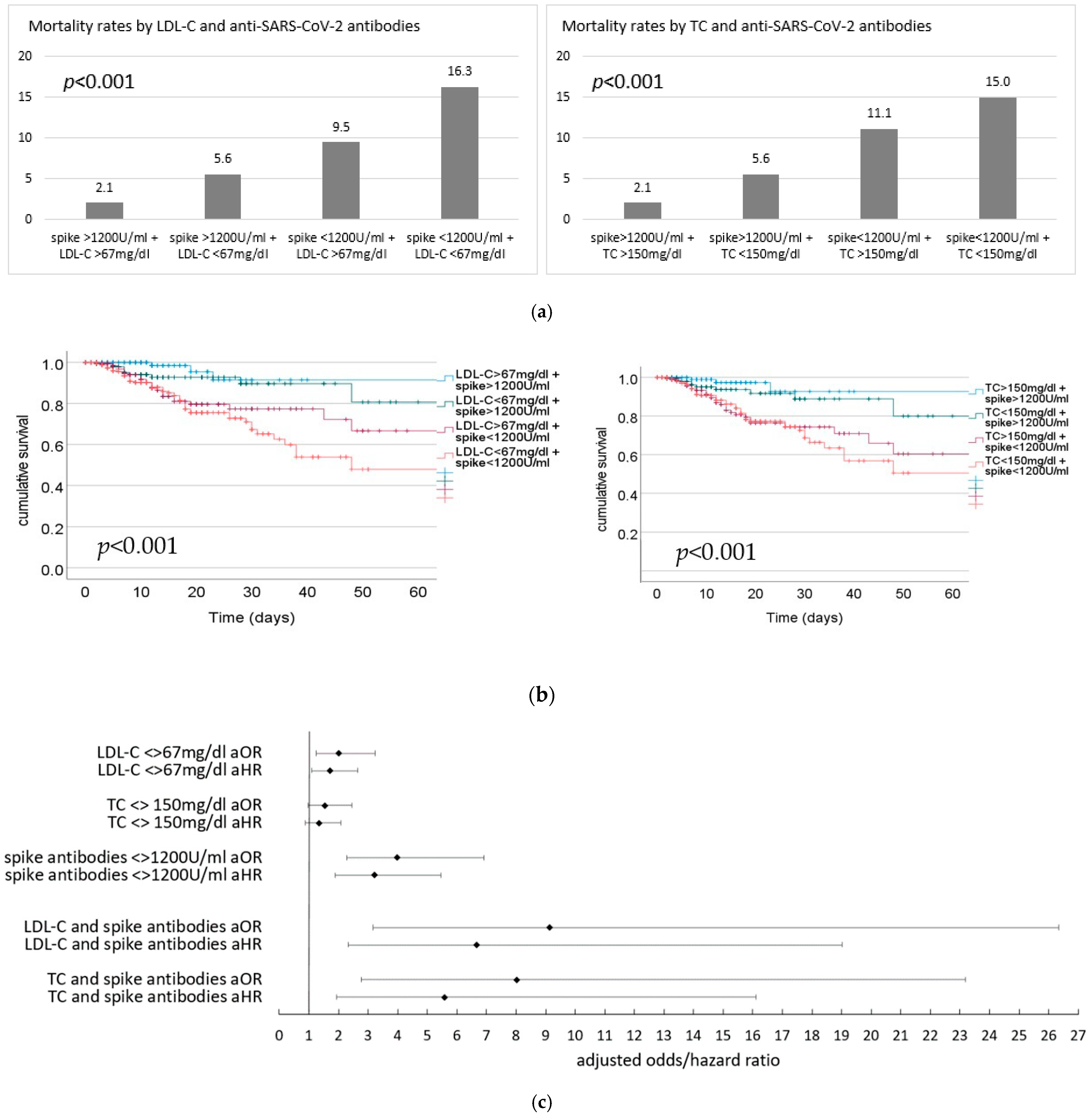
To this end, lipid levels and anti-SARS-CoV-2 spike antibodies were grouped into high and low categories. Lipid parameters and antibodies were then combined as follows—high/high, high/low, low/high, and high/high. Next, mortality rates and survival over time were calculated for all combinations. Results are depicted in Figure 2.
The lowest mortality rates occurred in patients with high levels of anti-SARS-CoV-2 antibodies and high levels of either LDL-C or total cholesterol. In contrast, patients with both low levels of LDL-C or total cholesterol and low levels of anti-SARS-CoV-2 antibodies showed the highest mortality rates.
For LDL-C, mortality rates varied between 2.1% for high levels of both LDL-C and anti-SARS-CoV-2 antibodies and 16.3% for low levels of LDL-C and anti-SARS-CoV-2 antibodies (aOR 9.137, 95%CI 3.169–26.343, p < 0.001; aHR 6.659, 95%CI 2.332–19.010, p < 0.001). Accordingly, for total cholesterol and anti-SARS-CoV-2 antibodies, mortality rates varied between 2.1% and 15.0% (aOR 8.012, 95%CI 2.769–23.181, p < 0.001; aHR 5.576, 95%CI 1.930–16.113, p = 0.002).

4. Discussion

In this prospective, multicenter cohort study on 1152 hospitalized patients with COVID-19, we are the first to show that lipid and anti-SARS-CoV-2 spike antibody levels are strongly associated with COVID-19 mortality.
Mortality rates were lowest in patients who had high levels of both LDL-C or TC and anti-SARS-CoV-2 spike antibodies. Conversely, patients who had low levels of both parameters registered the highest mortality rates.
Separately, plasma lipids and anti-SARS-CoV-2 antibodies have previously been shown to be associated with COVID-19 mortality. For instance, a systematic review and meta-analysis comprising more than 250.000 patients reported that severe COVID-19 patients had lower total cholesterol, HDL-C, and LDL-C levels than patients with non-severe courses [19]. A previous observational study from Spain described worse outcomes in hospitalized patients with low LDL-C and high triglyceride levels [13]. While we also observed worse outcomes in patients with low LDL-C levels, triglyceride levels did not differ significantly by outcome in our study population. This discrepancy may stem from differences in disease severity or fasting state at the time of sample collection. Our findings are in accordance with results from a meta-analysis that described lower total cholesterol, LDL-C, and HDL-C but not triglycerides in hospitalized patients with severe COVID-19 [20].
The pathophysiological role of lipoproteins in COVID-19 is still incompletely understood [5]. With regard to plasma lipid levels, it has previously been suggested that lipoproteins, especially HDL, play a crucial role in the removal of pathogen-associated lipids and toxins in severe infections such as sepsis [1,2,3,4]. Lower lipid levels in patients with COVID-19 may therefore indicate an impairment of this important biological process. Alternatively, higher lipid levels may simply be indicative of a better general condition of the individual, as low plasma lipid levels have been attributed to a hypermetabolic state and undernutrition in infected patients [2]. In addition to reduced levels of lipoproteins in COVID-19 patients, there is evidence to suggest that the function of lipoproteins may also be impaired. A study by Begue et al. reported that HDL particles from severely ill COVID-19 patients were less protective against permeability, VE-cadherin disorganization, and apoptosis than in healthy controls [22].
One of the main function of antibodies is the neutralization of pathogens. Previous studies have reported a positive correlation between neutralizing antibodies and protection against COVID-19 [23,24,25,26]. In addition, the administration of neutralizing monoclonal antibodies had positive effects in the prevention and treatment of COVID-19 [27,28,29]. Previous studies have demonstrated a protective effect of booster vaccinations against severe illness and hospitalization due to COVID-19 in the general population [30,31,32]. We previously reported that antibody levels on hospital admission are inversely related to in-hospital mortality in patients with COVID-19 [35].
Hypothetically, lipoproteins could be involved in the removal of SARS-CoV-2 particles that have previously been neutralized by antibodies. However, it is currently unclear if the association between lipoprotein and antibody levels and COVID-19 mortality indicates a pathophysiological mechanism or rather if they are representative of a better general condition that, in turn, improves chances of survival.
One of the major strengths of this study is its high recruitment rate, which reduces the risk of selection bias. Moreover, this investigation is based on a hard endpoint (in-hospital mortality) which is less susceptible to assessment bias because it does not require subjective clinical patient assessments [38]. In addition, we adjusted for multiple potential confounders, including statin therapy and known risk factors for COVID-19, including age, type II diabetes, and obesity [37,39].
With regard to limitations, this study was designed with a focus on hospitalized patients and its results therefore may not be applicable to outpatients. However, as severely ill patients are more likely to be treated in a hospital, we deemed it important to focus on this patient group. While we do not have follow-up data after discharge, this study was designed to examine the relationship between plasma lipoproteins, anti-SARS-CoV-2 antibodies, and mortality and thus centers on severely ill, hospitalized patients. Further, this study was conducted with residual sample material only; therefore, fasting periods could not be observed before sample collection, which may have affected triglyceride measurements in particular.
While we did see significant differences in outcome with decreasing concentrations of LDL-C, HDL-C, and total cholesterol, additional studies are required for determining a protective cutoff against negative outcomes in COVID-19. Our data suggest an association between lipoprotein and antibody levels and mortality in COVID-19. As both lipoproteins and antibodies have previously been associated with the removal of pathogens, a synergistic pathophysiological mechanism is theoretically possible. However, further studies are required to confirm such a relationship and to elucidate the exact mechanisms involved.
Understanding the combined role of lipoproteins and antibodies in severe infections, such as COVID-19, may help identify patients at risk, thereby allowing timely adjustment of therapy, and provide new options for future pharmacological therapeutic approaches.
In conclusion, our data suggest that LDL-C and TC-levels, combined with anti-SARS-CoV-2 spike antibodies, are a strong predictor of in-hospital mortality. Patients with low levels of LDL-C and total cholesterol combined with low levels of anti-SARS-CoV-2 antibodies exhibited the highest mortality rates.

Author Contributions

S.M. formal analysis, project administration, writing—original draft; C.H.S. resources, writing—review and editing; M.F. resources, writing—review and editing; A.L. writing—review and editing; H.D. resources, writing—review and editing; P.F. conceptualization, formal analysis, methodology, resources, supervision, writing—review and editing. All authors have accepted responsibility for the entire content of this manuscript and approved its submission. All authors have read and agreed to the published version of the manuscript.

Funding

This research received no external funding.

Institutional Review Board Statement

The local Institutional Review Board, Ethikkommission Vorarlberg, Roemerstrasse 15, A-6901 Bregenz, deemed the study exempt from review and waived the need to obtain informed consent from the study participants because the study was purely observational in character and did not directly experiment on humans or use human tissue samples (only residual material from blood samples was used for testing). This study was carried out in accordance with the Declaration of Helsinki of 1975 (revised 2013) and Good Clinical Research Practice.

Data Availability Statement

As personal individual information is included in the dataset, the data pertaining to this investigation is not publicly available to protect study participant privacy. However, an anonymised version will be shared upon reasonable request to the corresponding author.

Conflicts of Interest

The authors declare no conflict of interest.

Abbreviations

COVID-19coronavirus disease 2019
SARS-CoV-2severe acute respiratory syndrome coronavirus 2
PCRpolymerase chain reaction
ICUintensive care unit
OR, aOR, HRodds ratio, adjusted odds ratio, hazard ratio
CIconfidence interval
LDL-Clow density lipoprotein
HDL-Chigh density lipoprotein
BMIbody mass index
COPDchronic obstructive pulmonary disease
CADcoronary artery disease
TIAtransient ischemic attack
CVDcerebrovascular disease

References

  1. Tanaka, S.; Couret, D.; Tran-Dinh, A.; Duranteau, J.; Montravers, P.; Schwendeman, A.; Meilhac, O. High-density lipoproteins during sepsis: From bench to bedside. Crit. Care 2020, 24, 134. [Google Scholar] [CrossRef] [Green Version]
  2. Trinder, M.; Boyd, J.H.; Brunham, L.R. Molecular regulation of plasma lipid levels during systemic inflammation and sepsis. Curr. Opin. Lipidol. 2019, 30, 108–116. [Google Scholar] [CrossRef] [PubMed]
  3. Levels, J.H.; Abraham, P.R.; van den Ende, A.; van Deventer, S.J. Distribution and kinetics of lipoprotein-bound endotoxin. Infect. Immun. 2001, 69, 2821–2828. [Google Scholar] [CrossRef] [PubMed] [Green Version]
  4. Biller, K.; Fae, P.; Germann, R.; Drexel, H.; Walli, A.K.; Fraunberger, P. Cholesterol rather than procalcitonin or C-reactive protein predicts mortality in patients with infection. Shock 2014, 42, 129–132. [Google Scholar] [CrossRef]
  5. Abu-Farha, M.; Thanaraj, T.A.; Qaddoumi, M.G.; Hashem, A.; Abubaker, J.; Al-Mulla, F. The Role of Lipid Metabolism in COVID-19 Virus Infection and as a Drug Target. Int. J. Mol. Sci. 2020, 21, 3544. [Google Scholar] [CrossRef]
  6. Marsh, M.; Helenius, A. Virus entry: Open sesame. Cell 2006, 124, 729–740. [Google Scholar] [CrossRef] [Green Version]
  7. Mercer, J.; Helenius, A. Virus entry by macropinocytosis. Nat. Cell Biol. 2009, 11, 510–520. [Google Scholar] [CrossRef]
  8. Bagam, P.; Singh, D.P.; Inda, M.E.; Batra, S. Unraveling the role of membrane microdomains during microbial infections. Cell Biol. Toxicol. 2017, 33, 429–455. [Google Scholar] [CrossRef] [PubMed]
  9. Taube, S.; Jiang, M.; Wobus, C.E. Glycosphingolipids as receptors for non-enveloped viruses. Viruses 2010, 2, 1011–1049. [Google Scholar] [CrossRef] [Green Version]
  10. Chen, P.; Wu, M.; He, Y.; Jiang, B.; He, M.-L. Metabolic alterations upon SARS-CoV-2 infection and potential therapeutic targets against coronavirus infection. Signal Transduct. Target Ther. 2023, 8, 237. [Google Scholar] [CrossRef]
  11. Nagy, P.D.; Strating, J.R.P.M.; van Kuppeveld, F.J.M. Building Viral Replication Organelles: Close Encounters of the Membrane Types. PLoS Pathog. 2016, 12, e1005912. [Google Scholar] [CrossRef] [PubMed] [Green Version]
  12. Hsu, N.-Y.; Ilnytska, O.; Belov, G.; Santiana, M.; Chen, Y.-H.; Takvorian, P.M.; Pau, C.; van der Schaar, H.; Kaushik-Basu, N.; Balla, T.; et al. Viral reorganization of the secretory pathway generates distinct organelles for RNA replication. Cell 2010, 141, 799–811. [Google Scholar] [CrossRef] [PubMed] [Green Version]
  13. Masana, L.; Correig, E.; Ibarretxe, D.; Anoro, E.; Arroyo, J.A.; Jericó, C.; Guerrero, C.; Miret, M.; Näf, S.; Pardo, A.; et al. Low HDL and high triglycerides predict COVID-19 severity. Sci. Rep. 2021, 11, 7217. [Google Scholar] [CrossRef] [PubMed]
  14. Wang, H.; Yuan, Z.; Pavel, M.A.; Jablonski, S.M.; Jablonski, J.; Hobson, R.; Valente, S.; Reddy, C.B.; Hansen, S.B. The role of high cholesterol in age-related COVID19 lethality. Cold Spring Harbor Laboratory. bioRxiv 2021. [Google Scholar] [CrossRef]
  15. Xu, E.; Xie, Y.; Al-Aly, Z. Risks and burdens of incident dyslipidaemia in long COVID: A cohort study. Lancet Diabetes Endocrinol. 2023, 11, 120–128. [Google Scholar] [CrossRef]
  16. Durrington, P. Blood lipids after COVID-19 infection. Lancet Diabetes Endocrinol. 2023, 11, 68–69. [Google Scholar] [CrossRef]
  17. Liu, Y.; Pan, Y.; Yin, Y.; Chen, W.; Li, X. Association of dyslipidemia with the severity and mortality of coronavirus disease 2019 (COVID-19): A meta-analysis. Virol. J. 2021, 18, 157. [Google Scholar] [CrossRef] [PubMed]
  18. Ceasovschih, A.; Sorodoc, V.; Shor, A.; Haliga, R.E.; Roth, L.; Lionte, C.; Aursulesei, V.O.; Sirbu, O.; Culis, N.; Shapieva, A.; et al. Distinct Features of Vascular Diseases in COVID-19. J. Inflamm. Res. 2023, 16, 2783–2800. [Google Scholar] [CrossRef]
  19. Chidambaram, V.; Geetha, H.S.; Kumar, A.; Majella, M.G.; Sivakumar, R.K.; Voruganti, D.; Mehta, J.L.; Karakousis, P.C. Association of Lipid Levels With COVID-19 Infection, Disease Severity and Mortality: A Systematic Review and Meta-Analysis. Front. Cardiovasc. Med. 2022, 9, 862999. [Google Scholar] [CrossRef] [PubMed]
  20. Zinellu, A.; Paliogiannis, P.; Fois, A.G.; Solidoro, P.; Carru, C.; Mangoni, A.A. Cholesterol and Triglyceride Concentrations, COVID-19 Severity, and Mortality: A Systematic Review and Meta-Analysis With Meta-Regression. Front. Public Health 2021, 9, 705916. [Google Scholar] [CrossRef]
  21. Aparisi, Á.; Martín-Fernández, M.; Ybarra-Falcón, C.; Gil, J.F.; Carrasco-Moraleja, M.; Martínez-Paz, P.; Cusácovich, I.; Gonzalo-Benito, H.; Fuertes, R.; Marcos-Mangas, M.; et al. Dyslipidemia and Inflammation as Hallmarks of Oxidative Stress in COVID-19: A Follow-Up Study. Int. J. Mol. Sci. 2022, 23, 15350. [Google Scholar] [CrossRef] [PubMed]
  22. Begue, F.; Tanaka, S.; Mouktadi, Z.; Rondeau, P.; Veeren, B.; Diotel, N.; Tran-Dinh, A.; Robert, T.; Vélia, E.; Mavingui, P.; et al. Altered high-density lipoprotein composition and functions during severe COVID-19. Sci. Rep. 2021, 11, 2291. [Google Scholar] [CrossRef]
  23. Khoury, D.S.; Cromer, D.; Reynaldi, A.; Schlub, T.E.; Wheatley, A.K.; Juno, J.A.; Subbarao, K.; Kent, S.J.; Triccas, J.A.; Davenport, M.P. Neutralizing antibody levels are highly predictive of immune protection from symptomatic SARS-CoV-2 infection. Nat. Med. 2021, 27, 1205–1211. [Google Scholar] [CrossRef]
  24. Gilbert, P.B.; Montefiori, D.C.; McDermott, A.B.; Fong, Y.; Benkeser, D.; Deng, W.; Zhou, H.; Houchens, C.R.; Martins, K.; Jayashankar, L.; et al. Immune correlates analysis of the mRNA-1273 COVID-19 vaccine efficacy clinical trial. Science 2022, 375, 43–50. [Google Scholar] [CrossRef]
  25. Lumley, S.F.; O’Donnell, D.; Stoesser, N.E.; Matthews, P.C.; Howarth, A.; Hatch, S.B.; Marsden, B.D.; Cox, S.; James, T.; Warren, F.; et al. Antibody Status and Incidence of SARS-CoV-2 Infection in Health Care Workers. N. Engl. J. Med. 2021, 384, 533–540. [Google Scholar] [CrossRef]
  26. Harvey, R.A.; Rassen, J.A.; Kabelac, C.A.; Turenne, W.; Leonard, S.; Klesh, R.; Meyer, W.A.; Kaufman, H.W.; Anderson, S.; Cohen, O.; et al. Association of SARS-CoV-2 Seropositive Antibody Test With Risk of Future Infection. JAMA Intern. Med. 2021, 181, 672–679. [Google Scholar] [CrossRef]
  27. Cohen, M.S.; Nirula, A.; Mulligan, M.J.; Novak, R.M.; Marovich, M.; Yen, C.; Stemer, A.; Mayer, S.M.; Wohl, D.; Brengle, B.; et al. Effect of Bamlanivimab vs Placebo on Incidence of COVID-19 Among Residents and Staff of Skilled Nursing and Assisted Living Facilities: A Randomized Clinical Trial. JAMA 2021, 326, 46–55. [Google Scholar] [CrossRef]
  28. Du, L.; Yang, Y.; Zhang, X. Neutralizing antibodies for the prevention and treatment of COVID-19. Cell Mol. Immunol. 2021, 18, 2293–2306. [Google Scholar] [CrossRef]
  29. Kim, C.; Ryu, D.-K.; Lee, J.; Kim, Y.-I.; Seo, J.-M.; Kim, Y.-G.; Jeong, J.-H.; Kim, M.; Kim, J.-I.; Kim, P.; et al. A therapeutic neutralizing antibody targeting receptor binding domain of SARS-CoV-2 spike protein. Nat. Commun. 2021, 12, 288. [Google Scholar] [CrossRef] [PubMed]
  30. Abu-Raddad, L.J.; Chemaitelly, H.; Ayoub, H.H.; AlMukdad, S.; Yassine, H.M.; Al-Khatib, H.A.; Smatti, M.K.; Tang, P.; Hasan, M.R.; Coyle, P.; et al. Effect of mRNA Vaccine Boosters against SARS-CoV-2 Omicron Infection in Qatar. N. Engl. J. Med. 2022, 386, 1804–1816. [Google Scholar] [CrossRef] [PubMed]
  31. Bar-On, Y.M.; Goldberg, Y.; Mandel, M.; Bodenheimer, O.; Freedman, L.; Kalkstein, N.; Mizrahi, B.; Alroy-Preis, S.; Ash, N.; Milo, R.; et al. Protection of BNT162b2 Vaccine Booster against COVID-19 in Israel. N. Engl. J. Med. 2021, 385, 1393–1400. [Google Scholar] [CrossRef] [PubMed]
  32. Menni, C.; May, A.; Polidori, L.; Louca, P.; Wolf, J.; Capdevila, J.; Hu, C.; Ourselin, S.; Steves, C.J.; Valdes, A.M.; et al. COVID-19 vaccine waning and effectiveness and side-effects of boosters: A prospective community study from the ZOE COVID Study. Lancet Infect. Dis. 2022, 22, 1002–1010. [Google Scholar] [CrossRef] [PubMed]
  33. Cossiga, V.; Capasso, M.; Guarino, M.; Loperto, I.; Brusa, S.; Cutolo, F.M.; Attanasio, M.R.; Lieto, R.; Portella, G.; Morisco, F. Safety and Immunogenicity of Anti-SARS-CoV-2 Booster Dose in Patients with Chronic Liver Disease. J. Clin. Med. 2023, 12, 2281. [Google Scholar] [CrossRef] [PubMed]
  34. Valeanu, L.; Andrei, S.; Morosanu, B.; Longrois, D.; Bubenek-Turconi, S.-I.; Covati-Ro, C. The COVID-19 Vaccination Coverage in ICU Patients with Severe COVID-19 Infection in a Country with Low Vaccination Coverage-A National Retrospective Analysis. J. Clin. Med. 2023, 12, 1749. [Google Scholar] [CrossRef] [PubMed]
  35. Mink, S.; List, W.; Hoefle, G.; Frick, M.; Suessenbacher, A.; Winder, T.; Fetz, C.; Boesl, A.; Saely, C.H.; Drexel, H.; et al. Evaluation of SARS-CoV-2 antibody levels on hospital admission as a correlate of protection against mortality. J. Intern. Med. 2023, 293, 694–703. [Google Scholar] [CrossRef] [PubMed]
  36. Steenblock, C.; Schwarz, P.E.H.; Ludwig, B.; Linkermann, A.; Zimmet, P.; Kulebyakin, K.; Tkachuk, V.A.; Markov, A.G.; Lehnert, H.; de Angelis, M.H.; et al. COVID-19 and metabolic disease: Mechanisms and clinical management. Lancet Diabetes Endocrinol. 2021, 9, 786–798. [Google Scholar] [CrossRef]
  37. Clark, A.; Jit, M.; Warren-Gash, C.; Guthrie, B.; Wang, H.H.X.; Mercer, S.W.; Sanderson, C.; McKee, M.; Troeger, C.; Ong, K.L.; et al. Global, regional, and national estimates of the population at increased risk of severe COVID-19 due to underlying health conditions in 2020: A modelling study. Lancet Glob. Health 2020, 8, e1003–e1017. [Google Scholar] [CrossRef] [PubMed]
  38. Drexel, H.; Rosano, G.M.C.; Lewis, B.S.; Huber, K.; Vonbank, A.; Dopheide, J.F.; Mader, A.; Niessner, A.; Savarese, G.; Wassmann, S.; et al. The age of randomized clinical trials: Three important aspects of randomized clinical trials in cardiovascular pharmacotherapy with examples from lipid and diabetes trials. Eur. Heart J. Cardiovasc. Pharmacother. 2020, 6, 97–103. [Google Scholar] [CrossRef]
  39. Kwok, S.; Adam, S.; Ho, J.H.; Iqbal, Z.; Turkington, P.; Razvi, S.; Le Roux, C.W.; Soran, H.; Syed, A.A. Obesity: A critical risk factor in the COVID-19 pandemic. Clin. Obes. 2020, 10, e12403. [Google Scholar] [CrossRef]
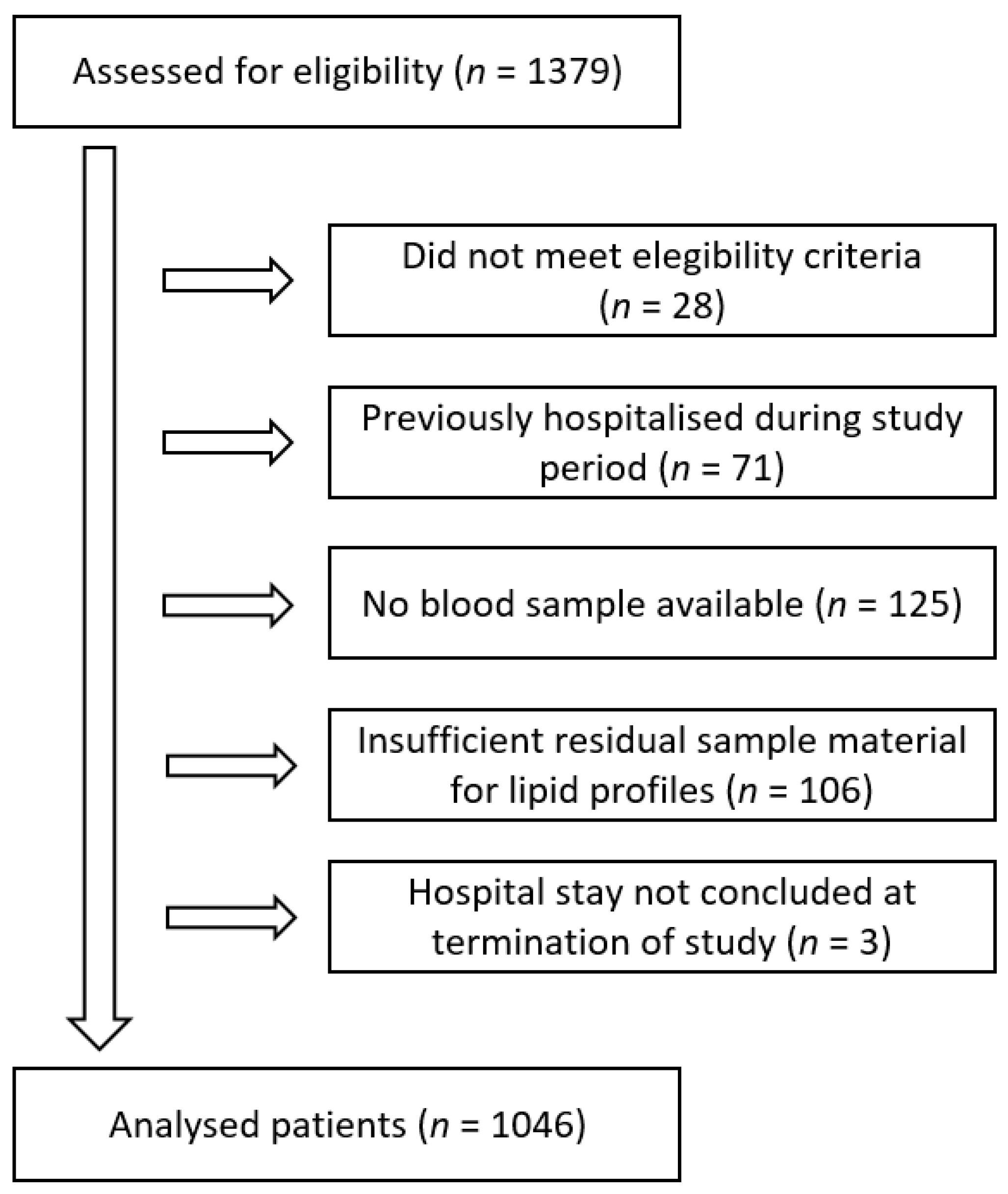
Figure 1. Patient flow diagram.
Jcm 12 05068 g001
Figure 2. (a) Mortality rates (%) by LDL-C (left)/TC (right) and anti-SARS-CoV2-spike antibodies. TC total cholesterol. (b) Cumulative survival over time in days depending on lipid and anti-SARS-CoV2 spike antibody levels; LDL-C (left) and TC (right). TC total cholesterol. (c) Adjusted odds and hazard ratios for in-hospital mortality for lipid levels (below vs. above the respective median value), anti SARS-CoV2 antibodies (<>1200 U/mL) as well as aOR/aHR for low vs. high levels of both lipid level and anti-SARS-CoV2 spike antibodies. TC total choles-terol, spike antibodies—anti-SARS-CoV2-spike antibodies.
Jcm 12 05068 g002
Table 1. Patient characteristics and outcomes by LDL-C and total cholesterol levels. Quantitative results are given as means ± standard deviation; lipid levels are given in mg/dL. BMI: body mass index, DM: diabetes mellitus, CAD: coronary artery disease, COPD: chronic obstructive pulmonary disease, TIA: transient ischemic attack, CVD: cerebrovascular disease, CT: cycle threshold, bold print: statistically significant.
Whole Cohort
n = 1152
LDL-C
<67 mg/dL
n = 525
LDL-C
>67 mg/dL
n = 520
p-ValueTC
<150 mg/dL
n = 520
TC
>150 mg/dL
n = 526
p-Value
age (years)66.8 ± 20.367.7 ± 19.866.0 ± 19.90.11668.2 ± 19.165.5 ± 20.50.038
male gender (%)53.256.649.60.02444.561.9<0.001
BMI (kg/m2)27.1 ± 6.527.7 ± 6.926.6 ± 5.80.01227.4 ± 6.726.8 ± 6.10.135
DM (%)23.930.517.3<0.00129.218.6<0.001
hypertension (%)50.555.045.80.00356.044.9<0.001
CAD (%)21.625.516.3<0.00126.915.0<0.001
heart failure (%)7.28.06.00.1967.76.30.368
COPD (%)9.611.28.30.10610.88.70.270
asthma (%)2.42.32.70.6732.32.70.713
renal disease (%)22.927.718.6<0.00127.219.10.002
stroke/TIA/CVD (%)11.715.28.80.00216.57.6<0.001
mortality (%)10.212.66.7<0.00111.77.60.024
CT value21.3 ± 6.621.3 ± 6.521.2 ± 6.50.65221.5 ± 6.621.0 ± 6.50.224
total cholesterol155.6 ± 50.3120.2 ± 2.2191.4 ± 42.3<0.001116.9 ± 22.7193.9 ± 39.7<0.001
LDL-C71.0 ± 37.042.7 ± 15.699.5 ± 29.6<0.00144.9 ± 18.196.8 ± 32.4<0.001
HDL-C37.7 ± 14.834.2 ± 13.241.4 ± 15.4<0.00132.8 ± 11.342.7 ± 16.1<0.001
triglycerides 139.8 ± 83.8132.7 ± 80.6146.9 ± 86.5<0.001119.0 ± 51.7160.3 ± 103<0.001
statin therapy (%)25.736.016.5<0.00135.417.3<0.001
Table 2. Odds ratios and adjusted odds ratios for each lipid parameter both for the continuous variable in decrements of 10 mg/dL (left) and for the z-scores of each variable (right). Adjusted odds ratios were adjusted for age, obesity, diabetes type II, and statin therapy. OR: odds ratio, aOR: adjusted odds ratio, HR: hazard ratio, ICU: intensive care unit, bold print—statistically significant.
OR, 95%CIp-ValueaOR, 95%CIp-ValueOR z-Scores, 95%CIp-ValueaOR z-Scores, 95%CIp-Value
LDL-C LDL-C
mortality1.16, 1.08–1.24<0.0011.16, 1.07–1.25<0.0011.73, 1.34–2.23<0.0011.73, 1.30–2.31<0.001
mortality, HR1.04, 1.02–1.06<0.0011.04, 1.02–1.06<0.0011.16, 1.09–1.23<0.0011.15, 1.08–1.23<0.001
HDL-C HDL-C
mortality1.23, 1.05–1.440.0121.28, 1.07–1.530.0081.35, 1.07–1.710.0121.44, 1.10–1.880.008
mortality, HR1.11, 1.06–1.16<0.0011.12, 1.07–1.17<0.0011.16, 1.09–1.24<0.0011.18, 1.10–1.26<0.001
total cholesterol total cholesterol
mortality1.09, 1.04–1.14<0.0011.08, 1.03–1.140.0031.51, 1.20–1.92<0.0011.49, 1.14–1.940.003
mortality, HR1.03, 1.02–1.04<0.0011.03, 1.01–1.04<0.0011.15, 1.09–1.23<0.0011.14, 1.07–1.22<0.001
Disclaimer/Publisher’s Note: The statements, opinions and data contained in all publications are solely those of the individual author(s) and contributor(s) and not of MDPI and/or the editor(s). MDPI and/or the editor(s) disclaim responsibility for any injury to people or property resulting from any ideas, methods, instructions or products referred to in the content.