Article

Treatment of COVID-19 during the Acute Phase in Hospitalized Patients Decreases Post-Acute Sequelae of COVID-19

by
Diana Badenes Bonet
1,2,3,
Oswaldo Antonio Caguana Vélez
1,2,3,
Xavier Duran Jordà
3,4,
Merce Comas Serrano
5,
Margarita Posso Rivera
5,
Mireia Admetlló
1,
Anna Herranz Blasco
1,
Elisa Cuadrado Godia
6,
Ester Marco Navarro
7,
Gemma Martin Ezquerra
8,
Zenaida Pineiro Aguin
2,9,
Maria Cinta Cumpli Gargallo
1,
Jose Gregorio Gonzalez Garcia
1,2,3,
Eva Balcells Vilarnau
1,2,3,10,
Diego Rodriguez Chiaradia
1,2,3,10,
Xavier Castells
5,
Joaquim Gea
1,2,3,10,
Juan P. Horcajada
2,3,10,11 and
Judit Villar-García
2,3,11,*,† on behalf of MAR Post-COVID-19 Unit
1
Respiratory Medicine Department, Hospital del Mar, 08003 Barcelona, Spain
2
Department of Medicine and Life Sciences, Universitat Pompeu Fabra (UPF), 08003 Barcelona, Spain
3
Hospital del Mar Medical Research Institute (IMIM), 08003 Barcelona, Spain
4
Assessoria Metodològica i Bioestadística (AMIB), 08003 Barcelona, Spain
5
Epidemiology and Evaluation Department, Hospital del Mar, REDISSEC, RICAPPS, 08003 Barcelona, Spain
6
Neurology Department, Hospital del Mar, 08003 Barcelona, Spain
7
Physical Medicine and Rehabilitation Department, Hospital del Mar, 08003 Barcelona, Spain
8
Dermatology Department, Hospital del Mar, 08003 Barcelona, Spain
9
Otorrinolaringology Department, Hospital del Mar, 08003 Barcelona, Spain
10
Centro de Investigación en Red de Enfermedades Respiratorias, (CIBERES), Instituto de Salud Carlos III (ISCIII), 08003 Barcelona, Spain
11
Infectious Diseases Department, Hospital del Mar, 08003 Barcelona, Spain
*
Author to whom correspondence should be addressed.
Collaborators of the Group Name is provided in the Appendix A.
J. Clin. Med. 2023, 12(12), 4158; https://doi.org/10.3390/jcm12124158
Submission received: 22 April 2023 / Revised: 31 May 2023 / Accepted: 12 June 2023 / Published: 20 June 2023
(This article belongs to the Special Issue Epidemiology and Clinical Characteristics of COVID-19)

Abstract

:
Background: The post-acute sequelae of SARS-CoV-2 (PASC) infection have caused a significant impact on our health system, but there is limited evidence of approved drugs focused on its prevention. Our objective was to identify risk factors that can determine the presence of PASC, with special attention to the treatment received in the acute phase, and to describe the profile of persistent symptoms in a multidisciplinary Post-Coronavirus Disease-19 (COVID-19) Unit. Methods: This one-year prospective observational study included patients following an acute COVID-19 infection, irrespective of whether they required hospital admission. A standardized symptom questionnaire and blood sampling were performed at the first follow-up visit, and demographic and clinical electronic data were collected. We compared subjects with PASC with those who had fully recovered. Multivariate logistic regression was performed to identify factors associated with PASC in hospitalized patients, and Kaplan–Meier curves were used to assess duration of symptoms according to disease severity and treatments received in the acute phase. Results: 1966 patients were evaluated; 1081 had mild disease, 542 moderate and 343 severe; around one third of the subjects had PASC, and were more frequently female, with obesity, asthma, and eosinophilia during acute COVID-19 disease. Patients who received treatment with dexamethasone and remdesivir during the course of the acute illness showed a lower median duration of symptoms, compared with those who received none of these treatments. Conclusion: Treatment with dexamethasone and/or remdesivir may be useful to reduce the impact of PASC secondary to SARS-CoV-2 infection. In addition, we identified female gender, obesity, asthma, and disease severity as risk factors for having PASC.

1. Introduction

Post-Acute sequelae of SARS-CoV-2 infection (PASC) currently affects millions of people worldwide [1,2]. Even in mild cases of SARS-CoV-2 infection, medium and long term health problems have been described, leading to significant health resource expenditure [3]. The lack of a consistent definition for PASC or long-term COVID-19 makes comparison between studies difficult. To address these challenges, a universal definition has been proposed, emphasizing symptoms persisting beyond one month (mild cases) or three months (severe cases) with a significant impact on quality of life [4]. Studies on long COVID-19 have revealed various findings related to immunology, virology, vascular issues, neurological symptoms, and myalgic encephalomyelitis/chronic fatigue syndrome. Multi-organ damage has been observed, with a significant overlap in symptoms [5]. Consequently, different studies have been published describing persistent symptoms in approximately one third of the subjects, with dyspnea, fatigue, exercise intolerance, joint pain, headache, ‘brain fog’ and psychological symptoms being the main reported ones [3,6,7]. Therefore, there is a growing interest in identifying the risk factors of developing PASC. Several publications have identified disease severity, female sex, age, body mass index, the presence of asthma and the number of symptoms during the first week of disease as potential factors that could be used to estimate the individual’s risk for PASC [8,9]. Moreover, a study identified an immunoglobulin (Ig) signature, based on total IgM and IgG3 levels, which, combined with other factors such as age, history of asthma and symptoms during the primary infection, was able to predict the risk of PASC independently of the time-point of blood sampling [10].
Despite the important impact of PASC on our health systems, there is a lack of studies specifically designed to evaluate whether treatments administered in the acute phase of COVID-19 disease can reduce the risk of PASC. There is evidence from a study that demonstrated that patients treated with interferon β-1b and antiviral therapy in the acute phase showed a greater probability of recovering their initial health status [11]. Furthermore, there is growing evidence supporting the effectiveness of antiviral therapies in reducing the risk of PASC. Specifically, the use of molnupiravir within five days of a positive SARS-CoV-2 test result has been associated with a reduced risk of PASC, post-acute death, and post-acute hospital admission in individuals with at least one risk factor for severe COVID-19 [12]. Similarly, the combined antivirals nirmatrelvir–ritonavir have also shown a reduction in the risk of PASC, post-acute death, and post-acute hospital admission in outpatients [13].
For now, there are no specific drug recommendations centered on PASC prevention. Current approved treatments, such as interleukin-6 receptor antagonists and dexamethasone, have been demonstrated to improve outcomes during hospitalization and survival during the acute infection in moderate and severe cases [14,15]. Moreover, new antiviral therapies are recommended for patients with risk of progression to severe COVID-19 since they can also reduce hospitalizations and deaths [16]. However, PASC can also occur in young people without risk factors.
On this point, and to understand the PASC prevalence, its risk factors and the potential impact of treatment received better, it is imperative to appropriately design follow-up strategies optimizing health resources.
Thus, our primary objective was to identify potential risk factors that can determine the presence and duration of PASC, with a special focus on the treatment received during the acute phase. Our second objective was to describe persistent symptoms and their duration in a multidisciplinary Post-COVID-19 Unit.

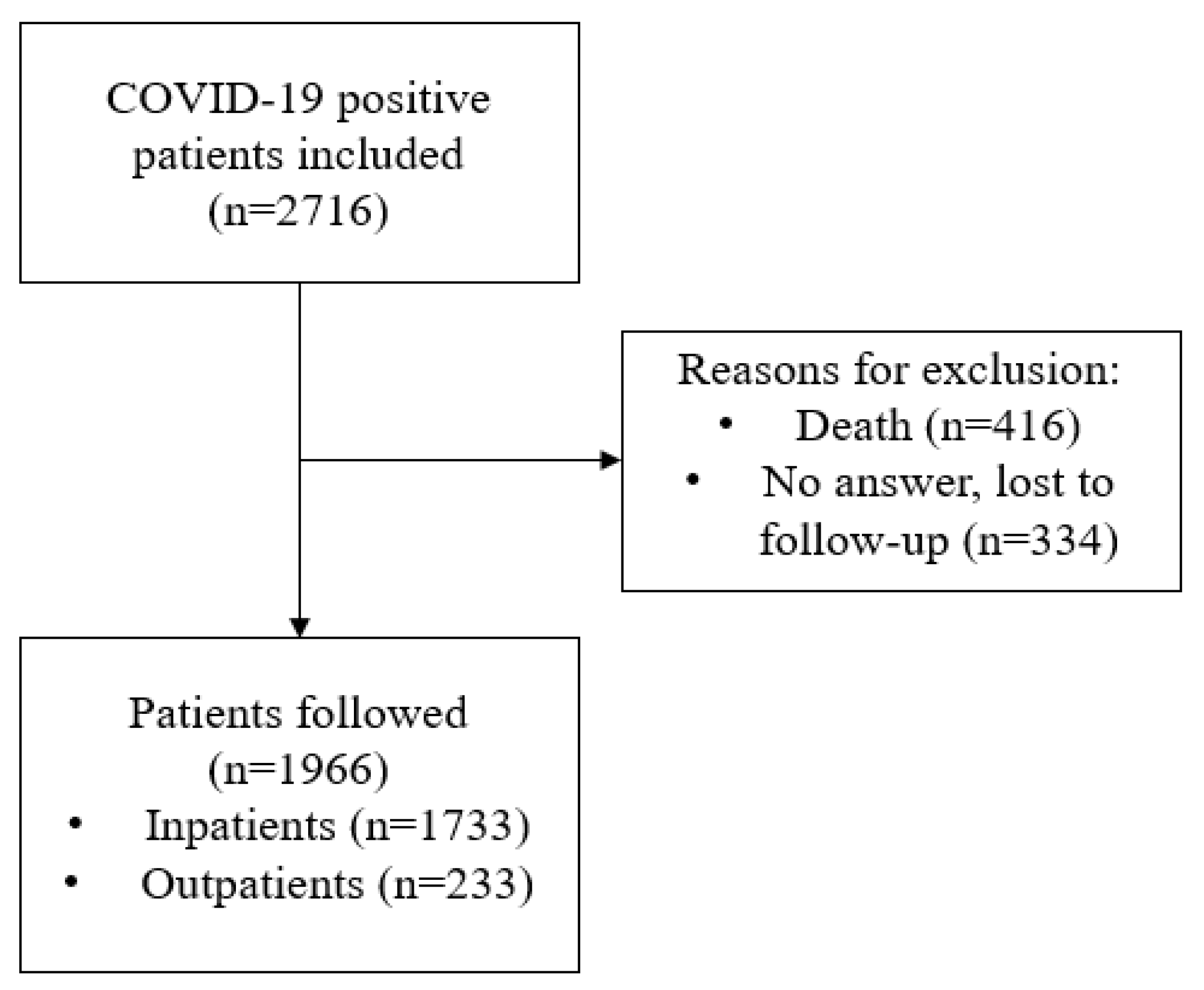
2. Materials and Methods

2.1. Study Design

This is a single-center prospective observational study carried out according to the principles of the Declaration of Helsinki for human investigations and approved by the local Ethics Committee (nº 2020/9455). Patients with a positive SARS-CoV-2 PCR test were offered the chance of participating at the first follow-up visit, and were then finally included after giving their verbal informed consent. A standardized symptom questionnaire (Table S1) was administered in the first follow-up visit to all the participants and demographic and clinical electronic data were collected. Blood samples were obtained from outpatients and from subjects who required hospitalization. Finally, participants were evaluated every 3 months and symptom persistence was evaluated and registered (Figure S1).

2.2. Study Participants

2.2.1. Inclusion Criteria

We included patients who had suffered from mild disease (not hospitalized) and those individuals with mild, moderate, or severe pneumonia due to COVID-19 (all of them admitted to our hospital) and were visited in our multidisciplinary Post-COVID-19 unit from June to December 2020 and followed-up during 1 year. Those who required hospitalization were followed in the Post-COVID-19 Unit, irrespective of whether they had persistent symptoms, whereas patients referred from primary care were symptomatic. Inclusion criteria were to have had a COVID-19 infection, confirmed with polymerase chain reaction (PCR), during the first and second wave of the pandemic in our center (March to December 2020). The predominant SARS-CoV-2 variants that circulated at that time were lineage B.1.1.7 (88%) followed by B. 1.177/A222V (7.5%). This study was conducted during the early phases of the COVID-19 vaccination programs and most of our patients had not been vaccinated at the time of data collection.

2.2.2. Exclusion Criteria

Subjects who refused to participate, who died during the follow-up, as well as those who we were unable to contact after hospital discharge were excluded.

2.3. COVID-19 Severity Groups

Participants were stratified by the acute COVID-19 infection severity into (a) mild disease: patients without need of supplemental oxygen therapy, (b) moderate disease: hospitalized patients who required supplemental low-flow oxygen therapy (FiO2 < 0.40) and (c) severe disease: hospitalized patients with supplemental high-flow oxygen therapy (FiO2 > 0.40), high-flow nasal cannula oxygen therapy (HFNC) or non-invasive and/or invasive mechanical ventilation [17].

2.4. Treatment Received for SARS-CoV-2 Infection

In our center, treatment modality has been protocolized since April 2020. Antivirals were indicated (remdesivir 200 mg the first day and 100 mg/day for 5 days) to patients with low-flow oxygen requirements to maintain SpO2 > 94%. In addition, treatment with heparin was indicated at a prophylactic dose of 40 mg/day in patients with D-dimer levels < 2000 mcg/L, intermediate doses of 1 mg/Kg/day with D-dimer levels > 2000 mcg/L, and anticoagulant doses of 1 mg/Kg/12 h in patients diagnosed with acute pulmonary embolism. Subjects with moderate–severe disease received treatment with dexamethasone 6 mg/day for 10 days and tocilizumab was posteriorly administered only in cases of worsening of respiratory symptoms, FiO2 > 35% and IL-6 levels > 40 pg/mL, according to our hospital protocol and available guidelines at that moment [18,19]. Moreover, subjects with organizing pneumonia received treatment with methylprednisolone that was progressively tapered at follow-up. Outpatients included in the study received no therapy.

2.5. Symptom Assessment

A structured questionnaire (including 22 symptoms) was performed at the initial visit (Table S1) by trained interviewers of the Post-COVID-19 Unit, and symptom persistence was registered at each follow-up visit to record symptom duration. The presence of PASC was considered according to the current definitions at the beginning of the pandemic, including ≥2 persistent symptoms affecting any organ or system after 4 weeks of the acute infection, having excluded other potential etiologies [20,21].

2.6. Blood Sampling

In hospitalized subjects, the main data from blood analyses, both at baseline and at the peak of disease severity, were collected. Moreover, another blood test was performed at inclusion at the first follow-up visit. In outpatients, only one blood sample was collected at the inclusion. The blood test parameters included C-reactive protein, ferritin, procalcitonin, interleukin-6 (IL-6), leucocyte count, creatinine, lactate dehydrogenase (LDH), glucose, total proteins, D-dimer, and platelets.

2.7. Statistical Analysis

Categorical variables were expressed as numbers and percentages, whereas continuous variables were presented as mean (standard deviation, SD), or median (25th and 75th percentiles, P25–P75) when the normality assumption was not fulfilled. Differences between groups were checked with Pearson’s Chi Squared test for categorical variables, and unpaired Student’s t-test or one-way ANOVA for continuous variables meeting the normality assumption, or non-parametric Mann–Whitney U test when normality could not be assumed. Significant differences were considered at the 0.05 p level. Multivariate logistic regression was performed to check factors associated with PASC in hospitalized patients. Variables in the multivariate analysis were introduced based on clinical criteria considering those that were statistically significant or nearly significant (p, 0.5–0.1) in the bivariate analysis. Results from this analysis were reported as odds ratios (OR) with their 95% confidence interval (CI). Kaplan–Meier curves were used to compare duration of symptoms by disease severity and the effects of treatments used in the acute phase. The log-rank test was performed to assess differences in symptom duration for each drug between its use or not in every group of disease severity. STATA version 15.1 (StataCorp, College Station, TX, USA) was used for statistical analysis.

3. Results

The study cohort included 2716 subjects referred after hospitalization and from primary care following a confirmed SARS-CoV-2 infection. Subjects who died or were lost during the follow-up (416 and 334, respectively) were excluded. In total, 1966 patients were finally followed-up at the Post-COVID-19 Unit (Figure 1).

3.1. Baseline Characteristics

The clinical characteristics and persisting symptoms of the total population are summarized in Table 1. The mean age was 56.4 years, and 50% were females. As far as disease severity is concerned, 54.9% had mild disease, whereas 27.6 and 17.5% showed moderate or severe disease, respectively. The most frequent comorbidity was arterial hypertension, followed by psychiatric disorders and diabetes mellitus, and all comorbidities were more frequent in subjects with severe disease. Around a third of the subjects experienced persisting symptoms at the follow-up, this being more frequent in the severe disease group. Dyspnea, fatigue and myalgia were the most symptomatic manifestations.

3.2. PASC in Hospitalized Patients

3.2.1. Clinical Data

Comparing subjects with PASC with those that completely recovered after hospitalization (Table 2), the former were older, more frequently females and had a higher frequency of obesity and asthma than the latter. COVID-19 severity was also associated with a higher PASC frequency. The most frequent persisting symptoms were again dyspnea, fatigue and myalgia, followed by cough, anxiety and depression.

3.2.2. Laboratory Data

Subjects with PASC had a significantly lower total lymphocyte count, lower Cd4 and Cd8, and higher interleukin 6 (IL-6), lactate dehydrogenase (LDH) and platelet count at baseline than those without. Laboratory results at the peak of the acute disease evidenced that the PASC group showed higher neutrophil, lymphocyte, eosinophil and platelet counts, as well as LDH levels, than subjects that completely recovered (Table 3).

3.2.3. PASC Prediction

Table 4 summarizes the results of the multivariate logistic regression model to identify predictors of PASC in hospitalized patients. In our cohort, female sex, severe disease, and asthma increased the risk of having PASC. Since the most severe patients usually show greater changes in inflammatory parameters, our multivariate analysis included this factor to avoid a potential bias. Although results were not statistically significant, a strong trend for having a higher risk of PASC was observed for eosinophil and neutrophil counts at the peak point of the acute disease.

3.3. PASC in Outpatients

A total of 233 patients did not require hospitalization but were followed-up at the Post-COVID-19 Unit of our center (Table 5). Those subjects with PASC were more frequently female and had more psychiatric disorders than those without, and the symptoms more frequently displayed were dyspnea, fatigue and myalgia, again followed by cough, anxiety and depression. No statistically significant differences were found in the follow-up laboratory parameters (Table S2).

3.4. Treatment Received

Given that treatment received is highly dependent on disease severity, we analyzed the effect of treatment with this in mind. In those cases that received treatment with remdesivir with a moderate or severe disease, a significant shorter duration of PASC was observed when compared with those not receiving the treatment (109 vs. 262 and 187 vs. 284 days, respectively). Similar results were found in patients who received dexamethasone, in cases with moderate or severe disease (133 vs. 271 and 266 vs. 309, respectively Table 6, Figure 2). Treatments with tocilizumab and methylprednisolone were not associated with a reduction in the duration of PASC symptoms.
Some patients received a combination of some of these therapies, according to disease severity. In total, 603 (71.2%) patients only received corticosteroids (dexamethasone, prednisone or methylprednisolone); 79 (9.3%) patients received corticosteroids and remdesivir and 187 (22.1%) patients received corticosteroids and tocilizumab (Table 6).

4. Discussion

This prospective study systematically described the profile of PASC one year after an acute COVID-19 infection in a large group of patients with a broad scope of clinical severity. Even more relevant is that, to our knowledge, this is the first prospective study in patients who required oxygen therapy and hospital admission to show that some treatments used during the acute phase reduces the risk of PASC. This is an important point, since the current approved treatments of COVID-19 infection are focused on preventing acute complications in people with risk factors, and preventing PASC in mild cases in an outpatient setting. In our study, we make a distinct contribution by focusing on a study cohort consisting of patients who presented in the acute phase of COVID-19, encompassing a range of severity from mild to moderate and severe cases. Our dataset includes data from hospitalized patients, providing valuable insights into the long-term health outcomes of this specific population. This comprehensive approach allows us to capture a broader spectrum of patients and provide a more comprehensive understanding of the long-term effects of COVID-19.
Indeed, a very large number of people at risk of PASC are relatively young without any comorbidities, so they currently do not meet the criteria for receiving treatment during the acute phase. However, there is very limited evidence on the association between treatments administered during the acute phase and predisposition of persistent symptoms [22,23,24,25]. There is an Italian cohort, where patients were treated in the acute phase of COVID-19 with monoclonal drugs, which obtained a 56% reduction in the risk of PASC appearance [26]; unlike our cohort, they required no oxygen therapy or hospital admission for SARS-CoV-2 infection. Our study shows that treatment with remdesivir and/or corticosteroids during the acute phase decreases the length of PASC in hospitalized subjects. Furthermore, corticosteroids or antivirals are more accessible worldwide than monoclonal drugs.
Remdesivir has been shown to be effective in slowing disease progression and shortening recovery time for COVID-19 patients [27], with the antiviral treatment being recommended for the patients with COVID-19 infection in national and international clinical guidelines, including WHO, NIH, NICE, ESCMID, IDSA, and SEIMC [18,19,28,29,30,31], although it is recommended to be restricted to patients with up to 7 days of symptoms, mild disease, but with no risk factors for progression to severe disease, and also in patients with moderate or severe infection requiring hospitalization [18,20]. The results of different studies led certain guidelines to recommend prolonging its administration in severe cases requiring invasive ventilation or ECMO or in patients with severe immunosuppression, since persistent viral replication beyond two weeks has been described in these groups [32]. The persistence of viral replication has also been involved in the pathophysiology of PASC. Therefore, it does not seem strange that the early administration of antiviral treatment can also prevent long-term symptoms [27,33]. Furthermore, some of the drugs currently in use to treat acute COVID-19 infection have potential anti-inflammatory and immunomodulatory effects that have already been analyzed in several cohorts and meta-analyses [22,34,35]. Those treatments, mainly tocilizumab and corticosteroids, are used for patients in the cytokine storm phase, and, although they have managed to reduce mortality when appropriately indicated [36], it is unknown to date whether they could also reduce the presence or length of PASC. However, immunoactivation and/or sustained inflammation have also been involved in the pathophysiology of PASC. Thus, it also seems reasonable that these treatments could be effective in reducing the impact of PASC. In fact, the inflammatory cascade that remains activated in certain susceptible individuals when they overcome the acute phase of COVID-19 infection includes cytokines and signaling molecules (such as interleukins 1β, -6, -8, -17 and tumor necrosis factor alpha) that have been involved in structural and functional abnormalities in respiratory and limb muscles, potentially partly explaining some PASC symptoms such as musculoskeletal discomfort, fatigue and dyspnea, which are among the most common persisting symptoms [37,38].
In the present study, we found that treatment with dexamethasone in the acute phase reduced the duration of PASC. However, treatment with methylprednisolone was not associated with a reduction in the long-term duration of symptoms, probably because it was intended to treat patients with severe pneumonia who were in the organizing/fibrosis phase. In the case of treatment with tocilizumab, we were unable to demonstrate effects on PASC. One possible hypothesis is that this would be explained by the action of this drug on a very specific pathway of IL6 and the use of a single dose with a short half-life.
According to the results of this study, and given both the high prevalence of PASC, which in many cases is disabling, and the burden that its care entails for the health system, administering treatment during the acute phase should be considered for all patients.
In this prospective study, we found that close to a quarter of the patients had at least one persistent symptom of COVID-19 months after the acute infection, and this is similar to the prevalence found in other series [6,9,16,39]. The same accounts for the main symptoms, which were dyspnea, fatigue, myalgia, persistent cough, anxiety and depression, were also similar to those described in several previous studies [16,40,41,42].
When comparing the present cohort with other series, the risk factors for PASC are relatively similar. In general, these studies identified age, female sex, comorbidities and severity of the disease during the acute process [43,44]. Age is probably an important predisposing factor, probably due to the lower regenerative capacity of the lung, in addition to underlying chronic pulmonary problems that could be associated [44,45]. A recent meta-analysis of patients identified several epidemiological and clinical risk factors associated with an increased risk of developing persistent symptoms of COVID-19, known as Post-COVID Conditions (PCC). Female sex, older age, higher BMI, smoking and preexisting comorbidities such as anxiety, depression, asthma, COPD, diabetes, IHD and immunosuppression were found to be significant risk factors for PCC. Hospitalization or ICU care during the acute phase of COVID-19 infection also increased the risk. These findings highlight the multifactorial nature of PCC and provide insights into its risk factors [46]. On the other hand, vaccination was found to have a protective role against PCC [46,47,48,49]. In our study, the severity of the acute disease was a predisposing factor for presenting PASC, related to the requirement for invasive and non-invasive respiratory support, as with other series [50,51,52,53,54]. Interestingly, asthma and obesity were the comorbidities that were associated with a higher risk of persistent symptoms after acute infection, regardless of the severity of the disease in hospitalized patients. These findings have also been found in other studies [8,10]. More severe COVID-19 disease has consistently been associated with both asthma and obesity. While the predisposition of the former has been related to increased concentrations of IL-4 [55,56]. Other frequent comorbidities also described in other series [6,9] were arterial hypertension, diabetes mellitus, COPD and anxiety-depressive disorders, but they did not show significant results in the present study, except in outpatients, in which subjects with PASC were more frequently female and had more psychiatric disorders than those without PASC.
Some laboratory findings linked to an inflammatory profile have also been found to increase the risk for PASC, such as a significant decrease in total lymphocyte counts—especially CD4+ T cells and CD8+ T cells—and an increase in LDH, CRP and IL-6 levels, as well as IL-1β, IL-6, TNF and IP10 [5,51,57,58,59,60], mostly in patients who required hospitalization. This could be due to the inflammatory and immunological pathophysiological mechanisms involved in the COVID-19 disease [61,62]. Moreover, our results also highlight the potential role of eosinophil counting during the acute disease in hospitalized patients. Counts of these cells were higher in our patients with PASC, and showed a tendency to be an independent factor of presenting persisting symptoms, even in the multivariate analysis, where variables that may be confounding factors were included. Some series have shown that, during the early phases of the acute phase, the levels of eosinophils are rather low, but could rise at 3 and 6 months [63,64,65]. Eosinophils play an important role in immunity; they eliminate bacteria and viruses, as well as behaving as an antigen-presenting cell [66]. However, the presence of eosinophils is also related to autoimmune and allergic diseases, which is consistent with the pathophysiology of COVID-19 and PASC [67] and may also justify the increased risk in patients with asthma. One of the hypotheses for the persistence of symptoms is the hyperactivation of the autoimmunity that the acute infection generates [68]. To our knowledge, this is also the first study that confers a prognostic value on the presence of eosinophils during acute infection, since it is related to persistent symptoms. No statistically significant differences were found in the follow-up laboratory parameters in outpatients, but in this group of patients the analytical study was not performed during the primary infection.

5. Strengths and Limitations

Our study investigates the impact of treatment during the acute phase on the risk of PASC and presents several strengths and limitations. One of the major strengths is that it is one of the few reports to highlight the potential benefit of treatment in the acute phase in reducing the risk of PASC. What is new is that the treatment decreases the incidence of PASC, including in patients without risk factors for severe COVID-19, for whom treatment is not currently recommended. This emphasizes the importance of treating patients who may not develop severe COVID-19 but are at risk for PASC. Additionally, our study benefits from a large and diverse population sample, encompassing individuals with varying severity levels of COVID-19, and a comprehensive and lengthy follow-up period that includes both clinical and laboratory parameters. Moreover, the standardized treatment protocols implemented since April 2020 have facilitated a more homogeneous comparison, with reliable data collected from electronic health records.
Our study has certain limitations that need to be acknowledged. Firstly, it is possible that patients who did not participate had milder symptoms compared to those who did, which could lead to an overestimation of the prevalence of prolonged symptoms, particularly among non-hospitalized individuals with mild COVID-19. Additionally, being a single-center study conducted during the early phase of the pandemic, the generalizability of our findings to patients infected with later variants of SARS-CoV-2 may be uncertain. We acknowledge that the lack of consideration for the specific viral variants circulating during the study period is a limitation of our research. In addition, the study was carried out during the initial period of the pandemic, before widespread vaccination campaigns. Therefore, the analysis did not include information on the vaccination status of the patients, which could have influenced treatment outcomes and tissue response. Moreover, it is important to recognize the absence of a control group as a notable limitation. However, it is important to note that despite this limitation, the study still provides valuable insights into the potential effects of the treatments on PASC. The findings should be interpreted cautiously, considering the absence of a control group [69].

6. Conclusions

Treatment with dexamethasone and remdesivir was associated with a shorter duration of symptoms after SARS-CoV-2 infection. In a 12-month follow-up more than one third of the subjects had persistent symptoms. We identified female sex, obesity, asthma, severity of the disease and eosinophilia as risk factors associated with the presence of PASC. In summary, early identification of people at a higher risk of PASC and an early initiation of antiviral and/or anti-inflammatory treatment may avoid the risk of long-term complications and reduce the burden that PASC places on the healthcare system. Further multicentric studies are needed to identify risk factors of PASC as well as to establish which therapies could reduce its risk.

Supplementary Materials

The following supporting information can be downloaded at: https://www.mdpi.com/article/10.3390/jcm12124158/s1, Figure S1: Follow-up protocol; Table S1: Standardized symptom assessment questionnaire after SARS-CoV-2 infection; Table S2: Outpatients laboratory results at follow-up (n = 233).

Author Contributions

Conceptualization, D.B.B., O.A.C.V. and J.V.-G.; methodology, X.D.J.; software, X.D.J.; validation, E.B.V., D.R.C., J.G. and J.P.H.; formal analysis, X.D.J., M.P.R., M.C.S. investigation, D.B.B., O.A.C.V. and J.V.-G.; resources, E.C.G., E.M.N., G.M.E., Z.P.A., M.C.C.G. and J.G.G.G.; data curation, M.A., A.H.B.; writing—original draft preparation, D.B.B., O.A.C.V. and J.V.-G.; writing—review and editing, D.B.B., O.A.C.V. and J.V.-G.; visualization, D.B.B., O.A.C.V. and J.V.-G.; supervision, D.B.B., J.V.-G., E.B.V., D.R.C., J.G., X.C. and J.P.H. All authors have read and agreed to the published version of the manuscript.

Funding

This research received no external funding.

Institutional Review Board Statement

The study carried out according to the principles of the Declaration of Helsinki for human investigations and approved by the local Ethics Committee (nº 2020/9455).

Informed Consent Statement

Not applicable.

Data Availability Statement

The data presented in this study are available on request from the corresponding author.

Acknowledgments

To the patients who have participated in this study and to the doctors, nurses, cleaners, clerks, pharmacists and other healthcare workers who have worked tirelessly during the COVID-19 pandemic.

Conflicts of Interest

The authors declare no conflict of interest.

Appendix A. MAR Post-COVID-19 Unit

Study sites and investigators: Abora Rial-Villavecchia, Jaime Rodríguez-Morera, Internal Medicine Department, Member of Unitat d’Expertesa Clínica (UEC) en malalties autoimmunes sistèmiques i vasculitis de l’Hospital del Mar, Barcelona, Spain. Bernat Recasens, Antía Moreira, Neurology Department, Neurovascular Research Group, Hospital del Mar Medical Research Institute (IMIM), Hospital del Mar, Barcelona, Spain. Maria Pilar Gracia-Arnillas, Intensive Care Unit, Hospital del Mar, Barcelona, Spain. Eva Rodríguez, Nefrology Department, Hospital del Mar, Barcelona, Spain. Gabriel Pedemonte-Sarrias, Otorrinolaringology Department, Hospital del Mar, Barcelona, Spain. Anna Guillen-Sola, Marta Tejero, Physical Medicine and Rehabilitation Department, Hospital del Mar, Barcelona, Spain. Antonio Salar, Carmen Jiménez, Hematology Department, Hospital del Mar, Barcelona, Spain. Silvia Oller, Angels Arroyo, Psiquiatry Department, Hospital del Mar, Barcelona, Spain. Andrea Izquierdo, Beatriz Vaquerizo, Cardiology Department, Hospital del Mar, Barcelona, Spain. Fabiola Ojeda, Josep Blanch, Reumatology Department, Hospital del Mar, Barcelona, Spain.

References

  1. Wang, D.; Hu, B.; Hu, C.; Zhu, F.; Liu, X.; Zhang, J.; Wang, B.; Xiang, H.; Cheng, Z.; Xiong, Y.; et al. Clinical Characteristics of 138 Hospitalized Patients With 2019 Novel Coronavirus-Infected Pneumonia in Wuhan, China. JAMA 2020, 323, 1061–1069. [Google Scholar] [CrossRef]
  2. Al-Aly, Z.; Xie, Y.; Bowe, B. High-dimensional characterization of post-acute sequelae of COVID-19. Nature 2021, 594, 259–264. [Google Scholar] [CrossRef] [PubMed]
  3. Menges, D.; Ballouz, T.; Anagnostopoulos, A.; Aschmann, H.E.; Domenghino, A.; Fehr, J.S.; Puhan, M.A. Burden of post-COVID-19 syndrome and implications for healthcare service planning: A population-based cohort study. PLoS ONE 2021, 16, e0254523. [Google Scholar] [CrossRef] [PubMed]
  4. Pan, D.; Pareek, M. Toward a Universal Definition of Post-COVID-19 Condition-How Do We Proceed? JAMA Netw. Open 2023, 6, e235779. [Google Scholar] [CrossRef]
  5. Davis, H.E.; McCorkell, L.; Vogel, J.M.; Topol, E.J. Long COVID: Major findings, mechanisms and recommendations. Nature reviews. Microbiology 2023, 21, 133–146. [Google Scholar] [CrossRef]
  6. Logue, J.K.; Franko, N.M.; McCulloch, D.J.; McDonald, D.; Magedson, A.; Wolf, C.R.; Chu, H.Y. Sequelae in Adults at 6 Months After COVID-19 Infection. JAMA Netw. Open 2021, 4, e210830. [Google Scholar] [CrossRef]
  7. Carfì, A.; Bernabei, R.; Landi, F.; Gemelli Against COVID-19 Post-Acute Care Study Group. Persistent Symptoms in Patients After Acute COVID-19. JAMA 2020, 324, 603–605. [Google Scholar] [CrossRef] [PubMed]
  8. Sudre, C.H.; Murray, B.; Varsavsky, T.; Graham, M.S.; Penfold, R.S.; Bowyer, R.C.; Pujol, J.C.; Klaser, K.; Antonelli, M.; Canas, L.S.; et al. Attributes and predictors of long COVID. Nat. Med. 2021, 27, 626–631. [Google Scholar] [CrossRef]
  9. Taquet, M.; Dercon, Q.; Luciano, S.; Geddes, J.R.; Husain, M.; Harrison, P.J. Incidence, co-occurrence, and evolution of long-COVID features: A 6-month retrospective cohort study of 273,618 survivors of COVID-19. PLoS Med. 2021, 18, e1003773. [Google Scholar] [CrossRef]
  10. Cervia, C.; Zurbuchen, Y.; Taeschler, P.; Ballouz, T.; Menges, D.; Hasler, S.; Adamo, S.; Raeber, M.E.; Bächli, E.; Rudiger, A.; et al. Immunoglobulin signature predicts risk of post-acute COVID-19 syndrome. Nat. Commun. 2022, 13, 446. [Google Scholar] [CrossRef]
  11. Tleyjeh, I.M.; Saddik, B.; AlSwaidan, N.; AlAnazi, A.; Ramakrishnan, R.K.; Alhazmi, D.; Aloufi, A.; AlSumait, F.; Berbari, E.; Halwani, R. Prevalence and predictors of Post-Acute COVID-19 Syndrome (PACS) after hospital discharge: A cohort study with 4 months median follow-up. PLoS ONE 2021, 16, e0260568. [Google Scholar] [CrossRef]
  12. Xie, Y.; Choi, T.; Al-Aly, Z. Molnupiravir and risk of post-acute sequelae of COVID-19: Cohort study. BMJ 2023, 381, e074572. [Google Scholar] [CrossRef]
  13. Xie, Y.; Choi, T.; Al-Aly, Z. Association of Treatment With Nirmatrelvir and the Risk of Post-COVID-19 Condition. JAMA Intern. Med. 2023, 183, 554–564. [Google Scholar] [CrossRef] [PubMed]
  14. RECOVERY Collaborative Group; Horby, P.; Lim, W.S.; Emberson, J.R.; Mafham, M.; Bell, J.L.; Linsell, L.; Staplin, N.; Brightling, C.; Ustianowski, A.; et al. Dexamethasone in Hospitalized Patients with COVID-19. N. Engl. J. Med. 2021, 384, 693–704. [Google Scholar] [CrossRef]
  15. RECOVERY Collaborative Group. Tocilizumab in patients admitted to hospital with COVID-19 (RECOVERY): A randomised, controlled, open-label, platform trial. Lancet 2021, 397, 1637–1645. [Google Scholar] [CrossRef] [PubMed]
  16. Hammond, J.; Leister-Tebbe, H.; Gardner, A.; Abreu, P.; Bao, W.; Wisemandle, W.; Baniecki, M.; Hendrick, V.M.; Damle, B.; Simón-Campos, A.; et al. Oral Nirmatrelvir for High-Risk, Nonhospitalized Adults with COVID-19. N. Engl. J. Med. 2022, 386, 1397–1408. [Google Scholar] [CrossRef] [PubMed]
  17. Sibila, O.; Molina-Molina, M.; Valenzuela, C.; Ríos-Cortés, A.; Arbillaga-Etxarri, A.; Torralba García, Y.; Díaz-Pérez, D.; Landete, P.; Mediano, O.; Tomás López, L.; et al. Spanish Society of Pulmonology and Thoracic Surgery (SEPAR) Consensus for Post-COVID-19 Clinical Follow-Up. Open Respir. Arch. 2020, 2, 278–283. [Google Scholar] [CrossRef]
  18. Arribas, J.R.; Carolina Garcia-Vidal, C.; Galán Montemayor, J.C.; Paño, J.R.; Rodríguez Baño, J. Recomendaciones SEIMC Para El Manejo Clínico de Pacientes Con COVID-19. Sociedad Española de Enfermedades Infecciosas y Microbiología Clínica 2:1–2. 2020. Available online: https://seimc.org/contenidos/documentoscientificos/recomendaciones/seimc-rc-2020-COVID19-manejoclinico.pdf (accessed on 1 May 2023).
  19. Lamontagne, F.; Agarwal, A.; Rochwerg, B.; Siemieniuk, R.A.; Agoritsas, T.; Askie, L.; Lytvyn, L.; Leo, Y.S.; Macdonald, H.; Zeng, L.; et al. A living WHO guideline on drugs for COVID-19. BMJ 2020, 370, m3379. [Google Scholar] [CrossRef] [PubMed]
  20. Seeßle, J.; Waterboer, T.; Hippchen, T.; Simon, J.; Kirchner, M.; Lim, A.; Müller, B.; Merle, U. Persistent Symptoms in Adult Patients 1 Year After Coronavirus Disease 2019 (COVID-19): A Prospective Cohort Study. Clin. Infect. Dis. Off. Publ. Infect. Dis. Soc. Am. 2022, 74, 1191–1198. [Google Scholar] [CrossRef] [PubMed]
  21. Nalbandian, A.; Sehgal, K.; Gupta, A.; Madhavan, M.V.; McGroder, C.; Stevens, J.S.; Cook, J.R.; Nordvig, A.S.; Shalev, D.; Sehrawat, T.S.; et al. Post-acute COVID-19 syndrome. Nat. Med. 2021, 27, 601–615. [Google Scholar] [CrossRef] [PubMed]
  22. Han, Q.; Zheng, B.; Daines, L.; Sheikh, A. Long-Term Sequelae of COVID-19: A Systematic Review and Meta-Analysis of One-Year Follow-Up Studies on Post-COVID Symptoms. Pathogens 2022, 11, 269. [Google Scholar] [CrossRef] [PubMed]
  23. Matuszewski, M.; Szarpak, L.; Rafique, Z.; Peacock, F.W.; Pruc, M.; Szwed, P.; Chirico, F.; Navolokina, A.; Ladny, J.R.; Denegri, A. Prediction Value of KREBS Von Den Lungen-6 (KL-6) Biomarker in COVID-19 Patients: A Systematic Review and Meta-Analysis. J. Clin. Med. 2022, 11, 6600. [Google Scholar] [CrossRef] [PubMed]
  24. Fialek, B.; De Roquetaillade, C.; Pruc, M.; Navolokina, A.; Chirico, F.; Ladny, J.R.; Peacock, F.W.; Szarpak, L. Systematic review with meta-analysis of mid-regional pro-adrenomedullin (MR-proadm) as a prognostic marker in COVID-19-hospitalized patients. Ann. Med. 2023, 55, 379–387. [Google Scholar] [CrossRef] [PubMed]
  25. Matuszewski, M.; Ładny, J.; Rafique, Z.; Peacock, F.; Pruc, M.; Gasecka, A.; Szwed, P.; Jankowski, L.; Chmielewski, J.; Panasiuk, L.; et al. Prediction value of soluble urokinase plasminogen activator receptor (suPAR) in COVID-19 patients—A systematic review and meta-analysis. Ann. Agric. Environ. Med. AAEM 2023, 30, 142–147. [Google Scholar] [CrossRef] [PubMed]
  26. Cimellaro, A.; Addesi, D.; Cavallo, M.; Spagnolo, F.; Suraci, E.; Cordaro, R.; Spinelli, I.; Passafaro, F.; Colosimo, M.; Pintaudi, M.; et al. Monoclonal Antibodies and Antivirals against SARS-CoV-2 Reduce the Risk of Long COVID: A Retrospective Propensity Score-Matched Case-Control Study. Biomedicines 2022, 10, 3135. [Google Scholar] [CrossRef]
  27. Beigel, J.H.; Tomashek, K.M.; Dodd, L.E.; Mehta, A.K.; Zingman, B.S.; Kalil, A.C.; Hohmann, E.; Chu, H.Y.; Luetkemeyer, A.; Kline, S.; et al. Remdesivir for the Treatment of COVID-19—Final Report. N. Engl. J. Med. 2020, 383, 1813–1826. [Google Scholar] [CrossRef]
  28. NIH Therapeutic Management of Adults with COVID-19. Available online: https://www.covid19treatmentguidelines.nih.gov/management/clinical-management/hospitalizedadults--therapeutic-management/ (accessed on 1 December 2022).
  29. NICE. Guideline [NG191]. COVID-19 Rapid Guideline: Managing COVID-19. Available online: https://app.magicapp.org/#/guideline/L4Qb5n/section/ERYAXn (accessed on 31 December 2022).
  30. Bartoletti, M.; Azap, O.; Barac, A.; Bussini, L.; Ergonul, O.; Krause, R.; Paño-Pardo, J.R.; Power, N.R.; Sibani, M.; Szabo, B.G.; et al. ESCMID COVID-19 living guidelines: Drug treatment and clinical management. Clin. Microbiol. Infect. Off. Publ. Eur. Soc. Clin. Microbiol. Infect. Dis. 2022, 28, 222–238. [Google Scholar] [CrossRef] [PubMed]
  31. IDSA. IDSA Guidelines on the Treatment and Management of Patients with COVID-19. Available online: https://www.idsociety.org/practice-guideline/covid-19-guideline-treatmentand-management/ (accessed on 31 December 2022).
  32. Garcia-Vidal, C.; Puerta-Alcalde, P.; Mateu, A.; Cuesta-Chasco, G.; Meira, F.; Lopera, C.; Monzo, P.; Santos-Bravo, M.; Duenas, G.; Chumbita, M.; et al. Prolonged viral replication in patients with hematologic malignancies hospitalized with COVID-19. Haematologica 2022, 107, 1731–1735. [Google Scholar] [CrossRef] [PubMed]
  33. Niknam, Z.; Jafari, A.; Golchin, A.; Danesh Pouya, F.; Nemati, M.; Rezaei-Tavirani, M.; Rasmi, Y. Potential therapeutic options for COVID-19: An update on current evidence. Eur. J. Med. Res. 2022, 27, 6. [Google Scholar] [CrossRef]
  34. Toussirot, E.; Marotte, H.; Mulleman, D.; Cormier, G.; Coury, F.; Gaudin, P.; Dernis, E.; Bonnet, C.; Damade, R.; Grauer, J.L.; et al. Increased high molecular weight adiponectin and lean mass during tocilizumab treatment in patients with rheumatoid arthritis: A 12-month multicentre study. Arthritis Res. Ther. 2020, 22, 224. [Google Scholar] [CrossRef]
  35. Castillero, E.; Alamdari, N.; Aversa, Z.; Gurav, A.; Hasselgren, P.O. PPARβ/δ regulates glucocorticoid- and sepsis-induced FOXO1 activation and muscle wasting. PLoS ONE 2013, 8, e59726. [Google Scholar] [CrossRef] [Green Version]
  36. WHO Rapid Evidence Appraisal for COVID-19 Therapies (REACT) Working Group; Sterne, J.A.C.; Murthy, S.; Diaz, J.V.; Slutsky, A.S.; Villar, J.; Angus, D.C.; Annane, D.; Azevedo, L.C.P.; Berwanger, O.; et al. Association Between Administration of Systemic Corticosteroids and Mortality Among Critically Ill Patients With COVID-19: A Meta-analysis. JAMA 2020, 324, 1330–1341. [Google Scholar] [CrossRef] [PubMed]
  37. Schakman, O.; Kalista, S.; Barbé, C.; Loumaye, A.; Thissen, J.P. Glucocorticoid-induced skeletal muscle atrophy. Int. J. Biochem. Cell Biol. 2013, 45, 2163–2172. [Google Scholar] [CrossRef] [PubMed]
  38. Hasan, L.K.; Deadwiler, B.; Haratian, A.; Bolia, I.K.; Weber, A.E.; Petrigliano, F.A. Effects of COVID-19 on the Musculoskeletal System: Clinician’s Guide. Orthop. Res. Rev. 2021, 13, 141–150. [Google Scholar] [CrossRef] [PubMed]
  39. Del Rio, C.; Collins, L.F.; Malani, P. Long-term Health Consequences of COVID-19. JAMA 2020, 324, 1723–1724. [Google Scholar] [CrossRef] [PubMed]
  40. Huang, C.; Huang, L.; Wang, Y.; Li, X.; Ren, L.; Gu, X.; Kang, L.; Guo, L.; Liu, M.; Zhou, X.; et al. 6-month consequences of COVID-19 in patients discharged from hospital: A cohort study. Lancet 2021, 397, 220–232. [Google Scholar] [CrossRef] [PubMed]
  41. Writing Committee for the COMEBAC Study Group; Morin, L.; Savale, L.; Pham, T.; Colle, R.; Figueiredo, S.; Harrois, A.; Gasnier, M.; Lecoq, A.L.; Meyrignac, O.; et al. Four-Month Clinical Status of a Cohort of Patients After Hospitalization for COVID-19. JAMA 2021, 325, 1525–1534. [Google Scholar] [CrossRef] [PubMed]
  42. Huang, L.; Li, X.; Gu, X.; Zhang, H.; Ren, L.; Guo, L.; Liu, M.; Wang, Y.; Cui, D.; Wang, Y.; et al. Health outcomes in people 2 years after surviving hospitalisation with COVID-19: A longitudinal cohort study. Lancet Respir. Med. 2022, 10, 863–876. [Google Scholar] [CrossRef] [PubMed]
  43. Liu, X.; Zhou, H.; Zhou, Y.; Wu, X.; Zhao, Y.; Lu, Y.; Tan, W.; Yuan, M.; Ding, X.; Zou, J.; et al. Risk factors associated with disease severity and length of hospital stay in COVID-19 patients. J. Infect. 2020, 81, e95–e97. [Google Scholar] [CrossRef]
  44. Wong, A.W.; Shah, A.S.; Johnston, J.C.; Carlsten, C.; Ryerson, C.J. Patient-reported outcome measures after COVID-19: A prospective cohort study. Eur. Respir. J. 2020, 56, 2003276. [Google Scholar] [CrossRef]
  45. Zhao, Y.M.; Shang, Y.M.; Song, W.B.; Li, Q.Q.; Xie, H.; Xu, Q.F.; Jia, J.L.; Li, L.M.; Mao, H.L.; Zhou, X.M.; et al. Follow-up study of the pulmonary function and related physiological characteristics of COVID-19 survivors three months after recovery. eClinicalMedicine 2020, 25, 100463. [Google Scholar] [CrossRef]
  46. Tsampasian, V.; Elghazaly, H.; Chattopadhyay, R.; Debski, M.; Naing TK, P.; Garg, P.; Clark, A.; Ntatsaki, E.; Vassiliou, V.S. Risk Factors Associated With Post-COVID-19 Condition: A Systematic Review and Meta-analysis. JAMA Int. Med. 2023, 183, e230750. [Google Scholar] [CrossRef] [PubMed]
  47. Xie, P.; Shi, X.; Zhu, B.; Zhao, W.; Li, X.; Zou, X.; Liu, G.; Han, X. COVID-19 vaccine uptake, reasons, and associated factors among older adults in Shenzhen, China. Hum. Vaccines Immunother. 2023, 19, 2196914. [Google Scholar] [CrossRef]
  48. Koller, A.; Szebeni, J. COVID-19 vaccines elicit effective IgG responses in an elderly thymus cancer patient with chemotherapy. Hum. Vaccines Immunother. 2023, 19, 2188035. [Google Scholar] [CrossRef] [PubMed]
  49. Fiolet, T.; Kherabi, Y.; MacDonald, C.J.; Ghosn, J.; Peiffer-Smadja, N. Comparing COVID-19 vaccines for their characteristics, efficacy and effectiveness against SARS-CoV-2 and variants of concern: A narrative review. Clin. Microbiol. Infect. Off. Publ. Eur. Soc. Clin. Microbiol. Infect. Dis. 2022, 28, 202–221. [Google Scholar] [CrossRef] [PubMed]
  50. Huang, L.; Yao, Q.; Gu, X.; Wang, Q.; Ren, L.; Wang, Y.; Hu, P.; Guo, L.; Liu, M.; Xu, J.; et al. 1-year outcomes in hospital survivors with COVID-19: A longitudinal cohort study. Lancet 2021, 398, 747–758. [Google Scholar] [CrossRef] [PubMed]
  51. Crook, H.; Raza, S.; Nowell, J.; Young, M.; Edison, P. Long covid-mechanisms, risk factors, and management. BMJ 2021, 374, n1648. [Google Scholar] [CrossRef]
  52. Lopez-Leon, S.; Wegman-Ostrosky, T.; Perelman, C.; Sepulveda, R.; Rebolledo, P.A.; Cuapio, A.; Villapol, S. More than 50 long-term effects of COVID-19: A systematic review and meta-analysis. Sci. Rep. 2021, 11, 16144. [Google Scholar] [CrossRef]
  53. Greenhalgh, T.; Knight, M.; A’Court, C.; Buxton, M.; Husain, L. Management of post-acute COVID-19 in primary care. BMJ 2020, 370, m3026. [Google Scholar] [CrossRef]
  54. Han, X.; Fan, Y.; Alwalid, O.; Li, N.; Jia, X.; Yuan, M.; Li, Y.; Cao, Y.; Gu, J.; Wu, H.; et al. Six-month Follow-up Chest CT Findings after Severe COVID-19 Pneumonia. Radiology 2021, 299, E177–E186. [Google Scholar] [CrossRef]
  55. Akdis, C.A.; Arkwright, P.D.; Brüggen, M.C.; Busse, W.; Gadina, M.; Guttman-Yassky, E.; Kabashima, K.; Mitamura, Y.; Vian, L.; Wu, J.; et al. Type 2 immunity in the skin and lungs. Allergy 2020, 75, 1582–1605. [Google Scholar] [CrossRef]
  56. Maurya, R.; Sebastian, P.; Namdeo, M.; Devender, M.; Gertler, A. COVID-19 Severity in Obesity: Leptin and Inflammatory Cytokine Interplay in the Link Between High Morbidity and Mortality. Front. Immunol. 2021, 12, 649359. [Google Scholar] [CrossRef] [PubMed]
  57. Liao, B.; Liu, Z.; Tang, L.; Li, L.; Gan, Q.; Shi, H.; Jiao, Q.; Guan, Y.; Xie, M.; He, X.; et al. Longitudinal clinical and radiographic evaluation reveals interleukin-6 as an indicator of persistent pulmonary injury in COVID-19. Int. J. Med. Sci. 2021, 18, 29–41. [Google Scholar] [CrossRef] [PubMed]
  58. Fialek, B.; Pruc, M.; Smereka, J.; Jas, R.; Rahnama-Hezavah, M.; Denegri, A.; Szarpak, A.; Jaguszewski, M.J.; Peacock, F.W.; Szarpak, L. Diagnostic value of lactate dehydrogenase in COVID-19: A systematic review and meta-analysis. Cardiol. J. 2022, 29, 751–758. [Google Scholar] [CrossRef] [PubMed]
  59. Szarpak, L.; Ruetzler, K.; Safiejko, K.; Hampel, M.; Pruc, M.; Kanczuga-Koda, L.; Filipiak, K.J.; Jaguszewski, M.J. Lactate dehydrogenase level as a COVID-19 severity marker. Am. J. Emerg. Med. 2021, 45, 638–639. [Google Scholar] [CrossRef] [PubMed]
  60. Ruetzler, K.; Szarpak, Ł.; Ładny, J.R.; Gąsecka, A.; Gilis-Malinowska, N.; Pruc, M.; Smereka, J.; Nowak, B.; Filipiak, K.J.; Jaguszewski, M.J. D-dimer levels predict COVID-19 severity and mortality. Kardiol. Pol. 2021, 79, 217–218. [Google Scholar] [CrossRef]
  61. Teuwen, L.A.; Geldhof, V.; Pasut, A.; Carmeliet, P. COVID-19: The vasculature unleashed. Nat. Rev. Immunol. 2020, 20, 389–391. [Google Scholar] [CrossRef]
  62. Anka, A.U.; Tahir, M.I.; Abubakar, S.D.; Alsabbagh, M.; Zian, Z.; Hamedifar, H.; Sabzevari, A.; Azizi, G. Coronavirus disease 2019 (COVID-19): An overview of the immunopathology, serological diagnosis and management. Scand. J. Immunol. 2021, 93, e12998. [Google Scholar] [CrossRef]
  63. Roca, E.; Ventura, L.; Zattra, C.M.; Lombardi, C. EOSINOPENIA: An early, effective and relevant COVID-19 biomarker? QJM Mon. J. Assoc. Physicians 2021, 114, 68–69. [Google Scholar] [CrossRef]
  64. Cazzaniga, M.; Fumagalli, L.A.M.; D’angelo, L.; Cerino, M.; Bonfanti, G.; Fumagalli, R.M.; Schiavo, G.; Lorini, C.; Lainu, E.; Terragni, S.; et al. Eosinopenia is a reliable marker of severe disease and unfavourable outcome in patients with COVID-19 pneumonia. Int. J. Clin. Pract. 2021, 75, e14047. [Google Scholar] [CrossRef]
  65. Lourda, M.; Dzidic, M.; Hertwig, L.; Bergsten, H.; Palma Medina, L.M.; Sinha, I.; Kvedaraite, E.; Chen, P.; Muvva, J.R.; Gorin, J.B.; et al. High-dimensional profiling reveals phenotypic heterogeneity and disease-specific alterations of granulocytes in COVID-19. Proc. Natl. Acad. Sci. USA 2021, 118, e2109123118. [Google Scholar] [CrossRef]
  66. Koenderman, L.; Siemers, M.J.; van Aalst, C.; Bongers, S.H.; Spijkerman, R.; Bindels, B.J.J.; Giustarini, G.; van Goor, H.M.R.; Kaasjager, K.A.H.; Vrisekoop, N. The Systemic Immune Response in COVID-19 Is Associated with a Shift to Formyl-Peptide Unresponsive Eosinophils. Cells 2021, 10, 1109. [Google Scholar] [CrossRef] [PubMed]
  67. Diny, N.L.; Rose, N.R.; Čiháková, D. Eosinophils in Autoimmune Diseases. Front. Immunol. 2017, 8, 484. [Google Scholar] [CrossRef] [PubMed] [Green Version]
  68. Karlsson, A.C.; Humbert, M.; Buggert, M. The known unknowns of T cell immunity to COVID-19. Sci. Immunol. 2020, 5, eabe8063. [Google Scholar] [CrossRef] [PubMed]
  69. Haslam, A.; Prasad, V. Comparability of Control and Comparison Groups in Studies Assessing Long COVID. Am. J. Med. 2023. Epub ahead of print. [Google Scholar] [CrossRef] [PubMed]
Figure 1. Flow diagram of subjects evaluated in the Post-COVID-19 Unit.
Jcm 12 04158 g001
Figure 2. Kaplan-Meier survival curves that integrate the different treatments administered or not during the acute phase, which compares the median duration days of persistent symptoms, according to the severity of acute COVID-19. Patients with moderate disease n = 542 (27.6%), with severe disease n = 343 (17.5%), all patients n = 1966. (A) Tocilizumab: the median duration of symptoms was 252 days vs. 226 with moderate disease and 269 days vs. 292 with severe disease; (B) Dexamethasone: the median duration of symptoms was 133 days vs. 271 with moderate disease and 266 days vs. 309 with severe disease. (C) Remdesivir: the median duration of symptoms was 109 days vs. 262 with moderate disease and 187 days vs. 284 with severe disease; (D) Methylprednisolone: the median duration of symptoms was 252 days vs. 226 with moderate disease and 294 days vs. 255 with severe disease.
Jcm 12 04158 g002
Table 1. Clinical characteristics and persisting symptoms of the total population according to acute COVID-19 severity.
All Patients n = 1966Mild n = 1081 (54.9%) (No Supplemental Oxygen FiO2 21%)Moderate n = 542 (27.6%) (Supplemental Oxygen FiO2 < 40%)Severe n = 343 (17.5%) (FiO2 > 40%, HFNC, NIV, IMV)p Value
Demographics
Age (years), mean (SD)56.4 (17.1)51 (16.5)63 (15.8)64 (14.0)<0.001
Females, n (%)974 (49.5)616 (56.9)228 (42.1)130 (37.9)<0.001
Comorbidities, n (%)
Arterial hypertension578 (29.4)181 (16.7)235 (43.3)162 (47.2)<0.001
Psychiatric disorders379 (19.2)182 (16.8)117 (21.6)80 (23.3) 0.348
Diabetes Mellitus258 (13.1)81 (7.5)108 (19.9)69 (20.1)<0.001
Obesity131 (6.6)36 (3.3)49 (9.0)46 (13.4)<0.001
Kidney failure166 (8.4)43 (3.9)60 (11.1)63 (18.3)<0.001
COPD122 (6.2)36 (3.3)52 (9.6)34 (9.9)<0.001
Asthma120 (6.1)64 (5.9)38 (7.0)18 (5.2) 0.492
Heart failure61 (3.1)11 (1.0)28 (5.2)22 (6.4)<0.001
Neoplasia46 (2.3)14 (1.3)20 (3.7)12 (3.5)0.003
Solid organ transplant11 (1.0)1 (0.9)4 (0.7)6 (1.7)0.004
Persisting symptoms, n (%)728 (37.0)370 (34.2)164 (30.2)194 (56.6)<0.001
Dyspnea472 (24.0)230 (21.3)108 (19.9)134 (39.1)<0.001
Fatigue391 (19.8)208 (19.2)88 (16.1)95 (27.7)<0.001
Myalgia237 (12.0)126 (11.6)47 (8.7)64 (18.6)<0.001
Anxiety symptoms227 (11.5)139 (12.8)48 (8.8)40 (11.6)0.035
Depressive symptoms198 (10.0)115 (10.6)44 (8.1)39 (11.3) 0.081
Memory loss177 (8.9)103 (9.5)39 (7.2)35 (10.2) 0.087
Motor dysfunction162 (8.2)72 (6.6)32 (5.9)58 (17.0)0.001
Palpitations129 (6.5)84 (7.7)21 (3.9)24 (7.0)0.008
Hair loss124 (6.3)64 (5.9)22 (4.1)38 (11.1)<0.001
Anosmia/hyposmia121 (6.2)79 (7.3)25 (4.6)17 (5.0)0.040
Headache119 (6.0)86 (8.0)16 (3.0)14 (4.1)<0.001
Persistent cough117 (5.9)67 (6.2)20 (3.7)30 (8.7)0.004
Ageusia/hypogeusia106 (5.4)65 (6.0)21 (3.9)20 (5.8) 0.083
Hypoesthesia103 (5.2)55 (5.1)15 (2.8)33 (9.6)<0.001
Dizziness/instability91 (4.6)46 (4.2)20 (3.7)25 (7.3)0.015
Audition loss73 (3.7)42 (3.8)15 (2.8)16 (4.6) 0.120
Reduced mobility61 (3.1)26 (2.4)17 (3.1)18 (5.2)0.015
Chest pain56 (2.8)29 (2.7)14 (2.6)13 (3.8) 0.168
Dysphagia52 (2.6)24 (2.2)14 (2.6)14 (4.1) 0.080
Skin alterations/mouth ulcers68 (3.5)39 (3.6)13 (2.4)16 (4.6) 0.076
Orthopnea38 (1.9)19 (1.8)10 (1.8)9 (2.6) 0.189
Fever30 (1.5)24 (2.2)3 (0.6)3 (0.9)0.013
Abbreviations: FiO2, oxygen inspiratory fraction; HFNC, high-flow nasal cannula; NIV, non-invasive ventilation; IMV, invasive mechanical ventilation; COPD, chronic obstructive pulmonary disease. Statistical significance (p < 0.05) is indicated by bold font.
Table 2. Characteristics of COVID-19 hospitalized patients with and without PASC (n = 1733).
Without PASC (n = 1389)With PASC (n = 344)p Value
Demographics
Age (years), mean (SD)56.5 (17.5)58.6 (15.6) 0.047
Females, n (%)624 (44.9)208 (60.5)<0.001
Comorbidities, n (%)
Arterial hypertension440 (31.7)126 (36.6) 0.135
Psychiatric disorders293 (21.1)83 (24.1) 0.278
Diabetes Mellitus202 (14.5)52 (15.1)0.835
Obesity91 (6.6)39 (11.3)0.004
Kidney failure130 (9.4)33 (9.6) 0.978
COPD91 (6.6)30 (8.7) 0.179
Asthma78 (5.6)38 (11.0)<0.001
Heart failure46 (3.3)14 (4.1) 0.593
Neoplasia38 (2.7)7 (2.0)0.459
Solid organ transplant6 (0.4)5 (1.5)0.058
Disease severity, n (%) <0.001
Mild754 (53.7)143 (41.0)
Moderate426 (30.3)97 (27.8)
Severe224 (16.0)109 (31.2)
Persisting symptoms, n (%)
Dyspnea164 (12.4)187 (56.7)<0.001
Fatigue108 (8.2)175 (53.0)<0.001
Myalgia56 (4.2)107 (32.4)<0.001
Persistent cough41 (3.1)43 (13.0)<0.001
Anxiety symptoms70 (5.3)89 (27.0)<0.001
Depressive symptoms61 (4.6)74 (24.0)<0.001
Memory loss65 (4.9)56 (17.0)<0.001
Hair loss57 (4.3)28 (8.5)<0.002
Headache34 (2.6)44 (13.3)<0.001
Hypoesthesia32 (2.4)36 (10.9)<0.001
Ageusia/hypogeusia39 (2.9)31 (9.4)<0.001
Anosmia/hyposmia46 (3.5)38 (11.5)<0.001
Motor dysfunction63 (4.8)59 (17.6)<0.001
Dysphagia21 (1.6)17 (5.1)<0.001
Chest pain24 (1.8)14 (4.2)0.009
Audition loss24 (1.8)26 (7.8)<0.001
Dizziness/instability33 (2.5)34 (10.1)<0.001
Reduced mobility26 (1.9)26 (7.8)<0.001
Orthopnea20 (1.5)11 (3.3)0.003
Palpitations34 (2.5)53 (15.8)<0.001
Skin alterations/mouth ulcers24 (1.8)13 (3.9)0.020
Fever7 (0.5)8 (2.4)<0.001
Abbreviations: PASC, post-acute sequelae of COVID-19; COPD, chronic obstructive pulmonary disease. Statistical significance (p < 0.05) is indicated by bold font.
Table 3. Laboratory data of patients evaluated in hospital setting (n = 1445).
Blood Test Data, Mean (SD) *Without PASC (n = 1159)With PASC (n = 286)p Value
Baseline value **
Glucose (mg/dL)124.77 (53.32)127.68 (54.98)0.414
Creatinine (mg/dL)0.97 (0.53)0.99 (0.87)0.526
LDH (U/L)282.79 (110.88)299.66 (125.97)0.029
Ferritin (ng/mL), median (p25–p75)696.50 (367.00–1180.50)764.53 (261.00–1203.50)0.743
IL-6 (pg/mL), median (p25–p75)23.63 (7.60–57.62)26.50 (11.40–75.42)0.008
Procalcitonin (ng/mL)0.70 (5.14)0.33 (1.54)0.400
CRP (mg/dL)7.79 (7.82)7.77 (7.63)0.968
Total proteins (g/dL)6.48 (0.61)6.42 (0.59)0.178
Total Lymphocytes (×103/μL)1.23 (0.84)1.08 (0.55)0.020
Lymphocytes T (×103/μL)1.28 (0.85)1.20 (0.61)0.130
CD4-T Lymphocyte (×103/μL)525.22 (320.77)477.34 (292.01)0.065
CD8-T Lymphocyte (×103/μL)286.37 (213.68)253.05 (179.30)0.051
Coefficient CD4/CD82.34 (1.60)2.39 (1.92)0.719
Neutrophils (×103/μL)5.32 (3.22)5.37 (2.93)0.811
Eosinophils (×103/μL)0.05 (0.09)0.04 (0.08)0.449
D-dimer (μg/L), median (p25–p75)630 (390–1050)600 (400–1040)0.612
Platelets (×103/μL)221.62 (85.71)236.62 (93.01)0.010
Peak value **
Glucose (mg/dL)116.20 (58.32)116.90 (57.92)0.861
Creatinine (mg/dL)1.04 (0.70)1.11 (1.02)0.169
LDH (U/L)304.75 (123.48)332.70 (144.98)0.001
Ferritin (ng/mL), median (p25–p75)723 (393–972)693 (308–951)0.688
IL-6 (pg/mL), median (p25–p75)16.60 (4.80–53.40)25.40 (6.50–71)0.120
Procalcitonin (ng/mL)0.72 (4.41)0.67 (1.83)0.905
CRP (mg/dL)6.31 (5.72)6.28 (5.43)0.924
Total proteins (g/dL)6.63 (0.57)6.59 (0.51)0.334
Total Lymphocytes (×103/μL)1.31 (1.04)1.16 (0.60)0.050
Lymphocytes T (×103/μL)1.82 (1.02)1.97 (0.91)0.019
CD4-T Lymphocyte (×103/μL)554.12 (340.29)513.64 (329.85)0.137
CD8-T Lymphocyte (×103/μL)300.81 (222.68)272.28 (187.62)0.101
Coefficient CD4/CD82.41 (1.62)2.48 (1.98)0.625
Neutrophils (×103/μL)611.07 (1658.34)990.23 (2753.85)0.029
Eosinophils (×103/μL)0.13 (0.19)0.19 (0.28)<0.001
D-dimer (μg/L), median (p25–p75)710 (440–950)740 (460–915)0.814
Platelets (×103/μL)325.19 (141.74)365.57 (150.91)<0.001
* Data expressed as mean and standard deviation unless otherwise specified. Abbreviations: PASC, post-acute sequelae of COVID-19; LDH, Lactate dehydrogenase; IL-6, Interleukin 6; CRP, c-reactive protein. ** Baseline laboratory values correspond to those obtained at hospital admission. Peak laboratory values correspond to the maximum clinical severity. Statistical significance (p < 0.05) is indicated by bold font.
Table 4. Multivariate logistic regression model of PASC at follow-up in subjects that required hospitalization.
n = 1432OR (95% CI)p-Value
Age1.00 (0.99, 1.01)0.862
Gender
Male1.00
Female2.01 (1.51, 2.67)<0.001
Asthma
No1.00
Yes2.04 (1.30, 3.20)0.002
Obesity
No1.00
Yes1.35 (0.88, 2.08) 0.170
Psychiatric disorders
No1.00
Yes1.11 (0.81, 1.51) 0.528
COVID-19 severity
Mild1.00
Moderate1.38 (0.97, 1.96) 0.074
Severe2.48 (1.63, 3.79)<0.001
Urea (peak), (mg/dL)
Urea (peak) < 491.00
Urea (peak) ≥491.02 (0.71, 1.46) 0.915
Lymphocytes T (peak), (×103/μL)
Lymphocytes T (peak) < 31.00
Lymphocytes T (peak) ≥ 31.09 (0.69, 1.74) 0.715
Eosinophils T (peak), (×103/μL)
Eosinophils T (peak) < 0.51.00
Eosinophils T (peak) ≥ 0.51.75 (0.99, 3.12) 0.056
Neutrophils (peak), (×103/μL)
Neutrophils (peak) < 71.00
Neutrophils (peak) ≥ 71.37 (0.98, 1.92) 0.068
Platelets (peak), (×103/μL)
Platelets (peak) < 4101.00
Platelets (peak) ≥ 4101.15 (0.84, 1.57) 0.382
Abbreviations: PASC, post-acute COVID-19 syndrome, OR: Odds Ratio; 95% CI: 95% confidence interval. Peak laboratory values correspond to the situation of maximum clinical severity. Statistical significance (p < 0.05) is indicated by bold font.
Table 5. Characteristics of COVID-19 outpatients with and without PASC (n = 233).
Without PASC (n = 112)With PASC (n = 121)p Value
Demographics
Age (years), mean (SD)52.30 (15.86)52.26 (15.05) 0.981
Females, n (%)
Males, n (%)
52 (46.4%)
60 (53.5%)
90 (74.4%)
31 (25.6%)
<0.001
<0.001
Comorbidities, n (%)
Arterial hypertension7 (6.3%)5 (4.1%) 0.465
Psychiatric disorders19 (18.1%)44 (38.9%)0.001
Diabetes Mellitus2 (1.8%)2 (1.7%) 0.938
Obesity0 (0.0%)1 (12.5%) 0.191
Kidney failure2 (15.4%)1 (12.5%) 0.854
COPD1 (0.9%)0 (0.0%) 0.298
Asthma2 (15.4%)2 (25.0%) 0.586
Heart failure1 (7.7%)0 (0.0%) 0.421
Neoplasia1 (0.9%)0 (0.0%) 0.298
Solid organ transplant0 (0%)0 (0%)
Persisting symptoms, n (%)
Dyspnea43 (41.0%)78 (69.0%)<0.001
Fatigue27 (25.7%)81 (71.7%)<0.001
Myalgia18 (17.1%)56 (49.6%)<0.001
Persistent cough10 (9.5%)23 (20.4%)0.026
Anxiety symptoms14 (13.3%)54 (47.8%)<0.001
Depressive symptoms19 (18.1%)44 (38.9%)0.001
Memory loss14 (13.3%)42 (37.2%)<0.001
Hair loss20 (19.0%)19 (16.8%) 0.667
Headache10 (9.5%)31 (27.4%)0.001
Hypoesthesia11 (10.5%)24 (21.2%)0.031
Ageusia/hypogeusia18 (17.1%)18 (15.9%) 0.809
Anosmia/hyposmia19 (18.1%)18 (15.9%) 0.670
Motor dysfunction19 (18.1%)21 (18.6%) 0.926
Dysphagia5 (4.8%)9 (8.0%) 0.335
Chest pain6 (5.7%)12 (10.6%) 0.189
Audition loss12 (11.4%)12 (10.6%) 0.849
Dizziness/instability6 (5.7%)19 (16.8%)0.010
Reduced mobility1 (1.0%)9 (8.0%)0.013
Orthopnea1 (1.0%)6 (5.3%) 0.068
Palpitations11 (10.5%)34 (30.1%)<0.001
Skin alterations/mouth ulcers12 (11.4%)19 (16.8%) 0.255
Fever1 (1.0%)15 (13.3%)<0.001
Abbreviations: PASC, post-acute COVID-19 syndrome; COPD, chronic obstructive pulmonary disease. Statistical significance (p < 0.05) is indicated by bold font.
Table 6. Median duration of symptoms in days, according to COVID-19 severity and treatments administered during the acute phase.
Moderate DiseaseSevere Disease
Treatments np50 (p25–p75) *np50 (p25–p75)
RemdesivirNo472262 (217, 304)296284 (216, 336)
Yes43109 (45, 143)36187 (131, 188)
Tocilizumab or SarilumabNo466256 (143, 303)193292 (216, 336)
Yes49252 (150, 298)139269 (175, 325)
Methylprednisolone or PrednisoneNo395256 (138, 300)126255 (158, 312)
Yes120252 (153, 308)206294 (220, 336)
DexamethasoneNo285271 (236, 308)87309 (239, 341)
Yes230133 (63, 298)245266 (158, 330)
* Data expressed as median of days and interquartile range [p50 (p25–p75)] unless otherwise specified.
Disclaimer/Publisher’s Note: The statements, opinions and data contained in all publications are solely those of the individual author(s) and contributor(s) and not of MDPI and/or the editor(s). MDPI and/or the editor(s) disclaim responsibility for any injury to people or property resulting from any ideas, methods, instructions or products referred to in the content.

