
Strategies for Facilitating Totally Percutaneous Transfemoral TAVR Procedures


Abstract
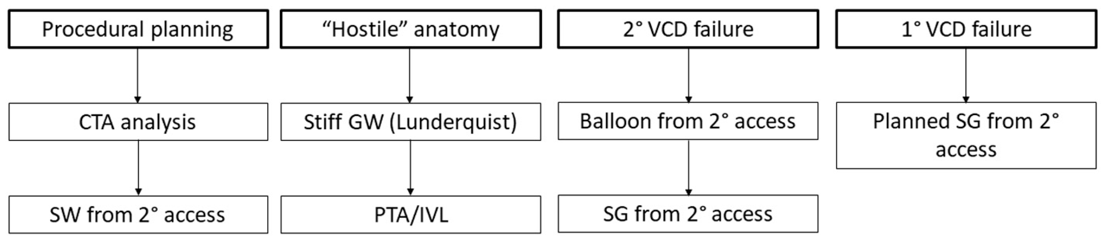
:1. Introduction
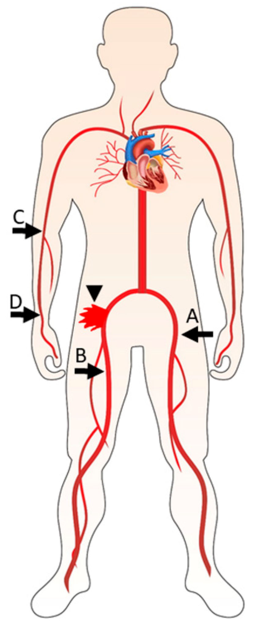
2. Vascular Complications Following TAVR
3. Vascular Closure Device Failure
4. Percutaneous Management of Vascular Complications
5. Vascular Repair by Stent Graft Implantation
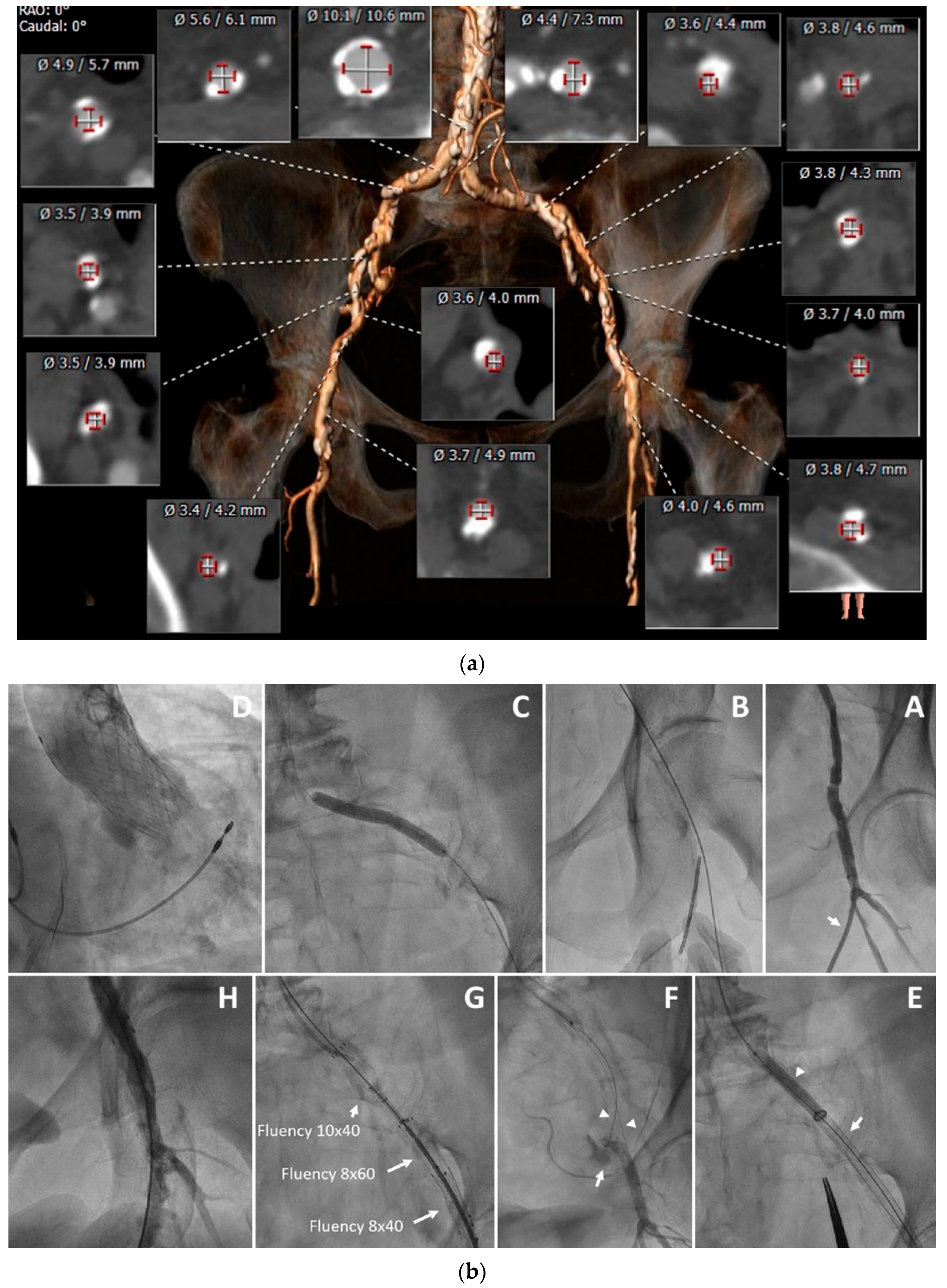
6. Technical Aspects of Stent Graft Implantation
7. Totally Percutaneous TAVR in Cases of Primary VCD Failure
8. Summary and Conclusions
Author Contributions
Funding
Institutional Review Board Statement
Informed Consent Statement
Conflicts of Interest
References
- Siontis, G.C.M.; Overtchouk, P.; Cahill, T.J.; Modine, T.; Prendergast, B.; Praz, F.; Pilgrim, T.; Petrinic, T.; Nikolakopoulou, A.; Salanti, G.; et al. Transcatheter aortic valve implantation vs. surgical aortic valve replacement for treatment of symptomatic severe aortic stenosis: An updated meta-analysis. Eur. Heart J. 2019, 40, 3143–3153. [Google Scholar] [CrossRef] [PubMed]
- Nguyen, V.; Willner, N.; Eltchaninoff, H.; Burwash, I.G.; Michel, M.; Durand, E.; Gilard, M.; Dindorf, C.; Iung, B.; Cribier, A.; et al. Trends in aortic valve replacement for aortic stenosis: A French nationwide study. Eur. Heart J. 2022, 43, 666–679. [Google Scholar] [CrossRef] [PubMed]
- Morello, A.; Corcione, N.; Ferraro, P.; Cimmino, M.; Pepe, M.; Cassese, M.; Frati, G.; Biondi-Zoccai, G.; Giordano, A. The best way to transcatheter aortic valve implantation: From standard to new approaches. Int. J. Cardiol. 2021, 322, 86–94. [Google Scholar] [CrossRef] [PubMed]
- Eugène, M.; Duchnowski, P.; Prendergast, B.; Wendler, O.; Laroche, C.; Monin, J.-L.; Jobic, Y.; Popescu, B.A.; Bax, J.J.; Vahanian, A.; et al. Contemporary Management of Severe Symptomatic Aortic Stenosis. J. Am. Coll. Cardiol. 2021, 78, 2131–2143. [Google Scholar] [CrossRef]
- Costa, G.; Bieliauskas, G.; Fukutomi, M.; Ihlemann, N.; Søndergaard, L.; De Backer, O. Feasibility and safety of a fully percutaneous transcatheter aortic valve replacement program. Catheter. Cardiovasc. Interv. 2021, 97, E418–E424. [Google Scholar] [CrossRef]
- Francone, M.; Budde, R.P.J.; Bremerich, J.; Dacher, J.N.; Loewe, C.; Wolf, F.; Natale, L.; Pontone, G.; Redheuil, A.; Vliegenthart, R.; et al. CT and MR imaging prior to transcatheter aortic valve implantation: Standardisation of scanning protocols, measurements and reporting-a consensus document by the European Society of Cardiovascular Radiology (ESCR). Eur. Radiol. 2020, 30, 2627–2650. [Google Scholar] [CrossRef] [Green Version]
- Kotronias, R.A.; Bray, J.J.; Rajasundaram, S.; Vincent, F.; Delhaye, C.; Scarsini, R.; Marin, F.; Terentes-Printzios, D.; Halcox, J.P.; Mamas, M.A.; et al. Ultrasound- Versus Fluoroscopy-Guided Strategy for Transfemoral Transcatheter Aortic Valve Replacement Access: A Systematic Review and Meta-Analysis. Circ. Cardiovasc. Interv. 2021, 14, e010742. [Google Scholar] [CrossRef]
- Witberg, G.; Tzalamouras, V.; Adams, H.; Patterson, T.; Roberts-Thomson, R.; Byrne, J.; Dworakowski, R.; MacCarthy, P.; Redwood, S.; Prendergast, B. Routine Ultrasound or Fluoroscopy Use and Risk of Vascular/Bleeding Complications After Transfemoral TAVR. JACC Cardiovasc. Interv. 2020, 13, 1460–1468. [Google Scholar] [CrossRef]
- Drafts, B.C.; Choi, C.H.; Sangal, K.; Cammarata, M.W.; Applegate, R.J.; Gandhi, S.K.; Kincaid, E.H.; Kon, N.; Zhao, D.X. Comparison of outcomes with surgical cut-down versus percutaneous transfemoral transcatheter aortic valve replacement: TAVR transfemoral access comparisons between surgical cut-down and percutaneous approach. Catheter. Cardiovasc. Interv. 2018, 91, 1354–1362. [Google Scholar] [CrossRef]
- Arnold, S.V.; Manandhar, P.; Vemulapalli, S.; Kosinski, A.; Desai, N.D.; Bavaria, J.E.; Cohen, D.J. Impact of Short-Term Complications of TAVR on Longer-Term Outcomes: Results from the STS/ACC Transcatheter Valve Therapy Registry. Eur. Heart J. Qual. Care Clin. Outcomes 2020, 7, 208–213. [Google Scholar] [CrossRef]
- Tamburino, C.; Capodanno, D.; Ramondo, A.; Petronio, A.S.; Ettori, F.; Santoro, G.; Klugmann, S.; Bedogni, F.; Maisano, F.; Marzocchi, A.; et al. Incidence and Predictors of Early and Late Mortality After Transcatheter Aortic Valve Implantation in 663 Patients with Severe Aortic Stenosis. Circulation 2011, 123, 299–308. [Google Scholar] [CrossRef] [PubMed] [Green Version]
- Thomas, M.; Schymik, G.; Walther, T.; Himbert, D.; Lefevre, T.; Treede, H.; Eggebrecht, H.; Rubino, P.; Michev, I.; Lange, R.; et al. Thirty-day results of the SAPIEN aortic Bioprosthesis European Outcome (SOURCE) Registry: A European registry of transcatheter aortic valve implantation using the Edwards SAPIEN valve. Circulation 2010, 122, 62–69. [Google Scholar] [CrossRef] [PubMed] [Green Version]
- Fanaroff, A.C.; Manandhar, P.; Holmes, D.R.; Cohen, D.J.; Harrison, J.K.; Hughes, G.C.; Thourani, V.H.; Mack, M.J.; Sherwood, M.W.; Jones, W.S.; et al. Peripheral Artery Disease and Transcatheter Aortic Valve Replacement Outcomes: A Report from the Society of Thoracic Surgeons/American College of Cardiology Transcatheter Therapy Registry. Circ. Cardiovasc. Interv. 2017, 10, e005456. [Google Scholar] [CrossRef] [PubMed]
- Darmoch, F.; Alraies, M.C.; Al-Khadra, Y.; Pacha, H.M.; Soud, M.; Kaki, A.; Rab, T.; Grines, C.L.; Bagur, R.; Kwok, C.S.; et al. Outcome of Transcatheter Aortic Valve Implantation in Patients with Peripheral Vascular Disease. Am. J. Cardiol. 2019, 124, 416–422. [Google Scholar] [CrossRef]
- Ueshima, D.; Barioli, A.; Fovino, L.N.; D’Amico, G.; Fabris, T.; Brener, S.J.; Tarantini, G. The impact of pre-existing peripheral artery disease on transcatheter aortic valve implantation outcomes: A systematic review and meta-analysis. Catheter. Cardiovasc. Interv. 2020, 95, 993–1000. [Google Scholar] [CrossRef]
- Scarsini, R.; De Maria, G.L.; Joseph, J.; Fan, L.; Cahill, T.J.; Kotronias, R.A.; Burzotta, F.; Newton, J.D.; Kharbanda, R.; Prendergast, B.; et al. Impact of Complications During Transfemoral Transcatheter Aortic Valve Replacement: How Can They Be Avoided and Managed? J. Am. Heart Assoc. 2019, 8, e013801. [Google Scholar] [CrossRef]
- Mach, M.; Okutucu, S.; Kerbel, T.; Arjomand, A.; Fatihoglu, S.G.; Werner, P.; Simon, P.; Andreas, M. Vascular Complications in TAVR: Incidence, Clinical Impact, and Management. J. Clin. Med. 2021, 10, 5046. [Google Scholar] [CrossRef]
- Sherwood, M.W.; Xiang, K.; Matsouaka, R.; Li, Z.; Vemulapalli, S.; Vora, A.N.; Fanaroff, A.; Harisson, J.K.; Thourani, V.H.; Holmes, D.; et al. Incidence, Temporal Trends, and Associated Outcomes of Vascular and Bleeding Complications in Patients Undergoing Transfemoral Transcatheter Aortic Valve Replacement: Insights from the Society of Thoracic Surgeons/American College of Cardiology Transcatheter Valve Therapies Registry. Circ. Cardiovasc. Interv. 2020, 13, e008227. [Google Scholar]
- Langouet, Q.; Martinez, R.; Saint-Etienne, C.; Soulami, R.B.; Harmouche, M.; Aupart, M. Incidence, predictors, impact, and treatment of vascular complications following transcatheter aortic valve implantation in a modern prospective cohort under real conditions. J. Vasc. Surg. 2020, 72, 2120–2129. [Google Scholar] [CrossRef]
- Hayashida, K.; Lefèvre, T.; Chevalier, B.; Hovasse, T.; Romano, M.; Garot, P.; Mylotte, D.; Uribe, J.; Farge, A.; Donzeau-Gouge, P.; et al. Transfemoral Aortic Valve Implantation: New Criteria to Predict Vascular Complications. JACC Cardiovasc. Interv. 2011, 4, 851–858. [Google Scholar] [CrossRef] [Green Version]
- Kadakia, M.B.; Herrmann, H.C.; Desai, N.D.; Fox, Z.; Ogbara, J.; Anwaruddin, S.; Jagasia, D.; Bavaria, J.E.; Szeto, W.Y.; Vallabhajosyula, P.; et al. Factors Associated with Vascular Complications in Patients Undergoing Balloon-Expandable Transfemoral Transcatheter Aortic Valve Replacement via Open Versus Percutaneous Approaches. Circ. Cardiovasc. Interv. 2014, 7, 570–576. [Google Scholar] [CrossRef] [PubMed] [Green Version]
- Batchelor, W.; Patel, K.; Hurt, J.; Totten, J.; Burroughs, P.; Smith, G.; Cuervo, M.; Davis, L.; Damluji, A.A.; Epps, K.; et al. Incidence, Prognosis and Predictors of Major Vascular Complications and Percutaneous Closure Device Failure Following Contemporary Percutaneous Transfemoral Transcatheter Aortic Valve Replacement. Cardiovasc. Revasc. Med. 2020, 21, 1065–1073. [Google Scholar] [CrossRef] [PubMed]
- Van Mieghem, N.M.; Tchetche, D.; Chieffo, A.; Dumonteil, N.; Messika-Zeitoun, D.; Van Der Boon, R.M.; Vahdat, O.; Buchanan, G.L.; Marcheix, B.; Himbert, D.; et al. Incidence, predictors, and implications of access site complications with transfemoral transcatheter aortic valve implantation. Am. J. Cardiol. 2012, 110, 1361–1367. [Google Scholar] [CrossRef] [PubMed]
- Généreux, P.; Head, S.J.; Van Mieghem, N.M.; Kodali, S.; Kirtane, A.J.; Xu, K.; Smith, C.; Serruys, P.W.; Kappetein, A.P.; Leon, M.B. Clinical Outcomes After Transcatheter Aortic Valve Replacement Using Valve Academic Research Consortium Definitions: A Weighted Meta-Analysis of 3519 Patients From 16 Studies. J. Am. Coll. Cardiol. 2012, 59, 2317–2326. [Google Scholar] [CrossRef] [Green Version]
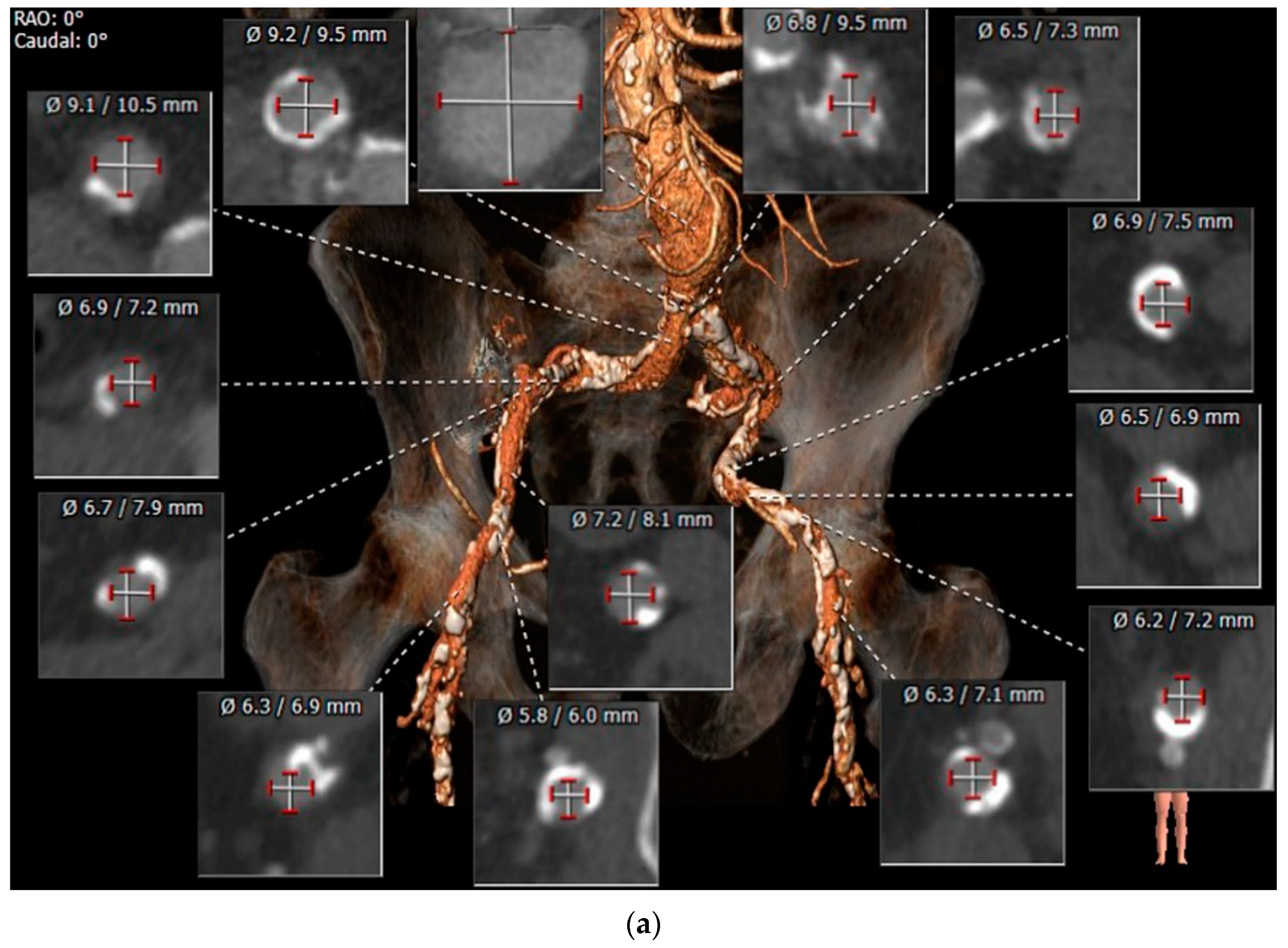
- Urbach, J.; Hou, C.R.; Lesser, J.R.; Stanberry, L.I.; Garberich, R.F.; Caye, D.; Sorajja, P.; Gössl, M. Computed Tomographic Angiography-Derived Risk Factors for Vascular Complications in Percutaneous Transfemoral Transcatheter Aortic Valve Implantation. Am. J. Cardiol. 2019, 124, 98–104. [Google Scholar] [CrossRef]
- Mach, M.; Poschner, T.; Hasan, W.; Szalkiewicz, P.; Andreas, M.; Winkler, B.; Grabenwöger, M. The Iliofemoral tortuosity score predicts access and bleeding complications during transfemoral transcatheter aortic valve replacement: DataData from the VIenna Cardio Thoracic aOrtic valve registrY (VICTORY). Eur. J. Clin. Investig. 2021, 51, e13491. [Google Scholar] [CrossRef]
- Kaluski, E.; Khan, S.U.; Singh, M.; Reitknecht, F.; Sattur, S.; Rogers, G.; Sporn, D. Iliofemoral peripheral orbital atherectomy for optimizing TAVR access: An innovative strategy in the absence of alternative access options. Cardiovasc. Revasc. Med. 2018, 19, 71–76. [Google Scholar] [CrossRef]
- Nardi, G.; De Backer, O.; Saia, F.; Søndergaard, L.; Ristalli, F.; Meucci, F.; Mattesini, A.; Demola, P.; Wang, X.; Al Jabri, A.; et al. Peripheral intravascular lithotripsy of iliofemoral arteries to facilitate transfemoral TAVI: A multicentre prospective registry. EuroIntervention 2022, 17, e1397–e1406. [Google Scholar] [CrossRef]
- Di Mario, C.; Goodwin, M.; Ristalli, F.; Ravani, M.; Meucci, F.; Stolcova, M.; Sardella, G.; Salvi, N.; Bedogni, F.; Berti, S.; et al. A Prospective Registry of Intravascular Lithotripsy-Enabled Vascular Access for Transfemoral Transcatheter Aortic Valve Replacement. JACC Cardiovasc. Interv. 2019, 12, 502–504. [Google Scholar] [CrossRef]
- Schneider, D.B.; Krajcer, Z.; Bonafede, M.; Thoma, E.; Hasegawa, J.; Bhounsule, P.; Thiel, E. Clinical and economic outcomes of ProGlide compared with surgical repair of large bore arterial access. J. Comp. Eff. Res. 2019, 8, 1381–1392. [Google Scholar] [CrossRef]
- Vierhout, B.P.; Pol, R.A.; El Moumni, M.; Zeebregts, C.J. Choice—Arteriotomy Closure Devices in EVAR, TEVAR, and TAVR: A Systematic Review and Meta-analysis of Randomised Clinical Trials and Cohort Studies. Eur. J. Vasc. Endovasc. Surg. Off. J. Eur. Soc. Vasc. Surg. 2017, 54, 104–115. [Google Scholar] [CrossRef] [PubMed] [Green Version]
- Babaliaros, V.; Devireddy, C.; Lerakis, S.; Leonardi, R.; Iturra, S.A.; Mavromatis, K.; Leshnower, B.G.; Guyton, R.A.; Kanitkar, M.; Keegan, P.; et al. Comparison of transfemoral transcatheter aortic valve replacement performed in the catheterization laboratory (minimalist approach) versus hybrid operating room (standard approach): Outcomes and cost analysis. JACC Cardiovasc. Interv. 2014, 7, 898–904. [Google Scholar] [CrossRef] [PubMed] [Green Version]
- Nakamura, M.; Chakravarty, T.; Jilaihawi, H.; Doctor, N.; Dohad, S.; Fontana, G.; Cheng, W.; Makkar, R.R. Complete percutaneous approach for arterial access in transfemoral transcatheter aortic valve replacement: A comparison with surgical cut-down and closure. Catheter. Cardiovasc. Interv. 2014, 84, 293–300. [Google Scholar] [CrossRef] [PubMed]
- Rymer, J.A.; Xiang, Q.; Wang, A.; Cohen, D.J.; Desai, N.D.; Kirtane, A.J.; Hughes, G.C.; Harrison, J.K.; Kosinski, A.S.; Vemulapalli, S. Factors Associated with and Outcomes of Aborted Procedures During Elective Transcatheter Aortic Valve Replacement. JACC Cardiovasc. Interv. 2019, 12, 1768–1777. [Google Scholar] [CrossRef]
- Van Kesteren, F.; van Mourik, M.S.; Vendrik, J.; Wiegerinck, E.M.; Henriques, J.P.; Koch, K.T.; de Winter, R.J.; Piek, J.J.; van Lienden, K.P.; Reekers, J.A.; et al. Incidence, Predictors, and Impact of Vascular Complications After Transfemoral Transcatheter Aortic Valve Implantation with the SAPIEN 3 Prosthesis. Am. J. Cardiol. 2018, 121, 1231–1238. [Google Scholar] [CrossRef]
- Chen, I.M.; Lee, T.H.; Chen, P.L.; Shih, C.C.; Chang, H.H. Factors in ProGlide(R) Vascular Closure Failure in Sheath Arteriotomies Greater than 16 French. Eur. J. Vasc. Endovasc. Surg. Off. J. Eur. Soc. Vasc. Surg. 2019, 58, 615–622. [Google Scholar] [CrossRef]
- Lee, C.H.; Ko, Y.-G.; Park, Y.; Shim, C.Y.; Hong, G.-R.; Lee, S.H.; Lee, S.; Jung, H.W.; Hong, S.-J.; Ahn, C.-M.; et al. Risk Factors for Closure Failure following Percutaneous Transfemoral Transcatheter Aortic Valve Implantation. Ann. Vasc. Surg. 2020, 66, 406–414. [Google Scholar] [CrossRef]
- Ruge, H.; Burri, M.; Erlebach, M.; Lange, R. Access site related vascular complications with third generation transcatheter heart valve systems. Catheter. Cardiovasc. Interv. 2020, 97, 325–332. [Google Scholar] [CrossRef]
- Al-Kassou, B.; Kandt, J.; Lohde, L.; Shamekhi, J.; Sedaghat, A.; Tabata, N.; Weber, M.; Sugiura, A.; Fimmers, R.; Werner, N.; et al. Safety and Efficacy of Protamine Administration for Prevention of Bleeding Complications in Patients Undergoing TAVR. JACC Cardiovasc. Interv. 2020, 13, 1471–1480. [Google Scholar] [CrossRef]
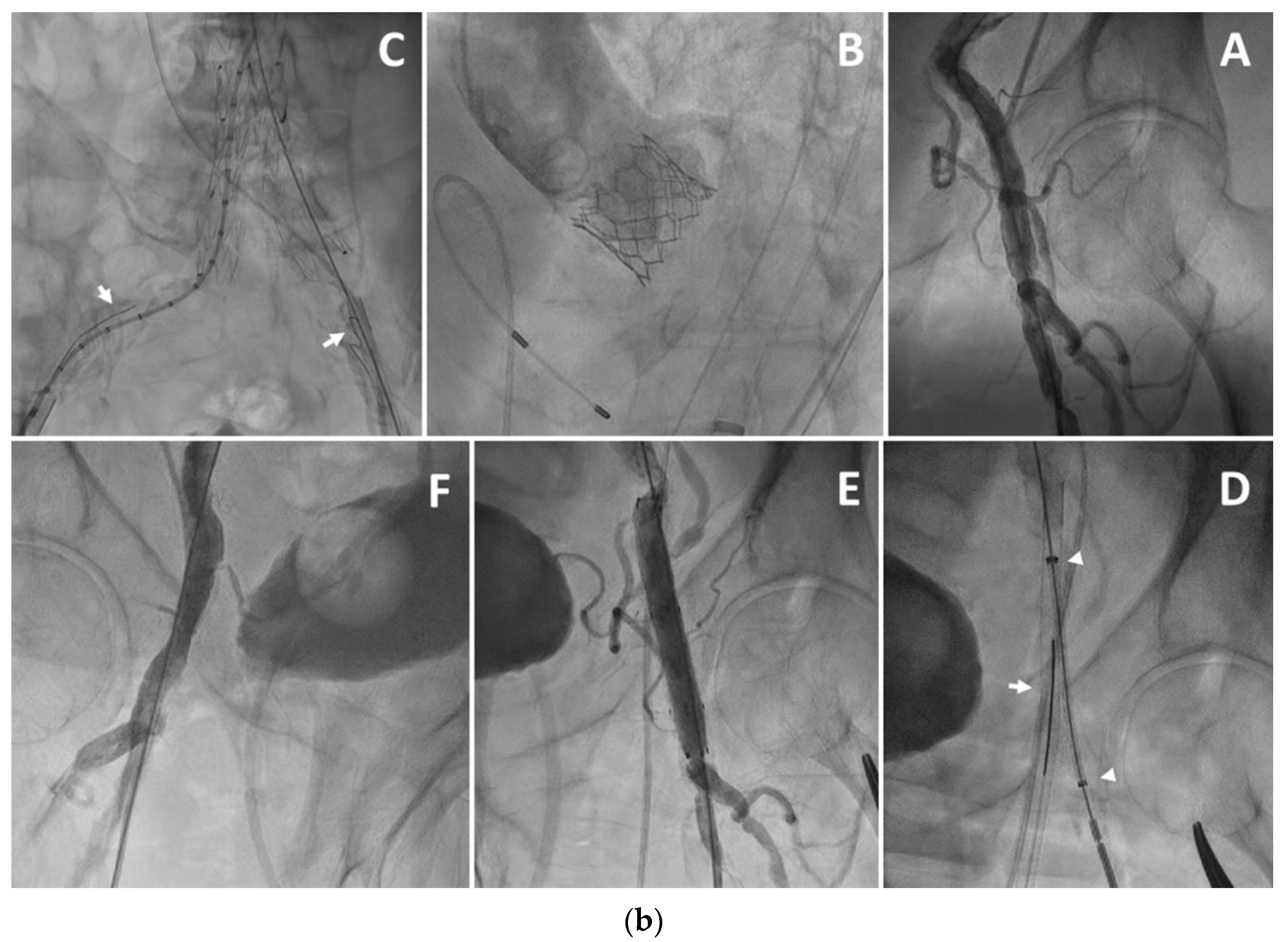
- Sliman, H.; Shiran, A.; Mannheim, D.; Avraham, E.; Karmeli, R.; Khader, N.; Zafrir, B.; Rubinshtein, R.; Jaffe, R. Retrograde Femoral Artery Stent-Graft Implantation for Treatment of Access-site Bleeding Following Transcatheter Aortic Valve Implantation. Isr. Med. Assoc. J. IMAJ 2019, 5, 322–325. [Google Scholar]
- Sanghvi, K.; Swarup, S.; Burns, P.; Kovach, R.; Ross, R.; Soussa, T. Prophylactic Retrograde Distal Common Femoral Access as a Bail-out Strategy in Patients with Increased Risk for Femoral Access Complication During Transfemoral Aortic Valve Replacement. Cardiovasc. Revasc. Med. 2019, 21, 481–485. [Google Scholar] [CrossRef] [PubMed]
- Khubber, S.; Bazarbashi, N.; Mohananey, D.; Kadri, A.; Gad, M.M.; Kaur, M.; Sammour, Y.M.; Lyden, M.; Ahuja, K.R.; Verma, B.; et al. Unilateral Access Is Safe and Facilitates Peripheral Bailout During Transfemoral-Approach Transcatheter Aortic Valve Replacement. JACC Cardiovasc. Interv. 2019, 12, 2210–2220. [Google Scholar] [CrossRef] [PubMed]
- Junquera, L.; Urena, M.; Latib, A.; Muñoz-Garcia, A.; Nombela-Franco, L.; Faurie, B.; Rodés-Cabau, J. Comparison of Transfemoral Versus Transradial Secondary Access in Transcatheter Aortic Valve Replacement. Circ. Cardiovasc. Interv. 2020, 13, e008609. [Google Scholar] [CrossRef] [PubMed]
- Lefèvre, G.; Jégou, A.; Dambrin, G.; Picard, F.; Anconina, J.; Pouzet, B.; Guesnier, L.; Khelifa, R.C.; Hilpert, L.; Doan, H.L.; et al. Comparison of TransFemoral Transcatheter Aortic Valve Replacement Performed with a Minimally Invasive Simplified Technique: “FAST” Versus a Standard Approach. J. Invasive Cardiol. 2019, 31, 300–306. [Google Scholar]
- Sliman, H.; Eitan, A.; Shiran, A.; Zafrir, B.; Jaffe, R. Transbrachial Secondary Vascular Access in Transcatheter Aortic Valve Replacement Procedures: A Single-Center Retrospective Analysis. Heart Lung Circ. 2022; in press. [Google Scholar] [CrossRef]
- Siracuse, J.J.; Gill, H.L.; Schneider, D.B.; Graham, A.R.; Connolly, P.H.; Jones, D.; Meltzer, A.J. Assessing the Perioperative Safety of Common Femoral Endarterectomy in the Endovascular Era. Vasc. Endovasc. Surg. 2014, 48, 27–33. [Google Scholar] [CrossRef]
- Mehta, M.; Zhou, Y.; Paty, P.S.; Teymouri, M.; Jafree, K.; Bakhtawar, H.; Hnath, J.; Feustel, P. Percutaneous common femoral artery interventions using angioplasty, atherectomy, and stenting. J. Vasc. Surg. 2016, 64, 369–379. [Google Scholar] [CrossRef]
- Stricker, H.; Spinedi, L.; Limoni, C.; Giovannacci, L. Stent-Assisted Angioplasty (SAA) at the Level of the Common Femoral Artery Bifurcation: Long-Term Outcomes. Cardiovasc. Interv. Radiol. 2020, 43, 541–546. [Google Scholar] [CrossRef] [Green Version]
- Changal, K.H.; Syed, M.; Dar, T.; Mangi, M.A.; Sheikh, M.A. Systematic Review and Proportional Meta-Analysis of Endarterectomy and Endovascular Therapy with Routine or Selective Stenting for Common Femoral Artery Atherosclerotic Disease. J. Interv. Cardiol. 2019, 2019, 1593401. [Google Scholar] [CrossRef]
- Wong, G.; Lahsaei, S.; Aoun, J.; Garcia, L.A. Management of common femoral artery occlusive disease: A review of endovascular treatment strategies and outcomes. Catheter. Cardiovasc. Interv. Off. J. Soc. Cardiac Angiogr. Interv. 2019, 93, 514–521. [Google Scholar] [CrossRef] [Green Version]
- Feldman, D.N.; Armstrong, E.J.; Aronow, H.D.; Gigliotti, O.S.; Jaff, M.R.; Klein, A.J.; Parikh, S.A.; Prasad, A.; Rosenfield, K.; Shishehbor, M.H.; et al. SCAI consensus guidelines for device selection in femoral-popliteal arterial interventions. Catheter. Cardiovasc. Interv. 2018, 92, 124–140. [Google Scholar] [CrossRef] [PubMed] [Green Version]
- Baltacioglu, F.; Cimsit, N.C.; Cil, B.; Cekirge, S.; Ispir, S. Endovascular stent-graft applications in latrogenic vascular injuries. Cardiovasc. Interv. Radiol. 2003, 26, 434–439. [Google Scholar] [CrossRef] [PubMed]
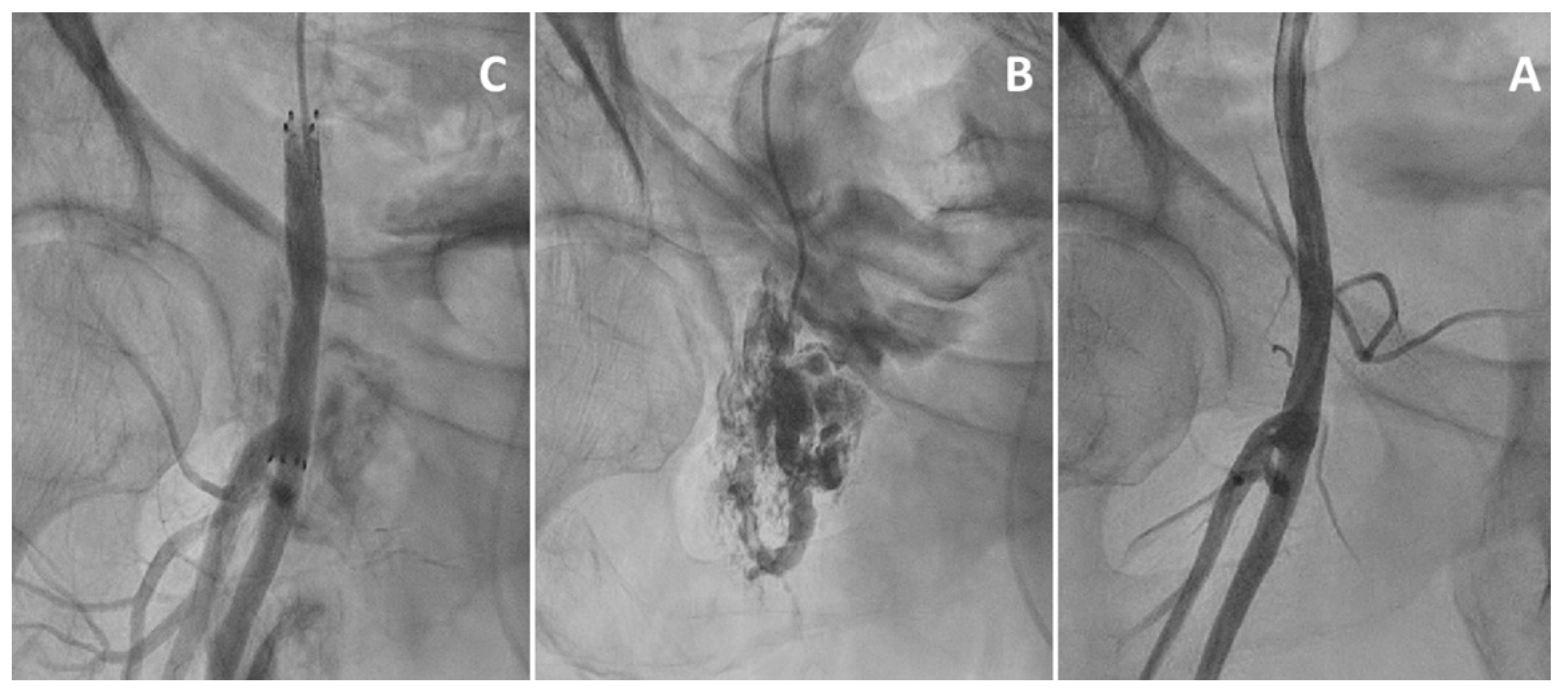
- Segal, A.; Flugelman, M.Y.; Khader, N.; Rubinshtein, R.; Lavi, I.; Karmeli, R.; Jubran, A.; Shiran, A.; Jaffe, R. Outcome of Stent Graft Implantation for Treatment of Access Site Bleeding After Transfemoral Transcatheter Aortic Valve Replacement. Am. J. Cardiol. 2017, 120, 456–460. [Google Scholar] [CrossRef] [PubMed]
- Sedaghat, A.; Neumann, N.; Schahab, N.; Sinning, J.-M.; Hammerstingl, C.; Pingel, S.; Schaefer, C.; Mellert, F.; Schiller, W.; Welz, A.; et al. Routine Endovascular Treatment with a Stent Graft for Access-Site and Access-Related Vascular Injury in Transfemoral Transcatheter Aortic Valve Implantation. Circ. Cardiovasc. Interv. 2016, 9, e003834. [Google Scholar] [CrossRef] [Green Version]
- Sedaghat, A.; Hansen, K.L.; Schahab, N.; May, M.C.; Weber, M.; Stundl, A.; Shamekhi, J.; Schaefer, C.; Nickenig, G.; Sinning, J.-M.; et al. Long-term follow-up after stent graft placement for access-site and access-related vascular injury during TAVI—The Bonn-Copenhagen experience. Int. J. Cardiol. 2019, 281, 42–46. [Google Scholar] [CrossRef]
- Steinvil, A.; Bernardo, N.; Rogers, T.; Koifman, E.; Buchanan, K.; Alraies, M.C.; Shults, C.; Torguson, R.; Okubagzi, P.G.; Pichard, A.D.; et al. Use of an ePTFE-covered nitinol self-expanding stent graft for the treatment off pre-closure device failure during transcatheter aortic valve replacement. Cardiovas. Revasc. Med. Incl. Mol. Interv. 2017, 18, 128–132. [Google Scholar] [CrossRef]
- Dencker, D.; Taudorf, M.; Luk, N.V.; Nielsen, M.B.; Kofoed, K.; Schroeder, T.V.; Søndergaard, L.; (Lönn), L.L.; De Backer, O. Frequency and Effect of Access-Related Vascular Injury and Subsequent Vascular Intervention After Transcatheter Aortic Valve Replacement. Am. J. Cardiol. 2016, 118, 1244–1250. [Google Scholar] [CrossRef]
- De Backer, O.; Arnous, S.; Sandholt, B.; Brooks, M.; Biasco, L.; Franzen, O.; Søndergaard, L. Safety and efficacy of using the Viabahn endoprosthesis for percutaneous treatment of vascular access complications after transfemoral aortic valve implantation. Am. J. Cardiol. 2015, 115, 1123–1129. [Google Scholar] [CrossRef]
- Shammas, G.A.; Harris, T.; Voelliger, C.M.; Shammas, A.N.; Jerin, M.; Shammas, N.W. Safety and Effectiveness of Closure Devices Applied to a Stented Common Femoral Artery: A Retrospective Analysis. Int. J. Angiol. 2016, 25, 165–167. [Google Scholar] [CrossRef] [Green Version]
- Eitan, A.; Shiran, A.; Sliman, H.; Zafrir, B.; Jaffe, R. Totally Percutaneous Transfemoral Transcatheter Aortic Valve Replacement Despite Failure to Deploy a Vascular Closure Device: A Single-Centre Case Series. Heart Lung Circ. 2022, 31, 390–394. [Google Scholar] [CrossRef]


| Valve | Valve Size | Sheath ID | Sheath OD | Vessel MLD | Manufacturer |
|---|---|---|---|---|---|
| Evolut R | 23/26/29 mm | 14F/4.7 mm | 6.0 mm | 5.0 mm | Medtronic |
| Evolut R | 34 mm | 16F/5.3 mm | 6.7 mm | 5.5 mm | Medtronic |
| Evolut Pro | 23/26/29 mm | 16F/5.3 mm | 6.7 mm | 5.5 mm | Medtronic |
| Sapien S3 | 23/26 mm | 14F/4.7 mm | 6.0 mm | 5.5 mm | Edwards |
| Sapien S3 | 29 mm | 16F/5.3 mm | 6.7 mm | 6.0 mm | Edwards |
Publisher’s Note: MDPI stays neutral with regard to jurisdictional claims in published maps and institutional affiliations. |
© 2022 by the authors. Licensee MDPI, Basel, Switzerland. This article is an open access article distributed under the terms and conditions of the Creative Commons Attribution (CC BY) license (https://creativecommons.org/licenses/by/4.0/).
Share and Cite
Eitan, A.; Sliman, H.; Shiran, A.; Jaffe, R. Strategies for Facilitating Totally Percutaneous Transfemoral TAVR Procedures. J. Clin. Med. 2022, 11, 2104. https://doi.org/10.3390/jcm11082104
Eitan A, Sliman H, Shiran A, Jaffe R. Strategies for Facilitating Totally Percutaneous Transfemoral TAVR Procedures. Journal of Clinical Medicine. 2022; 11(8):2104. https://doi.org/10.3390/jcm11082104
Chicago/Turabian StyleEitan, Amnon, Hussein Sliman, Avinoam Shiran, and Ronen Jaffe. 2022. "Strategies for Facilitating Totally Percutaneous Transfemoral TAVR Procedures" Journal of Clinical Medicine 11, no. 8: 2104. https://doi.org/10.3390/jcm11082104
APA StyleEitan, A., Sliman, H., Shiran, A., & Jaffe, R. (2022). Strategies for Facilitating Totally Percutaneous Transfemoral TAVR Procedures. Journal of Clinical Medicine, 11(8), 2104. https://doi.org/10.3390/jcm11082104