Article

WT1 Gene Mutations, rs16754 Variant, and WT1 Overexpression as Prognostic Factors in Acute Myeloid Leukemia Patients

by
Dorota Koczkodaj
1,
Szymon Zmorzyński
1,*,
Beata Grygalewicz
2,
Barbara Pieńkowska-Grela
2,
Wojciech Styk
3,
Sylwia Popek-Marciniec
1 and
Agata Anna Filip
1
1
Department of Cancer Genetics with Cytogenetic Laboratory, Medical University of Lublin, 20-059 Lublin, Poland
2
The Maria Sklodowska-Curie National Research Institute of Oncology, 02-781 Warszawa, Poland
3
Department of Psychology, Institute of Pedagogy and Psychology, Warsaw Management University, 03-772 Warszawa, Poland
*
Author to whom correspondence should be addressed.
J. Clin. Med. 2022, 11(7), 1873; https://doi.org/10.3390/jcm11071873
Submission received: 19 February 2022 / Revised: 17 March 2022 / Accepted: 25 March 2022 / Published: 28 March 2022
(This article belongs to the Section Hematology)

Abstract

:
(1) Background: The aim of our study was the complex assessment of WT1 variants and their expression in relation to chromosomal changes and molecular prognostic markers in acute myeloid leukemia (AML). It is the first multidimensional study in Polish AML patients; (2) Methods: Bone marrow aspirates of 90 AML patients were used for cell cultures (banding techniques and fluorescence in situ hybridization), and to isolate DNA (WT1 genotyping, array comparative genomic hybridization), and RNA (WT1 expression). Peripheral blood samples from 100 healthy blood donors were used to analyze WT1 rs16754; (3) Results: Allele frequency and distribution of WT1 variant rs16754 (A;G) did not differ significantly among AML patients and controls. Higher expression of WT1 gene was observed in AA genotype (of rs16754) in comparison with GA or GG genotypes—10,556.7 vs. 25,836.5 copies (p = 0.01), respectively. WT1 mutations were more frequent in AML patients under 65 years of age (p < 0.0001) and affected relapse-free survival (RFS). The presence of NPM1 or CEBPA mutations decreased the risk of WT1 mutation presence, odds ratio (OR) = 0.11, 95% CI 0.02–0.46, p = 0.002 or OR = 0.05, 95% CI 0.006–0.46, p = 0.002, respectively. We observed significantly higher WT1 expression in AML CD34+ vs. CD34−, −20,985 vs. 8304 (p = 0.039), respectively. The difference in WT1 expression between patients with normal and abnormal karyotype was statistically insignificant; (4) Conclusions: WT1 gene expression and its rs16754 variant at diagnosis did not affect AML outcome. WT1 mutation may affect RFS in AML.

1. Introduction

Acute myeloid leukemia (AML) affects the function of the hematopoietic system and is a genetically heterogeneous disease at cytogenetic and molecular levels [1]. AML development includes multistep events associated with point mutations, as well as chromosomal aberrations [2]. Mutations in genes encoding Wilms tumor 1 (WT1), nucleophosmin (NPM1), FMS-like tyrosine kinase-3 (FLT3), and CCAAT/enhancer-binding protein alpha (CEBPA) affect the pathogenesis of AML [3].
The WT1 gene (locus 11p13) encodes a transcription factor that regulates cell growth and differentiation [4]. It contains 10 exons, of which 4 (7–10) encode a DNA-binding domain [5]. The WT1 gene may possess both tumor suppressor and oncogenic functions in childhood Wilms tumors and leukemias, respectively [6]. WT1 gene mutations are found in 6–15% of newly diagnosed AML patients [3]. Hot spots were described in two exons (7 and 9), in which loss of function mutations can occur [7]. The alterations of the WT1 gene are associated with younger age, as well as the coexistence of FLT3 and CEBPA mutations [7]. The changes in the WT1 gene sequence may affect its expression [3]. The role of WT1 mutations in the pathogenesis of AML remains controversial [3].
Among many variants present in the WT1 gene, special attention has been paid to the single nucleotide polymorphism rs16754 located in exon 7 (at nucleotide 1297A>G) [8]. Some studies have suggested that rs16754 can be a negative prognostic factor in AML [3,9]. The prognosis in AML is affected adversely by WT1 gene overexpression [10,11]. The WT1 protein shows antiapoptotic activity. Moreover, higher WT1 expression is associated with lower cell differentiation [12]. However, the exact role of the WT1 gene in AML pathogenesis has not been completely revealed. The highest levels of WT1 expression were found in the M3 AML subgroup (according to French-American-British, FAB, classification) and were linked to a failure of achieving complete remission (CR), shorter overall survival (OS), and shorter progression-free survival [13,14]. It is possible that some changes in coding or noncoding regions of the WT1 gene or some chromosomal aberrations, especially in the form of microdeletions or microduplications, may affect WT1 gene expression. WT1 gene variants and their expression may affect AML prognosis and outcome [9]. However, the results remain controversial.
Abnormal divisions of leukemic cells are often the consequence of gene mutations whose products affect the signaling pathways associated with cell proliferation. The prognosis for AML patients is affected not only by WT1 gene mutation or abnormal expression but also by mutations of the FLT3, NPM1, and CEBPA genes which were also evaluated [15,16].
Most chromosomal aberrations implicate the appearance of certain gene rearrangements, which affect not only the pathogenesis of the disease but also the prognosis and response to the treatment. Clonal chromosomal abnormalities are identified in approximately 55% of adult AML patients and are recognized as independent prognostic factors [17]. Approximately 15% of patients with clonal changes have complex karyotype, i.e., three or more chromosomal alterations [18]. Patients with t(8;21), inv(16)/t(16;16), or t(15;17) have a favorable prognosis. The prognosis is poor for AML patients with complex karyotype or with the aberrations including inv(3)/t(3;3), t(6;9), monosomy 5 or del(5q), and monosomy 7 or del(7q) [19]. The cases of cytogenetically normal AML (CN-AML) are quite heterogeneous and, depending on cryptic molecular changes, are currently classified in the group of intermediate prognoses [20].
The aim of our study was the complex assessment of the WT1 rs16754 variant and WT1 mutations and expression in relation to chromosomal changes and molecular markers such as mutations in FLT3, NPM1, and CEBPA genes. Taking into account some of the controversies among current reports and conflicting results, we decided to analyze WT1 variants in the form of mutations and single nucleotide polymorphism, as well as WT1 gene expression in AML. Such multidimensional analysis has not yet been carried out among AML patients in the Polish population.

2. Materials and Methods

The study group consisted of 90 newly-diagnosed AML patients (42 women and 48 men) aged between 18 and 85 years, hospitalized at the Department of Hematooncology and Bone Marrow Transplantation, at the Medical University of Lublin between the years of 2008–2020 (Table 1). Control samples were of peripheral blood taken from 100 healthy blood donors at the Regional Blood Center of Kielce, Poland.
The preliminary diagnosis of AML was based on standard FAB (French-American-British) criteria [11]. On the basis of genetic changes detected at diagnosis, patients were stratified into one of four risk categories: favorable, intermediate-I, intermediate-II, and adverse, according to European Leukemia Net criteria [20]. The mean time of follow-up was 27 months.
With informed consent in accordance with the Declaration of Helsinki of 1975, revised in 2008, and after approval from the Medical University Ethics Committee (app. no.: KE-254/24/2011 and KE-0254/229/2013), the bone marrow aspirates were collected to heparin and EDTA collection tubes (Sarstedt, Nümbrecht, Germany), to be used for cell culture and for DNA, RNA isolation, respectively.
The treatment of patients generally depended on their age, performance status, and medical history. Patients aged 60 years or lower received induction regimen DAC (daunorubicin 60 mg/m2 on days 1 through 3, continuous infusion of cytarabine 200 mg/m2 on days 1 through 7, and cladribine 5 mg/m2 on days 1 through 5). Consolidation treatment consisted of one course of cytarabine 1.5 g/m2 on days 1 through 3 with mitoxantrone 10 mg/m2 on days 3 through 5 and one course of high-dose cytarabine 2 g/m2 twice daily on days 1, 3, and 5. Then those with favorable karyotype or not eligible for hematopoietic stem cell transplantation (HSCT) were treated for about two years with maintenance therapy based on cytarabine combined with daunorubicin or thioguanine; patients with intermediate or adverse karyotype were referred for allogeneic HSCT (allo-HSCT) or auto-HSCT in cases not eligible for allo-HSCT or lacking suitable donor. Patients aged over 60 years of age in good general condition and without significant comorbidities were treated with reduced induction regimen DA (daunorubicin 45 mg/m2 days 1 through 2 or 3 and continuous infusion of cytarabine 100 mg/m2 on days 1 through 5 or 7); consolidation and maintenance chemotherapy in this group was reduced as compared with younger patients. Finally, patients over 75 years or younger with significant comorbidities or in poor performance status received low-dose cytarabine (20 mg twice daily for 10 days) repeated every 4 weeks until disease progression. Another treatment option in this group of patients is an azacytidine-a hypomethylating agent used at a dose of 75 mg/m2 once a day for 7 days during a 28-day cycle. This treatment is continued until unacceptable toxicity or disease progression. Refractory and relapsed patients were treated with different regimens such as ICE (idarubicin, cytarabine, etoposide), CLAG (cladribine, cytarabine, G-CSF), or MEC (mitoxantrone, etoposide, cytarabine). Patients with promyelocytic leukemia received therapy based on idarubicin and ATRA (all-trans-retinoic acid).

2.1. Classical Cytogenetics and Fluorescence In Situ Hybridization (FISH)

Unstimulated cultures of both leukemic (from the first portion of the bone marrow aspirates) and normal (peripheral blood of healthy donors) cells were performed in 15 mL of growth medium (RPMI 1640 medium supplemented with 15% FCS, 100 U/mL penicillin, and 50 μg/mL streptomycin 50 μg/mL, Biomed). The cells were cultured for 24–72 h, and then cultures were terminated conventionally. Metaphase spreads were stained using banding techniques (GTG and RHG bands). Karyotypes were described according to the International System for Human Cytogenetic Nomenclature (ISCN) 2020 [4].
FISH was initially performed on uncultured cells from the diagnostic bone marrow specimens using following probes: 13q14.3, D13S319 and D13S25, 5q (5p15.31 and EGR1), 7q (7q22.1 and 7q31), 20q (20q12 and 20q13.2), EVI1 (3q26) breakapart, MLL (11q23), IGH/CCND1 (14q32.3/11q13), EGFR (7p11.2), EWSR1/ERG (21q22; 22q12), MDM2 (12q14.3-q15), IGH/BCL2 (14q32.3; 18q21) (all from Cytocell), dual-fusion probe the PML (15q22) and RARA (17q21.1) loci, 21q (D21S259, D21S341, D21S342, 21q22.13–q22.2), 17q23/17p12, TP53/CEP17, D5S23/D5S721 (5p15.2), EGR1 (5q31), CEP7/D7S486 (7q31), D8Z2 (CEP8), MYC (8q24.12–24.13), CDKN2A (9p21), MYB (6q23), CEP9 (9p11–q11), ETV6 (12p13), CEP15 (D15Z4), CEP17 (D17Z1), D20S108 (20q12), D13S319 (13q14.3)/13q34, RUNX1 (21q22), dual-fusion probe the BCR (22q11.2) and ABL (9q34) loci, AML1/ETO (21q22/8q22) (all from VYSIS, Abbott Laboratories), 1q21/SRD (1p36), hTERT 5p15/5q31, DEK/NUP214 (6p22; 9q34), FGFR1(8p12), MAF/IGH (16q23; 14q32), BCL1/IGH (11q13; 14q32), 22q11/22q13 (all from KREATECH Diagnostics). FISH was performed according to the manufacturer’s instructions. A total of 200 interphase cells per each sample were examined. The cutoff level for each individual probe was determined based on negative sample analysis and calculated as the mean ± 3SD.

2.2. DNA Isolation

DNA isolation from bone marrow aspirates (AML samples) and peripheral blood (control samples) was performed using a commercial kit (Qiagen, Hilden, Germany), according to the procedure provided by the manufacturer. The concentration and quality of isolated DNA were checked spectrophotometrically with NanoDrop 2000 (Thermo Scientific, Waltham, MA, USA). DNA was stored at −20 °C. DNA from bone marrow aspirates was used to analyze chromosomal aberrations (with array CGH method) and WT1 genotyping (with DNA sequencing method). DNA isolated from peripheral blood was used to analyze the WT1 rs16754 variant.

2.3. Array Comparative Genomic Hybridization (aCGH)

Each step of the aCGH method was performed according to the manufacturer’s (PerkinElmer, Waltham, MA, USA) instructions. The microarrays (Constitutional Chip 4.0) contained human DNA clones (about 100–300 kb long) derived from artificial bacterial chromosomes (BAC probes), which covered the entire human genome. The average resolution of the probes was less than 650 kb.
The process of array hybridization was carried out in HS 400 Pro hybridization station (Tecan, Mennedorf, Switzerland). Array scanning was performed in a ScanArrayGx scanner (PerkinElmer). The OneClick software (PerkinElmer) was used to analyze the results obtained with the aCGH technique. The results were described according to ISCN 2020 [4].

2.4. WT1 Genotyping—Analysis of WT1 Mutations and rs16754 Variant

The determination of all possible mutations in exons 7 and 9 and rs16754 variant (in exon 7) of the WT1 gene was performed using direct sequencing. Primer sequences and the procedure of sequencing were performed according to Summers et al., 2007 [21]. A total of 25 μL reaction mixture consisted of 100 ng genomic DNA, 10 µM of each primer, 0.25 mM dNTPs mixture, and 0.31 U of HD polymerase (Clontech) with 1 × PCR reaction buffer (Clontech). PCR reactions with modified thermal conditions were the same for exon 7 and 9 analysis: initial denaturation (98 °C-3 min), 35 cycles (98 °C-20 s, 60 °C-10 s, 72 °C-15 s), final elongation (72 °C-5 min). The PCR reactions were performed in Applied Biosystems 9700 Thermal Cycler. Sequencing PCR was performed using BigDye Terminator v3.1 Cycle Sequencing Kit (Applied Biosystems, Waltham, MA, USA) in a thermal cycler (as previously). The sequencing PCR product was purified using an exterminator kit (A&A Biotechnology, Gdynia, Poland). The sequencing run module was StdSeq50_POP7 in genetic analyzer 3130 (Applied Biosystems). The results were analyzed by use of Applied Biosystems software.

2.5. RNA Isolation and WT1 Expression

RNA isolation was carried out according to the procedure developed by Chomczynski and Sacchi [22]. The concentration of isolated RNA was checked spectrophotometrically using a NanoDrop device (Thermo Scientific). The quality of this nucleic acid was checked during electrophoresis in 2% agarose gel. RNA was stored at −80 °C.
After RNA isolation, the reverse transcription reaction was performed (A&A Biotechnology set). The real-time PCR reaction (Applied Biosystems 7500 Fast) was carried out on the cDNA (100 ng) template. The analysis of WT1 gene expression was performed using a WT1 ProfileQuant (Ipsogen, Marseille, France) kit. In real-time PCR, we used primers and probes with the sequences described by Cillioni et al. [23]. The ABL gene was the reference gene. The sequences of the primers and probes for ABL were used according to the protocol described by Beillard et al. [24]. In accordance with the LeukemiaNet guidelines, the detection of more than 250 copies of WT1/10,000 copies of the control ABL gene was defined as WT1 overexpression [23,25].

2.6. Analysis for Other Gene Mutations

FLT3 and NPM1 gene mutations were analyzed as previously described [26]. The detection of CEBPA gene mutation was according to Benthaus et al. 2008 [27].

2.7. Statistical Analysis

The efficacy of induction therapy was assessed using response criteria proposed by European Leukemia Net [20]. Overall survival (OS) was defined as the time from diagnosis to death from any cause. Relapse-free survival (RFS) was defined as time calculated from the achievement of remission until the date of relapse or death from any cause. The probabilities of OS and RFS rates were estimated using the Kaplan–Meier method. Genetic risk groups were compared with respect to these parameters using the log-rank test. In the case of WT1 rs16754 variant, deviations in genotype frequencies in controls (healthy blood donors) and cases (AML patients) from Hardy–Weinberg equilibrium (HWE) were assessed by Chi-squared test with Yates’s correction for the groups with less than five patients [28]. For 95% confidence interval (CI), we assumed p = 0.05 and χ2 = 3.84; therefore, if the χ2 ≤ 3.84 and the corresponding p ≥ 0.05, then the population is in HWE, as described previously by Zmorzynski et al., 2019 [29]. Differences with a p value less than 0.05 were considered statistically significant. Statistica software version 12.0 (Statsoft, Tulsa, AK, USA) was used for statistical analysis.

3. Results

The presented study included 90 AML patients, 42 males and 48 females, with a median age of 62.63 years. Cytogenetic and molecular analyzes were successful in all the individuals investigated within the study.

3.1. WT1 rs16754 Variant

The HWE test showed that the genotypic frequencies of the WT1 rs16754 variant were not in HWE for AML patients (Table 2). The differences in the genotypic and allelic frequencies of WT1 between the study and control groups were statistically insignificant (Table 3).
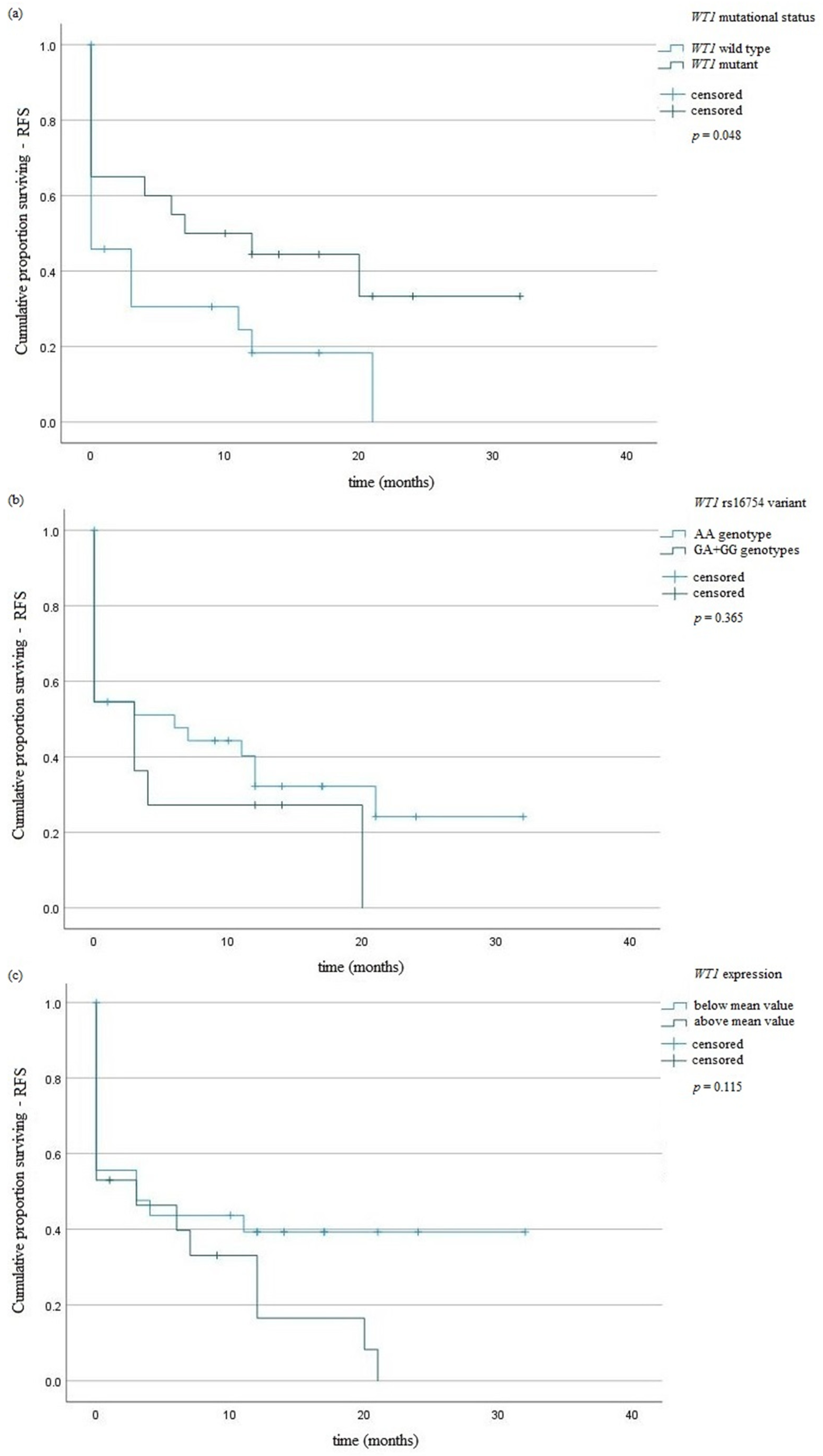
The studied WT1 variant did not influence the risk of AML. Genotypes AA predominated in the intermediate risk group (33/74). Genotypes AA and GA predominated in the adverse risk group (28/74 and 12/13, respectively). All cases with poor risks had homozygous GG (3/3). The presence of GA and GG genotypes did not affect the risk of WT1 mutations occurrence (p = 0.68). We observed higher expression of WT1 gene in patients with AA genotype in comparison with those with GA or GG genotypes—10,556.7 vs. 25,836.5 NCN copies (p = 0.01), respectively. The AA genotypes were associated with lower PLT numbers in comparison with GA + GG genotypes—78.37 vs. 137.18 (G/L), p = 0.02 (Table 4). Survival curves were defined by the Kaplan–Meier method and compared using the log-rank test. Among surviving patients, there were no statistically significant differences in OS and RFS between patients with AA genotype and those with GA or GG genotypes—p = 0.302 and p = 0.365, respectively (Figure 1 and Figure 2).

3.2. WT1 Mutations

WT1 mutations were found in 26/90 patients (28.9%). A total of 13 patients had WT1 mutations affecting exon 7 (14.4%), whereas 12 patients (13.3%) had WT1 mutations in exon 9. Only one patient had WT1 mutations in both exon 7 and 9 (1.1%). The total number of WT1 mutations was 24. We found the following mutations:
-
In exon 7: c.1375G>A (p.A399V), c.1334C>A (p.R385R), c.1382A>T (p.P401P), c.1389delA (p.N404H); c.1320G>A (p.P381S), c.1314C>T (p.V379I), c.1324indelGTACAAGAG/GTACAAGAGGGTACAAGAG (frameshift variant);
-
In exon 9: c.1590delC (p.L491X), c.1567G>A (p.R463P), c.1557T>A/C (p.T460S/A).
The most common mutations were substitutions (18/24, 75%). In contrast, deletions were found in 5 cases (5/24, 20.8%). One indel mutation was found (1/24, 4.1%).
WT1 mutations were present more frequently in AML patients with age under 65 years (p < 0.0001) (Table 4). The presence of NPM1 or CEBPA mutations decreased the risk of mutations occurrence in WT1 gene-OR = 0.11, 95% CI 0.02–0.46, p = 0.002 or OR = 0.05, 95% CI 0.006–0.46, p = 0.002, respectively (Table 4). WT1 mutation, as well as WT1 rs16754 genotypes, did not affect the OS of AML patients (Figure 1). We observed an association of WT1 mutations with RFS (Figure 2a). A univariate Cox analysis revealed that patients above 65 years of age had a 2.5-fold risk of death, p = <0.01 HR = 2.52 (Table 5).

3.3. WT1 Expression

The number of WT1 gene copies (normalized copy number, NCN) was calculated into 10,000 ABL gene copies. A value of >250 WT1 copies per 10,000 ABL copies was seen as a manifestation of overexpression of this WT1 gene. The WT1 overexpression was observed in all AML patients at the time of diagnosis. The mean of WT1 expression was 13,273.12 (range from 1342.9 to 172,093). In patients with normal karyotype and with chromosomal aberrations, we observed mean WT1 expression 12,747.44 (range 1342.9–172,093) and 13,798.79 (range 1458–105,842.5). The difference in WT1 expression between patients with normal and abnormal karyotypes was statistically insignificant (Table 4). We observed significantly higher WT1 expression in AML CD34 positive patients in comparison with AML CD34 negative individuals—20,985 (NCN) vs. 8304 (NCN), p = 0.039.

3.4. FLT3, NPM1, and CEBPA Mutations

Internal tandem duplication of FLT3 gene (FLT3-ITD) was observed in eight patients (8.8%). In all cases, FLT3-ITD change involved only one allele. In six patients with FLT3-ITD, complete remission could not be achieved. We did not observe a statistically significant difference between WT1 gene expression in patients with FLT3-ITD and patients with FLT3 wild type-13,628 vs. 13,238, p = 0.98. In patients with FLT3 mutation, hyperleukocytosis was observed in comparison with those with FLT3 wild type—49.5 vs. 152 G/L (p = 0.002), respectively. In patients who reached CR, a lower WBC number was observed—22.9 vs. 59 (G/L), p = 0.012. FLT3 mutation did not impact CR in AML patients (p = 0.81).
NPM1 mutational analysis revealed a type A mutation in exon 12 (956dupTCTG). This type of mutation was observed in 11 patients. Both FLT3-ITD and NPM1 mutations did not coexist together. The mean number of WT1 copies in patients with NPM1 mutation was lower than the median value (7663.26 NCN). However, this difference in mean WT1 NCN copies between AML patients with or without NPM1 mutation was statistically insignificant (p = 0.71). Similar results were observed in the case of patients with or without CEBPA mutation—7356 vs. 13,772 WT1 NCN (p = 0.43), respectively. CEBPA mutation coexisted with NPM1 mutation in three cases. In one AML patient, the presence of both–CEBPA mutation and FLT3-ITD was observed. A univariate Cox analysis identified NPM1 mutation impacting RFS of AML patients (p = 0.04, HR = 3.46) (Table 5). In multivariate Cox analysis, we did not observe any impact of WT1 mutation, WT1 overexpression, or molecular changes in FLT3, NPM1, and CEBPA genes on OS and RFS in AML patients.

3.5. Cytogenetic Aberrations

In our study, by use of conventional cytogenetics and aCGH, genomic imbalances were detected in 45 cases (50%). Owing to the aCGH technique, we detected an abnormal number of chromosomes that included gains and losses, as well as small structural changes in the form of duplications and deletions.
The aberrations detected with classical cytogenetic techniques included whole chromosomes. These changes were mostly related to the following chromosomes: −17 (11%); −18 (9%); −5 (8%), +8, −15, −16, −21 (7%); −3, +6, −7, −12, −14, −20, −22 (5%). Interstitial and terminal deletions were also detected (del(3q), del(5q), del(7q), del(11q), del(13q), del(17p)), translocations (t(3;15), t(5;7), t(8;16), t(8;21), t(9;22), t(10;12), t(11;11) and t(15;17)), inversions (inv(16)), isochromosomes (i(17q)) and the presence of marker chromosomes.
The chromosomal aberrations revealed by aCGH included small DNA fragments and were predominantly related to the following regions:
-
losses of (5)(q23q32)—20%;
-
losses of (7)(p12.3q36.3)—13%;
-
gains of (8)(q12.1q24.3)—10%;
-
gains of (11)(q12.2q14.1)—12.2%;
-
losses of (11)(q22q23.3)—14.4%;
-
losses of (17)(p13.3p13.1)—16.7%;
-
losses of (18)(p11.32q23)—11%;
-
gains of (22)(q12.3q13.2)—5.5%.
By using the aCGH technique, we have confirmed the occurrence of most changes detected by classical cytogenetic methods. In our study, we observed amplification of regions—3q26 (EVI1 gene locus), 8q24 (C-MYC gene locus), and 11q23 (MLL gene locus).
No chromosome X and Y aberrations were found using the microarray CGH method; however, such aberrations were seen in classical cytogenetics tests. In the case of deletions and additions, no statistically significant differences were found between the results obtained by classical cytogenetics (GTG and RHG) and aCGH.
The combination of conventional cytogenetics, FISH, and aCGH enabled us to find favorable karyotype in 13 patients (14.4%), intermediate karyotype in 34 patients (37.7%), and adverse karyotype in 43 patients (47.7%). The median OS was not reached in favorable-risk (26.6 months) and in adverse-risk group (12.8 months). It was reached in intermediate-risk group 39.7 months (p = 0.123 favorable vs. intermediate; p = 0.001 favorable vs. adverse; p = 0.001 intermediate vs. adverse). The median RFS (6.39 months) was not reached in adverse-risk group (1.8 month) in comparison with favorable-risk (10.5 months) and intermediate-risk (8.6 months) groups (p = 0.125 favorable vs. intermediate; p = 0.002 favorable vs. adverse; p = 0.017 intermediate vs. adverse).

4. Discussion

In our study, we analyzed the WT1 rs16754 variant, WT1 mutations, and WT1 gene expression taking into account chromosomal changes and molecular markers such as mutations in FLT3, NPM1, and CEBPA genes. To our knowledge, our investigation is the first multidimensional study including analysis of cryptic chromosomal and molecular changes in Polish AML patients.

4.1. WT1 rs16754 Variant

In our study, we found that genotypic frequencies of the WT1 gene were not in HWE. This suggests an association of WT1 rs16754 genotypes with the risk of disease development. However, in our study, we did not find the association of analyzed genotypes with AML risk. We cannot exclude, however, that the obtained result was due to sampling error. Moreover, analyzed genotypes did not affect OS or RFS of AML patients. It is consistent with results obtained by Kim et al. in Korean patients with AML [30]. In contrast, Long et al. in their meta-analysis observed that the WT1 rs16754 variant was associated with better survival of AML patients [9]. They concluded that the WT1 rs16754 variant was an independent favorable-risk marker [9]. Zhang et al. found that GG genotypes were associated with significantly higher WT1 expression at mRNA level in comparison with GA and AA genotypes [31]. In our study, we observed similar results. The expression of the WT1 gene was statistically higher in AML patients with AA genotype.

4.2. WT1 Mutations

We observed a higher frequency of WT1 mutations in AML patients under 65 years of age. This is in concordance with results obtained by Krauth et al. [32]. In their study of 3157 AML patients, they found that WT1 mutations were more frequent in younger AML patients and were associated with adverse effects in cytogenetically normal AML patients [32]. In concordance with other studies, we did not find a significant relationship between WT1 mutations and clinical characteristics, including gender, WBC and PLT count, and Hb concentration [3,33]. We did not find an association of WT1 mutations with normal karyotype. However, we observed a higher frequency of WT1 mutations in AML patients with NPM1 or CEBPA genes mutations. More frequent WT1 mutations were observed by Krauth and coworkers in CEBPA mutated AML [32]. In contrast, Hou et al. and Rostami et al. did not observe a relationship between WT1 mutations and the presence of mutations in genes-FLT3, NPM1, and CEBPA [3,34]. In other research, Toogeh and coworkers showed no correlation of WT1 mutations with FLT3-ITD [35]. In our study, we did not observe an association of WT1 mutations with FLT3-ITD. To assess the prognostic impact of WT1 mutations, we analyzed them taking into account OS and RFS. We found longer RFS in patients with WT1 mutation. However, this result is not consistent with previous reports [3,33,36,37]. This may be due to the fact that previous reports were conducted in other populations or as a result of sampling error.

4.3. WT1 Expression

The WT1 gene shows high expression in AML patients, and its expression at diagnosis may be an adverse predictor of disease outcome [38]. It has been confirmed that increased expression of the WT1 gene accelerated disease progression [38]. In our study, we observed WT1 overexpression in 97.7% of newly diagnosed AML patients, which is in agreement with previous studies [3,10,16]. In the presented material, all patients qualified for the study had completed chemotherapy. We observed significantly higher WT1 expression in AML CD34 positive patients in comparison with AML CD34 negative individuals. Yoon et al. carried out testing of 104 diagnosed AML patients with normal karyotype and showed that increased WT1 expression in bone marrow was significantly increased in AML patients [39]. We did not find a statistically significant difference in WT1 expression between patients with normal and abnormal karyotypes. Moreover, we did not observe an association between WT1 expression and OS or RFS. Results of our research confirm the data obtained by other authors [3,40].

4.4. Cytogenetic Analysis

We observed the occurrence of chromosome abnormalities, which were identified by conventional karyotype testing, aCGH, and were confirmed by FISH testing. Without conventional cytogenetics and FISH, we would not be able to detect and resolve balanced aberrations, such as translocations. Similar results were found in numerous studies [19,20]. Our data also support the importance of cytogenetic analysis as a component of the routine diagnostic workup of AML.
The difference in WT1 expression between patients with normal and abnormal karyotypes was statistically insignificant. Moreover, we did not observe an association between chromosomal aberrations and the presence of WT1 mutations.
The median OS was not reached in the favorable-risk group, as well as in the adverse-risk group. In the case of RFS, its median value was not reached in the adverse-risk group only.
The aberration frequently observed using aCGH was the loss of a long arm fragment of chromosome 5 (including NPM1 gene locus), which is in agreement with other studies [41,42]. Costa et al. and Rucker et al. detected losses of small regions in 5q [43,44]. Mehrotra et al. analyzed 48 AML patients and observed deletions in the 5q33.3 region [45]. Loss of this region was associated with the achievement of CR [45]. Veigaard et al. drew attention to the 5q23.1–q33.3 region, where they found deletions most frequently [46]. In addition, Schoch et al., in testing AML patients with complex karyotype, found deletions of the 5q14–q35 region, as we did in our study [47]. Itzhar et al. specified 5q31.3–q32 regions which were associated with AML development [48]. The deletion of the 5q31 region was associated with the aggressive course of AML, the occurrence of additional chromosomal aberrations, and an unfavorable prognosis [46]. We did not detect microdeletions or microduplications of loci, including WT1, FLT3, and CEBPA genes.

4.5. Clinical and Molecular Data

NPM1 mutations in AML patients are associated with the presence of normal karyotype and are rare in patients with genetic aberrations [49]. We did not notice shorter OS or RFS in patients with NPM1 mutation. The presence of this mutation is favorable and is associated with higher CR rates [4]. In CN-AML patients with NPM1 mutation, longer RFS is observed in comparison with that observed in patients with wild-type NPM1 [20]. In our study, we observed NPM1 gene mutations only in CN-AML. Patients with NPM1 mutations and lacking FLT3-ITD have a better prognosis [50]. The coexistence of NPM1 and FLT3-ITD mutations is a relatively common phenomenon, observed in about 18% of patients with CN-AML [51]. In our material, we did not find the coexistence of these mutations. Many studies have confirmed the correlation between FLT3-ITD and an increased number of leukocytes [52]. Our research has also shown that patients with FLT3-ITD mutations have increased leukocytosis. The FLT3-ITD adversely affects the response to treatment. However, in the presented material, we did not find an association between CR rate and the presence of the FLT3 mutation.

4.6. Limitations

The limitations of our study are associated with the number of AML patients recruited to the study, as well as an automated DNA sequencing method. The sample size was relatively small, in part due to the low incidence of the disease, as well as applied methods such as aCGH. Chromosomal aberrations are factors that need to be taken into account in determining the prognosis of AML patients. Due to their low resolution, the techniques of classical cytogenetics do not detect all alterations; therefore, other methods are used in diagnosing AML, such as PCR and FISH. However, both techniques require prior knowledge of DNA regions where the mutations have occurred. The 90 AML patients sufficed for most analyzes. However, some were not possible as a result of the low frequency of certain molecular changes. In the case of automated sequencing, it would be recommended to use the next-generation sequencing, which has higher resolution and enables analysis of all exons and introns of the WT1 gene. It is possible that mutations present in other coding regions or in regulatory regions of the WT1 gene were not detected in our analysis.

4.7. Conclusions

WT1 gene expression and its rs16754 variant at diagnosis did not affect outcomes in Polish AML patients. WT1 mutation may affect RFS in AML. In conclusion, we propose a combination of conventional cytogenetics, FISH, and array CGH as necessary tools to unravel the molecular karyotype of AML.

Author Contributions

Experiments and molecular analysis of WT1 variants and expression, S.Z. and S.P.-M.; mutation analysis, S.P.-M.; cytogenetic analysis (including classical cytogenetics, array CGH, and FISH) and results interpretation, D.K., B.G. and B.P.-G.; planning and supervision, A.A.F.; experimental data processing and analysis, S.Z. and W.S.; figure design, S.Z. and W.S.; writing—original draft preparation, review and editing, S.Z., D.K. and A.A.F. All authors have read and agreed to the published version of the manuscript.

Funding

This project was supported by the Medical University of Lublin, Poland (Grants MNmb 232, PBmb240 and DS 225).

Institutional Review Board Statement

The study obtained positive opinions (no. KE-254/24/2011 and KE-0254/229/2013) of the Bioethics Committee of Medical University of Lublin, according to the ethical standards established by Helsinki Declaration. All methods were performed in accordance with the relevant guidelines and regulations. All procedures performed in studies involving human participants were in accordance with the ethical standards of the institutional and national research committee and with the 1964 Helsinki declaration and its later amendments or comparable ethical standards.

Informed Consent Statement

Informed consent was obtained from all subjects involved in the study.

Data Availability Statement

The clinical data used to support the findings of this study are available from the corresponding author upon request.

Acknowledgments

Special thanks are due to Paul Dudek for proofreading and editing assistance, as well as substantive verification.

Conflicts of Interest

The authors declare no conflict of interest.

References

  1. Estey, E.; Döhner, H. Acute myeloid leukaemia. Lancet 2006, 368, 1894–1907. [Google Scholar] [CrossRef]
  2. Patel, J.P.; Gönen, M.; Figueroa, M.E.; Fernandez, H.; Sun, Z.; Racevskis, J.; Van Vlierberghe, P.; Dolgalev, I.; Thomas, S.; Aminova, O.; et al. Prognostic Relevance of Integrated Genetic Profiling in Acute Myeloid Leukemia. N. Engl. J. Med. 2012, 366, 1079–1089. [Google Scholar] [CrossRef] [PubMed] [Green Version]
  3. Rostami, S.; Kazemi, A.; Chahardouli, B.; Mohammadi, S.; Nikbakht, M.; Alizadeh, N.; Mousavi, A.; Alimoghaddam, K.; Ardestani, M.T. The Prognostic Impact of WT1 Expression Levels, Mutations, and SNP rs16754 in AML Patients: A Retrospective Cohort Study. J. Adv. Med. Biomed. Res. 2021, 29, 109–117. [Google Scholar] [CrossRef]
  4. Sugiyama, H. Wilms’ tumor gene WT1: Its oncogenic function and clinical application. Int. J. Hematol. 2001, 73, 177–187. [Google Scholar] [CrossRef]
  5. Döhner, H.; Weisdorf, D.J.; Bloomfield, C.D. Acute Myeloid Leukemia. N. Engl. J. Med. 2015, 373, 1136–1152. [Google Scholar] [CrossRef] [Green Version]
  6. Kang, H.J.; Park, J.H.; Chen, W.; Kang, S.I.; Moroz, K.; Ladanyi, M.; Lee, S.B. EWS-WT1 oncoprotein activates neuronal reprogramming factor ASCL1 and promotes neural differentiation. Cancer Res. 2014, 74, 4526–4535. [Google Scholar] [CrossRef]
  7. Rampal, R.; Figueroa, M.E. Wilms tumor 1 mutations in the pathogenesis of acute myeloid leukemia. Haematologica 2016, 101, 672–679. [Google Scholar] [CrossRef] [Green Version]
  8. Choi, Y.; Lee, J.-H.; Hur, E.-H.; Kang, M.J.; Kim, S.-D.; Lee, J.-H.; Kim, D.-Y.; Lim, S.-N.; Bae, K.-S.; Lim, H.-S.; et al. Single nucleotide polymorphism of Wilms’ tumor 1 gene rs16754 in Korean patients with cytogenetically normal acute myeloid leukemia. Ann. Hematol. 2011, 91, 671–677. [Google Scholar] [CrossRef]
  9. Long, J.; Fang, S.; Dai, Q.; Liu, X.; Zhu, W. The Wilms Tumor-1 (WT1) rs16754 polymorphism is a prognostic factor in acute myeloid leukemia (AML): A meta-analysis. Oncotarget 2016, 7, 32079–32087. [Google Scholar]
  10. Damm, F.; Heuser, M.; Morgan, M.; Yun, H.; Großhennig, A.; Göhring, G.; Schlegelberger, B.; Döhner, K.; Ottmann, O.; Lübbert, M.; et al. Single Nucleotide Polymorphism in the Mutational Hotspot of WT1 Predicts a Favorable Outcome in Patients With Cytogenetically Normal Acute Myeloid Leukemia. J. Clin. Oncol. 2010, 28, 578–585. [Google Scholar] [CrossRef]
  11. Bennett, J.M.; Catovsky, D.; Daniel, M.-T.; Flandrin, G.; Galton, D.A.G.; Gralnick, H.R.; Sultan, C. Proposals for the Classification of the Acute Leukaemias French-American-British (FAB) Co-operative Group. Br. J. Haematol. 1976, 33, 451–458. [Google Scholar] [CrossRef]
  12. King-Underwood, L.; Renshaw, J.; Pritchard-Jones, K. Mutations in the Wilms’ tumor gene WT1 in leukemias. Blood 1996, 87, 2171–2179. [Google Scholar] [CrossRef] [Green Version]
  13. Andersson, C.; Li, X.; Lorenz, F.; Golovleva, I.; Wahlin, A.; Li, A. Reduction in WT1 Gene Expression During Early Treatment Predicts the Outcome in Patients With Acute Myeloid Leukemia. Diagn. Mol. Pathol. 2012, 21, 225–233. [Google Scholar] [CrossRef] [PubMed]
  14. Luo, S.; Yu, K.; Yan, Q.-X.; Shen, Z.-J.; Wu, J.-B.; Chen, H.-M.; Gao, S.-M. Analysis of WT1 mutations, expression levels and single nucleotide polymorphism rs16754 inde novonon-M3 acute myeloid leukemia. Leuk. Lymphoma 2013, 55, 349–357. [Google Scholar] [CrossRef] [PubMed]
  15. Green, C.L.; Koo, K.K.; Hills, R.; Burnett, A.K.; Linch, D.C.; Gale, R.E. Prognostic Significance of CEBPA Mutations in a Large Cohort of Younger Adult Patients With Acute Myeloid Leukemia: Impact of Double CEBPA Mutations and the Interaction With FLT3 and NPM1 Mutations. J. Clin. Oncol. 2010, 28, 2739–2747. [Google Scholar] [CrossRef]
  16. Paschka, P.; Marcucci, G.; Ruppert, A.S.; Whitman, S.P.; Mrózek, K.; Maharry, K.; Langer, C.; Baldus, C.D.; Zhao, W.; Powell, B.L.; et al. Wilms’ Tumor 1 Gene Mutations Independently Predict Poor Outcome in Adults With Cytogenetically Normal Acute Myeloid Leukemia: A Cancer and Leukemia Group B Study. J. Clin. Oncol. 2008, 26, 4595–4602. [Google Scholar] [CrossRef] [PubMed]
  17. Marcucci, G.; Mrózek, K.; Ruppert, A.S.; Archer, K.J.; Pettenati, M.J.; Heerema, N.A.; Carroll, A.J.; Koduru, P.R.; Kolitz, J.E.; Sterling, L.J.; et al. Abnormal Cytogenetics at Date of Morphologic Complete Remission Predicts Short Overall and Disease-Free Survival, and Higher Relapse Rate in Adult Acute Myeloid Leukemia: Results From Cancer and Leukemia Group B Study 8461. J. Clin. Oncol. 2004, 22, 2410–2418. [Google Scholar] [CrossRef]
  18. Betz, B.L.; Hess, J.L. Acute Myeloid Leukemia Diagnosis in the 21st Century. Arch. Pathol. Lab. Med. 2010, 134, 1427–1433. [Google Scholar] [CrossRef]
  19. Grimwade, D.; Walker, H.; Oliver, F.; Wheatley, K.; Harrison, C.; Harrison, G.; Rees, J.; Hann, I.; Stevens, R.; Burnett, A.; et al. The importance of diagnostic cytogenetics on outcome in AML: Analysis of 1,612 patients entered into the MRC AML 10 trial. The Medical Research Council Adult and Children’s Leukaemia Working Parties. Blood 1998, 92, 2322–2333. Available online: http://www.ncbi.nlm.nih.gov/pubmed/9746770 (accessed on 10 February 2022). [CrossRef] [Green Version]
  20. Döhner, H.; Estey, E.H.; Amadori, S.; Appelbaum, F.R.; Büchner, T.; Burnett, A.K.; Dombret, H.; Fenaux, P.; Grimwade, D.; Larson, R.A.; et al. Diagnosis and management of acute myeloid leukemia in adults: Recommendations from an international expert panel, on behalf of the European LeukemiaNet. Blood 2010, 115, 453–474. [Google Scholar] [CrossRef]
  21. Summers, K.; Stevens, J.; Kakkas, I.; Smith, M.; Smith, L.L.; MacDougall, F.; Cavenagh, J.; Bonnet, D.; Young, B.D.; Lister, T.A.; et al. Wilms’ tumour 1 mutations are associated with FLT3-ITD and failure of standard induction chemotherapy in patients with normal karyotype AML. Leukemia 2007, 21, 550–551. [Google Scholar] [CrossRef]
  22. Chomczynski, P.; Sacchi, N. Single-step method of RNA isolation by acid guanidinium thiocyanate–phenol–chloroform extraction. Anal. Biochem. 1987, 162, 156–159. [Google Scholar] [CrossRef]
  23. Cilloni, D.; Gottardi, E.; De Micheli, D.; Serra, A.; Volpe, G.; Messa, F.; Rege-Cambrin, G.; Guerrasio, A.; Divona, M.; Coco, F.L.; et al. Quantitative assessment of WT1 expression by real time quantitative PCR may be a useful tool for monitoring minimal residual disease in acute leukemia patients. Leukemia 2002, 16, 2115–2121. [Google Scholar] [CrossRef] [Green Version]
  24. Beillard, E.; Pallisgaard, N.; Van Der Velden, V.H.J.; Bi, W.; Dee, R.; Van Der Schoot, E.; Delabesse, E.; MacIntyre, E.; Gottardi, E.; Saglio, G.; et al. Evaluation of candidate control genes for diagnosis and residual disease detection in leukemic patients using ‘real-time’ quantitative reverse-transcriptase polymerase chain reaction (RQ-PCR)—A Europe against cancer program. Leukemia 2003, 17, 2474–2486. [Google Scholar] [CrossRef] [Green Version]
  25. De Marchi, F.; Candoni, A.; Zannier, M.E.; Haley, L.; Lau, B.W.Y.; Fanin, R. Concomitant monitoring of WT1 and FLT3-ITD expression in FLT3-ITD acute myeloid leukemia patients: Which should we trust as a minimal residual disease marker? Am. J. Hematol. 2017, 92, E72–E74. [Google Scholar] [CrossRef] [Green Version]
  26. Koczkodaj, D.; Zmorzyński, S.; Michalak-Wojnowska, M.; Wasik-Szczepanek, E.; Filip, A.A. Examination of the FLT3 and NPM1 mutational status in patients with acute myeloid leukemia from southeastern Poland. Arch. Med Sci. 2016, 1, 120–128. [Google Scholar] [CrossRef] [PubMed] [Green Version]
  27. Benthaus, T.; Schneider, F.; Mellert, G.; Zellmeier, E.; Schneider, S.; Kakadia, P.M.; Hiddemann, W.; Bohlander, S.; Feuring-Buske, M.; Braess, J.; et al. Rapid and sensitive screening forCEBPAmutations in acute myeloid leukaemia. Br. J. Haematol. 2008, 143, 230–239. [Google Scholar] [CrossRef] [PubMed]
  28. Preacher, K.J. Calculation for the Chi-Square Test: An Interactive Calculation Tool for Chi-Square Tests of Goodness of Fit and Independence. 2019. Available online: http://quantpsy.org/ (accessed on 21 January 2022).
  29. Zmorzynski, S.; Szudy-Szczyrek, A.; Popek-Marciniec, S.; Korszen-Pilecka, I.; Wojcierowska-Litwin, M.; Luterek, M.; Chocholska, S.; Styk, W.; Swiderska-Kołacz, G.; Januszewska, J.; et al. ACE Insertion/Deletion Polymorphism (rs4646994) Is Associated With the Increased Risk of Multiple Myeloma. Front. Oncol. 2019, 9, 44. [Google Scholar] [CrossRef] [Green Version]
  30. Kim, N.; Kim, I.-S.; Chang, C.L.; Kang, J.-E.; Lee, E.Y.; Shin, H.-J. Risk-Reducing Genetic Variant of Wilms Tumor 1 Gene rs16754 in Korean Patients With BCR-ABL1-Negative Myeloproliferative Neoplasm. Ann. Lab. Med. 2015, 35, 348–351. [Google Scholar] [CrossRef]
  31. Zhang, D.-Y.; Yan, H.; Cao, S.; Zhang, W.; Li, X.-L.; Zeng, H.; Chen, X.-P. Wilms Tumor 1 rs16754 predicts favorable clinical outcomes for acute myeloid leukemia patients in South Chinese population. Leuk. Res. 2015, 39, 568–574. [Google Scholar] [CrossRef]
  32. Krauth, M.-T.; Alpermann, T.; Bacher, U.; Eder, C.; Dicker, F.; Ulke, M.; Kuznia, S.; Nadarajah, N.; Kern, W.; Haferlach, C.; et al. WT1 mutations are secondary events in AML, show varying frequencies and impact on prognosis between genetic subgroups. Leukemia 2014, 29, 660–667. [Google Scholar] [CrossRef] [PubMed]
  33. Gaidzik, V.I.; Schlenk, R.F.; Moschny, S.; Becker, A.; Bullinger, L.; Corbacioglu, A.; Krauter, J.; Schlegelberger, B.; Ganser, A.; Döhner, H.; et al. Prognostic impact of WT1 mutations in cytogenetically normal acute myeloid leukemia: A study of the German-Austrian AML Study Group. Blood 2009, 113, 4505–4511. [Google Scholar] [CrossRef] [Green Version]
  34. Hou, H.-A.; Huang, T.-C.; Lin, L.-I.; Liu, C.-Y.; Chen, C.-Y.; Chou, W.-C.; Tang, J.-L.; Tseng, M.-H.; Huang, C.-F.; Chiang, Y.-C.; et al. WT1 mutation in 470 adult patients with acute myeloid leukemia: Stability during disease evolution and implication of its incorporation into a survival scoring system. Blood 2010, 115, 5222–5231. [Google Scholar] [CrossRef] [PubMed] [Green Version]
  35. Toogeh, G.; Ramzi, M.; Faranoush, M.; Amirizadeh, N.; Haghpanah, S.; Moghadam, M.; Cohan, N. Prevalence and Prognostic Impact of Wilms’ Tumor 1 (WT1) Gene, Including SNP rs16754 in Cytogenetically Normal Acute Myeloblastic Leukemia (CN-AML): An Iranian Experience. Clin. Lymphoma Myeloma Leuk. 2016, 16, e21–e26. [Google Scholar] [CrossRef] [PubMed]
  36. Xu, J.; Zhang, Y.; Hu, J.; Ren, Y.; Wang, H. Clinical features and prognosis of normal karyotype acute myeloid leukemia pediatric patients with WT1 mutations: An analysis based on TCGA database. Hematology 2020, 25, 79–84. [Google Scholar] [CrossRef] [Green Version]
  37. Niktoreh, N.; Walter, C.; Zimmermann, M.; von Neuhoff, C.; von Neuhoff, N.; Rasche, M.; Waack, K.; Creutzig, U.; Hanenberg, H.; Reinhardt, D. Mutated WT1, FLT3-ITD, and NUP98-NSD1 Fusion in Various Combinations Define a Poor Prognostic Group in Pediatric Acute Myeloid Leukemia. J. Oncol. 2019, 2019, 1609128. [Google Scholar] [CrossRef] [Green Version]
  38. Di Stasi, A.; Jimenez, A.M.; Eminagawa, K.; Eal-Obaidi, M.; Erezvani, K. Review of the Results of WT1 Peptide Vaccination Strategies for Myelodysplastic Syndromes and Acute Myeloid Leukemia from Nine Different Studies. Front. Immunol. 2015, 6, 36. [Google Scholar] [CrossRef] [Green Version]
  39. Yoon, J.-H.; Kim, H.-J.; Shin, S.-H.; Yahng, S.-A.; Lee, S.-E.; Cho, B.-S.; Eom, K.-S.; Kim, Y.-J.; Lee, S.; Min, C.-K.; et al. Serial Measurement of WT1 Expression and Decrement Ratio Until Hematopoietic Cell Transplantation as a Marker of Residual Disease in Patients with Cytogenetically Normal Acute Myelogenous Leukemia. Biol. Blood Marrow Transplant. 2013, 19, 958–966. [Google Scholar] [CrossRef] [Green Version]
  40. Shimada, A.; Taki, T.; Koga, D.; Tabuchi, K.; Tawa, A.; Hanada, R.; Tsuchida, M.; Horibe, K.; Tsukimoto, I.; Adachi, S.; et al. High WT1 mRNA expression after induction chemotherapy and FLT3-ITD have prognostic impact in pediatric acute myeloid leukemia: A study of the Japanese Childhood AML Cooperative Study Group. Int. J. Hematol. 2012, 96, 469–476. [Google Scholar] [CrossRef] [PubMed]
  41. Suela, J.; Alvarez, S.; Cigudosa, J. DNA profiling by arrayCGH in acute myeloid leukemia and myelodysplastic syndromes. Cytogenet. Genome Res. 2007, 118, 304–309. [Google Scholar] [CrossRef]
  42. Evers, C.; Beier, M.; Poelitz, A.; Hildebrandt, B.; Servan, K.; Drechsler, M.; Germing, U.; Royer, H.-D.; Royer-Pokora, B. Molecular definition of chromosome arm 5q deletion end points and detection of hidden aberrations in patients with myelodysplastic syndromes and isolated del(5q) using oligonucleotide array CGH. Genes Chromosom. Cancer 2007, 46, 1119–1128. [Google Scholar] [CrossRef] [PubMed]
  43. Rücker, F.G.; Bullinger, L.; Schwaenen, C.; Lipka, D.B.; Wessendorf, S.; Fröhling, S.; Bentz, M.; Miller, S.; Scholl, C.; Schlenk, R.F.; et al. Disclosure of Candidate Genes in Acute Myeloid Leukemia With Complex Karyotypes Using Microarray-Based Molecular Characterization. J. Clin. Oncol. 2006, 24, 3887–3894. [Google Scholar] [CrossRef] [PubMed]
  44. Costa, A.R.S.; Belangero, S.; Melaragno, M.I.; Chauffaille, M.D.L. Additional chromosomal abnormalities detected by array comparative genomic hybridization in AML. Med. Oncol. 2012, 29, 2083–2087. [Google Scholar] [CrossRef] [PubMed]
  45. Mehrotra, M.; Luthra, R.; Ravandi, F.; Sargent, R.L.; Barkoh, B.A.; Abraham, R.; Mishra, B.M.; Medeiros, L.J.; Patel, K.P. Identification of clinically important chromosomal aberrations in acute myeloid leukemia by array-based comparative genomic hybridization. Leuk. Lymphoma 2014, 55, 2538–2548. [Google Scholar] [CrossRef] [PubMed] [Green Version]
  46. Veigaard, C.; Nørgaard, J.M.; Kjeldsen, E. Genomic profiling in high hyperdiploid acute myeloid leukemia: A retrospective study of 19 cases. Cancer Genet. 2011, 204, 516–521. [Google Scholar] [CrossRef]
  47. Schoch, C.; Kern, W.; Kohlmann, A.; Hiddemann, W.; Schnittger, S.; Haferlach, T. Acute myeloid leukemia with a complex aberrant karyotype is a distinct biological entity characterized by genomic imbalances and a specific gene expression profile. Genes Chromosom. Cancer 2005, 43, 227–238. [Google Scholar] [CrossRef]
  48. Itzhar, N.; Dessen, P.; Toujani, S.; Auger, N.; Preudhomme, C.; Richon, C.; Lazar, V.; Saada, V.; Bennaceur, A.; Bourhis, J.H.; et al. Chromosomal Minimal Critical Regions in Therapy-Related Leukemia Appear Different from Those of De Novo Leukemia by High-Resolution aCGH. PLoS ONE 2011, 6, e16623. [Google Scholar] [CrossRef] [Green Version]
  49. Thiede, C.; Steudel, C.; Mohr, B.; Schaich, M.; Schäkel, U.; Platzbecker, U.; Wermke, M.; Bornhäuser, M.; Ritter, M.; Neubauer, A.; et al. Analysis of FLT3-activating mutations in 979 patients with acute myelogenous leukemia: Association with FAB subtypes and identification of subgroups with poor prognosis. Blood 2002, 99, 4326–4335. [Google Scholar] [CrossRef] [Green Version]
  50. Park, B.; Chi, H.-S.; Park, S.-J.; Min, S.K.; Jang, S.; Park, C.-J.; Kim, D.-Y.; Lee, J.-H.; Lee, J.-H.; Lee, K.-H. Clinical Implications of Non-A-Type NPM1 and FLT3 Mutations in Patients with Normal Karyotype Acute Myeloid Leukemia. Acta Haematol. 2012, 127, 63–71. [Google Scholar] [CrossRef] [PubMed] [Green Version]
  51. Mrózek, K.; Marcucci, G.; Paschka, P.; Bloomfield, C.D. Advances in molecular genetics and treatment of core-binding factor acute myeloid leukemia. Curr. Opin. Oncol. 2008, 20, 711–718. [Google Scholar] [CrossRef] [Green Version]
  52. Fröhling, S.; Schlenk, R.F.; Breitruck, J.; Benner, A.; Kreitmeier, S.; Tobis, K.; Doöhner, H.; Doöhner, K. Prognostic significance of activating FLT3 mutations in younger adults (16 to 60 years) with acute myeloid leukemia and normal cytogenetics: A study of the AML Study Group Ulm. Blood 2002, 100, 4372–4380. [Google Scholar] [CrossRef] [Green Version]
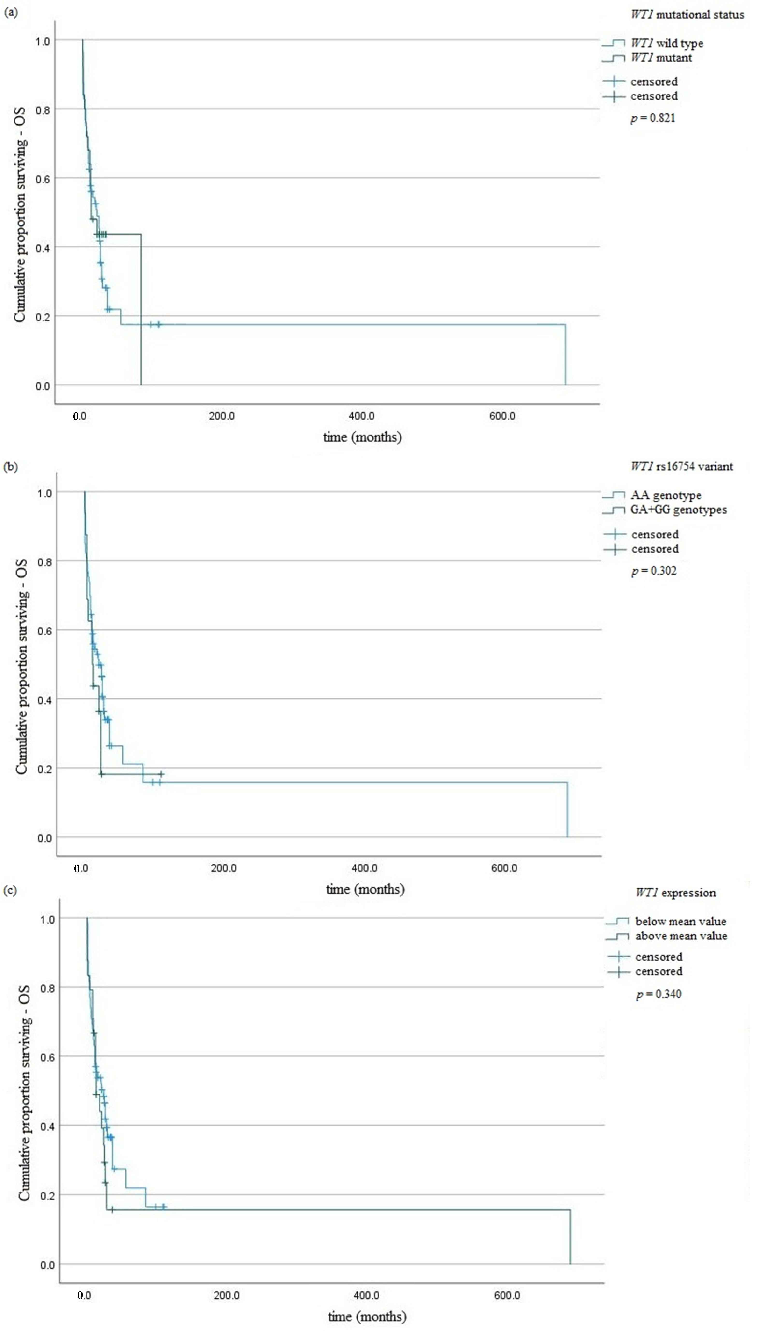
Figure 1. Kaplan–Meier analysis of OS in the group of AML patients taking into account: (a) WT1 mutation; (b) WT1 rs16754 variant; (c) WT1 overexpression.
Jcm 11 01873 g001
Figure 2. Kaplan–Meier analysis of RFS in the group of AML patients taking into account: (a) WT1 mutation; (b) WT1 rs16754 variant; (c) WT1 overexpression.
Jcm 11 01873 g002
Table 1. Clinical-laboratory characteristics of patients with AML.
Age median (range)62.63 (18–85)
Gender (%)
Male42 (47)
Female48 (53)
Laboratory parameters (range)
HB g/dL9.26 (4.8–9.7)
WBC G/L55.15 (0.8–94.84)
PLT G/L89.06 (6.0–783.0)
FAB subtype:
M06
M17
M27
M312
M442
M515
M61
Risk category:
favorable13
intermediate34
adverse43
Induction therapy:
DAC52
Idarubicin + ATRA6
AZA9
reduced DA15
low-dose cytarabine8
Stem cell transplantation:
no42
allogeneic37
autologous11
Cytogenetic:
Normal karyotype45
Abnormal karyotype45 (22 *)
FISH (%):
del(13)(q14.3)51 (56.6)
del(17)(p13.1)15 (16.7)
del(11)(q23)13 (14.4)
del(6)(q23)5 (5.5)
t(15;17)(q22;q21.1)6 (6.6)
BCR/ABL2 (2.2)
MLL amplification1 (1.1)
MYC amplification1 (1.1)
EVI1 amplification1 (1.1)
Immunophenotype—negativity/positivity
CD3451/39
CD3319/71
CD1466/24
Molecular variants—present/absent
FLT3-ITD8/82
NPM1 mutation11/79
CEBPA mutation7/83
* complex karyotype.
Table 2. Hardy–Weinberg equilibrium (HWE) for WT1 rs16754 variant in case and control groups according to expected (E) and observed (O) values.
GROUPSGENOTYPESTotalHWE p Value and χ2 *
WT1 rs16754 variant
-AAGAGG--
CONTROL
E79.2119.581.21100p = 0.44, χ2 = 0.57
O80182100
CASE
E70.218.551.2590p = 0.0007, χ2 = 11.5
O7411590
* if the χ2 ≤ 3.84 and the corresponding p ≥ 0.05, then the population is in HWE.
Table 3. The comparison of allele frequency and distribution of WT1 variant among AML patients and controls.
Gene Variants and AllelesAML n (%)Controls n (%)Odds Ratio95% CIp Values
Codominant model
AA74 (82.2%)80 (80%)1--
GA11 (12.2%)18 (18%)1.510.67–3.410.31
GG5 (5.5%)2 (2%)0.370.06–1.960.41
Dominant model
AA74 (82.2%)80 (80%)1--
GA + GG16 (17.7%)20 (20%)1.150.55–2.400.69
Recessive model
AA + GA85 (94.4%)98 (98%)1--
GG5 (5.5%)2 (2%)0.340.06–1.830.36
Total:90 (100%)100 (100%)
Alleles
A159 (88.3%)178 (89%)1--
G21 (11.7%)22 (11%)0.930.49–1.760.84
Total:180 (100%)200 (100%)
Table 4. Correlation of WT1 variants and overexpression with clinical characteristics in AML patients.
FeaturesWT1 AA GenotypeWT1 GA + GG Genotypep ValueWT1 MutatedWT1 Wild Typep ValueWT1 Expression *WT1 Expression **p Value
Gender
Male3480.7618380.3837120.61
Female4088262912
Age
Age < 65 years62120.632313<0.000155190.64
Age ≥ 65 years124351115
Cytogenetics
Normal karyotype35100.2716290.1632130.63
Abnormal karyotype39610353411
Point mutations
NPM1 wild type66130.6418610.00258210.75
NPM1 mutated838383
FLT3 wild type67150.9321610.0762200.25
FLT3 mutated715344
CEBPA wild type69140.8020630.00260230.74
CEBPA mutated526161
Clinical values
WBC median42.16113.590.0613.7461.190.6450.8274.630.96
Hb median9.189.740.338.859.330.279.289.170.17
PLT median78.37137.180.028690.280.1990.7984.450.45
* WT1 expression lower than medium value (NCN = 13,273.12). ** WT1 expression higher than medium value (NCN = 13,273.12).
Table 5. Analysis of prognostic impact on OS and RFS in AML patients.
FeatureUnivariate AnalysisMultivariate Analysis
p ValueHR95% CIp ValueHR95% CI
OS
Age<0.012.521.33–4,810.010.410.22–0.81
WT1 mutation0.821.070.59–2.330.740.890.48–1.52
WT1 overexpression0.350.760.44–1.340.540.840.45–1.82
NPM1 mutation0.132.030.81–5.100.311.730.61–4.93
FLT3-ITD0.760.860.35–2.170.610.780.29–1.93
CEBPA mutation0.541.440.45–4.620.960.970.27–3.27
Abnormal karyotype0.640.890.53–1.470.770.920.51–1.56
RFS
Age0.072.080.92–4.650.240.590.27–1.52
WT1 mutation0.111.840.59–3.250.761.130.36–2.18
WT1 overexpression0.190.630.31–1.270.590.790.33–1.83
NPM1 mutation0.043.461.03–6.550.172.750.92–5.45
FLT3-ITD0.580.740.26–2.130.380.570.15–1.99
CEBPA mutation0.162.370.70–7.950.621.420.39–5.75
Abnormal karyotype0.510.780.37–1.620.690.840.35–1.87
Publisher’s Note: MDPI stays neutral with regard to jurisdictional claims in published maps and institutional affiliations.