Article

Nonsurgical Periodontal Treatment Options and Their Impact on Subgingival Microbiota

1
Department of Operative Dentistry and Periodontology, Martin-Luther-University Halle-Wittenberg, 06108 Halle, Germany
2
Department of Operative Dentistry, Periodontology and Preventive Dentistry, University Hospital (RWTH) Aachen, 52074 Aachen, Germany
3
Private Practice, 52062 Aachen, Germany
*
Author to whom correspondence should be addressed.
J. Clin. Med. 2022, 11(5), 1187; https://doi.org/10.3390/jcm11051187
Submission received: 7 February 2022 / Revised: 18 February 2022 / Accepted: 21 February 2022 / Published: 23 February 2022
(This article belongs to the Special Issue Periodontal Diseases: Symptoms, Causes, Treatment, and Prevention)

Abstract

:
Background: Different periodontal treatment methods (quadrant-wise debridement, scaling and root planing (Q-SRP), full-mouth scaling (FMS), full-mouth disinfection (FMD), and FMD with adjuvant erythritol air-polishing (FMDAP)) were applied in periodontitis patients (stage III/IV). The study objective (substudy of ClinicalTrials.gov Identifier: NCT03509233) was to compare the impact of treatments on subgingival colonization. Methods: Forty patients were randomized to the treatment groups. Periodontal parameters and subgingival colonization were evaluated at baseline and 3 and 6 months after treatment. Results: Positive changes in clinical parameters were recorded in every treatment group during the 3-month follow-up period, but did not always continue. In three groups, specific bacteria decreased after 3 months; however, this was associated with a renewed increase after 6 months (FMS: Porphyromonas gingivalis; FMD: Eubacterium nodatum, Prevotella dentalis; and FMDAP: uncultured Prevotella sp.). Conclusions: The benefit of all clinical treatments measured after 3 months was associated with a decrease in pathogenic bacteria in the FMS, FMD, and FMDAP groups. However, after 6 months, we observed further improvement or some stagnation in clinical outcomes accompanied by deterioration of the microbiological profile. Investigating the subgingival microbiota might help appraise successful periodontal treatment and implement individualized therapy.

1. Introduction

Periodontitis, a prevalent inflammatory disease in humans, develops as a consequence of the interplay between subgingival dysbiosis of the oral microbiota and the resulting destructive host-immune response [1,2]. Among the possible therapy options, mechanical debridement applying scaling and root planing (SRP) represents an integral component of successful periodontal therapy [3]. To increase the effectiveness of classical quadrant-wise SRP (Q-SRP), various complementary therapy options have been discussed and evaluated.
It is known that pathobionts can colonize almost all niches of the oral cavity; therefore, a quadrant-wise approach is associated with the risk of recolonization [4]. To minimize this risk, full-mouth scaling (FMS) comprising SRP of all quadrants within 24 h was introduced. Supporting this, a full-mouth disinfection (FMD) approach has been proposed (combination of FMS and complementary chlorhexidine treatment) [5,6,7]. However, the evidence showing a clinical benefit of additional therapy options is inconsistent [8,9,10]. In a Cochrane Review, the authors conclude that there is no significant clinical benefit for FMS and FMD compared to Q-SRP [11]. Furthermore, the positive influence of additional mechanical intervention on the biofilm for clinical outcome was discussed. In this context, it was suggested that applying erythritol air-polishing (FMDAP) in addition to nonsurgical periodontal therapy might improve both clinical and microbiologic outcomes [12].
In the original study on which this substudy is based, Stein and colleagues demonstrated that FMDAP indeed enhances clinical outcome compared to SRP [13].
In addition, the importance of rehabilitating the subgingival dysbiosis has long been considered another indicator of successful nonsurgical periodontal therapy [14]. However, most studies focused only on select bacteria associated with periodontitis, with heterogeneous results (Porphyromonas gingivalis (Pg), Prevotella intermedia, Aggregatibacter actinomycetemcomitans (Aa) [14]; Aa, Pg, Tannerella forsythia (Tf), Treponema denticola (Td), Dialister pneumosintes [15]; Pg, Tf, Td [16]; and Pg, Tf, Fusobacterium nucleatum [17]).
To date, only few studies have taken a broader approach and investigated the impact of nonsurgical periodontal therapy on multiple subgingival pathogens. Rosalem et al. applied checkerboard DNA-DNA hybridization (40 subgingival species) [18]. They identified a reduction in bacteria of the orange and red complexes and an increase in Actinomyces species after periodontal therapy. In initial studies applying a comprehensive approach, next-generation sequencing (NGS) was used to investigate complex subgingival microbial changes due to nonsurgical periodontal therapy but without comparing different therapies [19,20,21].
The present study was initiated to narrow this gap. Consequently, the aim of this work was to compare the microbiological outcome of different, clinically successfully applied nonsurgical periodontal therapies (Q-SRP, FMD, FMS, and FMDAP) in patients with periodontitis stage III/IV using NGS methods. One hypothesis of the study was to expect no considerable differences in microbiological outcome comparing the four treatment options. Our second assumption, however, was that a considerable shift in biofilm composition would develop along with clinical improvement in the periodontium after mechanical treatment with all four treatment options.

2. Materials and Methods

2.1. Study Design

The present work comprises a substudy of a registered clinical trial database study “Anti-infectious Therapy of Periodontitis—Comparison of Different Clinical Strategies” (ClinicalTrials.gov: NCT03509233). The study was approved by the ethics committee of the University of Aachen, Germany (EK 046/16). Informed written consent was obtained from each patient. All investigations were carried out in accordance with the ethical guidelines of the “Declaration of Helsinki” and its amendment in “Tokyo and Venice”.
The study was designed as a prospective, randomized, blinded, four-arm, parallel-group study [11]. Patients were enrolled at two university dental departments (Department of Operative Dentistry and Periodontology, Martin-Luther-University, Halle/S, and Department of Operative Dentistry, Periodontology and Preventive Dentistry, University Hospital (RWTH), Aachen, Germany) after periodontal assessment and radiological examination. The present study included 40 patients, 10 in each treatment group.

2.2. Patients

Patient characteristics and treatment strategies (including calibration of the examiners) were described in detail by Stein et al., 2021 [13]. Patients were enrolled in the study between March/2017 and May/2020.
For inclusion in the study, patients had to be suffering from periodontitis stage III/IV [22]. They had to have at least 18 teeth, two multi-rooted and/or two single-rooted teeth in the first quadrant, with at least six sites with a probing depth (PPD) ≥ 6 mm. Radiological assessment had to show bone loss extending to the middle or apical third of the root.
Exclusion criteria were pregnancy, inability to give written informed consent, subgingival scaling and root planing within the last 6 months, use of antimicrobial rinsing solutions or intake of systemic antibiotics during the last 4 months, and intake of drugs that potentially cause gingival hyperplasia (e.g., hydantoin, nifedipine, and cyclosporin A).
Patients were classified as smokers if they smoked more than 10 cigarettes/day for more than 5 years [23,24]. All patients underwent supragingival debridement as an initial treatment. Patients received oral hygiene instructions including interdental care (at baseline and 3 and 6 months). Patients were given the same toothpaste and toothbrushes (meridol®; CP-GABA, Hamburg, Germany) and interdental brushes (elmex®, CP-GABA, Hamburg, Germany).
In the next step, the patients were randomized into four treatment groups [13]. For this purpose, sealed envelopes containing a code with the group allocation were used. An independent assistant had already randomized the patients before the appointment was made. In order to randomize for smoking, two different sets were used for each treatment group, an independent randomization assistant randomly drawing an envelope from the two sets for smokers and nonsmokers. The randomization assistant gave the allocation details only to the dentist who performed the treatment; this information was not reported to the clinical investigator. Each of the four treatment groups included the first 10 patients who successfully completed all follow-up examinations (3 months, 6 months) at the study centers in Halle and Aachen. The treatment groups were comprised of Q-SRP (quadrant-wise subgingival scaling and root planing clockwise in four sessions with an interval of 1 week between each quadrant without the use of antiseptics), FMS (full-mouth SRP within 24 h without the use of antiseptics), FMD (full-mouth SRP within 24 h with disinfection using antiseptic applications according to the protocol of Quirynen et al. (1998) [6]), and FMDAP (FMD combined with the use of subgingival erythritol air-polishing).

2.3. Clinical Periodontal Parameters

The dental examination involved determining the plaque index (PI) and gingival index (GI) [25], probing depth (PPD), clinical attachment level (CAL), and bleeding on probing (BOP) at six sites around each tooth (Parodontometer DB764R UNC 15; Aesculap, Tuttlingen, Germany). In addition, the proportion of nonbleeding, shallow sites (PPD ≤ 4 mm without bleeding on probing) according to the current periodontitis classification [22], was determined. All periodontal examinations were performed by one blinded examiner at each center at baseline and at 3 and 6 months after therapy. The comprehensive study protocol is available from the authors.

2.4. Statistical Analysis

Statistical analyses were performed using SPSS software (SPSS v.25.0 package; IBM, Chicago, IL, USA). Continuous data were evaluated for normal distribution using the Kolmogorov–Smirnov test and the Shapiro–Wilk test. Values of p < 0.05 were considered statistically significant. Differences in periodontal indices within the treatment groups compared to baseline were evaluated using the t-test (paired samples; Tukey–Kramer correction). The significance of differences in dental parameters among treatment groups at each time point was assessed by applying analysis of variance (ANOVA).
Differences in alpha diversity (Shannon indices) and beta diversity (UniFrac analyses) were assessed by Wilcoxon signed-ranks test and corrected for multiple comparisons using the Benjamini–Hochberg false discovery rate (FDR) of 5%. Differences in means among treatment groups (at all phylogenetic levels) were evaluated using a paired t-test, including FDR analysis.

2.5. Microbial Subgingival Sample Collection and DNA Isolation

Microbial samples were obtained from the deepest pocket of each quadrant at baseline (before periodontal therapy was performed) and after 3 and 6 months. All four bacterial plaque samples were pooled and microbial DNA was extracted using QIAamp® DNA mini kit (Qiagen, Hilden, Germany).
After verifying the quality and quantity (Qubit® dsDNA BR Assay Kit (ThermoFisher Scientific, Schwerte, Germany) and Qubit® Fluorometer (ThermoFisher Scientific, Schwerte, Germany); Bioanalyzer DNA 1000 chip (Agilent, Santa Clara, CA, USA), the DNA was shipped to Novogene Co., Ltd., Cambridge, UK for further analysis on dry ice.

2.6. Next Generation Sequencing

For NGS (Novogene Co., Ltd.), 16S V3/V4 regions were targeted to generate amplicons. Phusion® High-Fidelity PCR Master Mix (New England Biolabs, Ipswich, MA, USA) was used for amplification. PCR products were purified using the Qiagen Gel Extraction Kit (Qiagen, Hilden, Germany). Library preparation and indexing were carried out using NEBNext® UltraTM DNA Library Pre Kit (Illumina, San Diego, CA, USA). To quantify and normalize the library, the DNA was evaluated using a Qubit® 2.0 Fluorometer (Thermo Scientific, Waltham, MA, USA) and Agilent Bioanalyzer 2100 system (Agilent, Santa Clara, CA, USA). For sequencing analysis, the Illumina platform (Illumina, San Diego, CA, USA) was applied and 250 bp paired-end reads were generated.

2.7. Data Analysis

2.7.1. Paired-End Read Assembly

The reads were merged using FLASH (V1.2.7, http://ccb.jhu.edu/software/FLASH (accessed on 7 May 2021)) [26]. The raw tags were preprocessed and the quality filtered according to the QIIME workflow (V1.7.0, http://qiime.org/index.html (accessed on 7 May 2021)) [27]. Using the UCHIME algorithm, the tags were compared with a reference database for chimera depletion (http://www.drive5.com/usearch/manual/uchime_algo.html (accessed on 7 May 2021); http://drive5.com/uchime/uchime_download.html (accessed on 7 May 2021)) [28].

2.7.2. Operational Taxonomic Unit (OTU) Cluster and Species Annotation

Uparse software (Uparse V7.0.1001, http://drive 5.com/uparse (accessed on 7 May 2021)) was applied for sequence analysis [29]. Sequences showing >97% similarity were assigned to the same OTU.
For species annotation, we referred to the GreenGene Database (http://greengenes.lbl.gov{cgi-bin{nph-index.cgi (accessed on 7 May 2021)) based on the RDP classifier algorithm (V2.2, http://sourceforge.net/projects/rdp-classifier (accessed on 7 May 2021)) [30]. OTU abundance information was normalized according to a standard sequence corresponding to the sample with the least sequences. All subsequent analyses were assessed using output-normalized data.

2.7.3. Bioinformatics: Alpha and Beta Diversity

Alpha diversity was calculated with QIIME (V1.7.0) and displayed with R software (V2.15.3). The Shannon index was used to identify community diversity (http://mothur.org.wiki/Shannon (accessed on 7 May 2021)).
Beta diversity on both weighted and unweighted UniFrac was also calculated by applying the QIIME software (V1.7.0). The general distribution of the resulting bacterial community composition was evaluated using principal coordinates analysis (PCoA) with the R package ggplots2 (V2.15.3). The linear discriminant analysis effect size (LEfSe) pipeline was applied according to Galaxy (https://huttenhower.sph.harvard.edu/galaxy/ (accessed on 11 May 2021), https://huttenhower.sph.harvard.edu/galaxy/datasets/7c8874d23329d3dd/display/?preview=True (accessed on 11 May 2021)).

3. Results

3.1. Patient Characteristics

Patients were recruited from March/2017 until May/2020. The first 10 patients in each treatment group (40 patients in total) who completed the 3- and 6-month follow-up in Halle and Aachen were included in this substudy. Demographic parameters at baseline are displayed for all treatment groups in Table 1 (Table 1). Table 2 compares the full-mouth clinical parameters at baseline as well as at 3 and 6 months after periodontal treatment. In general, an improvement in periodontal parameters was documented after each of the four periodontal regimens (Table 2). A comparison of the different treatment groups showed that the positive changes were comparable in each group regarding reduction in PI, PPD, GI, and clinical attachment gain. However, there was no consistent improvement in all clinical parameters after 6 months (Table 2).

3.2. Microbiota Structure Analysis

The periodontal microbial composition was evaluated by comparing subgingival samples of periodontal patients randomized to one of four treatment procedures (Q-SRP, FMS, FMD, or FMDAP).

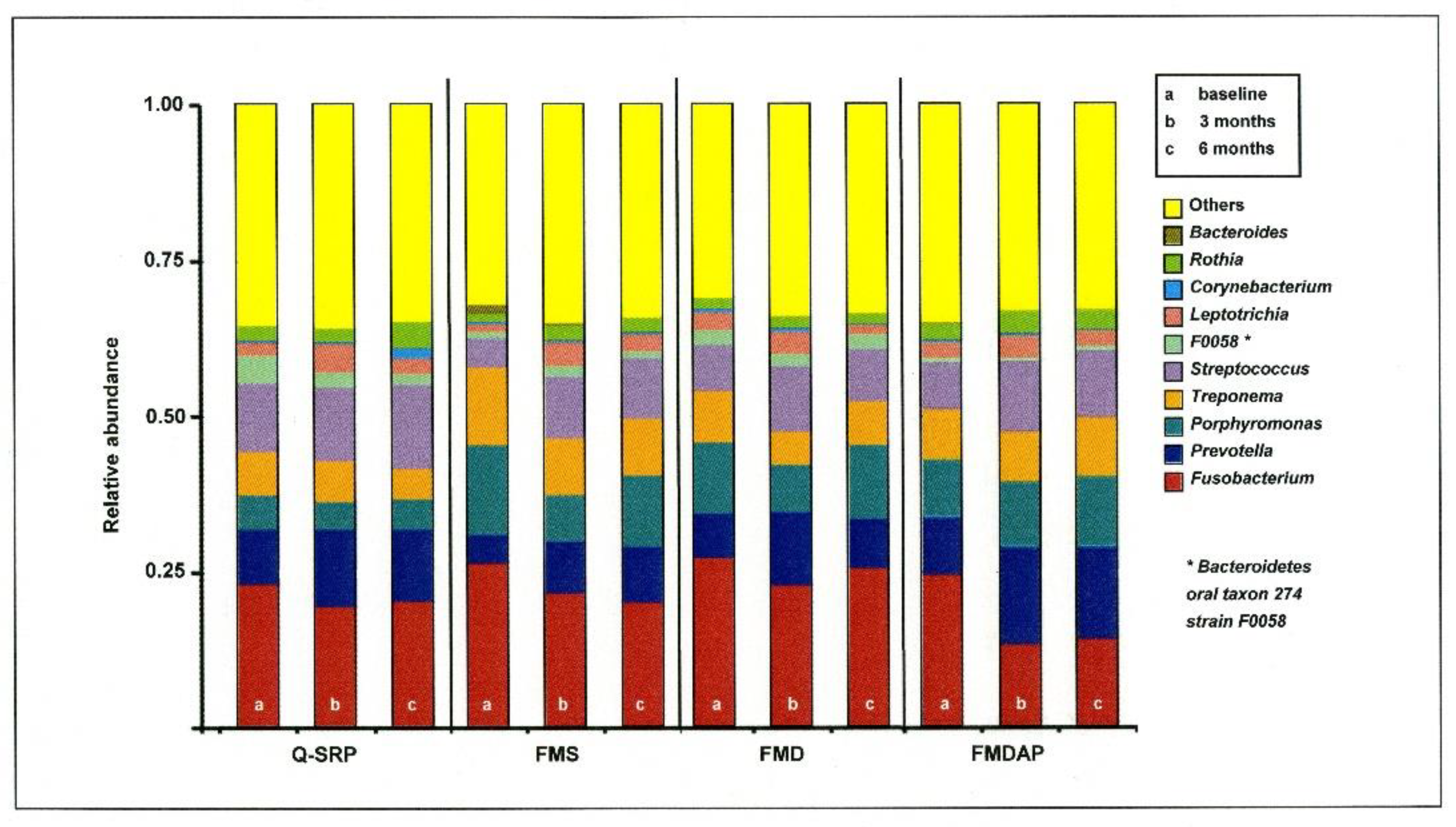
3.2.1. Relative Abundance in Relation to Periodontal Therapy

According to the species annotation results, the distribution within the different treatment groups was determined at the phylogenetic level over time. In Figure 1 the results are displayed in bar graphs for the top ten most abundant genera with (Figure 1). Among them, Fusobacterium, Prevotella, Porphyromonas, Treponema, and Streptococcus were found in the top five positions. Investigating the time course after periodontal therapy, no statistically significant differences were identified among these genera in all the periodontal treatment groups. However, a decrease in Fusobacterium (all treatment groups) and Treponema (except FMDAP group) was initially recorded 3 months after therapy, but this was associated with an increase after 6 months for Fusobacterium (Q-SRP, FMD, and FMDAP) and Treponema (FMD). However, the 6-month values remained slightly improved compared to the baseline values.
Interestingly, the pattern of Streptococcus was almost the opposite of that described for Fusobacterium.

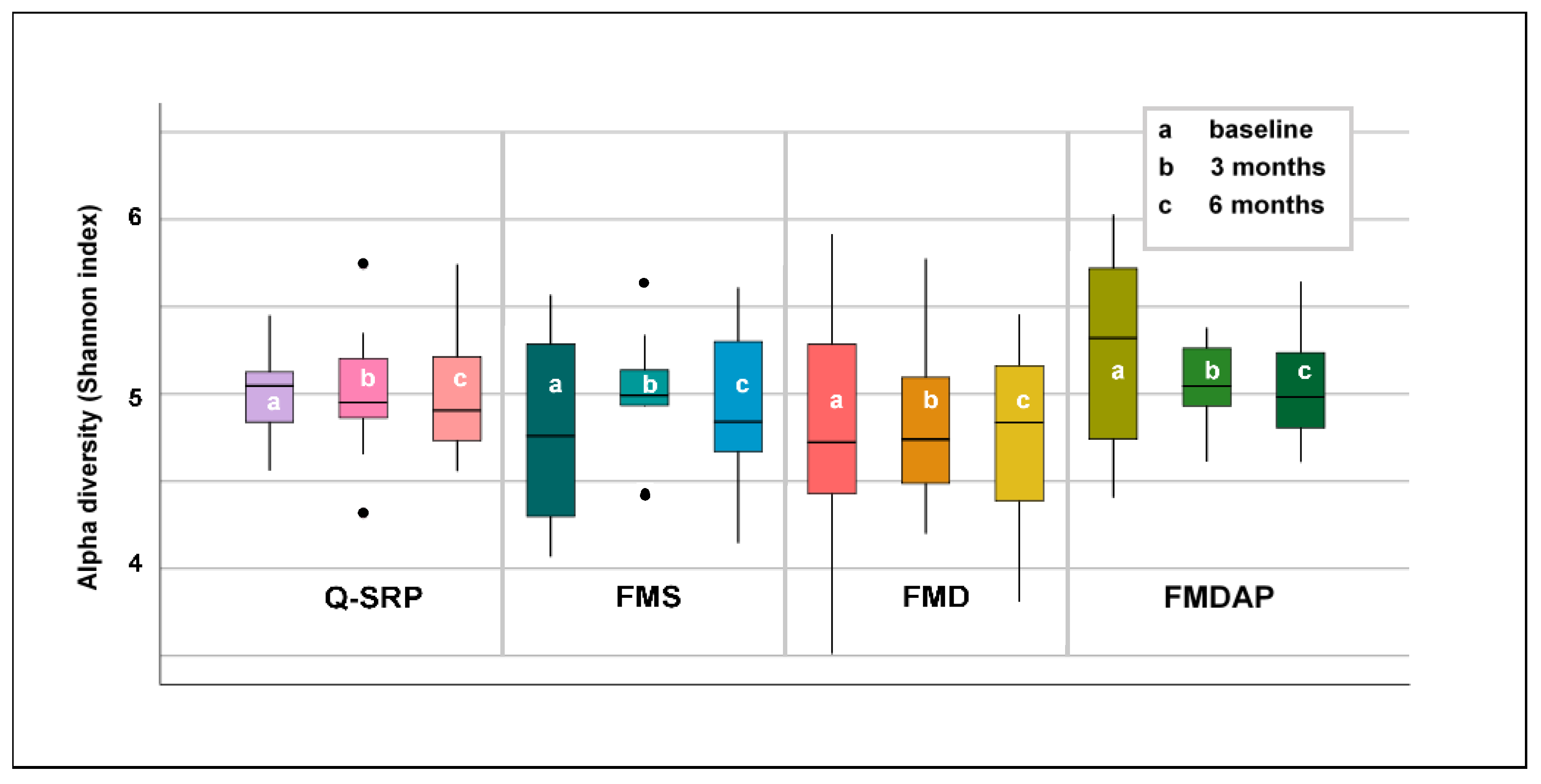
3.2.2. Alpha Diversity of Subgingival Microbiota Related to Periodontal Treatment

Differences in the alpha diversity within treatment groups were evaluated using the Shannon index (Figure 2). In conclusion, alpha diversity was not significantly changed by periodontal treatment after 3 and after 6 months. However, trends were noted. The alpha diversity in the subgingival microbiota of patients undergoing Q-SRP and FMDAP procedures decreased continuously. However, a comparable trend could not be observed for the other treatment groups. While alpha diversity initially increased in the FMS group, it decreased again after 6 months to almost the baseline level. In the FMD group, a continuous increase over time was recorded.

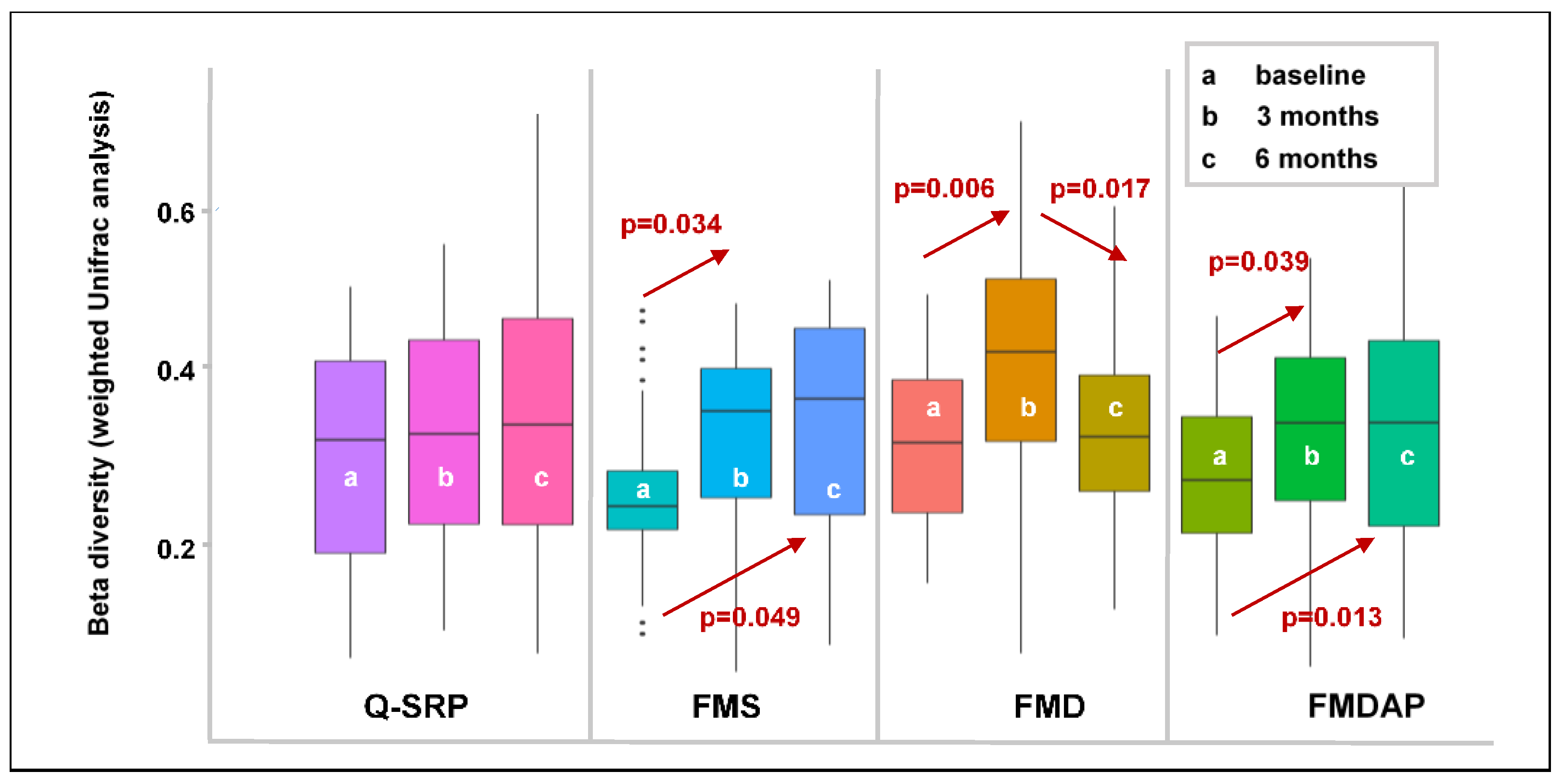
3.2.3. Effects of Periodontal Treatment on Beta Diversity

UniFrac is a widely used instrument to evaluate beta diversity, which involves the assessment of differences between communities. When considering the abundance of each taxon in a community, quantitative measures such as weighted UniFrac can be applied. When comparing community differences it was obvious that UniFrac distances increased in the FMS and FMDAP group over time after periodontal therapy (Figure 3). In the Q-SRP group a similar trend was shown. Only in the FMD treatment group was an initial increase in UniFrac distances observed 3 months after therapy combined with a decrease after another 3 months.
In the Q-SRP group, almost no significant deviation was shown at any of the phylogenetic levels. In the FMS group, changes were observed at all phylogenetic levels. At the species level, Porhyromonas gingivalis was significantly less prevalent after 3 months than at the time of treatment (p = 0.013). However, 6 months after treatment, the occurrence of this bacterium had increased again but was below the initial level (n.s.). In contrast, the occurrence of Campylobacter concisus had increased steadily 3 months (p = 0.040) and 6 months (p = 0.021) after treatment.
Patients undergoing FMD treatment initially showed a significant decrease in the occurrence of Eubacterium nodatum (p = 0.038) and Prevotella dentalis (p = 0.015) 3 months after treatment. However, colonization of these bacteria increased again 6 months after treatment, but did not reach the initial level (n.s.) (Table 3).
In the FMDAP group, the uncultured Prevotella sp. showed a course very similar to that of the two aforementioned bacteria in the FMD group; initially, a decrease (n.s.) was combined with a renewed increase (p = 0.025) (Table 3).
In PCoA, no discrimination of bacterial communities within the treatment groups was obvious regarding the different time points.
To illustrate the distribution of relative abundances within the treatment groups at different points in time, linear discriminant analysis (LDA) and LEfSe were applied. Here, we used these models for ranking bacterial species based on relative differences within the treatment groups. However, based on an LDA score of <4.0, no differences in the presence of bacteria at the phylogenetic species level were shown within any of the periodontal treatment groups.

4. Discussion

An important goal that has already been addressed in clinical trials is the issue of successful nonsurgical treatment of periodontitis [11,31]. In addition to improving the clinical periodontal parameters, the treatment effect on a shift in the subgingival bacterial spectrum is of great interest. In the present study, four different nonsurgical periodontal treatment strategies (Q-SRP, FMS, FMD, and FMDAP) were evaluated regarding changes in microbiological composition. This study is, to our knowledge, the first longitudinal study comparing the subgingival microbial outcome of different treatment options over a 6-month period. A profound change in subgingival microbial composition 3 months after treatment (FMS, FMD, and FMDAP) was shown. However, 6 months after treatment, a trend towards a profile that includes fewer disease-associated bacteria was again noticeable. Nevertheless, the microbial outcomes remained better than the baseline results.
In most studies investigating the microbiological effect of nonsurgical periodontal treatment, the focus has been on individual pathogens [14,15,16,17]. Newer analytical methods, such as 16S rRNA gene sequencing, can assess ecological changes that consider the local microbiota as a whole [32]. Therefore, this state-of-the-art approach was applied in the present study. Here, a NGS strategy using V3/V4 hypervariable regions, which combines advantages in sequencing depth, error rates, costs, and comparability with other studies, was applied [33]. From the perspective of molecular identification and quantification of defined disease-related taxa (such as P. gingivalis or P. intermedia), real-time quantitative polymerase chain reaction (qPCR) is the method of choice [34,35]. However, the present study focused on changes in the composition of the microbiota due to the different therapies and not on determining the absolute bacterial cell count.
The requirement for evaluating complex longitudinal study designs is inclusion of microbiota community variability [36]. A current approach reflecting microbiota diversity within a single sample is to assess alpha diversity over time after therapeutic intervention [36]. Shannon’s index, as one index for detecting alpha diversity, combines richness and diversity, implying that it reflects the variety of species and also takes the inequality between species abundance into account. In the present study, the alpha diversity decreased continuously after Q-SRP and FMDAP interventions (n.s., Figure 2). On the other hand, an increasing trend in alpha diversity after treatment was observed in patients receiving FMS or FMD therapy, although a continuous increase across all study time points was only demonstrated in FMD patients.
Two opposing hypotheses exist with regard to alpha diversity. One theory is that microbial diversity is higher in the subgingival plaque of patients with periodontitis [37,38], which is attributed to the greater pocket depth affecting the availability of nutrients and/or the impairment of the immune system, allowing the growth of a more diverse microbial community. In line with this theory, Johnston et al. detected a decrease in alpha diversity after nonsurgical periodontal treatment [20].
The second theory describes from a bacterial perspective that greater community diversity is associated with increased ecosystem resilience and a healthier condition [32]. This is in line with results of further studies that could confirm a disease-related decrease in alpha diversity compared to periodontally healthy individuals [39,40]. With the understanding that successful nonsurgical periodontal therapy should promote the subgingival microbiota from dysbiosis to a healthier bacterial situation, an increase in alpha diversity should be expected (contrary to the theory introduced by Shi et al., 2018 [37] and Abusleme et al., 2013 [38]). In summary, however, no conclusive assessment of the therapies implemented in this study can be derived due to the different explanatory models regarding alpha diversity.
It should be noted that alpha diversity values are not indicative of changes in microbial community composition [36]. For example, a microbial community can potentially completely shift its composition without sharing similar taxa, but still have similar alpha diversity dimensions. Therefore, in order to assess the clinical success of periodontal therapy in relation to the subgingival microbiota arrangement, changes in community composition (beta diversity) need to be evaluated. In the present study, weighted UniFrac analysis, including abundance and variety of species, was used to assess differences in beta diversity. Here, we could show an increase in beta diversity 3 months after nonsurgical periodontal therapy for all treatment groups (Q-SRP (n.s.), FMS (p = 0.034), FMD (p = 0.006), and FMDAP (p = 0.039), Figure 3). This is consistent with the clinical periodontal improvement measured in the four treatment groups in relation to the proportion of nonbleeding, shallow sites at 3 months (Table 2). In the FMS and FMDAP groups, beta diversity has increased significantly compared to baseline, even 6 months after treatment. Consistent with the increase in beta diversity, these two groups maintained their proportion of nonbleeding, shallow sites during the 6-month follow-up period (Table 2). However, in the FMD group, the rate of nonbleeding, shallow sites had decreased 6 months after treatment, which was accompanied by a significant decrease in beta diversity, too (Figure 3, Table 2). In the Q-SRP group, there was also a very slight but not significant trend for beta diversity to increase over the time, which was associated with an increase of proportion of nonbleeding, shallow sites 3 months after treatment. If previous studies on this aspect are taken as reference, the study designs and data are indeed inconsistent. On the one hand, clinical studies showed that changes in beta diversity were not in line with the periodontal outcome after nonsurgical periodontal therapy [19,41]. It should be noted, however, that none of these studies investigated different nonsurgical treatment approaches over 6 months as in the present study. On the other hand, Feres et al. showed that there was a clear distinction between pre- and post-treatment patients in terms of beta diversity [21]. However, a possible bias might have been introduced by the fact that patients were also treated with antibiotics in addition to nonsurgical periodontal therapy [21].
A more detailed assessment of the changes in microbiota composition reveals no significant changes in the Q-SRP group after treatment at all taxonomic levels. This is in line with the pattern of beta diversity, which, as mentioned above, has widely remained unchanged. A different pattern emerged in the other treatment groups. For these treatment groups, a decrease in pathogenic bacteria 3 months after treatment was initially shown, which was associated with a renewed increase 6 months after therapy. In the FMS group, Pg, the periodontal key pathogen [42], was significantly affected by periodontitis treatment. It has already been shown that the subgingival occurrence of Pg can be beneficially reduced by nonsurgical periodontal therapy [14]. Furthermore, in the FMD group differences in the prevalence of Eubacterium nodatum [43] and Prevotella dentalis [44] were noted. In the FMDAP group, uncultured Prevotella sp. also followed the course of the aforementioned bacteria (decrease 3 months after treatment and then an increase 6 months after therapy). However, it must be noted here that the uncultivated Prevotella sp. also include species that do not exhibit periodontal pathogenicity. These results regarding alterations in the microbiota over time after nonsurgical periodontitis treatment, including the gradual reintroduction of the baseline situation, have already been outlined in other studies of periodontitis treatment [45]. Based on the data obtained in the present study, we cannot confirm that the treatments investigated effectively perturbed the dysbiotic composition of the oral microbiota, which remains a condition associated with periodontal health to be promoted in the long term.

Study Limitations

The overall aim of this study was to determine longitudinal changes in clinical and microbial parameters after various nonsurgical periodontal treatments. However, there are certain limitations to the study design and the results should be interpreted in consideration of these factors.
One limiting factor is the lack of a healthy control group. It should be of interest not only to determine changes in the post-treatment microbiota but also to compare these changes with the microbiota associated with pristine periodontal health. Further studies should address this issue.
In addition, the patients were evaluated only at baseline and 3 and 6 months after treatment. By increasing the frequency of the examination intervals, possible shifts in the microbiota might be detected at an earlier stage. This knowledge could contribute to improved staging of a periodontal intervention. In a randomized clinical trial, Socransky et al. followed up patients over a period of 24 months after treatment and demonstrated in checkerboard DNA-DNA hybridization analyses that the change in select subgingival bacteria contributing to the stability of the periodontium was ensured over the study period [46]. Considering the challenge of keeping the periodontium healthy over a long period of time and reliably detecting a recurrence of periodontitis, studies with longer follow-up times should be conducted. Furthermore, no other factors that could influence periodontitis, its progression, and the outcome of nonsurgical periodontal treatment were considered in this study. For this reason, further large-scale studies should include the assessment of known periodontal risk modulators [47] such as smoking [48], gender [49], socioeconomic status [50], or oral hygiene management [51,52]. Therefore, these findings and their relevance for clinical practice should be considered on an individual basis [53].
Another limitation is that other substances whose effects on oral microbiota have already been demonstrated were not included in this study. Substances including paraprobiotics [54]; polyphenols, essential oils, and alkaloids [55]; and tea tree oil and ethanolic extract of propolis [56], aloe vera [57], or ozone [58] can be considered important adjuncts to periodontal therapy. For this reason, further studies should investigate such options for supplementing periodontal therapy.

5. Conclusions

In conclusion, we could confirm our hypothesis that the clinical success of nonsurgical periodontal treatment is associated with a change in microbial composition. The proportion of nonbleeding, shallow sites assessed by periodontal treatments with a full-mouth approach (FMS, FMD, and FMDAP) was accompanied by a reduction in the subgingival occurrence of bacteria presumably associated with periodontitis. Despite further improvement (FMS and FMDAP) or a slight decrease (FMD) in clinical outcomes, there was a clear recurrence of bacteria 6 months after therapy.
However, at this time, no clinical implications can be derived from the available microbiological data. The results from this study can be used to design large-scale prospective studies to investigate different nonsurgical periodontal treatment strategies, considering further influencing periodontal factors on the microbiota profile. This may open up opportunities to integrate the subgingival microbiota for diagnosis and risk assessment of periodontitis in the future. Investigating the subgingival microbiota might help appraise successful periodontal treatment and implement individualized therapy.

Author Contributions

All authors have made substantial contributions to conception and design of the study. J.M.S., A.S., D.K., S.S.Y.-M., F.S. and S.R. were involved in the recruitment and dental examination and therapy of the patients. S.S. and G.C. have been implicated in microbiological assessment. Mathematical analysis and interpretation of the data was performed by S.S., J.M.S., H.-G.S. and S.R., S.S., J.M.S., S.S.Y.-M., G.C. and S.R. were involved in drafting the manuscript and revising it critically. All authors have read and agreed to the published version of the manuscript.

Funding

The study was partly supported by E.M.S. Electro Medical Systems, Nyon/Switzerland and CP GABA GmbH, Hamburg/Germany. We acknowledge the financial support of the Open Access Publication Fund of the Martin-Luther-University Halle-Wittenberg.

Institutional Review Board Statement

The present study is a sub-study of the clinical trial database registered study “Anti-infectious Therapy of Periodontitis—Comparison of Different Clinical Strategies” (ClinicalTrials.gov: NCT03509233). The study was approved by the ethics committee of the University of Aachen, Germany (EK 046/16).

Informed Consent Statement

Informed written consent was obtained from each patient. All investigations were carried out in accordance with the ethical guidelines of the “Declaration of Helsinki” and its amendment in “Tokyo and Venice”.

Data Availability Statement

The comprehensive study protocol is available from the authors. Data can be provided by the authors upon request.

Acknowledgments

Authors wish to thank Johannes Brockmann, Aachen/Germany, Volker Beschow and Elisabeth Ehnert, Halle/Germany. Their support in recruitment and examination of study participants and co-ordination of all treatment procedures during the study is appreciated. We would like to thank all patients for participating in this study.

Conflicts of Interest

The authors declare no conflict of interest.

References

  1. Hajishengallis, G.; Liang, S.; Payne, M.A.; Hashim, A.; Jotwani, R.; Eskan, M.A.; McIntosh, M.L.; Alsam, A.; Kirkwood, K.L.; Lambris, J.D.; et al. Low-abundance biofilm species orchestrates inflammatory periodontal disease through the commensal microbiota and complement. Cell Host Microbe 2011, 10, 497–506. [Google Scholar] [CrossRef] [PubMed] [Green Version]
  2. Hajishengallis, G.; Darveau, R.P.; Curtis, M.A. The keystone pathogen hypothesis. Nat. Rev. Microbiol. 2011, 10, 717–725. [Google Scholar] [CrossRef] [PubMed]
  3. Khan, S.; Khalid, T.; Bettiol, S.; Crocombe, L.A. Non-surgical periodontal therapy effectively improves patient-reported outcomes: A systematic review. Int. J. Dent. Hyg. 2021, 19, 18–28. [Google Scholar] [CrossRef] [PubMed]
  4. Beikler, T.; Abdeen, G.; Schnitzer, S.; Sälzer, S.; Ehmke, B.; Heinecke, A.; Fleming, T.F. Microbiological shifts in intra- and extraoral habitats following mechanical periodontal therapy. J. Clin. Periodontol. 2004, 31, 777–783. [Google Scholar] [CrossRef] [PubMed]
  5. Quirynen, M.; Bollen, C.M.; Vandekerckhove, B.N.; Dekeyser, C.; Papaioannou, W.; Eyssen, H. Full- vs. partial-mouth disinfection in the treatment of periodontal infections: Short-term clinical and microbiological observations. J. Dent. Res. 1995, 74, 1459–1467. [Google Scholar] [CrossRef] [PubMed]
  6. Quirynen, M.; Mongardini, C.; van Steenberghe, D. The effect of a 1-stage full-mouth disinfection on oral malodor and microbial colonization of the tongue in periodontitis. A pilot study. J. Periodontol. 1998, 69, 374–382. [Google Scholar] [CrossRef] [PubMed]
  7. Quirynen, M.; De Soete, M.; Boschmans, G.; Pauwels, M.; Coucke, W.; Teughels, W.; van Steenberghe, D. Benefit of “one-stage full-mouth disinfection” is explained by disinfection and root planing within 24 hours: A randomized controlled trial. J. Clin. Periodontol. 2006, 33, 639–647. [Google Scholar] [CrossRef]
  8. Fang, H.; Han, M.; Li, Q.L.; Cao, C.Y.; Xia, R.; Zhang, Z.H. Comparison of full-mouth disinfection and quadrant-wise scaling in the treatment of adult chronic periodontitis: A systematic review and meta-analysis. J. Periodontal. Res. 2016, 51, 417–430. [Google Scholar] [CrossRef]
  9. Santuchi, C.C.; Cortelli, J.R.; Cortelli, S.C.; Cota, L.O.; Fonseca, D.C.; Alencar, C.O.; Costa, F.O. Scaling and Root Planing per Quadrant Versus One-Stage Full-Mouth Disinfection: Assessment of the Impact of Chronic Periodontitis Treatment on Quality of Life-A Clinical Randomized, Controlled Trial. J. Periodontol. 2016, 87, 114–123. [Google Scholar] [CrossRef]
  10. Fonseca, D.C.; Cortelli, J.R.; Cortelli, S.C.; Miranda Cota, L.O.; Machado Costa, L.C.; Moreira Castro, M.V.; Oliveira Azevedo, A.M.; Costa, F.O. Clinical and Microbiologic Evaluation of Scaling and Root Planing per Quadrant and One-Stage Full-Mouth Disinfection Associated with Azithromycin or Chlorhexidine: A Clinical Randomized Controlled Trial. J. Periodontol. 2015, 86, 1340–1351. [Google Scholar] [CrossRef]
  11. Eberhardt, J.; Jepsen, S.; Jervøe-Storm, P.M.; Needleman, I.; Worthington, H.V. Full-mouth treatment modalities (within 24 hours) for chronic periodontitis in adults. Cochrane Database Syst. Rev. 2015, 17, CD004622. [Google Scholar] [CrossRef] [PubMed] [Green Version]
  12. Jentsch, H.F.R.; Flechsig, C.; Kette, B.; Eick, S. Adjunctive air-polishing with erythritol in nonsurgical periodontal therapy: A randomized clinical trial. BMC Oral Health 2020, 20, 364. [Google Scholar] [CrossRef] [PubMed]
  13. Stein, J.M.; Michael, S.; Schittenhelm, F.; Reichert, S.; Kupitz, D.; Dommisch, H.; Kasaj, A.; Wied, S.; Vela, O.; Stratul, S. Comparison of three full-mouth concepts for the nonsurgical treatment of stage III and IV periodontitis—A randomized controlled trial. J. Clin. Periodontol. 2021, 48, 1516–1527. [Google Scholar] [CrossRef] [PubMed]
  14. Doungudomdacha, S.; Rawlinson, A.; Walsh, T.F.; Douglas, C.W.I. Effect of non-surgical periodontal treatment on clinical parameters and the numbers of Porphyromonas gingivalis, Prevotella intermedia and Actinobacillus actinomycetemcomitans at adult periodontitis sites. J. Clin. Periodontol. 2001, 28, 437–445. [Google Scholar] [CrossRef] [PubMed]
  15. Rodrigues, A.S.; Lourenção, D.S.; Neto, L.G.L.; Pannuti, C.M.; Hirata, R.D.C.; Hirata, M.H.; Lotufo, R.F.M.; De Micheli, G. Clinical and Microbiologic Evaluation, by Real-Time Polymerase Chain Reaction, of Non-Surgical Treatment of Aggressive Periodontitis Associated with Amoxicillin and Metronidazole. J. Periodontol. 2012, 83, 744–752. [Google Scholar] [CrossRef]
  16. Finoti, L.S.; Corbi, S.C.T.; Anovazzi, G.; Teixeira, S.R.L.; Capela, M.V.; Tanaka, M.H.; Kim, Y.J.; Orrico, S.R.P.; Cirelli, J.A.; Mayer, M.P.A.; et al. Pathogen levels and clinical response to periodontal treatment in patients with Interleukin 8 haplotypes. Pathog. Dis. 2013, 69, 21–28. [Google Scholar]
  17. Gul, S.S.; Griffiths, G.S.; Stafford, G.P.; Al-Zubidi, M.I.; Rawlinson, A.; Douglas, C.W.I. Investigation of a Novel Predictive Biomarker Profile for the Outcome of Periodontal Treatment. J. Periodontol. 2017, 88, 1135–1144. [Google Scholar] [CrossRef]
  18. Rosalem, W.; Rescala, B.; Teles, R.P.; Fischer, R.G.; Gustafsson, A.; Figueredo, C.M. Effect of non-surgical treatment on chronic and aggressive periodontitis: Clinical, immunologic, and microbiologic findings. J. Periodontol. 2011, 82, 979–989. [Google Scholar] [CrossRef]
  19. Chen, C.; Hemme, C.; Beleno, J.; Shi, Z.J.; Ning, D.; Qin, Y.; Tu, Q.; Jorgensen, M.; He, Z.; Wu, L.; et al. Oral microbiota of periodontal health and disease and their changes after nonsurgical periodontal therapy. ISME J. 2018, 12, 1210–1224. [Google Scholar] [CrossRef]
  20. Johnston, W.; Rosier, B.T.; Artacho, A.; Paterson, M.; Piela, K.; Delaney, C.; Brown, J.L.; Ramage, G.; Mira, A.; Culshaw, S. Mechanical biofilm disruption causes microbial and immunological shifts in periodontitis patients. Sci. Rep. 2021, 11, 9796. [Google Scholar] [CrossRef]
  21. Feres, M.; Retamal-Valdes, B.; Fermiano, D.; Faveri, M.; Figueiredo, L.C.; Mayer, M.P.A.; Lee, J.J.; Bittinger, K.; Teles, F. Microbiome changes in young periodontitis patients treated with adjunctive metronidazole and amoxicillin. J. Periodontol. 2021, 92, 467–478. [Google Scholar] [CrossRef] [PubMed]
  22. Papapanou, P.N.; Sanz, M.; Buduneli, N.; Dietrich, T.; Feres, M.; Fine, D.H.; Flemmig, T.F.; Garcia, R.; Giannobile, W.V.; Graziani, F.; et al. Periodontitis: Consensus report of workgroup 2 of the 2017 World Workshop on the Classification of Periodontal and Peri-Implant Diseases and Conditions. J. Periodontol. 2018, 89 (Suppl. 1), S173–S182. [Google Scholar] [CrossRef] [Green Version]
  23. Quirynen, M.; Mongardini, C.; De Soete, M.; Pauwels, M.; Coucke, W.; Van Eldere, J.; van Steenberghe, D. The role of chlorhexidine in the one-stage full-mouth disinfection treatment of patients with advanced adult periodontitis. Long-term clinical and microbiological observations. J. Clin. Periodontol. 2000, 27, 578–589. [Google Scholar] [CrossRef] [PubMed]
  24. Kinane, D.F.; Radvar, M. The effect of smoking on mechanical and antimicrobial periodontal therapy. J. Periodontol. 1997, 68, 467–472. [Google Scholar] [CrossRef] [PubMed]
  25. Löe, H. The gingival index, the plaque index and the retention index systems. J. Periodontol. 1967, 38, 610–616. [Google Scholar] [CrossRef]
  26. Magoc, T.; Salzberg, S.L. FLASH: Fast length adjustment of short reads to improve genome assemblies. Bioinformatics 2011, 27, 2957–2963. [Google Scholar] [CrossRef]
  27. Caporaso, J.G.; Kuczynski, J.; Stombaugh, J.; Bittinger, K.; Bushman, F.D.; Costello, E.K.; Fierer, N.; Peña, A.G.; Goodrich, J.K.; Gordon, J.I.; et al. QIJME allows analysis of high-throughput community sequencing data. Nat. Methods 2010, 7, 335–336. [Google Scholar] [CrossRef] [Green Version]
  28. Edgar, R.C.; Haas, B.J.; Clemente, J.C.; Quince, C.; Knight, R. UCHIME improves sensitivity and speed chimera detection. Bioinformatics 2011, 27, 2194–2200. [Google Scholar] [CrossRef] [Green Version]
  29. Edgar, R.C. UPARSE: Highly accurate OUT sequences from microbial amplicons reads. Nat. Methods 2013, 10, 996–998. [Google Scholar] [CrossRef]
  30. Wang, Q.; Garrity, G.M.; Tiedje, J.M.; Cole, J.R. Naive Bayesian classifier for rapid assignment of rRNA sequences into the new bacterial taxonomy. Appl. Environ. Microbiol. 2007, 73, 5261–5267. [Google Scholar] [CrossRef] [Green Version]
  31. Kwon, T.; Lamster, I.B.; Levin, L. Current concepts in the management of periodontitis. Intern. Dent. J. 2021, 71, 462–476. [Google Scholar] [CrossRef]
  32. Liu, G.; Luan, Q.; Chen, F.; Chen, Z.; Zhang, Q.; Yu, X. Shift in the subgingival microbiome following scaling and root planing in generalized aggressive periodontitis. J. Clin. Periodontol. 2018, 45, 440–452. [Google Scholar] [CrossRef] [PubMed]
  33. Papapanou, P.N.; Park, H.; Cheng, B.; Kokaras, A.; Paster, B.; Burkett, S.; Watson, C.W.; Annavajhala, M.K.; Uhlemann, A.C.; Noble, J.M. Subgingival microbiome and clinical periodontal status in an elderly cohort: The WHICAP ancillary study of oral health. J. Periodontol. 2020, 91 (Suppl. 1), S56–S67. [Google Scholar] [CrossRef] [PubMed]
  34. Sawant, S.; Dugad, J.; Parikh, D.; Singh, H. Absolute quantification of oral bacteria involved in oral cancer by real-time PCR. Med. Microecol. 2021, 7, 100034. [Google Scholar] [CrossRef]
  35. Abdelbary, M.M.H.; Schittenhelm, F.; Yekta-Michael, S.S.; Reichert, S.; Schulz, S.; Kasaj, A.; Braun, A.; Conrads, G.; Stein, J.M. Impact of three nonsurgical, full-mouth periodontal treatments on total bacterial load and selected pathobionts. Antibiotics 2022. under revision. [Google Scholar]
  36. Wagner, B.D.; Grunwald, G.K.; Zerbe, G.O.; Mikulich-Gilbertson, S.K.; Robertson, C.E.; Zemanick, E.T.; Harris, J.K. On the use of diversity measures in longitudinal sequencing studies of microbial communities. Front. Microbiol. 2018, 9, 1037. [Google Scholar] [CrossRef] [Green Version]
  37. Shi, M.; We, Y.; Hu, W.; Nie, Y.; Wu, X.; Lu, R. The subgingival microbiome of periodontal pockets with different probing depths in chronic and aggressive periodontitis: A pilot study. Front. Cell. Infect. Microbiol. 2018, 8, 124. [Google Scholar] [CrossRef]
  38. Abusleme, L.; Dupuy, A.K.; Dutzan, N.; Silva, N.; Burleson, J.A.; Strausbaugh, L.D.; Gamonal, J.; Diaz, P.I. The subgingival microbiome in health and periodontitis and its relationship with community biomass and infammation. ISME J. 2013, 7, 1016–1025. [Google Scholar] [CrossRef] [Green Version]
  39. Schulz, S.; Porsch, M.; Große, I.; Hoffmann, K.; Schaller, H.G.; Reichert, S. Comparison of the oral microbiome of patients with aggressive periodontitis and periodontitisfree subjects. Arch. Oral. Biol. 2019, 99, 169–176. [Google Scholar] [CrossRef]
  40. Deng, Z.L.; Szafranski, S.P.; Jarek, M.; Bhuju, S.; Wagner-Döbler, I. Dysbiosis in chronic periodontitis: Key microbial players and interactions with the human host. Sci. Rep. 2017, 7, 3703. [Google Scholar] [CrossRef] [Green Version]
  41. Schwarzberg, K.; Le, R.; Bharti, B.; Lindsay, S.; Casaburi, G.; Salvatore, F.; Saber, M.H.; Alonaizan, F.; Slots, J.; Gottlieb, R.A.; et al. The personal human oral microbiome obscures the effects of treatment on periodontal disease. PLoS ONE 2014, 9, e86708. [Google Scholar] [CrossRef] [PubMed]
  42. Rafiei, M.; Kiani, F.; Sayehmiri, F.; Sayehmiri, K.; Sheikhi, A.; Azodi, M.Z. Study of Porphyromonas gingivalis in periodontal diseases: A systematic review and meta-analysis. Med. J. Islam. Repub. Iran 2017, 31, 62. [Google Scholar] [CrossRef] [PubMed] [Green Version]
  43. Haffajee, A.D.; Teles, R.P.; Socransky, S.S. Association of Eubacterium nodatum and Treponema denticola with human periodontitis lesions. Oral. Microbiol. Immunol. 2006, 21, 269–282. [Google Scholar] [CrossRef] [PubMed]
  44. Ibrahim, M.; Subramanian, A.; Anishetty, S. Comparative pan genome analysis of oral Prevotella species implicated in periodontitis. Funct. Integr. Genom. 2017, 17, 513–536. [Google Scholar] [CrossRef] [PubMed]
  45. Nibali, L.; Sousa, V.; Davrandi, M.; Spratt, D.; Alyahya, Q.; Dopico, J.; Donos, N. Differences in the periodontal microbiome of successfully treated and persistent aggressive periodontitis. J. Clin. Periodontol. 2020, 47, 980–990. [Google Scholar] [CrossRef] [PubMed]
  46. Socransky, S.S.; Haffajee, A.D.; Teles, R.; Wennstrom, J.L.; Lindhe, J.; Bogren, A.; Hasturk, H.; van Dyke, T.; Wang, X.; Goodson, J.M. Effect of periodontal therapy on the subgingival microbiota over a 2-year monitoring period. I. Overall effect and kinetics of change. J. Clin. Periodontol. 2013, 40, 771–780. [Google Scholar] [CrossRef] [PubMed] [Green Version]
  47. Bouchard, P.; Carra, M.C.; Boillot, A.; Mora, F.; Rangé, H. Risk factors in periodontology: A conceptual framework. J. Clin. Periodontol. 2017, 44, 125–131. [Google Scholar] [CrossRef]
  48. Leite, F.R.M.; Nascimento, G.G.; Scheutz, F.; López, R. Effect of Smoking on Periodontitis: A Systematic Review and Meta-regression. Am. J. Prev. Med. 2018, 54, 831–841. [Google Scholar] [CrossRef]
  49. Ioannidou, E. The Sex and Gender Intersection in Chronic Periodontitis. Front. Public Health 2017, 5, 189. [Google Scholar] [CrossRef] [Green Version]
  50. Bartold, P.M. Lifestyle and periodontitis: The emergence of personalized periodontics. Periodontology 2000 2018, 78, 7–11. [Google Scholar] [CrossRef]
  51. Kinane, D.F.; Stathopoulou, P.G.; Papapanou, P.N. Periodontal diseases. Nat. Rev. Dis. Primers 2017, 3, 17038. [Google Scholar] [CrossRef]
  52. Ballini, A.; Di Cosola, M.; Saini, R.; Benincasa, C.; Aiello, E.; Marrelli, B.; Rajiv Saini, S.; Ceruso, F.M.; Nocini, R.; Topi, S.; et al. A Comparison of Manual Nylon Bristle Toothbrushes versus Thermoplastic Elastomer Toothbrushes in Terms of Cleaning Efficacy and the Biological Potential Role on Gingival Health. Appl. Sci. 2021, 11, 7180. [Google Scholar] [CrossRef]
  53. Inchingolo, F.; Martelli, F.S.; Gargiulo Isacco, C.; Borsani, E.; Cantore, S.; Corcioli, F.; Boddi, A.; Nguyễn, K.C.D.; De Vito, D.; Aityan, S.K.; et al. Chronic Periodontitis and Immunity, Towards the Implementation of a Personalized Medicine: A Translational Research on Gene Single Nucleotide Polymorphisms (SNPs) Linked to Chronic Oral Dysbiosis in 96 Caucasian Patients. Biomedicines 2020, 8, 115. [Google Scholar] [CrossRef]
  54. Butera, A.; Gallo, S.; Pascadopoli, M.; Maiorani, C.; Milone, A.; Alovisi, M.; Scribante, A. Paraprobiotics in Non-Surgical Periodontal Therapy: Clinical and Microbiological Aspects in a 6-Month Follow-Up Domiciliary Protocol for Oral Hygiene. Microorganisms 2022, 10, 337. [Google Scholar] [CrossRef]
  55. Furquim Dos Santos Cardoso, V.; Amaral Roppa, R.H.; Antunes, C.; Silva Moraes, A.N.; Santi, L.; Konrath, E.L. Efficacy of medicinal plant extracts as dental and periodontal antibiofilm agents: A systematic review of randomized clinical trials. J. Ethnopharmacol. 2021, 281, 114541. [Google Scholar] [CrossRef] [PubMed]
  56. Wiatrak, K.; Morawiec, T.; Rój, R.; Kownacki, P.; Nitecka-Buchta, A.; Niedzielski, D.; Wychowański, P.; Machorowska-Pieniążek, A.; Cholewka, A.; Baldi, D.; et al. Evaluation of Effectiveness of a Toothpaste Containing Tea Tree Oil and Ethanolic Extract of Propolis on the Improvement of Oral Health in Patients Using Removable Partial Dentures. Molecules 2021, 26, 4071. [Google Scholar] [CrossRef] [PubMed]
  57. Pradeep, A.R.; Agarwal, E.; Naik, S.B. Clinical and microbiologic effects of commercially available dentifrice containing aloe vera: A randomized controlled clinical trial. J. Periodontol. 2012, 83, 797–804. [Google Scholar] [CrossRef]
  58. Hayakumo, S.; Arakawa, S.; Takahashi, M.; Kondo, K.; Mano, Y.; Izumi, Y. Effects of ozone nano-bubble water on periodontopathic bacteria and oral cells-in vitro studies. Sci. Technol. Adv. Mater. 2014, 15, 55003. [Google Scholar] [CrossRef] [Green Version]
Figure 1. Microbial community composition at genus level (top 10 with the highest relative abundance). The top four genera were Fusobacterium, Prevotella, Porphyromonas, and Treponema. No statistically significant differences were identified among these genera within the periodontal treatment groups over time. Q-SRP: quadrant-wise subgingival scaling and root planing (SRP); FMS: full-mouth SRP without the use of antiseptics; FMD: full-mouth SRP within 24 h with disinfection using antiseptic applications; FMDAP: FMD combined with the use of subgingival erythritol air-polishing.
Jcm 11 01187 g001
Figure 2. Variations over time in alpha diversity (Shannon index) of patients’ subgingival microbiota considering periodontal treatment (comparing paired groups by applying Wilcoxon signed-ranks test). Q-SRP: quadrant-wise subgingival scaling and root planing (SRP); FMS: full-mouth SRP without the use of antiseptics; FMD: full-mouth SRP within 24 h with disinfection using antiseptic applications; FMDAP: FMD combined with subgingival erythritol air-polishing.
Jcm 11 01187 g002
Figure 3. Variations over time in beta diversity (weighted UniFrac analysis) of patients’ subgingival microbiota considering periodontal treatment (comparing of paired groups by applying Wilcoxon signed-ranks test, corrected for multiple comparisons using the Benjamini–Hochberg false discovery rate of 5%). Q-SRP: quadrant-wise subgingival scaling and root planing (SRP); FMS: full-mouth SRP without the use of antiseptics; FMD: full-mouth SRP within 24 h with disinfection using antiseptic applications; FMDAP: FMD combined with the use of subgingival erythritol air-polishing.
Jcm 11 01187 g003
Table 1. Patient data recorded at the baseline examination.
VariableQ-SRP
(n = 10)
FMS
(n = 10)
FMD
(n = 10)
FMDAP
(n = 10)
Age (years)58.1 ± 9.755.3 ± 11.858.9 ± 14.358.0 ± 12.6
Gender (male/female)8/26/44/65/5
Smokers (n)2441
Q-SRP: quadrant-wise subgingival scaling and root planing (SRP); FMS: full-mouth SRP without the use of antiseptics; FMD: full-mouth SRP within 24 h with disinfection using antiseptic applications; FMDAP: FMD combined with the use of subgingival erythritol air-polishing.
Table 2. Full-mouth clinical parameters at baseline and follow-up visits.
VariableTimepointQ-SRP
(n = 10)
FMS
(n = 10)
FMD
(n = 10)
FMDAP
(n = 10)
PIBaseline1.09 ± 0.351.35 ± 0.540.98 ± 0.601.14 ± 0.48
3 months0.55 ± 0.35 *0.78 ± 0.45 *0.52 ± 0.46 *0.49 ± 0.37 *
6 months0.60 ± 0.48 *0.92 ± 0.45 *0.82 ± 0.540.40 ± 0.25 *
GIBaseline1.28 ± 0.381.47 ± 0.681.22 ± 0.551.31 ± 0.47
3 months0.71 ± 0.56 *0.79 ± 0.58 *0.55 ± 0.52 *0.55 ± 0.45 *
6 months0.86 ± 0.68 *0.64 ± 0.51 *0.56 ± 0.43 *0.42 ± 0.32 *
PPD (mm)Baseline3.77 ± 0.743.92 ± 0.644.00 ± 0.493.91 ± 0.60
3 months3.32 ± 0.663.43 ± 0.60 *3.47 ± 0.47 *3.18 ± 0.69 *
6 months3.36 ± 0.623.35 ± 0.58 *3.30 ± 0.55 *3.18 ± 0.63 *
CAL (mm) Baseline4.36 ± 0.964.47 ± 0.525.10 ± 1.024.68 ± 0.75
3 months3.98 ± 0.82 *4.14 ± 0.49 *4.66 ± 0.93 *4.12 ± 0.65 *
6 months4.01 ± 0.81 *4.11 ± 0.644.63 ± 1.07 *4.15 ± 0.63 *
BOP (%)Baseline33.17 ± 14.8249.60 ± 23.0033.78 ± 14.5442.36 ± 19.26
3 months18.06 ± 11.88 *24.65 ± 20.54 *18.73 ± 12.12 *15.19 ± 15.65 *
6 months21.07 ± 11.00 *17.61 ± 17.4324.90 ± 17.67 *12.59 ± 13.78 *
NBSS (%)Baseline57.00 ± 17.1740.50 ± 21.1147.50 ± 16.8848.40 ± 18.93
3 months71.10 ±13.61 *64.50 ± 17.78 *70.40 ± 15.31 *75.50 ± 19.23 *
6 months69.80 ± 12.39 *70.80 ± 17.55 *66.6 ± 17.2779.20 ± 16.40 *
* Statistically significant differences (p < 0.05, t-test, paired sample) in comparison to baseline values; Q-SRP: quadrant-wise subgingival scaling and root planing (SRP); FMS: full-mouth SRP without the use of antiseptics; FMD: full-mouth SRP within 24 h with disinfection using antiseptic applications; FMDAP: FMD combined with the use of subgingival erythritol air-polishing; PI: plaque index; GI: gingival index; PPD: probing depth; CAL: clinical attachment level; BOP: bleeding on probing. NBSS: nonbleeding, shallow sites (PPD ≤ 4 mm without BOP).
Table 3. Occurrence of bacteria at baseline and follow-up visits in different treatment groups (only species are displayed that exhibit significant changes in the t-test, paired sample).
TreatmentTime PointBacterial Species
P. gingivalis change regarding baselineC. consius change regarding baseline
FMSBaseline
3 months
6 months
100%
41.7% *
75%
100%
128.6% *
300% *
E. nodatum change regarding baselineP. dentalis change regarding baseline
FMDBaseline
3 months
6 months
100%
33.3% *
66.7%
100%
25% *
50%
Uncultured Prevotella sp. change regarding baseline
FMDAPBaseline
3 months
6 months
100%
29.4% *
76.5%
* Statistically significant differences (p < 0.05, t-test, paired sample) in comparison to baseline values. FMS: full-mouth SRP without the use of antiseptics; FMD: full-mouth SRP within 24 h with disinfection using antiseptic applications; FMDAP: FMD combined with the use of subgingival erythritol air-polishing.
Publisher’s Note: MDPI stays neutral with regard to jurisdictional claims in published maps and institutional affiliations.

