
More Space, Less Noise—New-generation Low-Field Magnetic Resonance Imaging Systems Can Improve Patient Comfort: A Prospective 0.55T–1.5T-Scanner Comparison
 , ,
, ,
Abstract
1. Introduction
2. Materials and Methods
2.1. Study Population
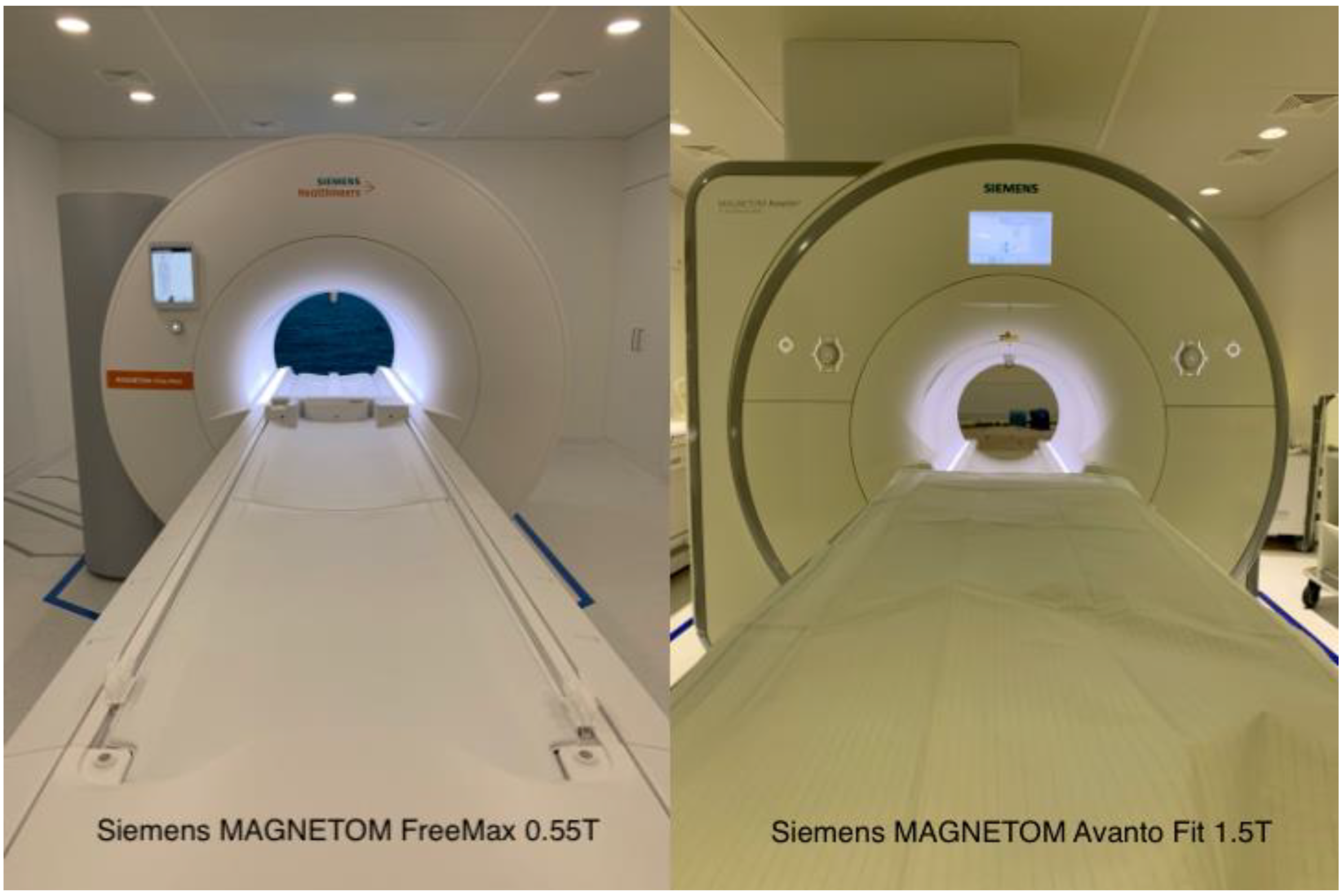
2.2. MR Imaging
2.3. Questionnaire
2.4. Noise Measurements
2.5. Statistics
3. Results
3.1. Questionnaire Results
3.2. Measurement of the Noise Levels
4. Discussion
Author Contributions
Funding
Institutional Review Board Statement
Informed Consent Statement
Data Availability Statement
Conflicts of Interest
References
- Mannaerts, C.K.; Kajtazovic, A.; Lodeizen, O.A.; Gayet, M.; Engelbrecht, M.R.; Jager, G.J.; Wijkstra, H.; de Reijke, T.M.; Beerlage, H.P. The Added Value of Systematic Biopsy in Men with Suspicion of Prostate Cancer undergoing Multiparametric MRI-Targeted Biopsy. In Urologic Oncology: Seminars and Original Investigations; Elsevier: Amsterdam, The Netherlands, 2019; p. 298.e1. [Google Scholar]
- Viallon, M.; Cuvinciuc, V.; Delattre, B.; Merlini, L.; Barnaure-Nachbar, I.; Toso-Patel, S.; Becker, M.; Lovblad, K.O.; Haller, S. State-of-the-art MRI techniques in neuroradiology: Principles, pitfalls, and clinical applications. Neuroradiology 2015, 57, 441–467. [Google Scholar] [CrossRef] [PubMed]
- Maron, M.S.; Maron, B.J. Clinical impact of contemporary cardiovascular magnetic resonance imaging in hypertrophic cardiomyopathy. Circulation 2015, 132, 292–298. [Google Scholar] [CrossRef] [PubMed]
- Palestro, C.J.; Love, C.; Miller, T.T. Imaging of musculoskeletal infections. Best Pract. Res. Clin. Rheumatol. 2006, 20, 1197–1218. [Google Scholar] [CrossRef] [PubMed]
- Dewey, M.; Schink, T.; Dewey, C.F. Claustrophobia during magnetic resonance imaging: Cohort study in over 55,000 patients. J. Magn. Reson. Imaging 2007, 26, 1322–1327. [Google Scholar] [CrossRef] [PubMed]
- Sadigh, G.; Applegate, K.E.; Saindane, A.M. Prevalence of Unanticipated Events Associated with MRI Examinations: A Benchmark for MRI Quality, Safety, and Patient Experience. J. Am. Coll. Radiol. 2017, 14, 765–772. [Google Scholar] [CrossRef] [PubMed]
- Eshed, I.; Althoff, C.E.; Hamm, B.; Hermann, K.-G.A. Claustrophobia and premature termination of magnetic resonance imaging examinations. J. Magn. Reson. Imaging 2007, 26, 401–404. [Google Scholar] [CrossRef] [PubMed]
- Alibek, S.; Vogel, M.; Sun, W.; Winkler, D.; Baker, C.A.; Burke, M.; Gloger, H. Acoustic noise reduction in MRI using Silent Scan: An initial experience. Diagn. Interv. Radiol. 2014, 20, 360–363. [Google Scholar] [CrossRef] [PubMed]
- Brunnquell, C.L.; Hoff, M.N.; Balu, N.; Nguyen, X.V.; Oztek, M.A.; Haynor, D.R. Making magnets more attractive: Physics and engi-neering contributions to patient comfort in MRI. Top Magn. Reason. Imaging 2020, 29, 167–174. [Google Scholar] [CrossRef] [PubMed]
- Iwan, E.; Yang, J.; Enders, J.; Napp, A.E.; Rief, M.; Dewey, M. Patient preferences for development in MRI scanner design: A survey of claustrophobic patients in a randomized study. Eur. Radiol. 2020, 31, 1325–1335. [Google Scholar] [CrossRef] [PubMed]
- Runge, V.M.; Heverhagen, J.T. Advocating the Development of Next-Generation, Advanced-Design Low-Field Magnetic Resonance Systems. Investig. Radiol. 2020, 55, 747–753. [Google Scholar] [CrossRef] [PubMed]
- Rusche, T.; Breit, H.C.; Bach, M.; Wasserthal, J.; Gehweiler, J.; Manneck, S.; Lieb, J.M.; De Marchis, G.M.; Psychogios, M.N.; Sporns, P.B. Potential of Stroke Imaging Using a New Prototype of Low-Field MRI: A Prospective Direct 0.55 T/1.5 T Scanner Comparison. J. Clin. Med. 2022, 11, 2798. [Google Scholar] [CrossRef] [PubMed]
- Heiss, R.; Grodzki, D.M.; Horger, W.; Uder, M.; Nagel, A.M.; Bickelhaupt, S. High-performance low field MRI enables visualization of persistent pulmonary damage after COVID-19. Magn. Reson. Imaging 2020, 76, 49–51. [Google Scholar] [CrossRef] [PubMed]
- Quirk, M.E.; Letendre, A.J.; Ciottone, R.A.; Lingley, J.F. Anxiety in patients undergoing MR imaging. Radiology 1989, 170, 463–466. [Google Scholar] [CrossRef] [PubMed]
- Cosmus, T.C.; Parizh, M. Advances in Whole-Body MRI Magnets. IEEE Trans. Appl. Supercond. 2010, 21, 2104–2109. [Google Scholar] [CrossRef]
- Vosshenrich, J.; Breit, H.-C.; Bach, M.; Merkle, E.M. Ökonomische Aspekte der Niederfeld-Magnetresonanztomographie. Der Radiol. 2022, 62, 400–404. [Google Scholar] [CrossRef] [PubMed]
- Hudson, D.; Heales, C.; Meertens, R. Review of claustrophobia incidence in MRI: A service evaluation of current rates across a multi-centre service. Radiography 2022, 28, 780–787. [Google Scholar] [CrossRef] [PubMed]
- Murphy, K.J.; Brunberg, J.A. Adult claustrophobia, anxiety and sedation in MRI. Magn. Reson. Imaging 1997, 15, 51–54. [Google Scholar] [CrossRef]
- Vahlensieck, M.; Schnieber, O. Routineperformance eines offenen Niederfeld-MRT-Geräts in der Beurteilung des Kniebinnenschadens und Vergleich mit Hochfeldsystemen. Orthopade 2003, 32, 175–178. [Google Scholar] [CrossRef] [PubMed]


| Bore Width (cm) | Field Strength (T) | Gradient Amplitude (mT/m) | Slew Rate (T/m/s) | |
|---|---|---|---|---|
| MAGNETOM Free Max | 80 | 0.55 | 26 | 45 |
| MAGNETOM Avanto FIT | 60 | 1.50 | 45 | 200 |
| Repitition Time (TR) (ms) | Echo time (TE) (ms) | Slice thickness (ST) (mm) | Resolution (mm2) | Field-of-view (FOV) (mm2) | Time of acquisition (TA) (min) | |||
|---|---|---|---|---|---|---|---|---|
| Lumbar spine | T1 Turbo spin echo (TSE) sagittal | 1.5T | 625 | 11 | 4 | 0.7 × 0.7 | 300 × 300 | 02:29 |
| 0.55T | 454 | 13 | 4 | 0.8 × 0.8 | 320 × 320 | 05:26 | ||
| T2 TSE sagittal | 1.5T | 3600 | 102 | 4 | 0.7 × 07 | 300 × 300 | 01:44 | |
| 0.55T | 3500 | 99 | 4 | 0.8 × 0.8 | 320 × 320 | 03:34 | ||
| T2 TSE axial | 1.5T | 4210 | 107 | 4 | 0.5 × 0.5 | 200 × 200 | 04:40 | |
| 0.55T | 5910 | 84 | 4 | 0.5 × 0.5 | 200 × 200 | 04:51 | ||
| T2 DIXON cor | 1.5T | 6630 | 90 | 5 | 0.8 × 0.8 | 300 × 300 | 05:00 | |
| Hip | TIRM cor | 1.5T | 4000 | 34 | 3.5 | 1.1 × 0.9 | 220 × 220 | 01:48 |
| 0.55T | 4440 | 35 | 3.5 | 1.6 × 1.1 | 220 × 220 | 02:48 | ||
| T2 TSE cor | 1.5T | 3400 | 73 | 3.5 | 0.6 × 0.5 | 220 × 220 | 01:25 | |
| 0.55T | 3220 | 77 | 3.5 | 1.0 × 0.7 | 220 × 220 | 02:21 | ||
| TIRM axial | 1.5T | 5210 | 54 | 5 | 0.8 × 0.6 | 180 × 180 | 03:33 | |
| 0.55T | 4260 | 25 | 5 | 1.1 × 0.9 | 220 × 220 | 04:58 | ||
| T1 TSE axial | 1.5T | 661 | 9.5 | 5 | 0.9 × 0.8 | 200 × 200 | 05:44 | |
| 0.55T | 517 | 9.4 | 5 | 1 × 0.7 | 220 × 220 | 03:40 | ||
| T1 TSE sagittal | 1.5T | 596 | 8.5 | 5 | 0.9 × 0.7 | 220 × 220 | 02:32 | |
| 0.55T | 517 | 9.4 | 5 | 1 × 0.7 | 220 × 220 | 03:40 | ||
| Head | Fluid attenuated inversion recovery (FLAIR) axial | 1.5T | 8510 | 112 | 3 | 0.9 x 0.9 | 187 × 230 | 03:26 |
| 0.55T | 7780 | 96 | 3 | 1.3 × 1 | 209 × 230 | 05:28 | ||
| Susceptibility weighted imaging (SWI) axial | 1.5T | 48 | 40 | 3 | 1.1 × 0.96 | 194 × 230 | 02:17 | |
| 0.55T | 172 | 100 | 3 | 0.9 x 0.8 | 201 × 230 | 02:23 | ||
| Diffusion weighted imaging (DWI) (echo-planar imaging(EPI)) axial | 1.5T | 6200 | 103 | 3 | 1.4 × 1.4 | 220 × 220 | 02:04 | |
| 0.55T | 7400 | 102 | 3 | 1.7 × 1.7 | 230 × 230 | 04:35 |
| Noise | Comfort | Space | Coil Comfort | Overall | |
|---|---|---|---|---|---|
| Head/C-Spine | 3.9 ± 0.7 | 3.9 ± 0.7 | 4.4 ± 0.8 | 3.6 ± 0.6 | 3.8 ± 0.6 |
| T-/L-Spine | 3.9 ± 0.6 | 3.6 ± 1.0 | 4.3 ± 0.7 | 3.5 ± 0.5 | 4.3 ± 0.7 |
| Hip | 4.1 ± 0.3 | 3.4 ± 0.5 | 4.8 ± 0.5 | 3.9 ± 0.4 | 4.5 ± 0.5 |
| Extremities | 3.8 ± 0.5 | 2.8 ± 0.5 | 3.4 ± 0.6 | 4.0 ± 0.0 | 3.4 ± 0.6 |
Publisher’s Note: MDPI stays neutral with regard to jurisdictional claims in published maps and institutional affiliations. |
© 2022 by the authors. Licensee MDPI, Basel, Switzerland. This article is an open access article distributed under the terms and conditions of the Creative Commons Attribution (CC BY) license (https://creativecommons.org/licenses/by/4.0/).
Share and Cite
Rusche, T.; Vosshenrich, J.; Winkel, D.J.; Donners, R.; Segeroth, M.; Bach, M.; Merkle, E.M.; Breit, H.-C. More Space, Less Noise—New-generation Low-Field Magnetic Resonance Imaging Systems Can Improve Patient Comfort: A Prospective 0.55T–1.5T-Scanner Comparison. J. Clin. Med. 2022, 11, 6705. https://doi.org/10.3390/jcm11226705
Rusche T, Vosshenrich J, Winkel DJ, Donners R, Segeroth M, Bach M, Merkle EM, Breit H-C. More Space, Less Noise—New-generation Low-Field Magnetic Resonance Imaging Systems Can Improve Patient Comfort: A Prospective 0.55T–1.5T-Scanner Comparison. Journal of Clinical Medicine. 2022; 11(22):6705. https://doi.org/10.3390/jcm11226705
Chicago/Turabian StyleRusche, Thilo, Jan Vosshenrich, David J. Winkel, Ricardo Donners, Martin Segeroth, Michael Bach, Elmar M. Merkle, and Hanns-Christian Breit. 2022. "More Space, Less Noise—New-generation Low-Field Magnetic Resonance Imaging Systems Can Improve Patient Comfort: A Prospective 0.55T–1.5T-Scanner Comparison" Journal of Clinical Medicine 11, no. 22: 6705. https://doi.org/10.3390/jcm11226705
APA StyleRusche, T., Vosshenrich, J., Winkel, D. J., Donners, R., Segeroth, M., Bach, M., Merkle, E. M., & Breit, H.-C. (2022). More Space, Less Noise—New-generation Low-Field Magnetic Resonance Imaging Systems Can Improve Patient Comfort: A Prospective 0.55T–1.5T-Scanner Comparison. Journal of Clinical Medicine, 11(22), 6705. https://doi.org/10.3390/jcm11226705