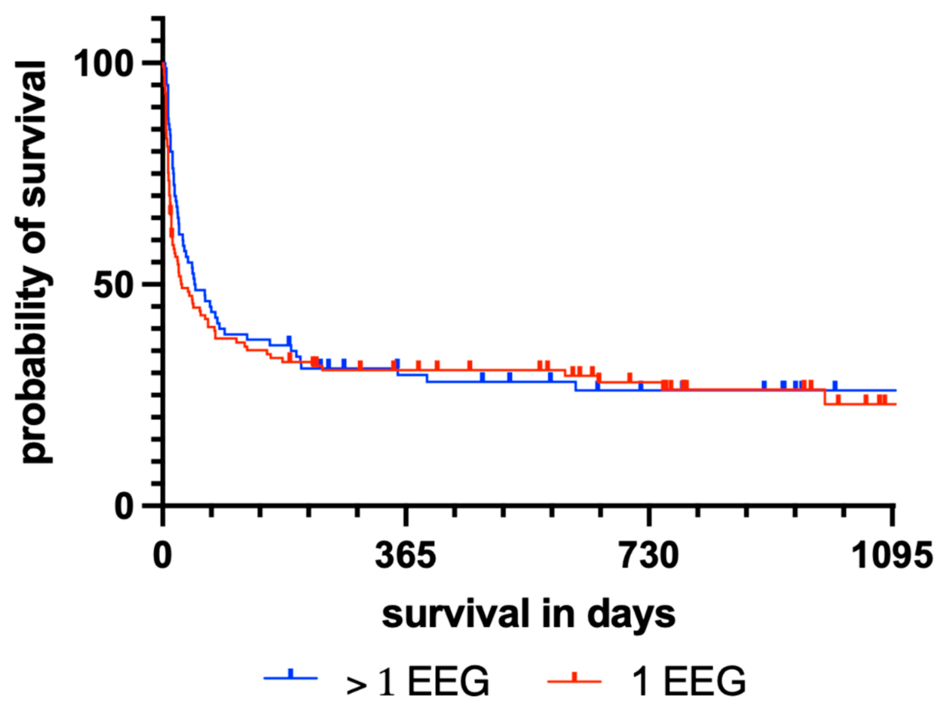
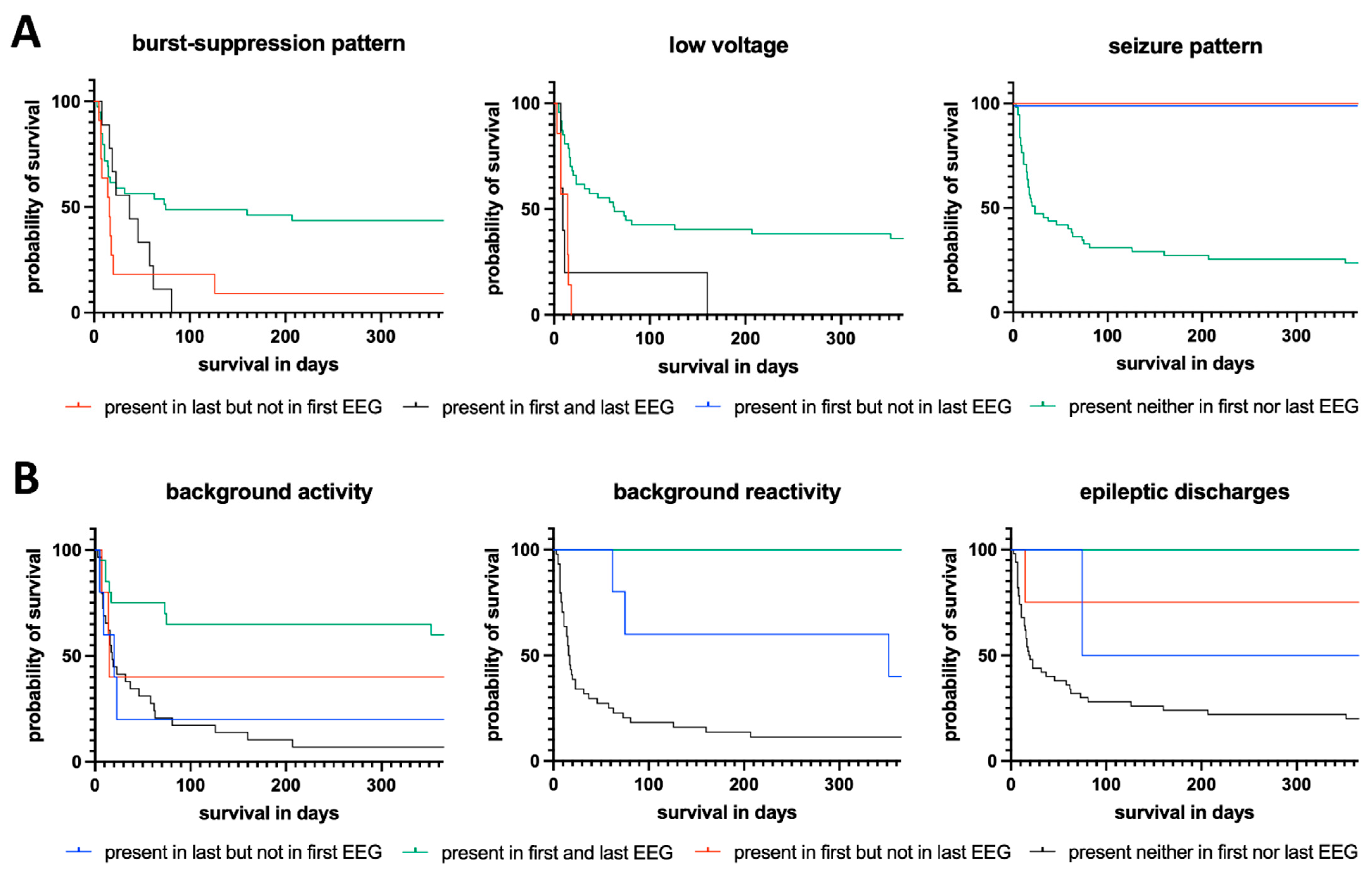
Repetitive Electroencephalography as Biomarker for the Prediction of Survival in Patients with Post-Hypoxic Encephalopathy
Abstract
1. Introduction
2. Materials and Methods
2.1. Study Setting and Design
2.2. Patient Care and EEG Recordings
2.3. Outcome Measures and Statistical Analysis
2.4. Analyzed Sociodemographic and Disease-Related Variables and Scale Levels
3. Results
3.1. Univariate Analysis of Reasons for More than One EEG Recording during Acute Neurocritical Care
3.2. Sociodemographic and Disease-Specific Characteristics
3.3. Univariate Analysis of the Predictive Properties of Repetitive EEGs during Acute Neurocritical Care
3.4. Multivariate Analysis of Predictive Properties of Repetitive EEGs during Acute Neurocritical Care
4. Discussion
5. Conclusions
Author Contributions
Funding
Institutional Review Board Statement
Informed Consent Statement
Data Availability Statement
Acknowledgments
Conflicts of Interest
Abbreviations
| ACNS | American Clinical Neurophysiology Society |
| BA | Background activity |
| BSP | Burst-suppression pattern |
| BSR | Brainstem reflex |
| CA | Cardiac arrest |
| CCI | Charlson comorbidity index |
| cEEG | Continuous electroencephalography |
| CI | Confidence interval |
| CPR | Cardiopulmonary Resuscitation |
| DGKN | German Society for Clinical Neurophysiology (Deutsche Gesellschaft für Klinische Neurophysiologie |
| ED | Epileptiform dischargers |
| EEG | Electroencephalography |
| HE | Hypoxic encephalopathy |
| HIS | Hospital information system |
| ICU | Intensive care unit |
| LV | Low voltage |
| MCRA | Multivariate Cox regression analysis |
| Med | Median |
| mRS | Modified rankin scale |
| n.a. | Not available |
| NCU | Neurocritical care units |
| NSE | Neuron-specific enolase |
| RECORD | REporting of studies Conducted using Observational Routinely-collected Data |
| rEEG | Repetitive electroencephalography |
| RPP | Rhythmic and periodic pattern |
| SARS-CoV-2 | Severe acute respiratory syndrome coronavirus type 2 |
| SD | Standard deviation |
| SE | Status epilepticus |
| SP | Seizure pattern |
| STROBE | Strengthening the Reporting of Observational Studies in Epidemiology |
| TTM | Therapeutic temperature management |
| UKGM | University Hospital Marburg |
References
- Grasner, J.T.; Bottiger, B.W.; Bossaert, L.; European Registry of Cardiac Arrest (EuReCa) ONE Steering Committee; EuReCa ONE Study Management Team. EuReCa ONE—ONE month—ONE Europe—ONE goal. Resuscitation 2014, 85, 1307–1308. [Google Scholar] [CrossRef] [PubMed]
- The Human Mortality Database. University of California, Berkley and Max-Planck-Institute for Demographic Research: 2013. Available online: https://www.demogr.mpg.de (accessed on 15 August 2022).
- Willems, L.M.; Trienekens, F.; Knake, S.; Beuchat, I.; Rosenow, F.; Schieffer, B.; Karatolios, K.; Strzelczyk, A. EEG patterns and their correlations with short- and long-term mortality in patients with hypoxic encephalopathy. Clin. Neurophysiol. 2021, 132, 2851–2860. [Google Scholar] [CrossRef] [PubMed]
- Coute, R.A.; Nathanson, B.H.; Panchal, A.R.; Kurz, M.C.; Haas, N.L.; McNally, B.; Neumar, R.W.; Mader, T.J. Disability-Adjusted Life Years Following Adult Out-of-Hospital Cardiac Arrest in the United States. Circ. Cardiovasc. Qual. Outcomes 2019, 12, e004677. [Google Scholar] [CrossRef] [PubMed]
- Coute, R.A.; Nathanson, B.H.; Mader, T.J.; McNally, B.; Kurz, M.C. Trend analysis of disability-adjusted life years following adult out-of-hospital cardiac arrest in the United States: A study from the CARES Surveillance Group. Resuscitation 2021, 163, 124–129. [Google Scholar] [CrossRef]
- Nolan, J.P.; Sandroni, C.; Bottiger, B.W.; Cariou, A.; Cronberg, T.; Friberg, H.; Genbrugge, C.; Haywood, K.; Lilja, G.; Moulaert, V.R.M.; et al. European Resuscitation Council and European Society of Intensive Care Medicine guidelines 2021: Post-resuscitation care. Intensive Care Med. 2021, 47, 369–421. [Google Scholar] [CrossRef]
- Allen, K.A.; Brandon, D.H. Hypoxic Ischemic Encephalopathy: Pathophysiology and Experimental Treatments. Newborn Infant Nurs. Rev. 2011, 11, 125–133. [Google Scholar] [CrossRef]
- Sekhon, M.S.; Ainslie, P.N.; Griesdale, D.E. Clinical pathophysiology of hypoxic ischemic brain injury after cardiac arrest: A "two-hit" model. Crit. Care 2017, 21, 90. [Google Scholar] [CrossRef]
- Geocadin, R.G.; Callaway, C.W.; Fink, E.L.; Golan, E.; Greer, D.M.; Ko, N.U.; Lang, E.; Licht, D.J.; Marino, B.S.; McNair, N.D.; et al. Standards for Studies of Neurological Prognostication in Comatose Survivors of Cardiac Arrest: A Scientific Statement From the American Heart Association. Circulation 2019, 140, e517–e542. [Google Scholar] [CrossRef]
- Beuchat, I.; Solari, D.; Novy, J.; Oddo, M.; Rossetti, A.O. Standardized EEG interpretation in patients after cardiac arrest: Correlation with other prognostic predictors. Resuscitation 2018, 126, 143–146. [Google Scholar] [CrossRef]
- Sivaraju, A.; Gilmore, E.J.; Wira, C.R.; Stevens, A.; Rampal, N.; Moeller, J.J.; Greer, D.M.; Hirsch, L.J.; Gaspard, N. Prognostication of post-cardiac arrest coma: Early clinical and electroencephalographic predictors of outcome. Intensive Care Med. 2015, 41, 1264–1272. [Google Scholar] [CrossRef]
- Hofmeijer, J.; Beernink, T.M.; Bosch, F.H.; Beishuizen, A.; Tjepkema-Cloostermans, M.C.; van Putten, M.J. Early EEG contributes to multimodal outcome prediction of postanoxic coma. Neurology 2015, 85, 137–143. [Google Scholar] [CrossRef] [PubMed]
- Zubler, F.; Steimer, A.; Kurmann, R.; Bandarabadi, M.; Novy, J.; Gast, H.; Oddo, M.; Schindler, K.; Rossetti, A.O. EEG synchronization measures are early outcome predictors in comatose patients after cardiac arrest. Clin. Neurophysiol. 2017, 128, 635–642. [Google Scholar] [CrossRef] [PubMed]
- Hofmeijer, J.; van Putten, M.J. EEG in postanoxic coma: Prognostic and diagnostic value. Clin. Neurophysiol. 2016, 127, 2047–2055. [Google Scholar] [CrossRef] [PubMed]
- Rossetti, A.O.; Carrera, E.; Oddo, M. Early EEG correlates of neuronal injury after brain anoxia. Neurology 2012, 78, 796–802. [Google Scholar] [CrossRef]
- Rossetti, A.O.; Oddo, M.; Logroscino, G.; Kaplan, P.W. Prognostication after cardiac arrest and hypothermia: A prospective study. Ann. Neurol. 2010, 67, 301–307. [Google Scholar] [CrossRef]
- Rossetti, A.O.; Urbano, L.A.; Delodder, F.; Kaplan, P.W.; Oddo, M. Prognostic value of continuous EEG monitoring during therapeutic hypothermia after cardiac arrest. Crit. Care 2010, 14, R173. [Google Scholar] [CrossRef]
- Hofmeijer, J.; Tjepkema-Cloostermans, M.C.; van Putten, M.J. Burst-suppression with identical bursts: A distinct EEG pattern with poor outcome in postanoxic coma. Clin. Neurophysiol. 2014, 125, 947–954. [Google Scholar] [CrossRef]
- Bermeo-Ovalle, A. Continuous EEG in ICU: Not a Luxury After All. Epilepsy Curr. 2021, 21, 21–23. [Google Scholar] [CrossRef]
- Biyani, S.; Arulprakash, N.; Thatikala, A.; Nalleballe, K.; Onteddu, S.; Shah, V. Underutilization of Neuro Critical Care during the COVID-19 Pandemic. Neurology 2022, 98, 450. [Google Scholar]
- Pressler, R.M.; Boylan, G.B.; Morton, M.; Binnie, C.D.; Rennie, J.M. Early serial EEG in hypoxic ischaemic encephalopathy. Clin. Neurophysiol. 2001, 112, 31–37. [Google Scholar] [CrossRef]
- Amorim, E.; Rittenberger, J.C.; Zheng, J.J.; Westover, M.B.; Baldwin, M.E.; Callaway, C.W.; Popescu, A.; Post Cardiac Arrest, S. Continuous EEG monitoring enhances multimodal outcome prediction in hypoxic-ischemic brain injury. Resuscitation 2016, 109, 121–126. [Google Scholar] [CrossRef] [PubMed]
- Muhlhofer, W.; Szaflarski, J.P. Prognostic Value of EEG in Patients after Cardiac Arrest-An Updated Review. Curr. Neurol. Neurosci. Rep. 2018, 18, 16. [Google Scholar] [CrossRef] [PubMed]
- Zachariah, J.; Rabinstein, A.A. The Reemergence of EEG Reactivity After Cardiac Arrest. Neurohospitalist 2017, 7, 137–140. [Google Scholar] [CrossRef] [PubMed]
- Fechner, A.; Hubert, K.; Jahnke, K.; Knake, S.; Konczalla, J.; Menzler, K.; Ronellenfitsch, M.W.; Rosenow, F.; Strzelczyk, A. Treatment of refractory and superrefractory status epilepticus with topiramate: A cohort study of 106 patients and a review of the literature. Epilepsia 2019, 60, 2448–2458. [Google Scholar] [CrossRef] [PubMed]
- Kortland, L.M.; Alfter, A.; Bahr, O.; Carl, B.; Dodel, R.; Freiman, T.M.; Hubert, K.; Jahnke, K.; Knake, S.; von Podewils, F.; et al. Costs and cost-driving factors for acute treatment of adults with status epilepticus: A multicenter cohort study from Germany. Epilepsia 2016, 57, 2056–2066. [Google Scholar] [CrossRef] [PubMed]
- Von Elm, E.; Altman, D.G.; Egger, M.; Pocock, S.J.; Gotzsche, P.C.; Vandenbroucke, J.P. Das Strengthening the Reporting of Observational Studies in Epidemiology (STROBE) Statement. Notfall Rettungsmed. 2008, 11, 260–265. [Google Scholar] [CrossRef]
- Nicholls, S.G.; Quach, P.; von Elm, E.; Guttmann, A.; Moher, D.; Petersen, I.; Sorensen, H.T.; Smeeth, L.; Langan, S.M.; Benchimol, E.I. The REporting of Studies Conducted Using Observational Routinely-Collected Health Data (RECORD) Statement: Methods for Arriving at Consensus and Developing Reporting Guidelines. PLoS ONE 2015, 10, e0125620. [Google Scholar] [CrossRef]
- Peberdy, M.A.; Callaway, C.W.; Neumar, R.W.; Geocadin, R.G.; Zimmerman, J.L.; Donnino, M.; Gabrielli, A.; Silvers, S.M.; Zaritsky, A.L.; Merchant, R.; et al. Part 9: Post-cardiac arrest care: 2010 American Heart Association Guidelines for Cardiopulmonary Resuscitation and Emergency Cardiovascular Care. Circulation 2010, 122, S768–S786. [Google Scholar] [CrossRef]
- Nielsen, N.; Wetterslev, J.; Cronberg, T.; Erlinge, D.; Gasche, Y.; Hassager, C.; Horn, J.; Hovdenes, J.; Kjaergaard, J.; Kuiper, M.; et al. Targeted temperature management at 33 degrees C versus 36 degrees C after cardiac arrest. N. Engl. J. Med. 2013, 369, 2197–2206. [Google Scholar] [CrossRef]
- Hirsch, L.J.; LaRoche, S.M.; Gaspard, N.; Gerard, E.; Svoronos, A.; Herman, S.T.; Mani, R.; Arif, H.; Jette, N.; Minazad, Y.; et al. American Clinical Neurophysiology Society’s Standardized Critical Care EEG Terminology: 2012 version. J. Clin. Neurophysiol. 2013, 30, 1–27. [Google Scholar] [CrossRef]
- Hirsch, L.J.; Fong, M.W.K.; Leitinger, M.; LaRoche, S.M.; Beniczky, S.; Abend, N.S.; Lee, J.W.; Wusthoff, C.J.; Hahn, C.D.; Westover, M.B.; et al. American Clinical Neurophysiology Society’s Standardized Critical Care EEG Terminology: 2021 Version. J. Clin. Neurophysiol. 2021, 38, 1–29. [Google Scholar] [CrossRef] [PubMed]
- Beuchat, I.; Alloussi, S.; Reif, P.S.; Sterlepper, N.; Rosenow, F.; Strzelczyk, A. Prospective evaluation of interrater agreement between EEG technologists and neurophysiologists. Sci. Rep. 2021, 11, 13406. [Google Scholar] [CrossRef]
- Yan, S.; Gan, Y.; Jiang, N.; Wang, R.; Chen, Y.; Luo, Z.; Zong, Q.; Chen, S.; Lv, C. The global survival rate among adult out-of-hospital cardiac arrest patients who received cardiopulmonary resuscitation: A systematic review and meta-analysis. Crit. Care 2020, 24, 61. [Google Scholar] [CrossRef] [PubMed]
- Westhall, E.; Rossetti, A.O.; van Rootselaar, A.F.; Wesenberg Kjaer, T.; Horn, J.; Ullen, S.; Friberg, H.; Nielsen, N.; Rosen, I.; Aneman, A.; et al. Standardized EEG interpretation accurately predicts prognosis after cardiac arrest. Neurology 2016, 86, 1482–1490. [Google Scholar] [CrossRef] [PubMed]
- Ruijter, B.J.; Hofmeijer, J.; Tjepkema-Cloostermans, M.C.; van Putten, M. The prognostic value of discontinuous EEG patterns in postanoxic coma. Clin. Neurophysiol. 2018, 129, 1534–1543. [Google Scholar] [CrossRef]
- Fantaneanu, T.A.; Sarkis, R.; Avery, K.; Scirica, B.M.; Hurwitz, S.; Henderson, G.V.; Lee, J.W. Delayed Deterioration of EEG Background Rhythm Post-cardiac Arrest. Neurocrit. Care 2017, 26, 411–419. [Google Scholar] [CrossRef]
- Azabou, E.; Navarro, V.; Kubis, N.; Gavaret, M.; Heming, N.; Cariou, A.; Annane, D.; Lofaso, F.; Naccache, L.; Sharshar, T. Value and mechanisms of EEG reactivity in the prognosis of patients with impaired consciousness: A systematic review. Crit. Care 2018, 22, 184. [Google Scholar] [CrossRef]
- Spalletti, M.; Carrai, R.; Scarpino, M.; Cossu, C.; Ammannati, A.; Ciapetti, M.; Tadini Buoninsegni, L.; Peris, A.; Valente, S.; Grippo, A.; et al. Single electroencephalographic patterns as specific and time-dependent indicators of good and poor outcome after cardiac arrest. Clin. Neurophysiol. 2016, 127, 2610–2617. [Google Scholar] [CrossRef]
- Soholm, H.; Kjaer, T.W.; Kjaergaard, J.; Cronberg, T.; Bro-Jeppesen, J.; Lippert, F.K.; Kober, L.; Wanscher, M.; Hassager, C. Prognostic value of electroencephalography (EEG) after out-of-hospital cardiac arrest in successfully resuscitated patients used in daily clinical practice. Resuscitation 2014, 85, 1580–1585. [Google Scholar] [CrossRef]
- Van Putten, M.J.; van Putten, M.H. Uncommon EEG burst-suppression in severe postanoxic encephalopathy. Clin. Neurophysiol. 2010, 121, 1213–1219. [Google Scholar] [CrossRef]
- Yang, Q.; Su, Y.; Hussain, M.; Chen, W.; Ye, H.; Gao, D.; Tian, F. Poor outcome prediction by burst suppression ratio in adults with post-anoxic coma without hypothermia. Neurol. Res. 2014, 36, 453–460. [Google Scholar] [CrossRef]
- Backman, S.; Cronberg, T.; Friberg, H.; Ullen, S.; Horn, J.; Kjaergaard, J.; Hassager, C.; Wanscher, M.; Nielsen, N.; Westhall, E. Highly malignant routine EEG predicts poor prognosis after cardiac arrest in the Target Temperature Management trial. Resuscitation 2018, 131, 24–28. [Google Scholar] [CrossRef]
- Mani, R.; Schmitt, S.E.; Mazer, M.; Putt, M.E.; Gaieski, D.F. The frequency and timing of epileptiform activity on continuous electroencephalogram in comatose post-cardiac arrest syndrome patients treated with therapeutic hypothermia. Resuscitation 2012, 83, 840–847. [Google Scholar] [CrossRef] [PubMed]
- Crepeau, A.Z.; Fugate, J.E.; Mandrekar, J.; White, R.D.; Wijdicks, E.F.; Rabinstein, A.A.; Britton, J.W. Value analysis of continuous EEG in patients during therapeutic hypothermia after cardiac arrest. Resuscitation 2014, 85, 785–789. [Google Scholar] [CrossRef] [PubMed]
- Crepeau, A.Z.; Rabinstein, A.A.; Fugate, J.E.; Mandrekar, J.; Wijdicks, E.F.; White, R.D.; Britton, J.W. Continuous EEG in therapeutic hypothermia after cardiac arrest: Prognostic and clinical value. Neurology 2013, 80, 339–344. [Google Scholar] [CrossRef]
- Kim, Y.J.; Kim, M.J.; Koo, Y.S.; Kim, W.Y. Background Frequency Patterns in Standard Electroencephalography as an Early Prognostic Tool in Out-of-Hospital Cardiac Arrest Survivors Treated with Targeted Temperature Management. J. Clin. Med. 2020, 9, 1113. [Google Scholar] [CrossRef] [PubMed]
- Alvarez, V.; Sierra-Marcos, A.; Oddo, M.; Rossetti, A.O. Yield of intermittent versus continuous EEG in comatose survivors of cardiac arrest treated with hypothermia. Crit. Care 2013, 17, R190. [Google Scholar] [CrossRef] [PubMed]
- Renzel, R.; Baumann, C.R.; Mothersill, I.; Poryazova, R. Persistent generalized periodic discharges: A specific marker of fatal outcome in cerebral hypoxia. Clin. Neurophysiol. 2017, 128, 147–152. [Google Scholar] [CrossRef]
- Lopez-Rolon, A.; Bender, A.; Project HOPE Investigator Group. Hypoxia and Outcome Prediction in Early-Stage Coma (Project HOPE): An observational prospective cohort study. BMC Neurol. 2015, 15, 82. [Google Scholar] [CrossRef] [PubMed]
- Geocadin, R.G.; Buitrago, M.M.; Torbey, M.T.; Chandra-Strobos, N.; Williams, M.A.; Kaplan, P.W. Neurologic prognosis and withdrawal of life support after resuscitation from cardiac arrest. Neurology 2006, 67, 105–108. [Google Scholar] [CrossRef] [PubMed]
- MacDarby, L.; Healy, M.; McHugh, J.C. EEG Availability in the Intensive Care Setting: A Multicentre Study. Neurocrit. Care 2021, 34, 287–290. [Google Scholar] [CrossRef] [PubMed]
- Kolls, B.J.; Lai, A.H.; Srinivas, A.A.; Reid, R.R. Integration of EEG lead placement templates into traditional technologist-based staffing models reduces costs in continuous video-EEG monitoring service. J. Clin. Neurophysiol. 2014, 31, 187–193. [Google Scholar] [CrossRef]
- Willems, L.M.; Balcik, Y.; Noda, A.H.; Siebenbrodt, K.; Leimeister, S.; McCoy, J.; Kienitz, R.; Kiyose, M.; Reinecke, R.; Schafer, J.H.; et al. SARS-CoV-2-related rapid reorganization of an epilepsy outpatient clinic from personal appointments to telemedicine services: A German single-center experience. Epilepsy Behav. 2020, 112, 107483. [Google Scholar] [CrossRef] [PubMed]
- Qasem, L.E.; Al-Hilou, A.; Zacharowski, K.; Funke, M.; Strouhal, U.; Reitz, S.C.; Jussen, D.; Forster, M.T.; Konczalla, J.; Prinz, V.M.; et al. Implementation of the "No ICU—Unless" approach in postoperative neurosurgical management in times of COVID-19. Neurosurg. Rev. 2022, 45, 3437–3446. [Google Scholar] [CrossRef] [PubMed]
- Rossetti, A.O.; Schindler, K.; Sutter, R.; Ruegg, S.; Zubler, F.; Novy, J.; Oddo, M.; Warpelin-Decrausaz, L.; Alvarez, V. Continuous vs Routine Electroencephalogram in Critically Ill Adults With Altered Consciousness and No Recent Seizure: A Multicenter Randomized Clinical Trial. JAMA Neurol. 2020, 77, 1225–1232. [Google Scholar] [CrossRef] [PubMed]
- Beuchat, I.; Rossetti, A.O.; Novy, J.; Schindler, K.; Ruegg, S.; Alvarez, V. Continuous Versus Routine Standardized Electroencephalogram for Outcome Prediction in Critically Ill Adults: Analysis From a Randomized Trial. Crit. Care Med. 2022, 50, 329–334. [Google Scholar] [CrossRef] [PubMed]
- Hill, C.E.; Blank, L.J.; Thibault, D.; Davis, K.A.; Dahodwala, N.; Litt, B.; Willis, A.W. Continuous EEG is associated with favorable hospitalization outcomes for critically ill patients. Neurology 2019, 92, e9–e18. [Google Scholar] [CrossRef]
- Benchimol, E.I.; Smeeth, L.; Guttmann, A.; Harron, K.; Moher, D.; Petersen, I.; Sorensen, H.T.; von Elm, E.; Langan, S.M.; RECORD Working Committee. The REporting of studies Conducted using Observational Routinely-collected health Data (RECORD) statement. PloS Med. 2015, 12, e1001885. [Google Scholar] [CrossRef]
- von Elm, E.; Altman, D.G.; Egger, M.; Pocock, S.J.; Gotzsche, P.C.; Vandenbroucke, J.P.; STROBE-Initiative. The Strengthening the Reporting of Observational Studies in Epidemiology (STROBE) statement: Guidelines for reporting observational studies. J. Clin. Epidemiol. 2008, 61, 344–349. [Google Scholar] [CrossRef]


| Rho a | p-Value a | |||
|---|---|---|---|---|
| Time of survival, days | −0.022 | 0.060 | ||
| Age, years | −0.047 | 0.371 | ||
| CPR duration, minutes | −0.140 | 0.214 | ||
| Modified Rankin Scale (mRS) | 0.091 | 0.072 | ||
| Charlson Comorbidity Index (CCI) | −0.043 | 0.397 | ||
| 1 EEG % (n) | >1 EEG % (n) | p-Value b | ||
| Intact brainstem reflexes (BSR) | Yes | 44.0 (11) | 56.0 (14) | 0.085 |
| No | 62.1 (108) | 37.9 (66) | ||
| Sex | Male | 63.2 (84) | 36.8 (49) | 0.170 |
| Female | 53.0 (35) | 47.0 (31) | ||
| Therapeutic temperature management (TTM) | Yes | 62.5 (70) | 37.5 (42) | 0.384 |
| No | 56.3 (45) | 43.8 (35) | ||
| CPR setting in hospital | Yes | 64.4 (47) | 35.6 (26) | 0.560 |
| No | 60.2 (71) | 39.8 (47) | ||
| EEG suppression (<2 µV) during first EEG | Yes | 100.0 (4) | 0.0 (0) | 0.980 |
| No | 58.9 (115) | 41.1 (80) | ||
| Preserved normal background activity during first EEG | Yes | 51.0 (48) | 49.0 (46) | 0.017 |
| No | 67.6 (71) | 32.4 (34) | ||
| Low voltage during first EEG | Yes | 74.4 (29) | 25.6 (10) | 0.039 |
| No | 56.3 (90) | 43.7 (70) | ||
| Burst-suppression pattern during first EEG | Yes | 25.9 (7) | 74.1 (20) | <0.001 |
| No | 65.1 (112) | 34.9 (60) | ||
| EEG reactivity during first EEG | Yes | 70.4 (50) | 29.6 (21) | 0.007 |
| No | 50.4 (59) | 49.6 (58) | ||
| Epileptiform discharges during first EEG | Yes | 71.4 (5) | 28.6 (2) | 0.523 |
| No | 59.4 (114) | 40.6 (78) | ||
| Seizure pattern during first EEG | Yes | 60.0 (6) | 40.0 (4) | 0.517 |
| No | 60.3 (114) | 39.7(75) | ||
| Sociodemographic Parameters | ||
|---|---|---|
| Age, years | Mean ± SD | 64.7 ± 11.7 |
| Median | 65.0 | |
| Range | 36–87 | |
| Sex, % (n) | Female | 39.0 (23) |
| Male | 61.0 (36) | |
| Modified Rankin Scale (mRS) | Mean ± SD | 1.1 ± 0.7 |
| Median | 1.0 | |
| Range | 0–2 | |
| Charlson Comorbidity Index (CCI) | Mean ± SD | 4.5 ± 2.5 |
| Median | 4.0 | |
| Range | 0–10 | |
| CPR parameters | ||
| CPR etiology | Primary cardiac | 76.3 (45) |
| Primary respiratory | 15.3 (9) | |
| Other | 8.4 (5) | |
| CPR duration, minutes | Mean ± SD | 23.8 ± 16.7 |
| Median | 20.0 | |
| Range | 2.0–90.0 | |
| CPR setting | In hospital | 27.1 (16) |
| Out of hospital | 64.4 (38) | |
| n.a. | 8.5 (5) | |
| Therapeutic temperature management (TTM) | Yes | 37.3 (22) |
| No | 62.7 (37) | |
| EEG parameters | ||
| Number of EEGs | Mean ± SD | 2.9 ± 1.5 |
| Median | 2.0 | |
| Range | 1.0–9.0 | |
| Time to first EEG, days | Mean ± SD | 4.3 ± 2.7 |
| Median | 4.0 | |
| Range | 1.0–11.0 | |
| Time to last EEG, days | Mean ± SD | 9.5 ± 3.3 |
| Median | 10.0 | |
| Range | 3.0–14.0 | |
| Hospitalization | ||
| Length of stay, days | Mean ± SD | 19.1 ± 14.6 |
| Median | 17.0 | |
| Range | 3.0–91.0 | |
| Mortality | ||
| Mortality % (n) | In hospital | 49.2 (29) |
| 30 days after discharge | 54.2 (32) | |
| 365 days post-CPR | 71.2 (42) | |
| Changes between First and Last EEGs | Patients | Mortality | Survival in Days | ||||
|---|---|---|---|---|---|---|---|
| % (n) | Rate, % (n) | Mean | ± SD | Med | 95% CI | p-Value a | |
| General trend | |||||||
| Improvement | 20.3 (12) | 38.5 (5) | 256.3 | 43.9 | - | - | 0.036 |
| Stable finding | 45.8 (27) | 81.5 (22) | 101.2 | 25.8 | 32.0 | 1.5–62.5 | |
| Worsening | 33.9 (20) | 75.0 (15) | 105.9 | 33.9 | 15.0 | 12.1–17.9 | |
| Burst-suppression pattern | |||||||
| Not in first but in last EEG | 18.6 (11) | 90.9 (10) | 54.8 | 31.2 | 16.0 | 6.3–25.7 | 0.016 |
| In first and last EEG | 15.3 (9) | 100.0 (9) | 38.9 | 8.2 | 37.0 | 0.0–77.9 | |
| In first but not in last EEG | 0.0 (0) | - | - | - | - | - | |
| Neither in first nor in last EEG | 66.1 (39) | 59.0 (23) | 178.8 | 20.4 | 32.0 | 0.0–73.9 | |
| Low-voltage EEG | |||||||
| Not in first but in last EEG | 11.9 (7) | 100.0 (7) | 11.1 | 2.1 | 14.0 | 5.8–22.2 | <0.001 |
| In first and last EEG | 8.5 (5) | 100.0 (5) | 38.8 | 30.3 | 9.0 | 4.7–13.3 | |
| In first but not in last EEG | 0.0 (0) | - | - | - | - | - | |
| Neither in first nor in last EEG | 79.7 (47) | 63.8 (30) | 162.9 | 23.7 | 63.0 | 24.0–102.0 | |
| Normal EEG background activity | |||||||
| Not in first but in last EEG | 8.5 (5) | 80.0 (4) | 84.4 | 62.8 | 20.0 | 0.0–43.6 | 0.002 |
| In first and last EEG | 33.8 (20) | 40.0 (8) | 247.0 | 35.9 | 15.0 | - | |
| In first but not in last EEG | 8.5 (5) | 60.0 (3) | 153.2 | 77.5 | 18.0 | 12.9–17.1 | |
| Neither in first nor in last EEG | 49.2 (29) | 93.1 (27) | 62.0 | 17.7 | 32.0 | 12.7–23.3 | |
| EEG reactivity | |||||||
| Not in first but in last EEG | 8.5 (5) | 60.0 (3) | 243.8 | 64.1 | 352.0 | 0.0–946.7 | <0.001 |
| In first and last EEG | 16.9 (10) | 0.0 (0) | 365 | - | 365 | - | |
| In first but not in last EEG | 0.0 (0) | - | - | - | - | - | |
| Neither in first nor in last EEG | 74.6 (44) | 88.6 (39) | 69.5 | 17.1 | 16.0 | 12.3–19.7 | |
| Epileptic discharges | |||||||
| Not in first but in last EEG | 3.4 (2) | 50.0 (1) | 220.0 | 102.5 | 75.0 | - | 0.031 |
| In first and last EEG | 5.1 (3) | 0.0 (0) | 365 | - | 365 | - | |
| In first but not in last EEG | 6.8 (4) | 25.0 (1) | 277.5 | 75.8 | 365 | - | |
| Neither in first nor in last EEG | 8.5 (50) | 80.0 (40) | 122.0 | 20.2 | 23.0 | 0.0–46.2 | |
| EEG seizure pattern | |||||||
| Not in first but in last EEG | 3.4 (2) | 0.0 (0) | 365.0 | 205.1 | - | - | 0.058 |
| In first and last EEG | 0.0 (0) | - | - | - | - | - | |
| In first but not in last EEG | 3.4 (2) | 0.0 (0) | 365.0 | - | 365.0 | - | |
| Neither in first nor in last EEG | 93.2 (55) | 76.4 (42) | 117.6 | 20.1 | 23.0 | 2.3–43.7 | |
| B a | Exp(B) a | 95% CI of Exp(B) a | p-Value a | |
|---|---|---|---|---|
| Burst suppression | ||||
| Not in first but in last EEG | 0.357 | 1.4 | 0.5–4.1 | 0.509 |
| In first and last EEG | 0.674 | 2.0 | 0.7–5.8 | 0.223 |
| Flat EEG <20 µV | ||||
| Not in first but in last EEG | 1.7 | 5.2 | 1.7–15.8 | 0.004 |
| In first and last EEG | 1.0 | 2.8 | 0.8–9.7 | 0.094 |
| Normal EEG background activity | ||||
| Not in first but in 2nd EEG | 0.862 | 2.4 | 0.6–8.9 | 0.201 |
| In first and last EEG | 0.532 | 1.7 | 0.6–5.2 | 0.351 |
| In first but not in last EEG | −0.293 | 0.746 | 0.2–3.0 | 0.683 |
| EEG reactivity | ||||
| Not in first but in last EEG | −1.1 | 0.3 | 0.1–1.2 | 0.099 |
| In first and last EEG | −12.9 | 0.0 | 0.0–0.0 | 0.950 |
| Epileptiform discharges | ||||
| Not in first but in last EEG | −1.4 | 0.2 | 0.0–2.3 | 0.209 |
| In first and last EEG | −0.4 | 0.6 | 0.0–0.0 | 0.999 |
| In first but not in last EEG | −1.4 | 0.3 | 0.0–2.1 | 0.202 |
| Seizure pattern | ||||
| Not in first but in last EEG | −0.432 | 0.6 | 0.0–0.0 | 0.999 |
| In first but not in last EEG | 0.0 | 1.0 | 0.0–0.0 | 1.000 |
Publisher’s Note: MDPI stays neutral with regard to jurisdictional claims in published maps and institutional affiliations. |
© 2022 by the authors. Licensee MDPI, Basel, Switzerland. This article is an open access article distributed under the terms and conditions of the Creative Commons Attribution (CC BY) license (https://creativecommons.org/licenses/by/4.0/).
Share and Cite
Willems, L.M.; Rosenow, F.; Knake, S.; Beuchat, I.; Siebenbrodt, K.; Strüber, M.; Schieffer, B.; Karatolios, K.; Strzelczyk, A. Repetitive Electroencephalography as Biomarker for the Prediction of Survival in Patients with Post-Hypoxic Encephalopathy. J. Clin. Med. 2022, 11, 6253. https://doi.org/10.3390/jcm11216253
Willems LM, Rosenow F, Knake S, Beuchat I, Siebenbrodt K, Strüber M, Schieffer B, Karatolios K, Strzelczyk A. Repetitive Electroencephalography as Biomarker for the Prediction of Survival in Patients with Post-Hypoxic Encephalopathy. Journal of Clinical Medicine. 2022; 11(21):6253. https://doi.org/10.3390/jcm11216253
Chicago/Turabian StyleWillems, Laurent M., Felix Rosenow, Susanne Knake, Isabelle Beuchat, Kai Siebenbrodt, Michael Strüber, Bernhard Schieffer, Konstantinos Karatolios, and Adam Strzelczyk. 2022. "Repetitive Electroencephalography as Biomarker for the Prediction of Survival in Patients with Post-Hypoxic Encephalopathy" Journal of Clinical Medicine 11, no. 21: 6253. https://doi.org/10.3390/jcm11216253
APA StyleWillems, L. M., Rosenow, F., Knake, S., Beuchat, I., Siebenbrodt, K., Strüber, M., Schieffer, B., Karatolios, K., & Strzelczyk, A. (2022). Repetitive Electroencephalography as Biomarker for the Prediction of Survival in Patients with Post-Hypoxic Encephalopathy. Journal of Clinical Medicine, 11(21), 6253. https://doi.org/10.3390/jcm11216253


