Article

Effects of Toxic Lithium Levels on ECG—Findings from the LiSIE Retrospective Cohort Study

1
Sunderby Research Unit, Department of Clinical Sciences, Psychiatry, Umeå University, 90187 Umeå, Sweden
2
Department of Public Health and Clinical Medicine, Medicine, Umeå University, 90187 Umeå, Sweden
3
Department of Clinical Sciences, Karolinska Institutet, Danderyd University Hospital, 18288 Stockholm, Sweden
4
Department of Psychiatry, Sunderby Hospital, 97180 Luleå, Sweden
5
Department of Clinical Sciences, Psychiatry, Umeå University, 90187 Umeå, Sweden
6
Sunderby Research Unit, Department of Public Health and Clinical Medicine, Umeå University, 90187 Umeå, Sweden
*
Author to whom correspondence should be addressed.
J. Clin. Med. 2022, 11(19), 5941; https://doi.org/10.3390/jcm11195941
Submission received: 19 August 2022 / Revised: 26 September 2022 / Accepted: 3 October 2022 / Published: 8 October 2022
(This article belongs to the Section Cardiology)

Abstract

:
(1) Background: Few studies have explored the impact of lithium intoxication on the heart. (2) Methods: We examined electrocardiogram (ECG) changes associated with lithium intoxication in the framework of the LiSIE (Lithium—Study into Effects and Side Effects) retrospective cohort study. We analysed ECGs before, during, and after intoxication. (3) Results: Of the 1136 patients included, 92 patients had experienced 112 episodes of lithium intoxication. For 55 episodes, there was an ECG available at the time; for 48 episodes, there was a reference ECG available before and/or after the lithium intoxication. Lithium intoxication led to a statistically significant decrease in heart rate from a mean 76 beats/min (SD 16.6) before intoxication to 73 beats/min (SD 17.1) during intoxication (p = 0.046). QTc correlated only weakly with lithium concentration (ρ = 0.329, p = 0.014). However, in 24% of lithium intoxication episodes, there were QT prolongations. In 54% of these, QTc exceeded 500 ms; patients with chronic intoxications being more affected. (4) Conclusions: Based on summary statistics, effects of lithium intoxication on HR and QTc seem mostly discrete and not clinically relevant. However, QT prolongation can carry a risk of becoming severe. Therefore, an ECG should always be taken in patients presenting with lithium intoxication.

1. Introduction

Lithium is a mainstay pharmacological treatment of bipolar disorder (BD), either during acute manic episodes, or as long-term maintenance treatment. In this capacity, the UK National Institute for Clinical Excellence (NICE) has endorsed lithium as a first line treatment [1] in 2014. Lithium is also used as an augmentation treatment for depression when antidepressants do not suffice. Observational and trial evidence suggest that, compared to other mood stabilisers, lithium is therapeutically superior for the prevention of suicide and of relapse into affective episodes [2,3,4,5,6].
Despite its clinical usefulness, lithium is not without problems. Lithium can affect kidney, thyroid, and parathyroid function, all of which require regular monitoring. A further problem is the narrow therapeutic window of lithium, with only a small difference between therapeutic and toxic concentrations. Depending on indications, the therapeutic lithium serum concentration (s-Li) lies between 0.4–1.2 mmol/L. A s-Li ≥ 1.5 mmol/L is seen as the cut-off point for a clinically significant intoxication [7]. At higher serum concentrations, lithium intoxications may become life-threatening. In our previous work, there was one case of intoxication with a s-Li ≥ 1.5 mmol/L per 100 patient-years of lithium treatment. About one third of intoxicated patients required intensive care [7].
The effects of lithium on the heart have rarely been studied. Due to its chemical proximity to sodium, lithium can be expected to affect cardiac conduction [8]. Particularly at supratherapeutic concentrations, lithium may affect cardiac conduction, leading to prolongation of the QT interval, bradycardia, and T-wave changes [9,10]. Lithium may even give rise to a Brugada type-1 pattern [11]. A retrospective study from France, analysing 128 patients with lithium intoxication admitted to intensive care (ICU) between 1992 and 2013, found cardiovascular complications in 24% of cases [12]. Acute care physicians and intensivists need to understand these potential cardiac risks to manage patients with lithium intoxication safely.

Aims and Hypothesis

We conducted the current study to examine the impact of toxic s-Li on the heart. Specifically, we tested the hypothesis that higher s-Li would lead to clinically relevant electrocardiogram (ECG) changes.

2. Materials and Methods

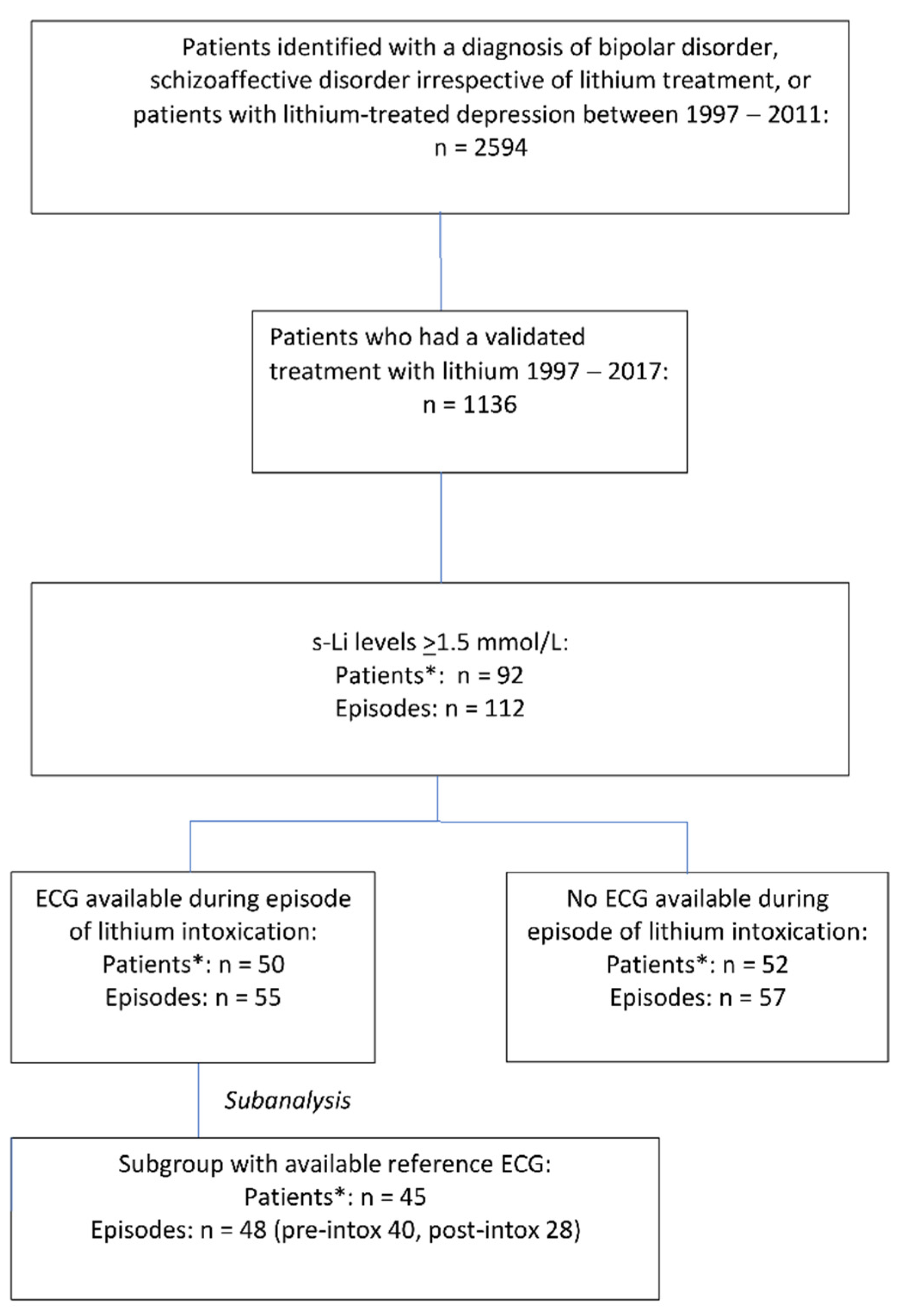
2.1. Study Design and Setting

This study was part of the LiSIE (Lithium—Study into Effects and Side Effects) retrospective cohort study based on a review of medical records. LiSIE aimed at identifying the best treatment for BD and related disorders, such as schizoaffective disorder (SZD), considering both effects and side effects. The study was conducted according to the guidelines of the Declaration of Helsinki and was approved by the Regional Ethics Review Board at Umeå University, Umeå Sweden (DNR 2010-227-31M, DNR 2011-228-32M, DNR 2014-10-32M, DNR 2018-76-32M).
Within the framework of this retrospective cohort study, we examined ECG at the time of lithium intoxication and compared these with reference ECG, taken before (ECGPRE-INTOX) and after (ECGPOST-INTOX) intoxication. The study was first piloted [13] and then conducted according to the STROBE (Strengthening the Reporting of Observational Studies in Epidemiology) checklist (Supplementary materials).

2.2. Participants

LiSIE invited all individuals in the Swedish regions of Västerbotten and Norrbotten ≥ 18 years of age, who had (a) received a diagnosis of BD (ICD F31), SZD (ICD F25) between 1997 and 2011, or (b) used lithium as a mood stabiliser between 1997 and 2011, for instance in the context of a depressive illness [7,14,15]. Participants were informed about the nature of the study in writing and provided verbal informed consent. The consent was documented in our research files, dated, and signed by the research worker who obtained the consent. In accordance with the ethics approval granted, for deceased patients, no consent was obtained. Recruitment was concluded by the end of 2012. The cohort was locked at this point; no new patients were included in the study thereafter.

2.3. Inclusion Criteria

We included all patients from the LiSIE cohort who (a) lived in the Swedish region of Norrbotten, (b) had experienced a documented episode of lithium intoxication with s-Li ≥1.5 mmol/L at any time between 1997 and 2017, and (c) had an ECG recorded at the time of the intoxication.

2.4. Exclusion Criteria

We excluded episodes of supratherapeutic s-Li when it was clear that these were only transient and had not given rise to an intoxication. This could for instance occur when patients had taken their prescribed lithium by mistake before their blood-test, resulting in measurement of peak (highest) rather than trough (lowest) concentrations.

2.5. Data Sources/Measurement

Lithium concentrations were obtained from a central laboratory database. ECGs were extracted from the electronic case records. Where necessary, we complemented with data manually extracted from hardcopy case records. Prior to analysis, we anonymised the data.

2.6. Outcome Variables

The outcome of this study was ECG changes observed in the context of a lithium intoxication. During the 21-year review period, some patients had repeated episodes of lithium intoxication, which were not temporally related. Therefore, we analysed ECG changes per episode and not per patient.

ECG Selection

For ECGINTOX, we chose the ECG taken at the time of the lithium intoxication. If several ECGs were available, we chose the ECG closest in time to the s-Li measurement. We then analysed reference ECGs available before intoxication, pre-intoxication, and/or after the intoxication, post-intoxication (ECGPRE-INTOX, ECGPOST-INTOX) (Table 1). Using an ECG ruler, we analysed all ECGs regarding heart rate (HR), rhythm, PQ interval, QRS duration, QT interval, ST segment and T-wave changes, U-wave presence, and pacemaker rhythm (Table 2). QT interval was corrected for HR (QTc), using a calculator for Bazett’s formula [16]. We further explored occurrence of QT prolongation as a dichotomous yes/no variable for the QTc value and left ventricular enlargement according to Sokolow-Lyon criteria [17]. For men, we used 0.450 s as cut-off point for QT prolongation, and for women we used 0.470 s [18]. All ECGs were coded according to the Minnesota Code Manual of Electrocardiographic Findings [19]. A specialist in cardiology double-checked all ECGs with unclear pathological changes.

2.7. Exposure Variables

2.7.1. Serum Lithium Concentrations

For ECGINTOX, to count as a clinically significant lithium intoxication, s-Li had to reach at least 1.5 mmol/L [7,20]. We chose the s-Li closest to the time of the ECG recording. Each episode of lithium intoxication was categorized as acute, acute on therapeutic, or chronic. We defined intoxications as acute when s-Li rose to ≥1.5 mmol/L within 24 h after ingestion of a supratherapeutic lithium dose (accidental or non-accidental overdose) in a lithium naïve patient. We defined intoxications as acute on therapeutic when s-Li rose to ≥1.5 mmol/L within 24 h after ingestion of a lithium overdose in a patient already treated with lithium at the time. As these intoxications were comparable in character, we merged them into one “acute” category. We regarded an intoxication as chronic when a recent overdose was ruled out [7]. Finally, we stratified severity of lithium intoxication into two categories: less severe with a lithium concentration ≤ 2.5 mmol/L, and severe with a lithium concentration > 2.5 mmol/L [7,21,22].
For the reference ECG, we chose the lithium concentration logged in the medical notes nearest in time to the respective ECG, in a range between 0.2 mmol/L (minimal evidence of exposure) to 1.2 mmol/L (maximal therapeutic concentration).

2.7.2. Concomitant Use of Medicines with Potential QT Prolonging Effect

We recorded the use of medicines that could potentially prolong QT or induce torsades de pointes (TdP). These medicines were classified into three risk categories [23,24].
  • Known TdP risk: medicines known to prolong the QT interval with a known risk of TdP, even if taken as recommended.
  • Possible TdP risk: medicines known to prolong the QT interval with a current lack of evidence for a risk of TdP if taken as recommended.
  • Conditional TdP risk: medicines that may cause TdP if taken (a) in excessive dosage, (b) in conjunction with other medicines that may directly or indirectly contribute to QT prolongation, or (c) by patients with additional risk factors, such as congenital long QT interval, extreme bradycardia, or hypokalaemia.

2.7.3. Concomitant Use of Medicines with Other Cardiac Effects

We recorded antiarrhythmics and antihypertensives as potential cofounders of ECG changes.

2.7.4. Cardiovascular Comorbidities and Risk Factors

We recorded the presence of cardiovascular comorbidities and risk factors coinciding with any of the included ECGs. Such conditions included cerebral stroke/transient ischemic attack (TIA), acute myocardial infarction/ischemic heart disease, heart failure, angina pectoris, arrhythmias, arterial hypertension, pacemaker use, hyperlipidaemia, and diabetes mellitus.

2.7.5. Potassium Concentrations

Where available, we also recorded potassium concentration (s-K) at the time around intoxication. We defined hypo- and hyperkalaemia according to the laboratory s-K reference intervals in use at the time of intoxication. From 1999 to May 2004, this was <3.5 mmol/L for hypokalaemia and >4.7 mmol/L for hyperkalaemia. From June 2004, this was <3.6 mmol/L for hypokalaemia and >4.6 mmol/L for hyperkalaemia. For the years 1997 to 1998, information for the reference intervals was not available. For these years, we used the reference intervals valid between 1999 and May 2004.

2.8. Bias

We checked for selection bias in the LiSIE cohort. In accordance with the ethics approval granted, we compared age, sex, maximum recorded lithium, and creatinine concentrations in anonymous form for consenting and non-consenting patients. There were no significant differences. For patients with lithium intoxication, we compared age and sex distribution between episodes with ECG and without ECG. Again, we did not find any significant differences. However, the mean lithium concentration was significantly higher in the group having available ECG (p = 0.003). This means that our sample was biased toward more severe lithium intoxications.

2.9. Missing Data

By default, ECGINTOX was available for all included cases. Reference ECGs were not available for all cases. We conducted separate subgroup-analyses for episodes with ECGINTOX and ECGPRE-INTOX or ECGPOST-INTOX.

2.10. Statistical Methods

We reported data at episode level. First, we conducted a descriptive analysis of all variables. For continuous variables, we reported mean and standard deviation (SD), as well as median and minimum/maximum (min/max). For comparison of continuous variables between ECGINTOX and ECGPRE-INTOX or ECGPOST-INTOX, we used Wilcoxon rank test for paired data. For correlation between s-Li and HR or QTc at intoxication, we used Spearman Rank correlation (ρ) accounting for the non-normal distribution of the data. For categorical variables, we reported proportions. For comparison of categorical variables between ECGINTOX and ECGPRE-INTOX or ECGPOST-INTOX, we used McNemar’s test. We also conducted a univariate analysis with logistic regression to explore which exposure variables were associated with QT prolongation at intoxication. We abstained from multivariate analysis because the sample size was too small for the number of variables to be fitted. This would have resulted in overfitting of the model. The significance level was set to 0.05 throughout. The statistical analysis was conducted with IBM SPSS Statistics (Version 27, IBM, Armonk, NY, USA).

3. Results

Of 1136 patients (673 females, 59%) exposed to lithium between 1997 and 2017, 92 patients had experienced 112 episodes of lithium intoxication with lithium concentrations ≥ 1.5 mmol/L. Seventeen patients had more than one episode of intoxication. ECGINTOX was available for 55 episodes in 50 patients. For 48 episodes, there was a reference ECG available, 40 ECGPRE-INTOX and 28 ECGPOST-INTOX (Figure 1).

3.1. Baseline Characteristics

The 55 episodes occurred at a mean age of 55.2 years and a median of 56.0 years. Seventy-one percent of the patients were women, and 64% of episodes were chronic intoxications. The mean maximum s-Li was 2.4 mmol/L, and the median was 2.1 mmol/L. Twenty-nine percent of intoxications were severe. The highest s-Li recorded was 9.3 mmol/L in the context of an acute intoxication. In 49% of all episodes, ECG and s-Li were obtained within 90 min of each other. Fifty-one percent of patients had cardiovascular comorbidities, and 24% were receiving anti-arrhythmic medication at the time. Based on 51 episodes with available s-K, 24% had hyperkalaemia and 15% had hypokalaemia (Table 3). One patient died within one day of the lithium intoxication, but the death was considered unrelated to the intoxication.

3.2. ECG Changes at Lithium Intoxication

There was a wide variation of ECG changes, with QT prolongation and T-wave inversion being most common (Table 4).

3.3. Heart Rate and Rhythm
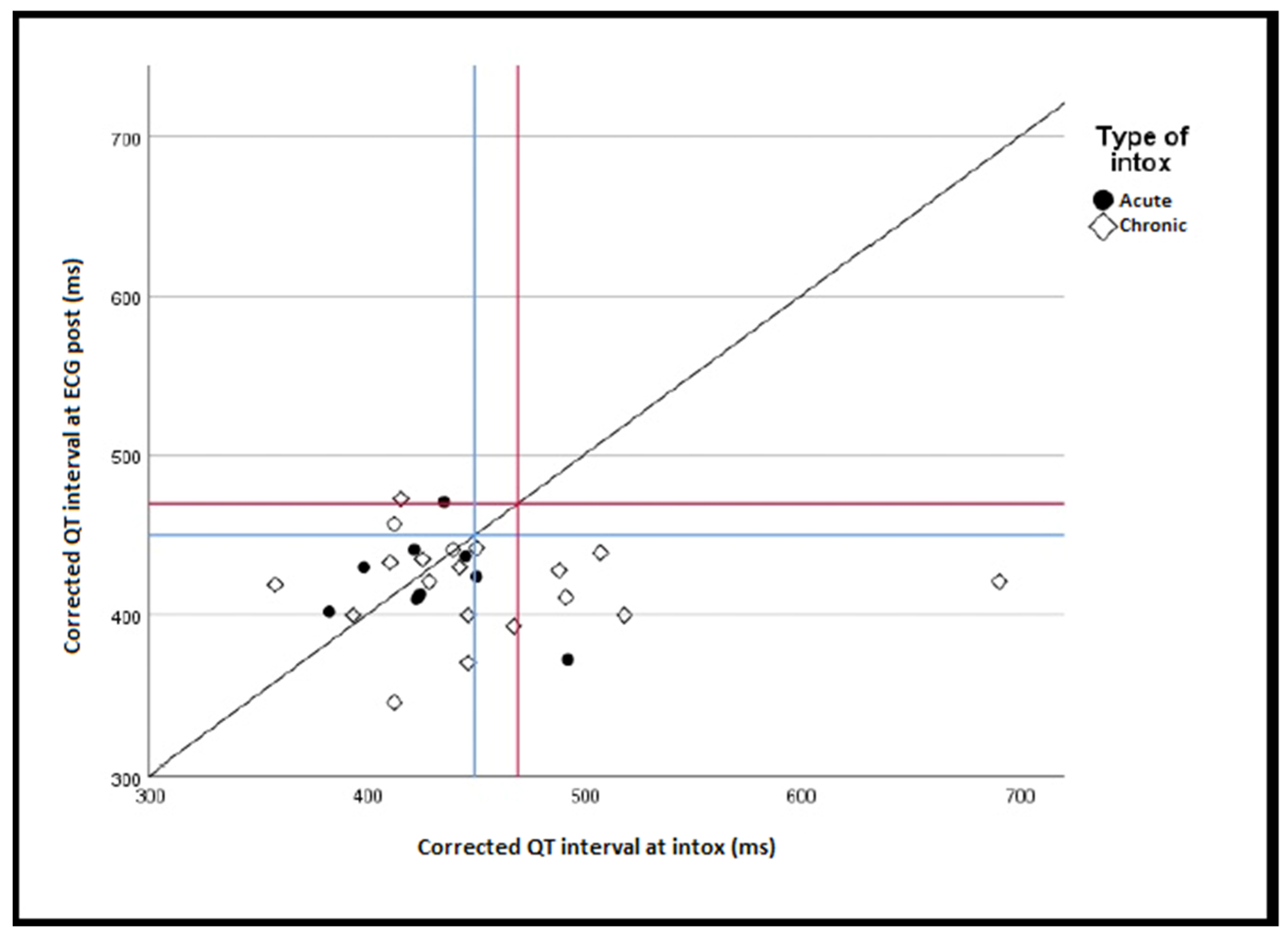
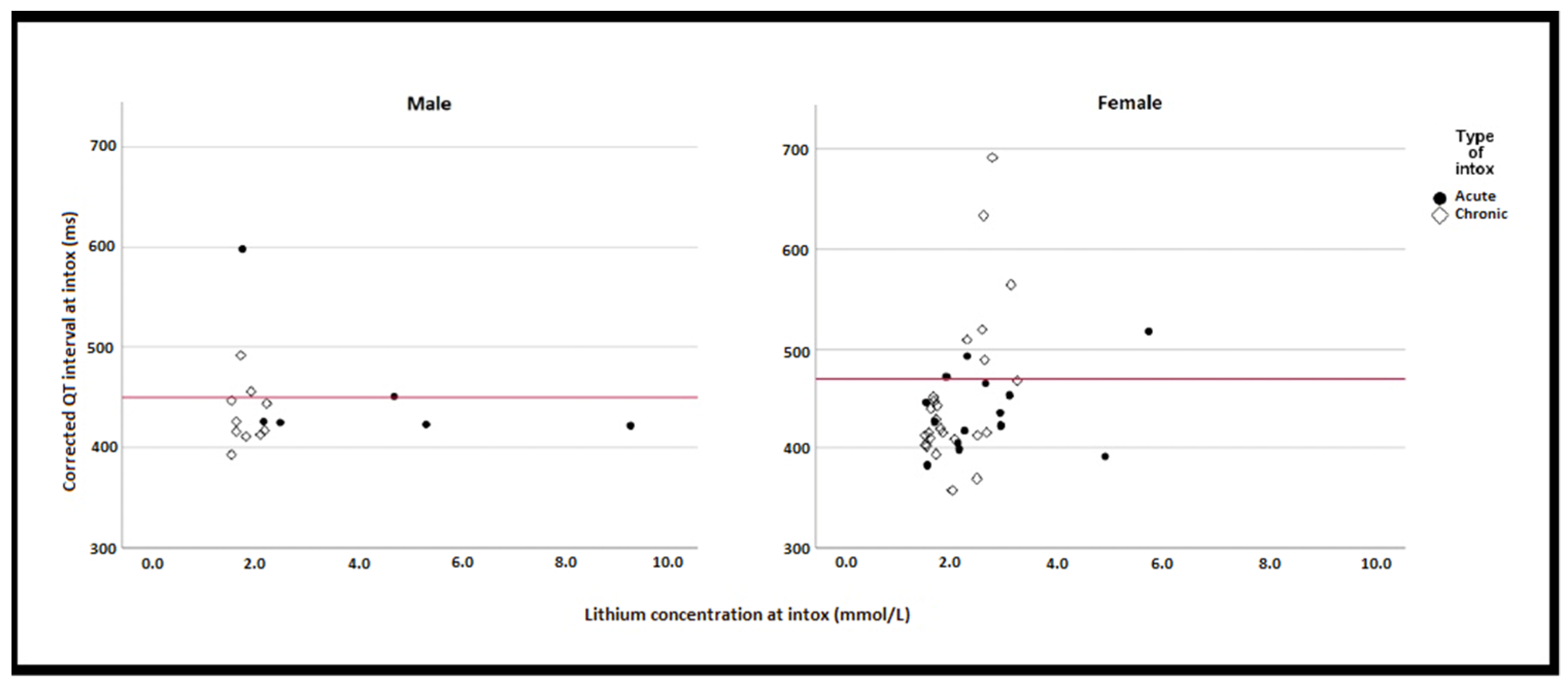
The mean HR at intoxication was 73 beats/min (SD 17.1) and the median 71 beats/min (min 42, max 119). The mean HR pre-intoxication was 76 beats/min (SD 16.6) and the median 78 beats/min (min 45, max 115). The mean HR post-intoxication was 78 beats/min (SD 16.1) and the median 79 beats/min (min 52, max 107). There was a significant decrease in HR during intoxication compared to ECGPRE-INTOX (p = 0.046), but not to ECGPOST-INTOX (p = 0.614). HR on ECGINTOX did not correlate with s-Li at the time of intoxication (ρ = 0.051, p = 0.710). Sinus rhythm was present in 48 (87%) of the episodes. In one of these, a one-beat sinus arrest occurred. Of the remaining seven episodes, four showed atrial fibrillations. In one of these, this was a new phenomenon. Two episodes recorded a nodal rhythm. In one of these, this was a new phenomenon. One episode had pacemaker rhythm. Two episodes had prolonged PQ interval. In one of these, this was a new phenomenon.

3.4. QT Interval

The mean QTc at intoxication was 0.446 s (SD 0.06). The mean QTc pre-intoxication was 0.426 s (SD 0.03). The mean QTc post-intoxication was 0.420 s (SD 0.03). QTc recorded on ECGINTOX was not significantly longer compared to ECGPRE-INTOX or ECGPOST-INTOX for the whole sample (p = 0.104, p = 0.070), men (p = 0.367, p = 0.263), women (p = 0.136, p = 0.113) or acute intoxications only (p = 0.556, p = 0.859). For chronic intoxications, QTc was significantly longer in ECGINTOX compared to ECGPOST-INTOX (p = 0.040), but not compared to ECGPRE-INTOX (p = 0.145) (Figure 2).
QTc on ECGINTOX correlated only weakly with s-Li at intoxication (ρ = 0.329, p = 0.014) (Figure 3). However, there were 13 (24%) episodes with prolonged QTc on ECGINTOX. Of these, nine (69%) occurred in women and eight (62%) in the context of a chronic intoxication (Figure 3). A U-wave was present in three (23%) episodes (Table 5). In seven (54%) of the 13 episodes, QTc exceeded 500 ms. Of these, five (71%) concerned chronic intoxications. The largest QTc increase was 258 ms, from 433 ms in the ECGPRE-INTOX to 691 ms at intoxication. In seven of the 13 episodes, QTc reverted to normal in the ECGPOST-INTOX. For all 13 episodes, both mean and median s-K were 3.7 mmol/L (SD 0.70, min 2.80, max 4.80 mmol/L). Seven patients had at least one known cardiovascular condition at time of lithium intoxication. None had diabetes. In six episodes, patients had been prescribed additional medicines with known or possible risk of TdP (Table 5).
On univariate analysis, only severe lithium intoxication and hypokalaemia were significantly associated with QT prolongation (Table 6).

3.5. T-Wave Inversion

T-wave inversion was found in 42% of episodes at the time of intoxication. There was no significant difference in the presence of T-wave inversion between ECGINTOX and ECGPRE-INTOX (p = 0.454), or ECGPOST-INTOX (p = 0.344).

4. Discussion

In our study, lithium intoxication led to a significant decrease in heart rate. However, the difference was small and not clinically relevant in most cases. QTc correlated only weakly with s-Li. For acute intoxications, there were no statistically significant differences in mean QTc between ECGINTOX and the reference ECGs. For chronic intoxications, there was a statistically significant difference in mean QTc and ECGPOST-INTOX, but not ECGPRE-INTOX. Since our study covered a fairly small number of patients, the absence of significant mean differences could be the result of the limited sample size.
However, about a quarter of all patients experienced QT prolongations in connection with a lithium intoxication. This was more common in chronic intoxications, but it also occurred in acute on therapeutic or acute intoxications. In approximately half of the intoxications with QT prolongation, QTc exceeded 500 ms. A QTc ≥500 ms has been identified as a powerful predictor of 30-day all cause-mortality [25]. In our study, a substantial number of patients had therefore been at risk of life-threatening complications, which could be easily missed when only exploring the summary statistics. On univariate analysis, severity of lithium intoxication and hypokalaemia were significantly associated with QT prolongation. This is in line with clinical experience. To our knowledge, this is the first study systematically exploring ECG changes during lithium intoxications and comparing these with reference ECGs before and/or after.
The aforementioned French study, exploring ECG changes associated with lithium intoxications in an ICU setting, reported 20% collapse/shock, 2% asystole, and 2% sudden bradycardia associated with either a third-degree atrioventricular block or a sinoatrial block [12]. In this study, only 27% had chronic intoxications. Of patients with s-Li > 5.2 mmol/L, 46% were not dialyzed. Another study examined ECG changes associated with therapeutic s-Li in 53 patients four and twelve months after starting lithium. At four- and twelve-month follow-ups, significant decreases in HR and T-wave flattening were noted. Additionally, at 12-month follow-up, the PQ interval was significantly longer. However, there were no R, ST segment, or QTc changes [26].
Distinguishing between acute and chronic intoxications is of clinical relevance. In acute intoxications, s-Li poorly reflects intracellular lithium levels for two reasons. Firstly, lithium may not yet have been absorbed enterally. Secondly, in patients with normal renal function, during transition into the intracellular space, lithium will continuously be cleared. Chronic intoxications often occur in patients with declining renal function [7]. Here, s-Li rises more slowly in the blood. This gives lithium time to reach the intracellular space and unfold its toxic effects [22,27,28]. Patients with chronic intoxications may therefore be at particular risk. Once lithium has entered the intracellular space, it is not easily cleared [12,22,27,28]. This raises the question at which s-Li patients should be offered extracorporeal toxin removal (ECTR), such as dialysis, to clear lithium. Depending on the presence or absence of renal impairment, thresholds between 4 and 5.2 mmol/L have been recommended [12,29]. Our own work, with a higher proportion of chronic intoxications, suggests a lower threshold to decrease the risk of cardiac complication and mortality [7,22].
Being observational, our current study depended on the quality of the information recorded in the medical records. ECG was available in only about half of all episodes, not all of which had reference ECG available. The quality was generally good. We only selected episodes with s-Li ≥1.5 mmol/L to avoid a bias towards mild and borderline intoxications. This bias towards more severe intoxications ensured that we did not underestimate the impact of lithium on cardiac conduction. The clinical course of lithium intoxications is often difficult at the time of presentation. Early dialysis may prevent the development of more serious cardiac complications associated with higher lithium concentrations. In our region, the threshold for dialysis was low [7]. In our sample, dialysis was used as a treatment for intoxication in 11 episodes. The lowest s-Li at which dialysis was used was 1.9 mmol/L. Therefore, our findings should not be extrapolated to clinical settings that employ higher thresholds of s-Li for dialysis [22].

5. Conclusions

Our findings suggest that lithium intoxication can affect HR and QTc. On summary statistics, these changes seem mostly discrete and not clinically relevant for many patients. However, some patients develop severe QT prolongations. This can occur in all types of intoxications—chronic, acute on therapeutic, or acute. Higher s-Li and hypokalaemia may add to the risk of prolonged QT. Therefore, an ECG should always be taken with patients who present with lithium intoxication.

Supplementary Materials

The following supporting information can be downloaded at: https://www.mdpi.com/article/10.3390/jcm11195941/s1, File S1: STROBE Statement—checklist for our study.

Author Contributions

Conceptualization of the study: M.O. and U.W.; methodology: P.T., M.O., U.W., M.S., K.L., R.L. and M.M.; validation of data: P.T., M.O., K.L., M.S. and U.W.; formal analysis: P.T., R.L. and U.W.; data curation: M.O. and U.W.; writing—original draft preparation: P.T., MO. and U.W.; writing—review and editing: P.T., M.O., K.L., M.S., M.M., R.L. and U.W.; visualization: P.T., R.L. and U.W.; supervision: M.O. and U.W. All authors have read and agreed to the published version of the manuscript.

Funding

This work was supported by grants (NLL-931604, NLL-941888, NLL-969413) from the Research & Development Fund of Norrbotten Region, Research and Innovation Unit and the Department of Psychiatry, Sunderby Hospital, both Region Norrbotten, Sweden.

Institutional Review Board Statement

The study was conducted according to the guidelines of the Declaration of Helsinki and approved by the Regional Ethics Review Board at Umeå University, Sweden (DNR 2010-227-31M, DNR 2011-228-32M, DNR 2014-10-32M, DNR 2018-76-32M).

Informed Consent Statement

Informed consent was obtained from all subjects in the study. In accordance with the ethics approval granted, no consent was obtained for deceased patients.

Data Availability Statement

The datasets generated and/or analysed during the current study are not publicly available due to lack of ethics committee permission and not having been part of the consent process. The structures of the dataset and the coding specification are available from the authors. Any other reasonable request will be raised with the Swedish Ethical Review Authority and the healthcare provider.

Conflicts of Interest

U.W. has received funding for educational activities on behalf of Norrbotten Region (Masterclass Psychiatry Programme 2014–2018, EAPM 2016 Luleå, Sweden): Astra Zeneca, Janssen, Eli Lilly, Novartis, Otsuka/Lundbeck, Servier, Shire and Sunovion. U.W. has received lecture honoraria from Lundbeck and Janssen. U.W. also serves on a Scientific Committee for Janssen, receiving an honorarium for this activity. M.O. has been a scientific advisory board member of Astra Zeneca Sweden 2018–2020. P.T., K.L., M.S., M.M., and R.L. declare that there are no competing interests.

References

  1. NICE. Bipolar Disorder: Assessment and Management. Clinical Guideline 185; National Institute for Health and Care Excellence: London, UK, 2014; Available online: https://www.nice.org.uk/guidance/cg185 (accessed on 30 November 2021).
  2. Kirov, G. Lithium plus valproate combination therapy versus monotherapy for relapse prevention in bipolar I disorder (BALANCE): A randomised open-label trial. Lancet 2010, 375, 385–395. [Google Scholar] [CrossRef]
  3. Antolín-Concha, D.; Lähteenvuo, M.; Vattulainen, P.; Tanskanen, A.; Taipale, H.; Vieta, E.; Tiihonen, J. Suicide mortality and use of psychotropic drugs in patients hospitalized due to bipolar disorder: A Finnish nationwide cohort study. J. Affect. Disord. 2020, 277, 885–892. [Google Scholar] [CrossRef]
  4. Öhlund, L.; Ott, M.; Bergqvist, M.; Oja, S.; Lundqvist, R.; Sandlund, M.; Renberg, E.S.; Werneke, U. Clinical course and need for hospital admission after lithium discontinuation in patients with bipolar disorder type I or II: Mirror-image study based on the LiSIE retrospective cohort. BJPsych Open 2019, 5, e101. [Google Scholar] [CrossRef] [PubMed]
  5. Joas, E.; Karanti, A.; Song, J.; Goodwin, G.M.; Lichtenstein, P.; Landén, M. Pharmacological treatment and risk of psychiatric hospital admission in bipolar disorder. Br. J. Psychiatry 2017, 210, 197–202. [Google Scholar] [CrossRef]
  6. Hayes, J.F.; Marston, L.; Walters, K.; Geddes, J.R.; King, M.; Osborn, D.P. Lithium vs. valproate vs. olanzapine vs. quetiapine as maintenance monotherapy for bipolar disorder: A population-based UK cohort study using electronic health records. World Psychiatry 2016, 15, 53–58. [Google Scholar] [CrossRef] [Green Version]
  7. Ott, M.; Stegmayr, B.; Renberg, E.S.; Werneke, U. Lithium intoxication: Incidence, clinical course and renal function—A population-based retrospective cohort study. J. Psychopharmacol. 2016, 30, 1008–1019. [Google Scholar] [CrossRef] [Green Version]
  8. Mehta, N.; Vannozzi, R. Lithium-induced electrocardiographic changes: A complete review. Clin. Cardiol. 2017, 40, 1363–1367. [Google Scholar] [CrossRef] [Green Version]
  9. Hsu, C.-H.; Liu, P.-Y.; Chen, J.-H.; Yeh, T.-L.; Tsai, H.-Y.; Lin, L.-J. Electrocardiographic Abnormalities as Predictors for Over-Range Lithium Levels. Cardiology 2005, 103, 101–106. [Google Scholar] [CrossRef] [PubMed]
  10. Mamiya, K.; Sadanaga, T.; Sekita, A.; Nabeyama, Y.; Yao, H.; Yukawa, E. Lithium concentration correlates with QTc in patients with psychosis. J. Electrocardiol. 2005, 38, 148–151. [Google Scholar] [CrossRef] [PubMed]
  11. Darbar, D.; Yang, T.; Churchwell, K.; Wilde, A.A.M.; Roden, D.M. Unmasking of Brugada Syndrome by Lithium. Circulation 2005, 112, 1527–1531. [Google Scholar] [CrossRef] [PubMed]
  12. Vodovar, D.; El Balkhi, S.; Curis, E.; Deye, N.; Mégarbane, B. Lithium poisoning in the intensive care unit: Predictive factors of severity and indications for extracorporeal toxin removal to improve outcome. Clin. Toxicol. 2016, 54, 615–623. [Google Scholar] [CrossRef]
  13. Ström, M. ECG Changes during Lithium Intoxication. 2017. Available online: https://www.diva-portal.org/smash/record.jsf?dswid=-4106&pid=diva2%3A1141514 (accessed on 17 May 2022).
  14. Öhlund, L.; Ott, M.; Oja, S.; Bergqvist, M.; Lundqvist, R.; Sandlund, M.; Renberg, E.S.; Werneke, U. Reasons for lithium discontinuation in men and women with bipolar disorder: A retrospective cohort study. BMC Psychiatry 2018, 18, 37. [Google Scholar] [CrossRef] [Green Version]
  15. Lieber, I.; Ott, M.; Öhlund, L.; Lundqvist, R.; Eliasson, M.; Sandlund, M.; Werneke, U. Lithium-associated hypothyroidism and potential for reversibility after lithium discontinuation: Findings from the LiSIE retrospective cohort study. J. Psychopharmacol. 2019, 34, 293–303. [Google Scholar] [CrossRef]
  16. Bazett, H.C. An analysis of the time-relations of the electrocardiograms. Heart 1920, 7, 353–370. [Google Scholar] [CrossRef]
  17. Sokolow, M.; Lyon, T.P. The ventricular complex in left ventricular hypertrophy as obtained by unipolar precordial and limb leads. Am. Hear. J. 1949, 37, 161–186. [Google Scholar] [CrossRef]
  18. Sverker, J. Klinisk EKG-DIAGNOSTIK 2.0., 1st ed.; Elander: Mölnlycke, Sweden, 2012; pp. 26–35. [Google Scholar]
  19. Prineas, R.; Crow, R.; Blackburn, H. The Minnesota Code Manual of Electrocardiographic Findings; John Wright-PSG, Inc.: Littleton, MA, USA, 1982; Available online: http://users.stlcc.edu/departments/fvbio/ECG_MNcode.pdf (accessed on 17 May 2022).
  20. Chen, K.-P.; Shen, W.W.; Lu, M.-L. Implication of serum concentration monitoring in patients with lithium intoxication. Psychiatry Clin. Neurosci. 2003, 58, 25–29. [Google Scholar] [CrossRef] [Green Version]
  21. Wood, A.J.; Price, L.H.; Heninger, G.R. Lithium in the Treatment of Mood Disorders. New Engl. J. Med. 1994, 331, 591–598. [Google Scholar] [CrossRef]
  22. Ott, M.; Stegmayr, B.; Salander Renberg, E.; Werneke, U. Prognosis and outcome of severe lithium poisoning: Authors’ reply. J. Psychopharmacol. 2017, 31, 1275–1277. [Google Scholar]
  23. Woosley, R.L.; Heise, C.W.; Gallo, T.; Tate, J.; Woosley, D.; Romero, K.A. QTdrugs List [Internet]. Available online: https://crediblemeds.org (accessed on 25 November 2021).
  24. Danielsson, B.; Collin, J.; Bergman, G.J.; Borg, N.; Salmi, P.; Fastbom, J. Antidepressants and antipsychotics classified with torsades de pointes arrhythmia risk and mortality in older adults—A Swedish nationwide study. Br. J. Clin. Pharmacol. 2016, 81, 773–783. [Google Scholar] [CrossRef] [PubMed] [Green Version]
  25. Gibbs, C.; Thalamus, J.; Kristoffersen, D.T.; Svendsen, M.V.; Holla, L.; Heldal, K.; Haugaa, K.; Hysing, J. QT prolongation predicts short-term mortality independent of comorbidity. Europace 2019, 21, 1254–1260. [Google Scholar] [CrossRef] [PubMed]
  26. Bucht, G.; Smigan, L.; Wahlin, A.; Eriksson, P. ECG Changes during Lithium Therapy. Acta Medica Scand. 2009, 216, 101–104. [Google Scholar] [CrossRef]
  27. Baird-Gunning, J.; Lea-Henry, T.; Hoegberg, L.C.G.; Gosselin, S.; Roberts, D.M. Lithium Poisoning. J. Intensiv. Care Med. 2016, 32, 249–263. [Google Scholar] [CrossRef]
  28. Waring, W.S. Delayed Cardiotoxicity in Chronic Lithium Poisoning: Discrepancy between Serum Lithium Concentrations and Clinical Status. Basic Clin. Pharmacol. Toxicol. 2007, 100, 353–355. [Google Scholar] [CrossRef]
  29. Decker, B.S.; Goldfarb, D.S.; Dargan, P.I.; Friesen, M.; Gosselin, S.; Hoffman, R.S.; Lavergne, V.; Nolin, T.D.; Ghannoum, M.; EXTRIP Workgroup. Extracorporeal Treatment for Lithium Poisoning: Systematic Review and Recommendations from the EXTRIP Workgroup. Clin. J. Am. Soc. Nephrol. 2015, 10, 875–887. [Google Scholar] [CrossRef]
Figure 1. Selection of study sample. n: number. * Some patients had more than one episode of lithium intoxication.
Jcm 11 05941 g001
Figure 2. Relationship between QTc after and at the time of lithium intoxication (n = 28). Blue lines at 450 ms indicating cut-off point for QT prolongation among men. Red lines at 470 ms indicating cut-off point for QT prolongation among women. Intox: intoxication; ms: millisecond.
Jcm 11 05941 g002
Figure 3. Relationship between lithium concentrations and QTc. Red lines indicating cut-off point for QT prolongation (450 ms for men and 470 ms for women). Intox: intoxication; ms: millisecond.
Jcm 11 05941 g003
Table 1. Selection criteria for ECG.
ECGCriteria
ECGINTOXAcute intoxication: ECG within 24 h from detected s-Li ≥ 1.5 mmol/L
Chronic intoxication: ECG within four days from detected s-Li ≥ 1.5 mmol/L
ECGPRE-INTOXECG taken at least one day before lithium intoxication
ECGPOST-INTOXECG taken later than four days after lithium intoxication
ECG: electrocardiogram; s-Li: serum lithium concentration.
Table 2. Criteria for ECG changes.
ECG ChangesCriteria
PQ intervalProlonged if >220 ms
Heart rateBradycardia < 50 b/min
Tachycardia > 100 b/min
QT prolongationQTc > 470 ms for females, QTc > 450 ms for males
f a U-wave was present, the QT interval was measured from the beginning of the Q-wave to the nadir on the U-wave.
ST segment elevationIf present in two adjacent leads with ≥2 mm for males and ≥1.5 mm for females in V2-V3, or ≥1 mm in other leads.
ST segment depressionIf present with ≥1 mm in two adjacent leads
T-wave changesIf inverted
U-waveIf present
ECG: electrocardiogram; ms: millisecond; b/min: beats per minute; QTc: corrected QT interval; mm: millimetre.
Table 3. Baseline characteristics of the sample at time of intoxication (n = 55 episodes).
Age, mean (SD)55.2 (18.5) years
Age, median (min-max)56.0 (22–86) years
Sex, n (%)
Male
Female

16 (29)
39 (71)
Type of intoxication, n (%)
Acute
Chronic

20 (36)
35 (64)
Lithium concentration at time of intoxication, mean (SD)2.4 (1.3) mmol/L
Lithium concentration at time of intoxication, median (min-max)2.1 (1.5–9.3) mmol/L
Severity of intoxication, n (%)
Severe (>2.5 mmol/L)
Moderate (≤2.5 mmol/L)

16 (29)
39 (71)
Dialysis treatment, n (%)11 (20)
Lithium concentration in episodes treated with dialysis, median (min-max)2.7 (1.9–9.3) mmol/L
Lithium concentration in episodes treated without dialysis, median (min-max)1.8 (1.5–5.3) mmol/L
Time between lithium serum concentration measurement and ECG, mean (SD)1.0 (8.0) h
Pre-existing cardiac and vascular comorbidities at time of intoxication, n (%)
Total
Cerebral stroke/TIA
Ischemic heart disease
Heart failure
Angina pectoris
Atrial fibrillation
Bradycardia
Hypertension
Pace-maker use

28 (51)
5 (9)
3 (5)
6 (11)
2 (4)
3 (5)
1 (2)
25 (45)
1 (2)
Cardiac risk factors at time of intoxication, n (%)
Diabetes mellitus type 1
Diabetes mellitus type 2
Hyperlipidaemia

3 (5)
7 (13)
8 (15)
Presence of medications with known risk of TdP, n (%) a12 (22)
Presence of antiarrhythmic medication including beta blockers, n (%) a13 (24)
Presence of antihypertensive medication excluding beta-blockers, n (%) a23 (42)
Potassium at time of intoxication, mean (SD) b4.3 (0.8) mmol/L
Potassium concentration at time of intoxication, median (min-max) b4.2 (2.8–7.3) mmol/L
Hypokalaemia b,c, n (%)8 (15)
Hyperkalaemia b,d, n (%)13 (24)
n: number; SD: standard deviation; ECG: electrocardiogram; TIA: transient ischemic attack; TdP: torsades de pointes. a episodes could have medications from more than one category. b available for 51 episodes. c <3.5 mmol/L from January 1999 until June 2004, <3.6 mmol/L since June 2004. d >4.7 mmol/L from January 1999 until June 2004, >4.6 mmol/L since June 2004.
Table 4. ECG changes at time of intoxication (n = 55 episodes).
Heart rate, n (%)
Bradycardia
Tachycardia

3 (5)
3 (5)
Rhythm, n (%)
Sinus rhythm
  • One beat sinus arrest
Nodal rhythm
Atrial fibrillation
Pacemaker rhythm

48 (87)
1 (2)
2 (4)
4 (7)
1 (2)
PQ interval, n (%)
Prolongation a
  • AV block type 1

2 (4)
2 (4)
Branch block, n (%)
LBBB
RBBB

1 (2)
2 (4)
QT prolongation, n (%)13 (24)
ST segment elevation, n (%) b0 (0)
ST segment depression, n (%) b2 (4)
T-wave inversion, n (%) c23 (42)
U-waves, n (%)4 (7)
ECG: electrocardiogram; n: number; LBBB: left bundle branch block, RBBB: right bundle branch block. a available for 48 episodes with sinus rhythm. b available for 53 episodes, excluding one telemetry ECG and one ECG with LBBB. c available for 54 episodes, excluding one telemetry ECG.
Table 5. Episodes of lithium intoxication with QT prolongation.
Episode (n = 13)Type of Intoxications-Li
(mmol/L)
s-K
(mmol/L)
QTc at Intoxication (s)QTc in Reference ECG (s)Other ECG FindingsDrugs with Known or Possible Risk of TdPDrugs with Conditional Risk of TdPCardiac Comorbidity
1Chronic2.782.900.691Pre: 0.433
Post: 0.421
AripiprazoleBendroflumethiazide
Furosemide
Hypertension
Angina pectoris
2Chronic2.623.700.633 U-wave
Inverted T-wave
NoneNoneNone
3Acute on therapeutic1.763.400.598 TachycardiaCitalopram
Haloperidol
Chlorprothixene
NoneNone
4Chronic3.133.600.564Pre: 0.440RBBB
Inverted T-wave
NoneOmeprazole
Furosemide
Hypertension
5Chronic2.593.700.519Pre: 0.394
Post: 0.400
BradycardiaNoneBendroflumethiazideHypertension
6Acute on therapeutic5.734.700.517Pre: 0.443 Levomepromazine
Promethazine
Clomipramine
Loperamide
None
7Chronic2.303.900.508Pre: 0.543
Post: 0.439
Atrial PM rhythm
U-wave
VenlafaxineOlanzapineHeart failureSick sinus syndrome (PM)
8Acute2.303.100.493Post: 0.372 NoneNone None
9Chronic1.734.800.492Pre: 0.461
Post: 0.411
LBBB
Inverted T-wave
AV block I
NoneOlanzapine
Omeprazole
Hypertension
10Chronic2.642.800.489 Pre: 0.400
Post: 0.428
U-wave
Inverted T-wave
Citalopram
Clozapine
NoneNone
11Acute on therapeutic1.914.300.472Pre: 0.422LVHNoneHydroxyzine
Formoterol
Hypertension
12Chronic1.933.000.456Pre: 0.468 ClozapineLoperamideHypertension
13Acute on therapeutic4.694.600.451Post: 0.424TachycardiaNoneClomipramineNone
n: number; s-Li: serum lithium concentration; s-K: serum potassium concentration; s: second; QTc: corrected QT interval; ECG: electrocardiogram; TdP: torsades de pointes; LBBB: left bundle branch block; RBBB: right bundle branch block; AV block: atrioventricular block; LVH: left ventricle hypertrophy; PM: pacemaker.
Table 6. Factors associated with QT prolongation at intoxication.
OR [95% CI] p
Lithium concentration (mmol/L)

>2.5 mmol/L = Severe
≤2.5 mmol/L = Moderate (baseline)


4.28 [1.15–15.95]


0.030
Age
≥65 years
<65 (baseline)

1.25 [0.35–4.53]

0.734
Sex
Female
Male (baseline)

0.90 [0.23–3.49]

0.879
Type of intoxication
Chronic
Acute (baseline)

0.89 [0.25–3.21]

0.857
Potassium concentration (mmol/L)

Hypokalaemia a
Hyperkalaemia b 
Normal (baseline)


6.67 [1.23–36.06]
0.73 [0.13–4.19]


0.028
0.722
Co-medication with drugs with known or possible risk for QT prolongation
Yes
No (baseline)

0.53 [0.15–1.85]

0.318
Concomitant use of medicines with other cardiac effects
Yes
No (baseline)

1.17 [0.34–4.06]

0.809
Cardiovascular comorbidities and risk factors
Yes
No (baseline)

1.17 [0.34–4.06]

0.809
OR: odds ratio; CI: confidence interval; a <3.5 mmol/L from January 1999 until June 2004, <3.6 mmol/L since June 2004; b >4.7 mmol/L from January 1999 until June 2004, >4.6 mmol/L since June 2004.
Publisher’s Note: MDPI stays neutral with regard to jurisdictional claims in published maps and institutional affiliations.

