Article

Intra Articular Injection of Autologous Microfat and Platelets-Rich Plasma in the Treatment of Wrist Osteoarthritis: A Pilot Study

by
Alice Mayoly
1,
Marie Witters
1,
Elisabeth Jouve
2,
Cécilia Bec
3,
Aurélie Iniesta
1,
Najib Kachouh
1,
Julie Veran
3,
Fanny Grimaud
3,
Anouck Coulange Zavarro
3,
Rémi Fernandez
4,5,
David Bendahan
5,
Laurent Giraudo
3,
Chloé Dumoulin
3,
Christophe Chagnaud
4,
Dominique Casanova
6,
Florence Sabatier
3,7,
Régis Legré
1,
Charlotte Jaloux
1 and
Jérémy Magalon
3,7,*
1
Department of Hand and Limb Reconstructive Surgery, Hôpital de la Timone, Assistance Publique-Hôpitaux de Marseille, 13005 Marseille, France
2
Pharmacometry, Clinical Investigation Center—Center for Clinical Pharmacology and Therapeutic Evaluations (CIC-CPCET), Clinical Pharmacology and Pharmacovigilance Department, Hôpital de la Timone, AP-HM, 13005 Marseille, France
3
Therapy Cell Laboratory, Hôpital de la Conception, AP-HM, INSERM CIC BT 1409, 13005 Marseille, France
4
Radiology Department, Hôpital de la Conception, Assistance Publique-Hôpitaux de Marseille, 13005 Marseille, France
5
Biological and Medical Magnetic Resonance Center, 13005 Marseille, France
6
Department of Plastic and Reconstructive Surgery, Hôpital de la Conception, Assistance Publique-Hôpitaux de Marseille, 13005 Marseille, France
7
C2VN, INSERM 1263, INRA 1260, Aix-Marseille University, 13005 Marseille, France
*
Author to whom correspondence should be addressed.
J. Clin. Med. 2022, 11(19), 5786; https://doi.org/10.3390/jcm11195786
Submission received: 13 July 2022 / Revised: 23 September 2022 / Accepted: 27 September 2022 / Published: 29 September 2022

Abstract

:
No injection treatment has been proven to be effective in wrist osteoarthritis. When conservative measures fail, its management involves invasive surgery. Emergence of biotherapies based on adipose derived stem cells (ADSC) offers promising treatments for chondral degenerative diseases. Microfat (MF) and platelets-rich plasma (PRP) mixture, rich in growth factors and ADSC could be a minimally invasive injectable option in the treatment of wrist osteoarthritis. The aim of this uncontrolled prospective study was to evaluate the safety of a 4 mL autologous MF-PRP intra-articular injection, performed under local anesthesia. The secondary purpose was to describe the clinical and MRI results at 12 months of follow-up. Patients’ data collected were: occurrence of adverse effects, Visual analog scale (VAS), Disabilities of the Arm, Shoulder and Hand score (DASH) and Patient-Rated Wrist Evaluation (PRWE) scores, wrist strength, wrist range of motion and 5-level satisfaction scale. No serious adverse event was recorded. A statistically significant decrease in pain, DASH, PRWE and force was observed at each follow-up. Our preliminary results suggest that intra-articular autologous MF and PRP injection may be a new therapeutic strategy for wrist osteoarthritis resistant to medical symptomatic treatment prior to surgical interventions.

Graphical Abstract

1. Introduction

Wrist osteoarthritis (OA) is a progressive non-inflammatory joint disease involving radiocarpal and/or midcarpal cartilages. It is one of the most common afflictions encountered by hand surgeons. Although it can be well tolerated for years, it leads to long-term severe functional impairments including pain and loss of strength and motion. Wrist OA is usually secondary to post-traumatic sequelae with two main etiologies: malunions of the distal radial articular surface due to intra-articular fractures, and disturbance of carpus biomechanics due to peri-scaphoid lesions (scapho-lunate ligament rupture named scapho-lunate advanced collapse or SLAC, and scaphoid non-union named scaphoid non-union advanced collapse or SNAC). Rarely, wrist OA is due to idiopathic causes like carpal avascular necrosis (Kienböck’s or Preiser’s disease) or congenital wrist abnormalities (Madelung’s deformity) [1,2,3,4]. Conservative treatments by splints, analgesics and anti-inflammatory drugs are considered the first line of treatment whereas surgical procedures are indicated in cases of refractory pain [3,5]. Corticosteroid and hyaluronic acid injections, which are widely used in gonarthrosis, have limited evidence of their effectiveness in wrist OA [6,7,8]. Surgical treatments can reduce patients pain and preserve wrist function but results are not always optimal [9,10,11,12,13].
There is a real therapeutic gap between conservative treatments and invasive surgeries in the treatment of wrist OA. Finding a minimally invasive therapy that would delay the need for surgical treatment is a challenge in the management of this condition. Biological injectable options and cell therapy have recently emerged in treatment of chondral degenerative diseases with promising results. Among them, Platelet-Rich-Plasma (PRP), defined as an autologous plasma suspension of platelets, is used in reparative and regenerative processes due to the high concentration of growth factors contained in platelets [14,15,16]. PRP has been the subject of increasing clinical interest in the treatment of chondral degenerative diseases with satisfactory results particularly in gonarthrosis [16,17,18,19,20]. Another innovative option is related to the use of adipose tissue which is easily harvested with liposuction. Stromal vascular fraction (SVF) cells contained in adipose tissue include mesenchymal multipotent stem cells (Adipose-derived stem cells: ADSCs) which are capable of differentiating into cartilage cells [21,22,23,24,25]. The term microfat (MF) was recently described and relates to a dedicated adipose tissue harvesting procedure using a multiperforated cannula with holes of 1 mm selecting only small fat lobules [26]. MF has better trophic and mechanical properties than conventionally harvested adipose tissue. It allows better adhesion and migration of multipotent stem cells from adipose tissue and has better fluidity which allows it to be injected intra-articularly with small gauge needles [27].
The combination of autologous microfat and PRP (MF-PRP), respectively rich in multipotent stem cells and growth factors, aims to create an optimal environment for cartilage cell regeneration. This regenerative product has been previously described in racehorses with good results associated with efficacy as assessed by significant reduction in lameness and an early return to competition [28]. Given that this product requires a minimally invasive procedure performed under local anesthesia in a half-outpatient setting, the combination of autologous MF-PRP is a promising therapy for wrist OA. As far as the regulatory aspect is concerned, certain issues related to the classification of this product as an Advanced Therapy Medicinal Product (ATMP) have been circumvented because of the exhaustive biological characterisation described previously [29].
Our hypothesis is that standardized intra-articular injection of this innovative treatment (Autologous MF-PRP) could relieve pain and improve function in patients with wrist OA resistive to previous well-conducted conservative treatment. The aim of this study was to assess feasibility and safety of autologous MF-PRP intra-articular injection in the treatment of wrist OA in these patients. The secondary purpose was to provide preliminary clinical results and Magnetic Resonance Imaging (MRI) findings after a 12 months follow-up.

2. Materials and Methods

2.1. Study Design and Patient’s Selection

A single site uncontrolled feasibility trial of phase I–IIa ran from June 2017 to February 2019 and was performed in collaboration between the Hand and Limb Reconstructive Surgery department of La Timone University Hospital, the Plastic and Reconstructive Surgery Department of La Conception University Hospital, and the Cell Therapy Department of La Conception University Hospital. After a first screening, the suitability of patients for inclusion was assessed by a hand surgeon, according to the inclusion and exclusion criteria presented in Table 1. The protocol was approved by a national ethics committee (authorization #16-65 from Comité de Protection des Personnes Sud Méditerranée #1) and national health regulatory authority (Agence Nationale de Sécurité du Médicament et des Produits de Santé, authorization #160879A-12) and registered in Clinicaltrials.gov website (NCT03164122 and EudraCT # 2016-002648-18). The study was carried out according to the Declaration of Helsinki and with the principles of Good Clinical Practice. All patients gave written informed consent before their participation.

2.2. Surgical Procedure

The procedure consisted of two consecutive surgical steps performed under local anesthesia in an operation theatre and the preparation of the experimental products performed in the cell therapy department. The first surgical step consisted of MF and blood harvesting and the second step correspond to the reinjection of the autologous MF-PRP product inside the wrist. The entire procedure was carried out over half a day in an outpatient setting.

2.3. Blood and Adipose Tissue Harvesting

The harvesting procedures were performed after skin decontamination (antiseptic foaming solution, rinsing with sterile water, drying, antiseptic foaming solution, rinsing with sterile water, drying and alcoholic dermal antiseptic). 18 mL of blood was collected by venipuncture using a 21-gauge needle filling one 20-mL syringe containing 2 mL of Anticoagulant Citrate Dextrose Solution, Solution A (ACD-A) (Fidia, Abano Terme, Italy). The harvesting of adipose tissue was performed on the inner side of the knees after infiltration with a local anesthetic solution composed of 140 mL of injectable serum and 60 mL of xylocaine® (10 mg/mL) with adrenaline (0.005 mg/mL). 50 mg of adipose tissue were harvested using a Hapifat® kit (Benew Medical, Melesse, France). A St’rim cannula (14 gauge, 2-mm external diameter, 8 holes of 0.58 mm square) was both connected to a 10 mL syringe and a purification Puregraft 50® bag through a Fat Lock System® (Figure 1). Both products were immediately packed in a sterile bag before being transferred to the GMP cell therapy facility where all procedures were performed within a class A microbiological safety cabinet.

2.4. Preparation of Experimental Products: MF-PRP

The blood was transferred into the Hy-tissue 20 PRP device (Fidia, Abano Terme, Italy) before centrifugation using the Omnigrafter 3.0 (Fidia, Abano Terme, Italy) and PRP Large Volume Cycle (3200 rpm for 10 min). All plasma was recovered using a 10 mL syringe through the Push-out system. 300 µL of whole blood and PRP preparation were sampled to determine platelet, leukocyte and RBC concentrations.
The harvested adipose tissue was purified twice using a 1:1 rinsing with saline solution [30] allowing elimination of fluid excess, lipid, blood cells, and fragments through filtration by the Puregraft bag membrane.
Final experimental products were packaged in two 5 mL syringes, each containing 2 mL of PRP and purified MF. These 2 syringes were connected to each other by a 3-way valve in closed position.

2.5. Biological Characterization of Injected Products

Cell count: whole blood and each PRP preparation were sampled to determine platelets, leukocytes and red blood cells counts using automated hematology blood cell analyzers Sysmex XN-10 (Sysmex, Kobe, Japan) in accordance with recently published guidelines [31].
Microbiological assay: 250 µL of PRP or MF were sampled in Bactec culture bottles (Peds Plus Aerobic/F and Plus Anaerobic/F culture vials, containing each 40 mL of medium). The Bactec method (Becton Dickinson, Sparks, MD, USA) uses a computer-controlled incubation/detection system. The media used contained proprietary factors designed to inactivate a wide variety of antibacterial and antifungal agents. Bactec culture bottles were incubated at 37 °C for a total of 10 days, and automated readings were taken every 10 min. Detection of organisms resulted in an audible alarm and automatic recording at detection.
Mixed products Growth Factors Release Measurement. 500 μL of MF and PRP were mixed and then placed in a 48-well collagen coated plate (CellAffix, APSciences, Columbia, MD, USA) and incubated for 30 min at 37 °C with 5% CO2 to allow contact with collagen. After this incubation, 500 μL of non-supplemented Dulbecco’s Modified Eagle Medium (DMEM) (Gibco, Thermo Fisher Scientific, Waltham, MA, USA) was added to the mixture and incubated for 24 h at 37 °C with 5% CO2. After 24 h the samples were centrifuged (Multifuge Heraus 3 S-R centrifuge, Thermo Scientific, Indianapolis, IN, USA) at 1500 rpm for 5 min to remove MF, and samples were stored at −80 °C until used for analysis. A combination of 12 cytokines and growth factors classified as inflammatory (Interleukin-1β, Tumor Necrosis Factor-α, Interleukin-6, Inteferon γ), anti-inflammatory (Interleukin-1 Receptor antagonist, Interleukin-10) and regenerative (Platelet Derived Growth Factor AA-BB or AB-BB, Vascular Endothelial Growth Factor, Nerve Growth Factor, Epidermal Growth Factor, Fibroblast Growth Factor 2, Transforming Growth Factor β1) were measured using a Magpix instrument (Luminex xMAP Technology, Luminex Inc., Austin, TX, USA) allowing simultaneous measurement of the different analytes in small sample volume.

2.6. Intra-Articular Injection of MF-PRP

The two 5 mL syringes were connected and mixed by gently shaking back and forth ten times to obtain a final homogeneously mixed product of 4 mL. After 4 steps of skin decontamination and local anesthesia with xylocaine® (10 mg/mL) without adrenaline at the dorsal side of the wrist, 4 mL of MF-PRP was injected with a 16-gauge needle into the radiocarpal joint near the scapholunate ligament. The injection site was located 10 mm distal and 5 mm ulnar to the radial tubercle. The exact location of the needle was verified under fluoroscopic guidance (Zhiem Imaging solo E5830-SD-H6 Ziehm Imaging GmbH, Nuremberg, Germany). Pain according to the Visual Analog Scale (VAS) was also recorded during intra-articular injection. An analgesic immobilisation procedure with a specific cast providing compression, stabilization and cryotherapy (IGLOO®, IGPO, Implants Service Orthopédie, 91130 Ris-Orangis) was immediately put in place for 7 days. Patients went home with instructions to limit the overuse of the wrist and to use paracetamol or ice on the injected area to relieve pain if necessary. Non-steroidal anti-inflammatory drugs were prohibited for 7 days following the injection. During the follow-up, no treatment restriction was applied and, thereafter, a gradual return to normal sporting or recreational activities was tolerated.

2.6.1. Evaluation Tools and Follow-Up

Patients were prospectively assessed at baseline and at 7 days and 1, 3, 6 and 12 months after injection. The primary endpoint was the safety of the treatment evaluated by the occurrence of adverse events up to one month after injection. Secondary clinical endpoints were assessed at 3, 6 and 12 months and included subjective pain rating by a VAS (0–100 mm), functional evaluation utilising the Disabilities of the Arm, Shoulder and Hand score (DASH) and the Patient-Rated Wrist Evaluation (PRWE), objective wrist strength measured by the Jamar hydraulic Hand dynamometer (average score of 3 consecutive measurements), objective wrist range of motion measured using a goniometer and patient’s satisfaction rated on a 5-level scale: Very dissatisfied, Dissatisfied, Neutral, Satisfied and Very satisfied.

2.6.2. MRI Acquisition and Analysis

MRI examination was performed on a Verio 3 T Siemens MRI scanner (Siemens, Munich, Germany) with an 8-channel transmitter/receiver flexible coil wrapped around the wrist. Patients were in the supine position with the hand attached to a plastic support located a few centimetres above the abdominal wall. The carpal bone region was scanned coronally using a 2D T1 TSE sequence (TR = 500 ms, TE = 11 ms, voxel size = 0.4 × 0.4 × 2.5 mm3), a 2D fat saturated TSE sequence (TR = 3000 ms, TE = 46 ms, voxel size = 0.3 × 0.3 × 2.5 mm3) and a 3D VIBE sequence (TR = 10 ms, TE = 4 ms, voxel size = 0.5 × 0.5 × 0.5 mm3).
Pre- and post-injection (12 months) MRI were anonymized and randomized. The 3D sequences after reformatting in the same section were read in a blinded way by a senior radiologist specialized in osteoarticular imaging. Two parameters were studied between pre and post MRIs: the structural modification of the cartilage of the joint by analysing change in the thickness of the radioscaphoid space in the presence of oedema. The radioscaphoid space was measured between the line joining the subchondral bone plate of the scaphoid to the radius (hyposignal line under the cartilage of the 2 bones) facing sector II and III of the scaphoid [32]. Both parameters were classified as improved, impaired or stable for each patient.
The degree of confidence of the radiologist was scored between 0 and 2 using a Likert scale (0 = uncertain, 1=possible and 2 = certain).

2.6.3. Statistical Analysis

The sample size was fixed at 12 patients without formal calculation (no available data) based on typical sample sizes in early evaluation of a new biological and cell therapy.
Quantitative variables are described as mean ± standard deviation (SD) with median [minimum–maximum] in supplementary tables. Categorical variables are described as numbers and percentages. Absolute changes at 3-, 6- and 12-months post-treatment are calculated from baseline. Safety analyses were only descriptive. To assess preliminary efficacy of this treatment, changes were tested (H0: change = 0) using the Wilcoxon signed rank test. A stepdown Bonferroni correction was applied to reduce the chance of a type one error for multiplicity. Responder patients were defined according to the minimal clinically important differences (MCID) of scores and a test of binomial proportion (p = 0.5) was performed. Longitudinal changes on the DASH of 10.83 points and on the PRWE of 11.5 points represent the MCID [33,34]. The proportion of responder patients on wrist strength was compared between affected vs normal wrist using the McNemar exact test.

3. Results

3.1. Characteristics of Patients

Twelve patients (8 men and 4 women) aged of 53.8 ± 14.8 years were included in the study and followed until 12 months. They had wrist OA of grade 3 or 4 according to Kellgren and Lawrence classification with a mean duration of disease of 6.6 ± 9.1 years since diagnosis. The wrist OA was due to SLAC (n = 4), SNAC (n = 4) and radius malunions (n = 4). The radiocarpal joint only was affected in 8 cases, while both radial and mid-carpal joints were involved in 4 cases. The dominant hand was affected in 7 patients. The baseline characteristics of the patients are presented in Table 2.

3.2. Biological Characteristics of Mixed Products

Table 3 summarises biological characteristics of MF-PRP. The final average volume injected of the mixed product was 3.6 ± 0.4 mL. The injected experimental product contained 741 ± 162 millions of platelets representing 95.3 ± 2.8% of the injected PRP cells compared to red blood cells (4.5 ± 2.7%) and leukocytes (0.1 ± 0.1%) corresponding to a high purity PRP according to the DEPA (Dose of injected platelets, Efficiency of production, Purity of the PRP, Activation of the PRP) classification [35]. The main regenerative growth factor (GF) released by the MF-PRP mix were fibroblast growth factor 2 (FGF-2) (6828 ± 3345 pg/mL), transforming growth factor β1 (TGF-β1) (5019 ± 2891 pg/mL) while the IL-6 inflammatory cytokine was also highly released (4669 ± 3187 pg/mL). However, the presence of the anti-inflammatory (interleukin-1 receptor antagonist) IL1Ra cytokine (155 ± 149 pg/mL) was detected with a mean IL1Ra/IL1-β ratio of 88.7 ± 108.4. The main nonconformity observed was the absence of sterility on MF in 2/12 batches.

3.3. Safety

No serious adverse event was recorded. The main side-effect was pain at the harvesting adipose tissue sites with a mean VAS value of 28.3 ± 15.9 (0–50) seven days after injection. This pain was relieved by grade 1 analgesics and completely resolved after one month follow-up. Half of the patients reported no pain or mild pain (VAS less than 3) during intra articular injection. Two patients reported moderate pain (VAS from 3 to 6) and four patients reported pain greater than 6. In all cases, the pain disappeared completely within one minute after the injection and all patients were able to resume their regular activities one week after the injection. Bacteriological contamination of the MF was reported in two batches. The bacterial species found in both samples were Staphylococcus epidermidis in one and propionibacterium acnes in the other. No clinical symptoms were observed but both patients were treated with antibiotics for 10 days as a precaution.

3.4. Clinical Assessment

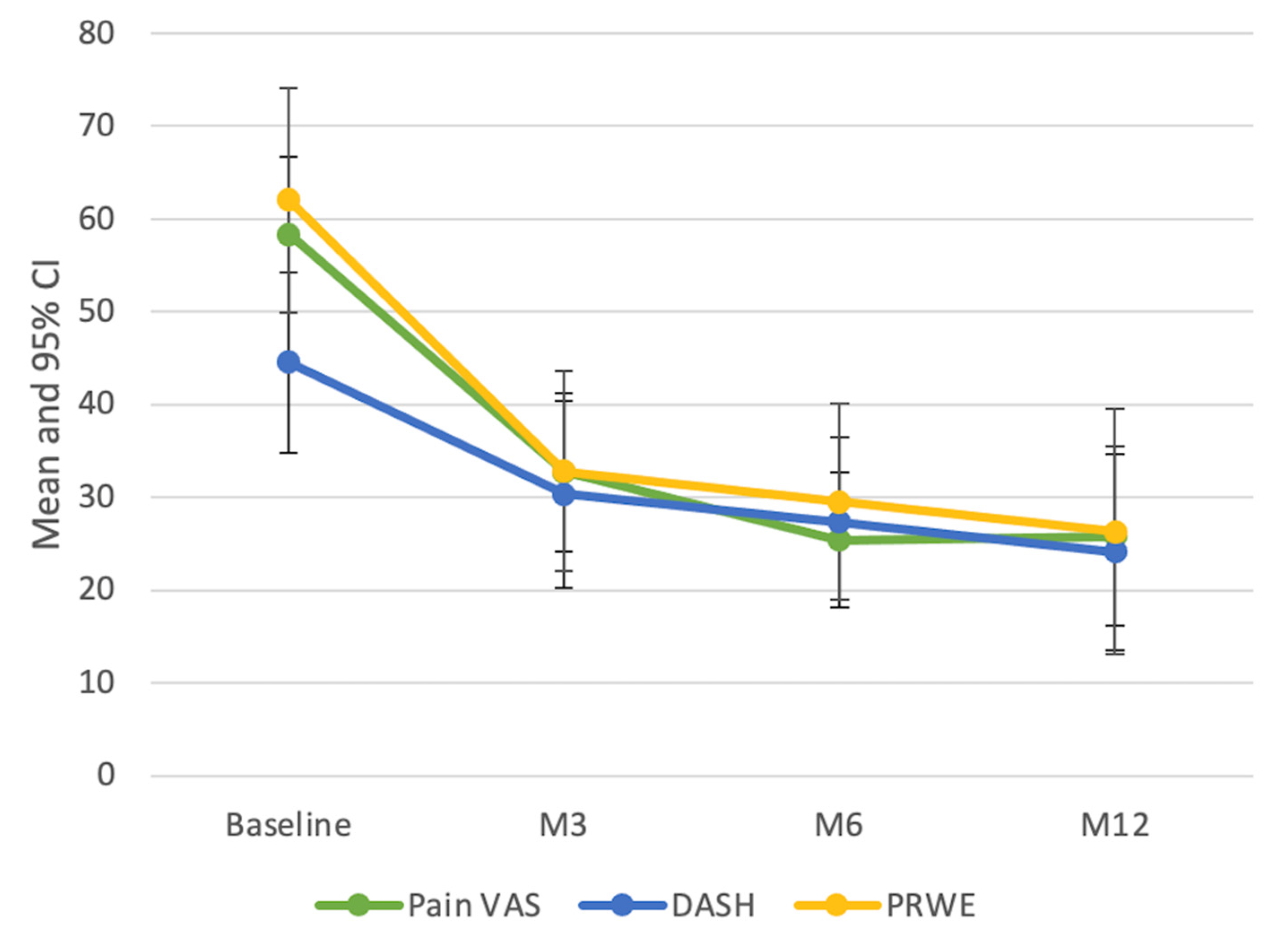
A statistically significant improvement in pain according VAS was observed at each follow-up (Figure 2 and Supplementary Table S1) with a mean of 58.3 ± 13.2 at baseline decreasing to 25.8 ± 15.2 at one year. Seven patients had an improvement in pain greater than 30 at one year after injection. DASH and PRWE scores were also significantly improved at each follow-up and the MCID was reached in 9 (75%) of patients for the DASH and 11 (91.7%) for the PRWE at 12 months. A statistically significant increase in strength of the injured wrist was observed at each follow-up with a mean change of 8.3 ± 6.9 kg at one year from baseline (Figure 3 and Supplementary Table S2) versus 5.4 ± 4.9 kg for the normal wrist. Comparison between patients with radiocarpal OA and both radio- and midcarpal OA did not reveal any significant difference on VAS pain, DASH and PRWE scores and wrist strength (Supplementary Tables S3 and S4). The injured wrist treated with MF-PRP mix also significantly improved ulnar inclination up to 6 months with persistence at 12 months whereas the normal wrist did not change over time. No significant increase was observed for flexion, extension or radial inclination of the injured wrist. Regarding patient satisfaction at one year, 9 were satisfied or very satisfied and 3 patients were neutral.

3.5. MRI Assessment

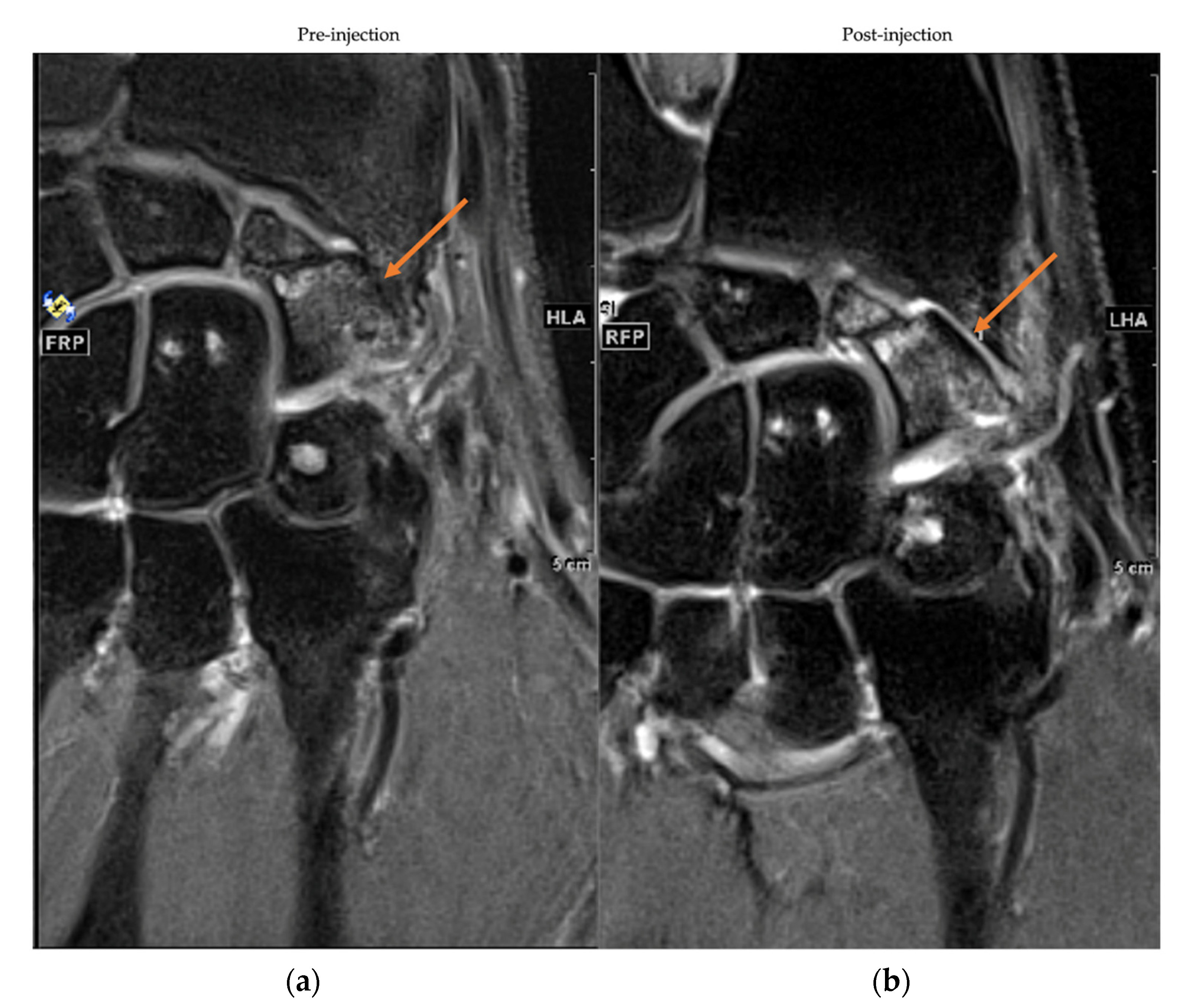
Qualitative MRI analysis of cartilage structural changes at 12 months showed improvement in 3 patients (25%) with a Likert confidence scale of 1 (n = 2) and 0 (n = 1). No change was noted in 6 patients (50%) with Likert confidence scale of 2 (n = 1), 1 (n = 2) and 0 (n = 3) while possible impairment was observed in 3 patients (25%) with uncertainty for all patients. Regarding the variation in oedema, the radiologist confidence level was higher (11/12 assessment score 2 on the Likert scale): 3 (25%) patients were improved, 7 (58 %) showed no difference while 2 (17%) patients were impaired. Figure 4 illustrates the pre- and post-injection MRIs of one patient showing improvement in cartilage structural changes.

4. Discussion

The aim of this study was to evaluate the feasibility and the safety of intra articular injection MF-PRP in treatment of wrist OA. The only side effect reported in our cohort was pain at the harvesting adipose sites which was relieved with grade 1 analgesics and completely resolved after 1 month. This adverse event is commonly observed after any liposuction and also reported in other similar studies [36,37,38,39]. This is consistent with the good tolerance observed after injection of MF-PRP mixture into the joints of racehorses [28]. More generally, no infectious or neoplastic complications related to intra-articular injection of any orthobiological product (PRP, ADSCs, SVF or unfractionated adipose tissue) have been reported in the literature [40,41,42,43] whereas the most common complications reported were early pain and swelling of the injected joint [36,40,44,45,46,47,48]. The latter side effect is usually observed after joint injection, regardless of the product injected. In fact, in comparative studies evaluating SVF or ADSC injection versus placebo or hyaluronic acid, pain and swelling of the injected joint were reported in both groups [37,46,49,50,51].
Pain and swelling are common after injection in the knee but rather rare in carpo-metacarpal injection [36]. Delayed tenosynovitis and tendonitis at 6–8 weeks of injection have been described by one author in 20% of patient after injection of SVF and PRP in various joints and mainly concerned elderly patients [40].
A bacteriological contamination of MF was identified in two cases without clinical consequence. As a precaution, we treated these patients with antibiotics after medical advice from infectious disease specialists. Although this aspect raises concerns about patient safety, this type of contamination has been previously described by Louis et al. also using MF (4/30 products) and Garza et al. using SVF (2/26 products) [52,53]. None of these events were associated with clinical infections.
Many regenerative therapies, based on bone marrow or adipose tissue-derived multipotent stem cells capable of differentiating into cartilage cells have been evaluated for the treatment of OA. Adipose tissue is considered a valuable source of multipotent stem cells with up to 40 times the concentration of bone marrow [54]. The most popular strategy was to directly inject a homogeneous suspension of multipotent stem cells (MSCs). Several comparative studies have recorded the superiority of this treatment over placebo or hyaluronic acid in the treatment of knee OA [46,49,50,51]. This technique requires several complex and expensive purification and expansion steps by culture in an approved manufacturing center lasting two to three weeks [55]. In contrast, the preparation of MF can be done in one hour, allowing for liposuction and reinjection to be performed in the same procedure. The procedure requires the use of a dedicated cannula to select small (600 µm) fat lobules followed by a purification step using the Puregraft® bag membrane required to remove oily substances and blood residues [26]. Although the manufacturing slightly differs with microfragmented fat (MFAT) [36,38,41,56,57,58,59], both techniques generally select small fat lobules that retain intact MSCs in the vascular niche of the adipose stroma during the preparation process [59].
Regarding the efficacy of autologous unfractionated adipose tissue for pain relief, several authors have reported injections in OA of the thumb carpometacarpal joint, but this has never been performed in wrist OA. In 2017, Herold et al. were the first to study autologous unfractionated adipose tissue obtained by liposuction and centrifugation in the treatment of carpometacarpal OA of the thumb [60]. The mean change at 12 months from baseline in pain at stress according in VAS measured pain with movement was 53, 20, and 29 mm for stage 2, 3 and 4 according to Eaton /Littler classification, respectively. Haas et al. also treated thumb carpometacarpal OA from stage 1 to 3 (stage 3 = 58%) using unfractionated adipose tissue without centrifugation step and reported an average reduction of pain under stress of 29 mm at 12 months [38]. More recently, Froschauer et al., reported long term effect of unfractionated adipose tissue without centrifugation in stage 2 and 3 thumb carpometacarpal with the main reduction of pain being 50 mm at two years after the injection [39]. All these results are consistent with the pain VAS improvement reported in our study (32.4 ± 17.8 mm 12 months after injection) where all treated patients had advanced wrist OA (grade 3 or 4 according to Kellgren and Lawrence).
None of the studies mentioned prior used combined adipose tissue with PRP. The use of MF-PRP should improve clinical outcomes as it has been reported that PRP increases MSC proliferation and chondrogenic differentiation in vitro [16,61,62,63,64,65] and may improve cartilage regeneration. These properties combined with the release of platelet growth factors participating in the tissue healing process and inducing an anti-inflammatory effect in chondral disease could be important in the treatment of OA [66,67,68]. From a clinical perspective, only two studies reported the use of PRP combined with unmanipulated adipose tissue in knee OA. Both Louis et al. and Pintat et al. reported similar clinical improvement in WOMAC scores of approximatively 20 points at 6 months which was maintained up to one year [52,69]. We observed a similar improvement in the functional status of the wrist up to one year with a mean change of 20 ± 11.8 and 35.7 ± 19.5 in DASH and PRWE scores respectively. Interestingly, Louis et al. compared the injection of MF mixed or not with PRP, and found no significant difference between the experimental groups. This raises the question of the necessity of using PRP in the study product, although Louis et al. conceded that their trial had a limited number of patients treated, and could have been underpowered. Both studies also reported MRI findings showing a decrease in relaxation time, a reduction in the severity of the grade of OA and an improvement in joint space, but without reaching statistical significance. Nevertheless, performing MRI of the wrist was not an easy task compared to MRI of the knee. The small size of the carpal bones requires higher accuracy and image quality to be able to analyze a structural change of the cartilage. In addition, due to the constant micromovements of the carpal bones, an image registration process was required to make the pre- and post-injection MRI slices stackable. This process deteriorated the image quality and made comparative MRI analysis difficult, leading to a low level of confidence from the blinded radiologist for the evaluation procedure. In future experiments, the use of a rigid cast should be validated to ensure that patients are scanned in the same position during repeated measurements.
Wrist denervation is classically one of the first surgical option offered for severe and persistent wrist OA. It is the only surgical procedure that preserves the joint anatomy and mobility of the wrist. Although this surgical procedure has shown globally encouraging results in chronic wrist OA, results are heterogeneous regarding different parameters such as improvement in mean pain scores (36–92%), or increase in grip strength (7–64%) and failure rate (re-operation, persistent pain or worse,6 to 29%) [70]. In our study, intra-articular injection of MF-PRP resulted in a 56% improvement in pain VAS and a 32% improvement in grip strength. Interestingly, Erne et al. compared the efficiency of autologous fat injection with the standard surgical procedure (Lundsborg’s resection arthroplasty) in the treatment of carpometacarpal OA of the thumb [71]. Adipose tissue injection was associated with a shorter delay before obtaining a complete relief of pain. Moreover, injection of MF-PRP is less invasive and likely to have a curative effect, allowing a potential regeneration of the cartilage whereas wrist denervation is a purely palliative analgesic treatment.
The major limitation of our study is the absence of a placebo group, which is a significant weakness given the considerable placebo effect in OA care [72]. Indeed, studies comparing long-term results with more accessible and cost-effective injectable drugs (steroid, HA or PRP alone) will be necessary to ensure the value of MSC-PRP injection in wrist OA. Given the complexity and size of the wrist articular cartilage, MRI quantification is complex and requires technological improvements to provide more valuable and objective data of the structural effects of MF-PRP injection on wrist cartilage. Finally, some of the patients included presented both radiocarpal and midcarpal wrist OA, whereas the MF-PRP injection was systematically performed in the radiocarpal joint. Further investigations of possible communication between both joints are necessary to ensure proper product distribution, especially when patients present associated SLAC or SNAC. This is further strengthened by the absence of clinical significance between these two groups. This issue should be clarified before conducting future studies.
Moreover, no data is available to clearly explain the mechanism of action of MF-PRP in OA although the main hypothesis is linked to anti-inflammatory properties as well as regenerative capacity likely due to the ADSCs kept intact into MF and growth factors contained in the PRP.

5. Conclusions

MF-PRP injection in wrist OA is a safe procedure and provides significant clinical benefit on function and pain relief for up to 12 months. Larger and placebo-controlled studies are required to confirm whether these new strategies can improve pain relief and wrist function and induce significant and measurable structural benefit, ultimately delaying and avoiding the need for a major surgical procedure.

Supplementary Materials

The following supporting information can be downloaded at: www.mdpi.com/article/10.3390/jcm11195786/s1. Table S1: Patient-related Outcomes over the 12 months post treatment; Table S2: Clinical measures over the 12 months post treatment; Table S3: Patient-related Outcomes over the 12 months post treatment by localization; Table S4: Clinical measures over the 12 months post treatment by localization.

Author Contributions

Conceptualization, A.M., M.W., A.I. and N.K.; methodology, E.J.; validation, J.M., C.J. and R.L.; formal analysis, E.J.; investigation, J.V. and F.G.; resources, A.C.Z., L.G. and C.D.; data curation, R.F.; writing—original draft preparation, A.M.; writing—review and editing, C.B., A.M. and D.B.; supervision, J.M. and C.J.; project administration, C.C., D.C. and F.S. All authors have read and agreed to the published version of the manuscript.

Funding

This research received no external funding.

Institutional Review Board Statement

The protocol was approved by a national ethics committee (authorization #16-65 from Comité de Protection des Personnes Sud Méditerranée #1) and national health regulatory authority (Agence Nationale de Sécurité du Médicament et des Produits de Santé, authorization #160879A-12) and registered in Clinicaltrials.gov website (NCT03164122 and EudraCT # 2016-002648-18). The study was carried out according to the Declaration of Helsinki and with the principles of Good Clinical Practice. All patients gave written informed consent before their participation.

Informed Consent Statement

Written informed consent was obtained from all subjects involved in the study.

Data Availability Statement

Main data presented in this study are contained within this article and Supplementary Materials. The data that support the findings of the study are available by a request to the corresponding author.

Acknowledgments

Medical devices Hy Tissue 20 ® device (Proteal, Barcelona, Spain now acquired by Fidia, Abano Terme, Italy) and Hapi® fat (Benew Medical, Melesse, France) kit were given for free by companies.

Conflicts of Interest

J.M. received honorarium for educational support from FIDIA, HORIBA ARTHREX and MACOPHARMA. These manufacturers had no role in the design of the study; in the collection, analyses, or interpretation of data; in the writing of the manuscript; or in the decision to publish the results. Other authors did not declare any conflict of interest.

References

  1. Stanley, J.K. Degenerative arthritis of the wrist. Curr. Orthop. 1999, 13, 290–296. [Google Scholar] [CrossRef]
  2. Laulan, J.; Marteau, E.; Bacle, G. Wrist osteoarthritis. Orthop. Traumatol. Surg. Res. 2015, 101, S1–S9. [Google Scholar] [CrossRef] [PubMed]
  3. Weiss, K.E.; Rodner, C.M. Osteoarthritis of the wrist. J. Hand Surg. 2007, 32, 725–746. [Google Scholar] [CrossRef]
  4. Watson, H.K.; Ballet, F.L. The SLAC wrist: Scapholunate advanced collapse pattern of degenerative arthritis. J. Hand Surg. 1984, 9, 358–365. [Google Scholar] [CrossRef]
  5. Lane, N.; Shidara, K.; Wise, B. Osteoarthritis year in review 2016: Clinical. Osteoarthr. Cartil. 2017, 25, 209–215. [Google Scholar] [CrossRef] [PubMed]
  6. Kolasinski, S.L.; Neogi, T.; Hochberg, M.C.; Oatis, C.; Guyatt, G.; Block, J.; Callahan, L.; Copenhaver, C.; Dodge, C.; Felson, D.; et al. 2019 American College of Rheumatology/Arthritis Foundation Guideline for the Management of Osteoarthritis of the Hand, Hip, and Knee. Arthritis Care Res. 2020, 72, 149–162. [Google Scholar] [CrossRef]
  7. Mahendira, D.; Towheed, T. Systematic review of non-surgical therapies for osteoarthritis of the hand: An update. Osteoarthr. Cartil. 2009, 17, 1263–1268s. [Google Scholar] [CrossRef]
  8. Lue, S.; Koppikar, S.; Shaikh, K.; Mahendira, D.; Towheed, T.E. Systematic review of non-surgical therapies for osteoarthritis of the hand: An update. Osteoarthr. Cartil. 2017, 25, 1379–1389. [Google Scholar] [CrossRef]
  9. Fuchsberger, T.; Gonser, P.; Boesch, C.; Tonagel, F.; Fischborn, T.; Schaller, H.; Haerle, M. Corrigendum to Patient-rated long-term results after complete denervation of the wrist. J. Plast. Reconstr. Aesthetic Surg. 2018, 71, 1381. [Google Scholar] [CrossRef]
  10. Sauerbier, M.; Kluge, S.; Bickert, B.; Germann, G. Subjective and objective outcomes after total wrist arthrodesis in patients with radiocarpal arthrosis or Kienböck’s disease. Chir. Main 2000, 19, 223–231. [Google Scholar] [CrossRef]
  11. Nair, R. Review Article: Total Wrist Arthroplasty. J. Orthop. Surg. 2014, 22, 399–405. [Google Scholar] [CrossRef] [PubMed]
  12. Sagerfors, M.; Gupta, A.; Brus, O.; Pettersson, K. Total Wrist Arthroplasty: A Single-Center Study of 219 Cases With 5-Year Follow-up. J. Hand Surg. 2015, 40, 2380–2387. [Google Scholar] [CrossRef] [PubMed]
  13. Herzberg, G.; Boeckstyns, M.; Sorensen, A.I.; Axelsson, P.; Kroener, K.; Liverneaux, P.; Obert, L.; Merser, S. “Remotion” Total Wrist Arthroplasty: Preliminary Results of a Prospective International Multicenter Study of 215 Cases. J. Wrist Surg. 2012, 1, 17–22. [Google Scholar] [CrossRef]
  14. Sánchez-González, D.J.; Méndez-Bolaina, E.; Trejo-Bahena, N.I. Platelet-Rich Plasma Peptides: Key for Regeneration. Int. J. Pept. 2012, 2012, 532519. [Google Scholar] [CrossRef] [PubMed]
  15. Marx, R.E.; Carlson, E.R.; Eichstaedt, R.M.; Schimmele, S.R.; Strauss, J.E.; Georgeff, K.R. Platelet-rich plasma: Growth factor enhancement for bone grafts. Oral Surg. Oral Med. Oral Pathol. Oral Radiol. Endodontol. 1998, 85, 638–646. [Google Scholar] [CrossRef]
  16. Xie, X.; Zhang, C.; Tuan, R.S. Biology of platelet-rich plasma and its clinical application in cartilage repair. Arthritis Res. Ther. 2014, 16, 204–215. [Google Scholar] [CrossRef]
  17. Dai, W.L.; Zhou, A.G.; Zhang, H.; Zhang, J. Efficacy of Platelet-Rich Plasma in the Treatment of Knee Osteoarthritis: A Meta-analysis of Randomized Controlled Trials. Arthroscopy 2017, 33, 659–670.e1. [Google Scholar] [CrossRef]
  18. Meheux, C.J.; McCulloch, P.C.; Lintner, D.M.; Varner, K.E.; Harris, J.D. Efficacy of Intra-articular Platelet-Rich Plasma Injections in Knee Osteoarthritis: A Systematic Review. Arthroscopy 2016, 32, 495–505. [Google Scholar] [CrossRef]
  19. Khoshbin, A.; Leroux, T.; Wasserstein, D.; Marks, P.; Theodoropoulos, J.; Ogilvie-Harris, D.; Gandhi, R.; Takhar, K.; Lum, G.; Chahal, J. The Efficacy of Platelet-Rich Plasma in the Treatment of Symptomatic Knee Osteoarthritis: A Systematic Review With Quantitative Synthesis. Arthroscopy 2013, 29, 2037–2048. [Google Scholar] [CrossRef]
  20. Laudy, A.B.; Bakker, E.W.; Rekers, M.; Moen, M.H. Efficacy of platelet-rich plasma injections in osteoarthritis of the knee: A systematic review and meta-analysis. Br. J. Sports Med. 2015, 49, 657–672. [Google Scholar] [CrossRef] [Green Version]
  21. English, A.; Jones, E.A.; Corscadden, D.; Henshaw, K.; Chapman, T.; Emery, P.; McGonagle, D. A comparative assessment of cartilage and joint fat pad as a potential source of cells for autologous therapy development in knee osteoarthritis. Rheumatology 2007, 46, 1676–1683. [Google Scholar] [CrossRef] [PubMed]
  22. Maumus, M.; Manferdini, C.; Toupet, K.; Peyrafitte, J.-A.; Ferreira, R.; Facchini, A.; Gabusi, E.; Bourin, P.; Jorgensen, C.; Lisignoli, G.; et al. Adipose mesenchymal stem cells protect chondrocytes from degeneration associated with osteoarthritis. Stem Cell Res. 2013, 11, 834–844. [Google Scholar] [CrossRef] [PubMed]
  23. Erickson, G.R.; Gimble, J.M.; Franklin, D.M.; Rice, H.E.; Awad, H.; Guilak, F. Chondrogenic Potential of Adipose Tissue-Derived Stromal Cells in Vitro and in Vivo. Biochem. Biophys. Res. Commun. 2002, 290, 763–769. [Google Scholar] [CrossRef]
  24. Huang, J.I.; Zuk, P.A.; Jones, N.F.; Zhu, M.; Lorenz, H.P.; Hedrick, M.H.; Benhaim, P. Chondrogenic Potential of Multipotential Cells from Human Adipose Tissue. Plast. Reconstr. Surg. 2004, 113, 585–594. [Google Scholar] [CrossRef]
  25. Zuk, P.A.; Zhu, M.; Ashjian, P.; De Ugarte, D.A.; Huang, J.I.; Mizuno, H.; Alfonso, Z.C.; Fraser, J.K.; Benhaim, P.; Hedrick, M.H. Human Adipose Tissue Is a Source of Multipotent Stem Cells. Mol. Biol. Cell 2002, 13, 4279–4295. [Google Scholar] [CrossRef] [PubMed]
  26. Nguyen, P.S.; Desouches, C.; Gay, A.M.; Hautier, A.; Magalon, G. Development of micro-injection as an innovative autologous fat graft technique: The use of adipose tissue as dermal filler. J. Plast. Reconstr. Aesthetic Surg. 2012, 65, 1692–1699. [Google Scholar] [CrossRef]
  27. Alharbi, Z.; Opländer, C.; Almakadi, S.; Fritz, A.; Vogt, M.; Pallua, N. Conventional vs. micro-fat harvesting: How fat harvesting technique affects tissue-engineering approaches using adipose tissue-derived stem/stromal cells. J. Plast. Reconstr. Aesthetic Surg. 2013, 66, 1271–1278. [Google Scholar] [CrossRef]
  28. Bembo, F.; Eraud, J.; Philandrianos, C.; Bertrand, B.; Silvestre, A.; Veran, J.; Sabatier, F.; Magalon, G.; Magalon, J. Combined use of platelet rich plasma and micro-fat in sport and race horses with degenerative joint disease: Preliminary clinical study in eight horses. Muscle Ligaments Tendons J. 2016, 6, 198–204. [Google Scholar] [CrossRef]
  29. Mayoly, A.; Iniesta, A.; Curvale, C.; Kachouh, N.; Jaloux, C.; Eraud, J.; Vogtensperger, M.; Veran, J.; Grimaud, F.; Jouve, E.; et al. Development of Autologous Platelet-Rich Plasma Mixed-Microfat as an Advanced Therapy Medicinal Product for Intra-Articular Injection of Radio-Carpal Osteoarthritis: From Validation Data to Preliminary Clinical Results. Int. J. Mol. Sci. 2019, 20, 1111. [Google Scholar] [CrossRef]
  30. Sautereau, N.; Daumas, A.; Truillet, R.; Jouve, E.; Magalon, J.; Veran, J.; Casanova, D.; Frances, Y.; Magalon, G.; Granel, B. Efficacy of Autologous Microfat Graft on Facial Handicap in Systemic Sclerosis Patients. Plast. Reconstr. Surg. -Glob. Open 2016, 4, e660. [Google Scholar] [CrossRef]
  31. Graiet, H.; Lokchine, A.; Francois, P.; Velier, M.; Grimaud, F.; Loyens, M.; Berda-Haddad, Y.; Veran, J.; Dignat-George, F.; Sabatier, F.; et al. Use of platelet-rich plasma in regenerative medicine: Technical tools for correct quality control. BMJ Open Sport Exerc. Med. 2018, 4, e000442. [Google Scholar] [CrossRef] [PubMed]
  32. Schernberg, F. Classification des fractures du scaphoïde carpien. Etude anatomo-radiologique des traits [Classification of fractures of the carpal scaphoid. An anatomo-radiologic study of characteristics]. Rev. Chir. Orthop. Reparatrice Appar. Mot. 1988, 74, 693–695. [Google Scholar] [PubMed]
  33. Franchignoni, F.; Vercelli, S.; Giordano, A.; Sartorio, F.; Bravini, E.; Ferriero, G. Minimal Clinically Important Difference of the Disabilities of the Arm, Shoulder and Hand Outcome Measure (DASH) and Its Shortened Version (QuickDASH). J. Orthop. Sports Phys. Ther. 2014, 44, 30–39. [Google Scholar] [CrossRef] [PubMed]
  34. Walenkamp, M.M.; de Muinck Keizer, R.J.; Goslings, J.C.; Vos, L.M.; Rosenwasser, M.P.; Schep, N.W. The Minimum Clinically Important Difference of the Patient-rated Wrist Evaluation Score for Patients with Distal Radius Fractures. Clin. Orthop. Relat. Res. 2015, 473, 3235–3241. [Google Scholar] [CrossRef]
  35. Magalon, J.; Chateau, A.L.; Bertrand, B.; Louis, M.L.; Silvestre, A.; Giraudo, L.; Veran, J.; Sabatier, F. DEPA classification: A proposal for standardising PRP use and a retrospective application of available devices. BMJ Open Sport Exerc. Med. 2016, 2, e000060. [Google Scholar] [CrossRef]
  36. Barfod, K.W.; Blønd, L. Treatment of osteoarthritis with autologous and microfragmented adipose tissue. Dan. Med. J. 2019, 66, A5565. [Google Scholar]
  37. Hong, Z.; Chen, J.; Zhang, S.; Zhao, C.; Bi, M.; Chen, X.; Bi, Q. Intra-articular injection of autologous adipose-derived stromal vascular fractions for knee osteoarthritis: A double-blind randomized self-controlled trial. Int. Orthop. 2018, 43, 1123–1134. [Google Scholar] [CrossRef]
  38. Haas, E.M.; Eisele, A.; Arnoldi, A.; Paolini, M.; Ehrl, D.; Volkmer, E.; Giunta, R.E. One-Year Outcomes of Intraarticular Fat Transplantation for Thumb Carpometacarpal Joint Osteoarthritis: Case Review of 99 Joints. Plast. Reconstr. Surg. 2020, 145, 151–159. [Google Scholar] [CrossRef]
  39. Malanga, G.A.; Chirichella, P.S.; Hogaboom, N.S.; Capella, T. Clinical evaluation of micro-fragmented adipose tissue as a treatment option for patients with meniscus tears with osteoarthritis: A prospective pilot study. Int. Orthop. 2021, 45, 473–480. [Google Scholar] [CrossRef]
  40. Pak, J.; Chang, J.-J.; Lee, J.H.; Lee, S.H. Safety reporting on implantation of autologous adipose tissue-derived stem cells with platelet-rich plasma into human articular joints. BMC Musculoskelet. Disord. 2013, 14, 337. [Google Scholar] [CrossRef]
  41. Froschauer, S.M.; Holzbauer, M.; Wenny, R.; Schmidt, M.; Huemer, G.M.; Kwasny, O.; Duscher, D. Autologous Fat Transplantation for Thumb Carpometacarpal Joint Osteoarthritis (Liparthroplasty): A Case Series with Two Years of Follow-UP. J. Clin. Med. 2020, 10, 113. [Google Scholar] [CrossRef] [PubMed]
  42. Kemper, R.; Wirth, J.; Baur, E.M. Arthroscopic Synovectomy Combined with Autologous Fat Grafting in Early Stages of CMC Osteoarthritis of the Thumb. J. Wrist. Surg. 2018, 7, 165–171. [Google Scholar] [CrossRef]
  43. Hudetz, D.; Borić, I.; Rod, E.; Jeleč, Ž.; Kunovac, B.; Polašek, O.; Vrdoljak, T.; Plečko, M.; Skelin, A.; Polančec, D.; et al. Early results of intra-articular micro-fragmented lipoaspirate treatment in patients with late stages knee osteoarthritis: A prospective study. Croat. Med. J. 2019, 60, 227–236. [Google Scholar] [CrossRef]
  44. Adriani, E.; Moio, M.; Di Paola, B.; Salustri, W.; Alfieri, A.; Parisi, P.; Ruggiero, M.; Borab, Z.; Carlesimo, B. Percutaneous Fat Transfer to Treat Knee Osteoarthritis Symptoms: Preliminary Results. Joints 2017, 5, 89–92. [Google Scholar] [CrossRef] [PubMed]
  45. Roato, I.; Belisario, D.C.; Compagno, M.; Lena, A.; Bistolfi, A.; Maccari, L.; Mussano, F.; Genova, T.; Godio, L.; Perale, G.; et al. Concentrated adipose tissue infusion for the treatment of knee osteoarthritis: Clinical and histological observations. Int. Orthop. 2019, 43, 15–23. [Google Scholar] [CrossRef] [PubMed]
  46. Freitag, J.; Bates, D.; Wickham, J.; Shah, K.; Huguenin, L.; Tenen, A.; Paterson, K.; Boyd, R. Adipose-derived mesenchymal stem cell therapy in the treatment of knee osteoarthritis: A randomized controlled trial. Regen. Med. 2019, 14, 213–230. [Google Scholar] [CrossRef] [PubMed]
  47. Chen, Z.; Wang, C.; You, D.; Zhao, S.; Zhu, Z.; Xu, M. Platelet-rich plasma versus hyaluronic acid in the treatment of knee osteoarthritis: A meta-analysis. Medicine 2020, 99, e19388. [Google Scholar] [CrossRef]
  48. Zhao, J.; Huang, H.; Liang, G.; Zeng, L.F.; Yang, W.; Liu, J. Effects and safety of the combination of platelet-rich plasma (PRP) and hyaluronic acid (HA) in the treatment of knee osteoarthritis: A systematic review and meta-analysis. BMC Musculoskelet Disord. 2020, 21, 224. [Google Scholar] [CrossRef]
  49. Kuah, D.; Sivell, S.; Longworth, T.; James, K.; Guermazi, A.; Cicuttini, F.; Wang, Y.; Craig, S.; Comin, G.; Robinson, D.; et al. Safety, tolerability and efficacy of intra-articular Progenza in knee osteoarthritis: A randomized double-blind placebo-controlled single ascending dose study. J. Transl. Med. 2018, 16, 49. [Google Scholar] [CrossRef]
  50. Lu, L.; Dai, C.; Zhang, Z.; Du, H.; Li, S.; Ye, P.; Fu, Q.; Zhang, L.; Wu, X.; Dong, Y.; et al. Treatment of knee osteoarthritis with intra-articular injection of autologous adipose-derived mesenchymal progenitor cells: A prospective, randomized, double-blind, active-controlled, phase IIb clinical trial. Stem Cell Res. Ther. 2019, 10, 143. [Google Scholar] [CrossRef]
  51. Lee, W.S.; Kim, H.J.; Kim, K.I.; Kim, G.B.; Jin, W. Intra-Articular Injection of Autologous Adipose Tissue-Derived Mesenchymal Stem Cells for the Treatment of Knee Osteoarthritis: A Phase IIb, Randomized, Placebo-Controlled Clinical Trial. Stem Cells Transl. Med. 2019, 8, 504–511. [Google Scholar] [CrossRef] [PubMed] [Green Version]
  52. Louis, M.L.; Dumonceau, R.G.; Jouve, E.; Cohen, M.; Djouri, R.; Richardet, N.; Jourdan, E.; Giraudo, L.; Dumoulin, C.; Grimaud, F.; et al. Intra-Articular Injection of Autologous Microfat and Platelet-Rich Plasma in the Treatment of Knee Osteoarthritis: A Double-Blind Randomized Comparative Study. Arthroscopy 2021, 37, 3125–3137.e3. [Google Scholar] [CrossRef] [PubMed]
  53. Garza, J.R.; Campbell, R.E.; Tjoumakaris, F.P.; Freedman, K.B.; Miller, L.S.; Maria, D.S.; Tucker, B.S. Clinical Efficacy of Intra-articular Mesenchymal Stromal Cells for the Treatment of Knee Osteoarthritis: A Double-Blinded Prospective Randomized Controlled Clinical Trial. Am. J. Sports Med. 2020, 48, 588–598. [Google Scholar] [CrossRef] [PubMed]
  54. Kern, S.; Eichler, H.; Stoeve, J.; Klüter, H.; Bieback, K. Comparative Analysis of Mesenchymal Stem Cells from Bone Marrow, Umbilical Cord Blood, or Adipose Tissue. Stem Cells 2006, 24, 1294–1301. [Google Scholar] [CrossRef]
  55. Jorgensen, C.; Djouad, F.; Bouffi, C.; Mrugala, D.; Noël, D. Multipotent mesenchymal stromal cells in articular diseases. Best Pract. Res. Clin. Rheumatol. 2008, 22, 269–284. [Google Scholar] [CrossRef]
  56. Mautner, K.; Bowers, R.; Easley, K.; Fausel, Z.; Robinson, R. Functional Outcomes Following Microfragmented Adipose Tissue Versus Bone Marrow Aspirate Concentrate Injections for Symptomatic Knee Osteoarthritis. Stem Cells Transl. Med. 2019, 8, 1149–1156. [Google Scholar] [CrossRef]
  57. Borić, I.; Hudetz, D.; Rod, E.; Jeleč, Ž.; Vrdoljak, T.; Skelin, A.; Polašek, O.; Plečko, M.; Trbojević-Akmačić, I.; Lauc, G.; et al. A 24-Month Follow-Up Study of the Effect of Intra-Articular Injection of Autologous Microfragmented Fat Tissue on Proteoglycan Synthesis in Patients with Knee Osteoarthritis. Genes 2019, 10, 1051. [Google Scholar] [CrossRef]
  58. Heidari, N.; Noorani, A.; Slevin, M.; Cullen, A.; Stark, L.; Olgiati, S.; Zerbi, A.; Wilson, A. Patient-Centered Outcomes of Microfragmented Adipose Tissue Treatments of Knee Osteoarthritis: An Observational, Intention-to-Treat Study at Twelve Months. Stem Cells Int. 2020, 2020, 8881405. [Google Scholar] [CrossRef]
  59. Tremolada, C.; Colombo, V.; Ventura, C. Adipose Tissue and Mesenchymal Stem Cells: State of the Art and Lipogems® Technology Development. Curr. Stem Cell Rep. 2016, 2, 304–312. [Google Scholar] [CrossRef]
  60. Herold, C.; Rennekampff, H.O.; Groddeck, R.; Allert, S. Autologous Fat Transfer for Thumb Carpometacarpal Joint Osteoarthritis: A Prospective Study. Plast. Reconstr. Surg. 2017, 140, 327–335. [Google Scholar] [CrossRef]
  61. Roux, C.; Pisani, D.F.; Ben Yahia, H.; Djedaini, M.; Beranger, G.E.; Chambard, J.-C.; Ambrosetti, D.; Michiels, J.-F.; Breuil, V.; Ailhaud, G.; et al. Chondrogenic potential of stem cells derived from adipose tissue: A powerful pharmacological tool. Biochem. Biophys. Res. Commun. 2013, 440, 786–791. [Google Scholar] [CrossRef] [PubMed]
  62. Van Pham, P.; Bui, K.H.-T.; Ngo, D.Q.; Vu, N.B.; Truong, N.H.; Phan, N.L.-C.; Le, D.M.; Duong, T.D.; Nguyen, T.D.; Le, V.T.; et al. Activated platelet-rich plasma improves adipose-derived stem cell transplantation efficiency in injured articular cartilage. Stem Cell Res Ther. 2013, 4, 91. [Google Scholar] [CrossRef] [PubMed]
  63. Mishra, A.; Tummala, P.; King, A.; Lee, B.; Kraus, M.; Tse, V.; Jacobs, C.R. Buffered platelet-rich plasma enhances mesenchymal stem cell proliferation and chondrogenic differentiation. Tissue Eng. Part C Methods 2009, 15, 431–435. [Google Scholar] [CrossRef] [PubMed]
  64. Atashi, F.; Jaconi, M.E.; Pittet-Cuénod, B.; Modarressi, A. Autologous platelet-rich plasma: A biological supplement to enhance adipose-derived mesenchymal stem cell expansion. Tissue Eng. Part C Methods 2015, 21, 253–262. [Google Scholar] [CrossRef] [PubMed]
  65. Rubio-Azpeitia, E.; Andia, I. Partnership between platelet-rich plasma and mesenchymal stem cells: In vitro experience. Muscles Ligaments Tendons J. 2014, 4, 52–62. [Google Scholar] [CrossRef]
  66. 66. Van Buul, G.M.; Koevoet, W.L.; Kops, N.; Koen Bos, P.; Verhaar, J.A.N.; Weinans, H.; Bernsen, M.R.; Van Osch, G.J.V.M. Platelet-rich plasma releasate inhibits inflammatory processes in osteoarthritic chondrocytes. Am. J. Sports Med. 2011, 39, 2362–2370. [Google Scholar] [CrossRef]
  67. Ter Huurne, M.; Schelbergen, R.; Blattes, R.; Blom, A.; de Munter, W.; Grevers, L.C.; Jeanson, J.; Noël, D.; Casteilla, L.; Jorgensen, C.; et al. Antiinflammatory and chondroprotective effects of intraarticular injection of adipose-derived stem cells in experimental osteoarthritis. Arthritis Rheum. 2012, 64, 3604–3613. [Google Scholar] [CrossRef]
  68. Nava, S.; Sordi, V.; Pascucci, L.; Tremolada, C.; Ciusani, E.; Zeira, O.; Cadei, M.; Soldati, G.; Pessina, A.; Parati, E.; et al. Long-Lasting Anti-Inflammatory Activity of Human Microfragmented Adipose Tissue. Stem Cells Int. 2019, 2019, 5901479. [Google Scholar] [CrossRef]
  69. Pintat, J.; Silvestre, A.; Magalon, G.; Gadeau, A.P.; Pesquer, L.; Perozziello, A.; Peuchant, A.; Mounayer, C.; Dallaudière, B. Intra-articular Injection of Mesenchymal Stem Cells and Platelet-Rich Plasma to Treat Patellofemoral Osteoarthritis: Preliminary Results of a Long-Term Pilot Study. J. Vasc. Interv. Radiol. 2017, 28, 1708–1713. [Google Scholar] [CrossRef]
  70. Chin, K.W.T.K.; Engelsman, A.F.; van Gulik, T.M.; Strackee, S.D. Selective denervation of the wrist for chronic pain: A systematic literature review. J. Hand Surg. Eur. Vol. 2020, 45, 265–272. [Google Scholar] [CrossRef]
  71. Erne, H.C.; Cerny, M.K.; Ehrl, D.; Bauer, A.T.; Schmauss, V.; Moog, P.; Broer, P.N.; Loew, S.; Schmauss, D. Autologous Fat Injection versus Lundborg Resection Arthroplasty for the Treatment of Trapeziometacarpal Joint Osteoarthritis. Plast. Reconstr. Surg. 2018, 141, 119–124. [Google Scholar] [CrossRef]
  72. Zhang, W.; Robertson, J.; Jones, A.C.; Dieppe, P.A.; Doherty, M. The placebo effect and its determinants in osteoarthritis: Meta-analysis of randomised controlled trials. Ann. Rheum. Dis. 2008, 67, 1716–1723. [Google Scholar] [CrossRef]
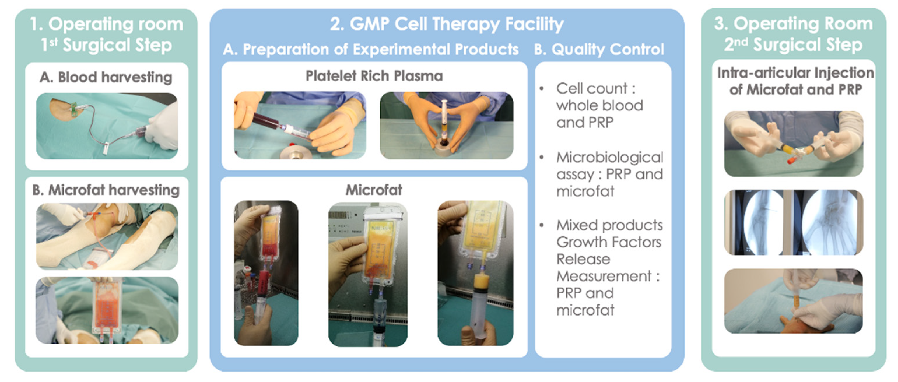
Figure 1. Procedure of preparation of experimental products: PRP and Microfat.
Jcm 11 05786 g001
Figure 2. Evolution of DASH and PRWE scores and VAS of pain during the 12 months of treatment.
Jcm 11 05786 g002
Figure 3. Wrist strength (a) and Ulnar inclination (b) over 12 months post treatment.
Jcm 11 05786 g003
Figure 4. MRIs frontal sections of pre-injection (a) and 12 months post-injection (b). Cartilage condition was assessed with a 2D proton density turbo spin echo (TSE) with fat saturation sequence. The arrow indicates the location of the joint space of interest.
Jcm 11 05786 g004
Table 1. Inclusion and Exclusion Criteria.
Inclusion Criteria
  -
Age between 18 and 75 years;
  -
Radio-carpal osteoarthritis grade 3 or 4 according to Kellgren and Lawrence classification resulting from post-traumatic malunion of an articular distal radius fracture or SLAC or SNAC;
  -
Failure of well-managed pharmacological treatment (analgesics, anti-inflammatory drugs, splinting, physiotherapy) defined as persistent daily painful condition > 40 mm according to visual analog scale (VAS);
  -
Body Mass Index ≥ 20 kg/m2;
  -
Hb > 10 g/dL;
  -
Negative pregnancy test;
  -
Written informed consent.
Exclusion Criteria
  -
Contraindications to MRI scanning;
  -
Thrombocytopenia < 150 G/L; thrombocytosis > 450 G/L; thrombopathy;
  -
Other coagulation disorders;
  -
Infectious disease or positive serology to VIH-1, HCV, HBV and syphilis;
  -
Chronic treatment with oral corticosteroids (or last dose taken less than 2 weeks before);
  -
Intra articular wrist injection of corticosteroid or hyaluronic acid less than 8 weeks before inclusion;
  -
Nonsteroidal anti-inflammatory drug or platelet inhibiting agent or antivitamin K treatment completed less than 2 weeks before injection;
  -
Fever or recent disease (<1 month before injection);
  -
Auto immune disease;
  -
Inflammatory arthritis;
  -
Immune deficit;
  -
History of or ongoing malignancy;
  -
Pregnancy;
  -
Patient under guardianship or involved in another clinical trial.
MRI: Magnetic Resonance Imaging; Hb: haemoglobin; VIH: human immunodeficiency virus; HCV: Hepatitis C Virus; HBV: Hepatitis B Virus.
Table 2. Baseline Characteristics of patients.
Number of Patients (n)12
Gender (women/men)4 (33)/8 (67)
Age (years)53.8 ± 14.8 (24–68)
BMI (kg/m2) 25.6 ± 3.8 (20–30.5)
Dominant hand: Right-handed 12 (100)
Affected hand: Right/Left7 (58)/5 (42)
Disease duration from diagnosis (years) 6.6 ± 9.1 (1–25)
Traumatic etiology:12 (100)
  -
SLAC
4 (33.3)
  -
SNAC
4 (33.3)
  -
Radius malunion
4 (33.3)
Osteoarthritis grade (Kellgren and Lawrence
classification), (0 to 4)
Grade 3: 3 (25)
Grade 4: 9 (75)
Localisation of osteoarthritis (X-ray)
  -
Radiocarpal joint
8 (67)
  -
Radiocarpal joint and midcarpal joint
4 (33)
Data are presented as mean ± standard deviation and (minimum–maximum) for continuous variable and n (%) of patients for categorical variables. SLAC: scapholunate advanced collapse; SNAC: Scaphoid nonunion advanced collapse.
Table 3. Biological characteristics of experimental products.
Mean ± SDMedian (Min–Max)
Total mean volume (mL)3.6 ± 0.43.8 (2.7–4)
Microbiological Assay, Free of germ
Microfat10 (83)
PRP12 (100)
Cells (millions; %)
Platelets741 ± 162; 95.3 ± 2.8715 (480–1094)
Red Blood Cells33.3 ± 17.7; 4.5 ± 2.730 (17.5–76)
Leukocytes1.2 ± 1.3; 0.1 ± 0.10.9 (0.1–4.1)
Ratio platelets/cc of fat410 ± 88407 (274–547)
Inflammatory cytokines (pg/mL)
IL-1b7.7 ± 16.61.8 (0–58)
TNFa25.3 ± 23.518.3 (0.6–84.7)
IL-64669 ± 31873861 (344–9960)
IFN γ4 ± 60.1 (0–18.9)
Anti-Inflammatory cytokines (pg/mL)
IL-1012.1 ± 8.511.1 (0–25.1)
IL-1ra155 ± 149125 (28–539)
Ratio IL1Ra/Il1b88.7 ± 108.439.9 (1.6–378.6)
Growth factors (pg/mL)
PDGF519 ± 340490 (62–1095)
VEGF-a28.2 ± 31.324.7 (0–101.8)
EGF131.8 ± 67.3135.3 (5.3–214.4)
FGF-26828 ± 33455904.9 (2410–12,999)
TGFB15019 ± 28915095.2 (1092–9271)
Data are presented as mean ± standard deviation and (minimum–maximum) for continuous variable and n (%) of patients for categorical variables. PRP: Platelet Rich Plasma; IL: interleukin; TNF-α: tumor necrosis factor-α; IFN-γ: interferon-γ; IL1Ra: interleukin-1 receptor antagonist; EGF: epidermal growth factor; VEGF: vascular endothelial growth factor; PDGF: platelet-derived growth factor; FGF2: fibroblast growth factor 2; TGF-β1: transforming growth factor β1.
Publisher’s Note: MDPI stays neutral with regard to jurisdictional claims in published maps and institutional affiliations.