

Correlation between Hypoperfusion Intensity Ratio and Functional Outcome in Large-Vessel Occlusion Acute Ischemic Stroke: Comparison with Multi-Phase CT Angiography

 ,
,
Abstract
:1. Introduction
2. Materials and Methods
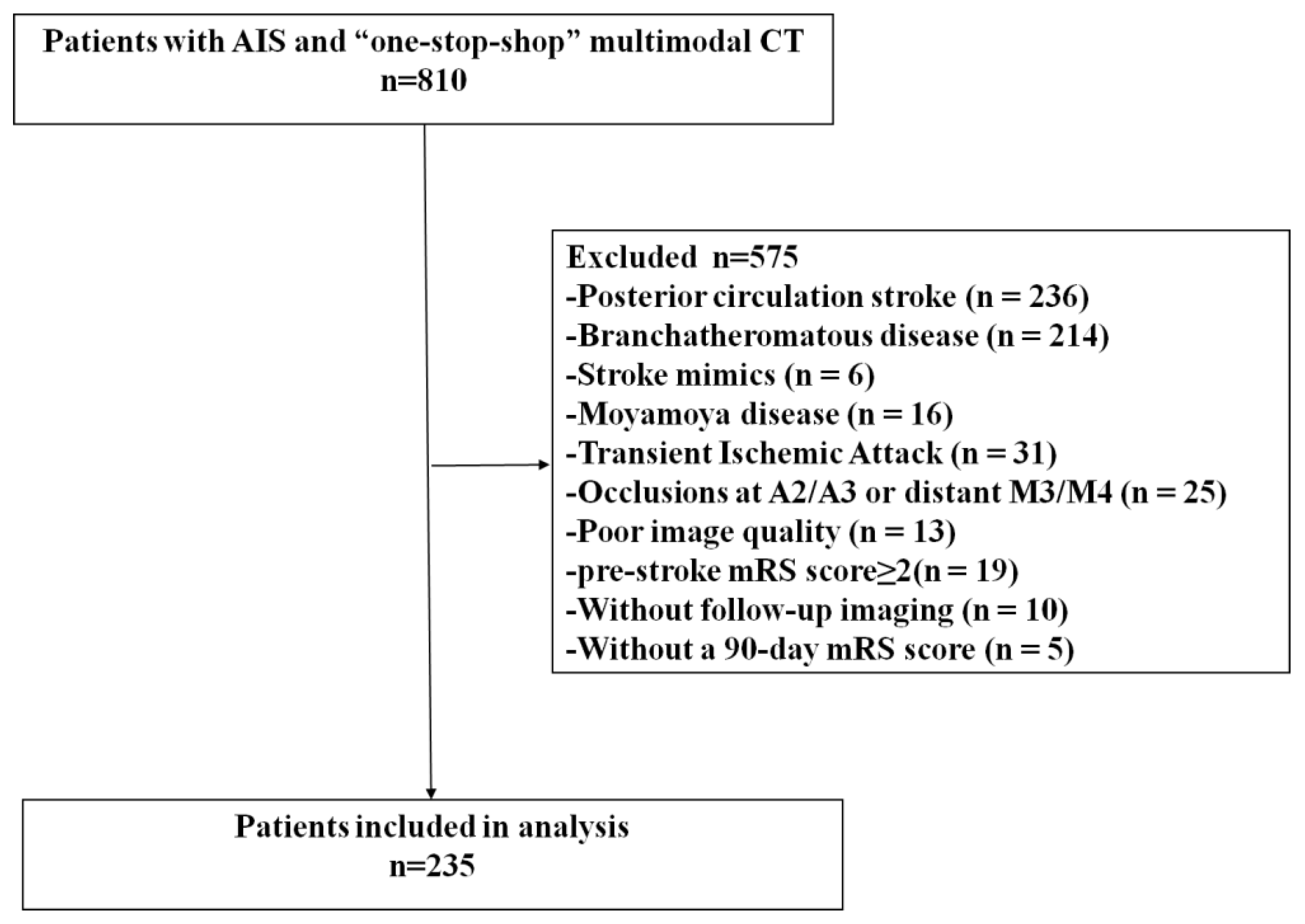
2.1. Study Design and Patient Selection
2.2. Imaging Protocol
2.3. Imaging Reconstruction and Interpretation
2.4. Statistical Analysis
3. Results
3.1. Patient Characteristics
3.2. Association between Baseline Radiologic Features and Functional Outcomes
3.3. Association between HIR, MCTA Collateral Score, and Functional Outcome
3.4. Predictive Ability of HIR and MCTA Collateral Score
4. Discussion
Limitations
5. Conclusions
Supplementary Materials
Author Contributions
Funding
Institutional Review Board Statement
Informed Consent Statement
Data Availability Statement
Acknowledgments
Conflicts of Interest
References
- Goyal, M.; Demchuk, A.M.; Menon, B.K.; Eesa, M.; Rempel, J.L.; Thornton, J.; Roy, D.; Jovin, T.G.; Willinsky, R.A.; Sapkota, B.L.; et al. Randomized assessment of rapid endovascular treatment of ischemic stroke. N. Engl. J. Med. 2015, 372, 1019–1030. [Google Scholar] [CrossRef] [PubMed]
- Goyal, M.; Menon, B.K.; van Zwam, W.H.; Dippel, D.W.J.; Mitchell, P.J.; Demchuk, A.M.; Dávalos, A.; Majoie, C.B.L.M.; van der Lugt, A.; de Miquel, M.A.; et al. Endovascular thrombectomy after large-vessel ischaemic stroke: A meta-analysis of individual patient data from five randomised trials. Lancet 2016, 387, 1723–1731. [Google Scholar] [CrossRef]
- Vagal, A.; Aviv, R.; Sucharew, H.; Reddy, M.; Hou, Q.; Michel, P.; Jovin, T.; Tomsick, T.; Wintermark, M.; Khatri, P. Collateral Clock Is More Important Than Time Clock for Tissue Fate. Stroke 2018, 49, 2102–2107. [Google Scholar] [CrossRef]
- Bang, O.Y.; Goyal, M.; Liebeskind, D.S. Collateral Circulation in Ischemic Stroke: Assessment Tools and Therapeutic Strategies. Stroke 2015, 46, 3302–3309. [Google Scholar] [CrossRef] [PubMed]
- Campbell, B.C.V.; Khatri, P. Stroke. Lancet 2020, 396, 129–142. [Google Scholar] [CrossRef]
- Rao, V.L.; Mlynash, M.; Christensen, S.; Yennu, A.; Kemp, S.; Zaharchuk, G.; Heit, J.J.; Marks, M.P.; Lansberg, M.G.; Albers, G.W. Collateral status contributes to differences between observed and predicted 24-h infarct volumes in DEFUSE 3. J. Cereb. Blood Flow Metab. 2020, 40, 1966–1974. [Google Scholar] [CrossRef]
- Liebeskind, D.S.; Woolf, G.W.; Shuaib, A.; Collaterals, C. Collaterals 2016: Translating the collaterome around the globe. Int. J. Stroke 2017, 12, 338–342. [Google Scholar] [CrossRef]
- Menon, B.K.; Sajobi, T.T.; Zhang, Y.; Rempel, J.L.; Shuaib, A.; Thornton, J.; Williams, D.; Roy, D.; Poppe, A.Y.; Jovin, T.G.; et al. Analysis of Workflow and Time to Treatment on Thrombectomy Outcome in the Endovascular Treatment for Small Core and Proximal Occlusion Ischemic Stroke (ESCAPE) Randomized, Controlled Trial. Circulation 2016, 133, 2279–2286. [Google Scholar] [CrossRef]
- Seker, F.; Potreck, A.; Mohlenbruch, M.; Bendszus, M.; Pham, M. Comparison of four different collateral scores in acute ischemic stroke by CT angiography. J. Neurointerv. Surg. 2016, 8, 1116–1118. [Google Scholar] [CrossRef]
- Menon, B.K.; D’Esterre, C.D.; Qazi, E.M.; Almekhlafi, M.; Hahn, L.; Demchuk, A.M.; Goyal, M. Multiphase CT Angiography: A New Tool for the Imaging Triage of Patients with Acute Ischemic Stroke. Radiology 2015, 275, 510–520. [Google Scholar] [CrossRef]
- Fasen, B.; Heijboer, R.J.J.; Hulsmans, F.H.; Kwee, R.M. Diagnostic performance of single-phase CT angiography in detecting large vessel occlusion in ischemic stroke: A systematic review. Eur. J. Radiol. 2021, 134, 109458. [Google Scholar] [CrossRef] [PubMed]
- Dundamadappa, S.; Iyer, K.; Agrawal, A.; Choi, D.J. Multiphase CT Angiography: A Useful Technique in Acute Stroke Imaging-Collaterals and Beyond. AJNR Am. J. Neuroradiol. 2021, 42, 221–227. [Google Scholar] [CrossRef]
- Calamante, F.; Christensen, S.; Desmond, P.M.; Ostergaard, L.; Davis, S.M.; Connelly, A. The physiological significance of the time-to-maximum (Tmax) parameter in perfusion MRI. Stroke 2010, 41, 1169–1174. [Google Scholar] [CrossRef] [PubMed]
- Olivot, J.M.; Mlynash, M.; Inoue, M.; Marks, M.P.; Wheeler, H.M.; Kemp, S.; Straka, M.; Zaharchuk, G.; Bammer, R.; Lansberg, M.G.; et al. Hypoperfusion intensity ratio predicts infarct progression and functional outcome in the DEFUSE 2 Cohort. Stroke 2014, 45, 1018–1023. [Google Scholar] [CrossRef] [PubMed]
- Mlynash, M.; Lansberg, M.G.; De Silva, D.A.; Lee, J.; Christensen, S.; Straka, M.; Campbell, B.C.; Bammer, R.; Olivot, J.M.; Desmond, P.; et al. Refining the definition of the malignant profile: Insights from the DEFUSE-EPITHET pooled data set. Stroke 2011, 42, 1270–1275. [Google Scholar] [CrossRef]
- Guenego, A.; Fahed, R.; Albers, G.W.; Kuraitis, G.; Sussman, E.S.; Martin, B.W.; Marcellus, D.G.; Olivot, J.M.; Marks, M.P.; Lansberg, M.G.; et al. Hypoperfusion intensity ratio correlates with angiographic collaterals in acute ischaemic stroke with M1 occlusion. Eur. J. Neurol. 2020, 27, 864–870. [Google Scholar] [CrossRef]
- Guenego, A.; Mlynash, M.; Christensen, S.; Kemp, S.; Heit, J.J.; Lansberg, M.G.; Albers, G.W. Hypoperfusion ratio predicts infarct growth during transfer for thrombectomy. Ann. Neurol. 2018, 84, 616–620. [Google Scholar] [CrossRef]
- Guenego, A.; Marcellus, D.G.; Martin, B.W.; Christensen, S.; Albers, G.W.; Lansberg, M.G.; Marks, M.P.; Wintermark, M.; Heit, J.J. Hypoperfusion Intensity Ratio Is Correlated with Patient Eligibility for Thrombectomy. Stroke 2019, 50, 917–922. [Google Scholar] [CrossRef]
- Lyndon, D.; van den Broek, M.; Niu, B.; Yip, S.; Rohr, A.; Settecase, F. Hypoperfusion Intensity Ratio Correlates with CTA Collateral Status in Large-Vessel Occlusion Acute Ischemic Stroke. AJNR Am. J. Neuroradiol. 2021, 42, 1380–1386. [Google Scholar] [CrossRef]
- Monteiro, A.; Cortez, G.M.; Greco, E.; Aghaebrahim, A.; Sauvageau, E.; Hanel, R.A. Hypoperfusion intensity ratio for refinement of elderly patient selection for endovascular thrombectomy. J. Neurointerv. Surg. 2021, 14, 242–247. [Google Scholar] [CrossRef]
- Murray, N.M.; Culbertson, C.J.; Wolman, D.N.; Mlynash, M.; Lansberg, M.G. Hypoperfusion Intensity Ratio Predicts Malignant Edema and Functional Outcome in Large-Vessel Occlusive Stroke with Poor Revascularization. Neurocrit. Care 2021, 35, 79–86. [Google Scholar] [CrossRef] [PubMed]
- Lin, L.; Chen, C.; Tian, H.; Bivard, A.; Spratt, N.; Levi, C.R.; Parsons, M.W. Perfusion Computed Tomography Accurately Quantifies Collateral Flow After Acute Ischemic Stroke. Stroke 2020, 51, 1006–1009. [Google Scholar] [CrossRef] [PubMed]
- Lin, L.; Yang, J.; Chen, C.; Tian, H.; Bivard, A.; Spratt, N.J.; Levi, C.R.; Parsons, M.W.; INSPIRE Study Group. Association of Collateral Status and Ischemic Core Growth in Patients with Acute Ischemic Stroke. Neurology 2021, 96, e161–e170. [Google Scholar] [CrossRef]
- Powers, W.J.; Rabinstein, A.A.; Ackerson, T.; Adeoye, O.M.; Bambakidis, N.C.; Becker, K.; Biller, J.; Brown, M.; Demaerschalk, B.M.; Hoh, B.; et al. Guidelines for the Early Management of Patients with Acute Ischemic Stroke: 2019 Update to the 2018 Guidelines for the Early Management of Acute Ischemic Stroke: A Guideline for Healthcare Professionals From the American Heart Association/American Stroke Association. Stroke 2019, 50, e344–e418. [Google Scholar] [CrossRef] [PubMed]
- Yang, Q.; Zheng, J.; Chen, W.; Chen, X.; Wen, D.; Chen, W.; Xiong, X.; Zhang, Z. Association Between Preadmission Metformin Use and Outcomes in Intensive Care Unit Patients with Sepsis and Type 2 Diabetes: A Cohort Study. Front. Med. 2021, 8, 640785. [Google Scholar] [CrossRef] [PubMed]
- Baek, J.H.; Kim, Y.D.; Lee, K.J.; Choi, J.K.; Baik, M.; Kim, B.M.; Kim, D.J.; Heo, J.H.; Nam, H.S. Low Hypoperfusion Intensity Ratio Is Associated with a Favorable Outcome Even in Large Ischemic Core and Delayed Recanalization Time. J. Clin. Med. 2021, 10, 1869. [Google Scholar] [CrossRef]
- Parish, J.M.; Strong, D.; Prasad, T.; Rhoten, J.B.; Clemente, J.D.; Defilipp, G.; Bernard, J.D.; Hines, A.; Stetler, W.R.; Karamchandani, R.R.; et al. Abstract P557: Hypoperfusion Intensity Ratio and Cerebral Blood Volume Index as Predictors of Outcome for Recanalized Middle Cerebral Artery Occlusions. Stroke 2021, 52, AP557. [Google Scholar] [CrossRef]
- Nomani, A.Z.; Kamtchum Tatuene, J.; Rempel, J.L.; Jeerakathil, T.; Winship, I.R.; Khan, K.A.; Buck, B.H.; Shuaib, A.; Jickling, G.C. Association of CT-Based Hypoperfusion Index with Ischemic Core Enlargement in Patients With Medium and Large Vessel Stroke. Neurology 2021, 97, e2079–e2087. [Google Scholar] [CrossRef]
- Menon, B.K.; Qazi, E.; Nambiar, V.; Foster, L.D.; Yeatts, S.D.; Liebeskind, D.; Jovin, T.G.; Goyal, M.; Hill, M.D.; Tomsick, T.A.; et al. Differential Effect of Baseline Computed Tomographic Angiography Collaterals on Clinical Outcome in Patients Enrolled in the Interventional Management of Stroke III Trial. Stroke 2015, 46, 1239–1244. [Google Scholar] [CrossRef]
- de Havenon, A.; Mlynash, M.; Kim-Tenser, M.A.; Lansberg, M.G.; Leslie-Mazwi, T.; Christensen, S.; McTaggart, R.A.; Alexander, M.; Albers, G.; Broderick, J.; et al. Results from DEFUSE 3: Good Collaterals Are Associated with Reduced Ischemic Core Growth but Not Neurologic Outcome. Stroke 2019, 50, 632–638. [Google Scholar] [CrossRef]
- Martinon, E.; Lefevre, P.H.; Thouant, P.; Osseby, G.V.; Ricolfi, F.; Chavent, A. Collateral circulation in acute stroke: Assessing methods and impact: A literature review. J. Neuroradiol. 2014, 41, 97–107. [Google Scholar] [CrossRef] [PubMed]
- Bivard, A.; Levi, C.; Lin, L.; Cheng, X.; Aviv, R.; Spratt, N.J.; Lou, M.; Kleinig, T.; O’Brien, B.; Butcher, K.; et al. Validating a Predictive Model of Acute Advanced Imaging Biomarkers in Ischemic Stroke. Stroke 2017, 48, 645–650. [Google Scholar] [CrossRef] [PubMed]
- García-Tornel, Á.; Campos, D.; Rubiera, M.; Boned, S.; Olivé-Gadea, M.; Requena, M.; Ciolli, L.; Muchada, M.; Pagola, J.; Rodriguez-Luna, D.; et al. Ischemic Core Overestimation on Computed Tomography Perfusion. Stroke 2021, 52, 1751–1760. [Google Scholar] [CrossRef] [PubMed]


| No. of Patients and Characteristics | ALL n = 235 (100%) | Favorable Outcome n = 127 (54%) | Unfavorable Outcome n = 108 (46%) | p-Value |
|---|---|---|---|---|
| Age, Median (IQR) | 66 [55.5, 73.0] | 65 [55.0, 72.0] | 66 [56.0, 74.2] | 0.27 |
| Male, n (%) | 178 (75.7) | 103 (81.1) | 75 (69.4) | 0.054 |
| Risk factors n (%) | ||||
| Hypertension, n (%) | 175 (74.5) | 90 (70.9) | 85 (78.7) | 0.221 |
| Diabetes, n (%) | 61 (26) | 26 (20.5) | 35 (32.4) | 0.054 |
| Hyperlipidemia, n (%) | 93 (39.6) | 52 (40.9) | 41 (38) | 0.74 |
| Prior stroke, n (%) | 53 (22.6) | 26 (20.5) | 27 (25) | 0.502 |
| Coronary artery disease, n (%) | 42 (17.9) | 27 (21.3) | 15 (13.9) | 0.194 |
| Valvular disease, n (%) | 43 (18.3) | 17 (13.4) | 26 (24.1) | 0.052 |
| Chronic heart failure, n (%) | 20 (8.5) | 7 (5.5) | 13 (12) | 0.121 |
| Atrial fibrillation, n (%) | 47 (20) | 17 (13.4) | 30 (27.8) | 0.01 |
| Smoke, n (%) | 82 (34.9) | 50 (39.4) | 32 (29.6) | 0.154 |
| Drink, n (%) | 36 (15.3) | 22 (17.3) | 14 (13) | 0.457 |
| Homocysteine, Median [IQR] | 13 [10.2, 16.4] | 12.9 [10.3, 16.2] | 13.2 [10.2, 16.8] | 0.457 |
| SBP, Median [IQR] | 147 [130, 164.5] | 144 [129.5, 165.5] | 150 [133, 163] | 0.579 |
| DBP, Median [IQR] | 87 [78, 96] | 87 [78, 96] | 88 [79, 98] | 0.997 |
| HR, Median [IQR] | 79 [72, 89] | 78 [71, 86] | 80 [75, 96] | 0.032 |
| Blood Glucose, Median [IQR] | 5.9 [5.2, 7.6] | 5.6 [5.0, 6.8] | 6.5 [5.7, 8.5] | <0.001 |
| Glycosylated hemoglobin, Median [IQR] | 5.8 [5.5, 6.2] | 5.8 [5.6, 6.1] | 5.8 [5.5, 6.4] | 0.515 |
| Time from last known well to CT (min) Median [IQR] | 600 [600, 900] | 540 [330, 840] | 660 [390, 960] | 0.134 |
| Baseline NIHSS, Median [IQR] | 10 [5, 16] | 8 [4, 12] | 15 [9, 19] | <0.001 |
| Imaging items | ||||
| ASPECTS on NCCT, Median [IQR] | 7 [6, 8] | 7 [7, 8] | 6 [5, 7] | <0.001 |
| Location of occlusion on mCTA, n (%) | 0.068 | |||
| ICA or Tandem | 85 (36.2) | 39 (30.7) | 46 (42.6) | |
| M1 | 102 (43.4) | 56 (44.1) | 46 (42.6) | |
| M2 or further distal | 48 (20.4) | 32 (25.2) | 16 (14.8) | |
| mCTA score, n (%) | <0.001 | |||
| 1 | 20 (8.5) | 0 (0) | 20 (18.5) | |
| 2 | 17 (7.2) | 2 (1.6) | 15 (13.9) | |
| 3 | 117 (49.8) | 63 (49.6) | 54 (50) | |
| 4 | 63 (26.8) | 46 (36.2) | 17 (15.7) | |
| 5 | 18 (7.7) | 16 (12.6) | 2 (1.9) | |
| Good collaterals (4–5) | 81 (34.5) | 62 (26.4) | 19 (8) | |
| Poor collaterals (1–3) | 154 (65.5) | 65 (27.7) | 89 (37.9) | |
| mCTA score, Median [IQR] | 3 [3, 4] | 3 [3, 4] | 3 [2, 3] | <0.001 |
| ischemic core volume (rCBF < 30%) (mL), Median [IQR] | 4.7 [1.8, 17.5] | 2.1 [1.0, 4.5] | 15.2 [5.5, 39.3] | <0.001 |
| Mismatch ratio, Median [IQR] | 13.8 [4.6, 33.5] | 22.9 [11.6, 45.6] | 5.8 [2.6, 14] | <0.001 |
| TMax > 6 s volume (mL), Median [IQR] | 78.9 [46.8, 121] | 59.0 [29.7, 89.2] | 97.5 [68.7, 142.2] | <0.001 |
| TMax > 10 s volume (mL), Median [IQR] | 17.6 [6.3, 39.4] | 7.1 [3.1, 13.2] | 39.6 [25.3, 65.2] | <0.001 |
| HIR, Median [IQR] | 0.2 [0.1, 0.4] | 0.1 [0.1, 0.2] | 0.4 [0.4, 0.5] | <0.001 |
| FIV, Median [IQR] | 26.8 [11.4, 76.2] | 12.6 [7.5, 18.4] | 78.9 [44.5, 165] | <0.001 |
| Type of treatment, n (%) | 0.358 | |||
| Intravenous thrombolysis | 31 (13.2) | 19 (15) | 12 (11.1) | |
| Bridging therapy | 14 (6.0) | 7 (5.5) | 7 (6.5) | |
| EVT | 73 (31.1) | 44 (34.6) | 29 (26.9) | |
| Supportive medical treatment only | 117 (49.8) | 57 (44.9) | 60 (55.6) | |
| 90 d_mRS, Median [IQR] | 2 [1, 4] | 1 [1, 2] | 4 [3, 5] | <0.001 |
| Variables | Crude OR, 95%CI | p-Value | Adjust OR, 95%CI | p-Value |
|---|---|---|---|---|
| Age | 1.01 (0.99–1.03) | 0.304 | 1.01 (0.95–1.06) | 0.908 |
| Gender | 1.89 (1.03–3.46) | 0.039 | 0.94 (0.22–4.05) | 0.929 |
| Blood Glucose | 1.13 (1.02–1.25) | 0.015 | 1.24 (0.98–1.57) | 0.075 |
| NIHSS | 1.15 (1.1–1.21) | <0.001 | 1.04 (0.95–1.15) | 0.354 |
| ASPECTS | 0.33 (0.25–0.45) | <0.001 | 0.49 (0.24–1) | 0.05 |
| mCTA score | 0.27 (0.18–0.42) | <0.001 | 0.44 (0.15–1.23) | 0.117 |
| rCBF < 30% | 1.09 (1.05–1.12) | <0.001 | 0.97 (0.92–1.01) | 0.166 |
| TMax > 6 s | 1.02 (1.01–1.02) | <0.001 | 1 (0.99–1.01) | 0.955 |
| HIR (per 0.01); | 1.3 (1.22–1.4) | <0.001 | 1.32 (1.21–1.45) | <0.001 |
| Variable | AUC | 95%CI | SE # | Youden Index | Associated Criterion | Sensitivity (%) | 95%CI (%) | Specificity (%) | 95%CI (%) | |
|---|---|---|---|---|---|---|---|---|---|---|
| HIR | 0.968 | 0.937, 0.987 | 0.0123 | Z = 7.493 | 0.881 | >0.3 | 88.89 | 81.4, 94.1 | 99.21 | 95.7, 100 |
| mCTA | 0.741 | 0.680, 0.795 | 0.0288 | p < 0.0001 | 0.3123 | <3 | 82.4 | 73.9, 89.1 | 48.8 | 39.9, 57.8 |
Publisher’s Note: MDPI stays neutral with regard to jurisdictional claims in published maps and institutional affiliations. |
© 2022 by the authors. Licensee MDPI, Basel, Switzerland. This article is an open access article distributed under the terms and conditions of the Creative Commons Attribution (CC BY) license (https://creativecommons.org/licenses/by/4.0/).
Share and Cite
Wan, Z.; Meng, Z.; Xie, S.; Fang, J.; Li, L.; Chen, Z.; Liu, J.; Jiang, G. Correlation between Hypoperfusion Intensity Ratio and Functional Outcome in Large-Vessel Occlusion Acute Ischemic Stroke: Comparison with Multi-Phase CT Angiography. J. Clin. Med. 2022, 11, 5274. https://doi.org/10.3390/jcm11185274
Wan Z, Meng Z, Xie S, Fang J, Li L, Chen Z, Liu J, Jiang G. Correlation between Hypoperfusion Intensity Ratio and Functional Outcome in Large-Vessel Occlusion Acute Ischemic Stroke: Comparison with Multi-Phase CT Angiography. Journal of Clinical Medicine. 2022; 11(18):5274. https://doi.org/10.3390/jcm11185274
Chicago/Turabian StyleWan, Zhifang, Zhihua Meng, Shuangcong Xie, Jin Fang, Li Li, Zhensong Chen, Jinwu Liu, and Guihua Jiang. 2022. "Correlation between Hypoperfusion Intensity Ratio and Functional Outcome in Large-Vessel Occlusion Acute Ischemic Stroke: Comparison with Multi-Phase CT Angiography" Journal of Clinical Medicine 11, no. 18: 5274. https://doi.org/10.3390/jcm11185274
APA StyleWan, Z., Meng, Z., Xie, S., Fang, J., Li, L., Chen, Z., Liu, J., & Jiang, G. (2022). Correlation between Hypoperfusion Intensity Ratio and Functional Outcome in Large-Vessel Occlusion Acute Ischemic Stroke: Comparison with Multi-Phase CT Angiography. Journal of Clinical Medicine, 11(18), 5274. https://doi.org/10.3390/jcm11185274