Machine Perfusion for Extended Criteria Donor Livers: What Challenges Remain?
Abstract
1. Introduction
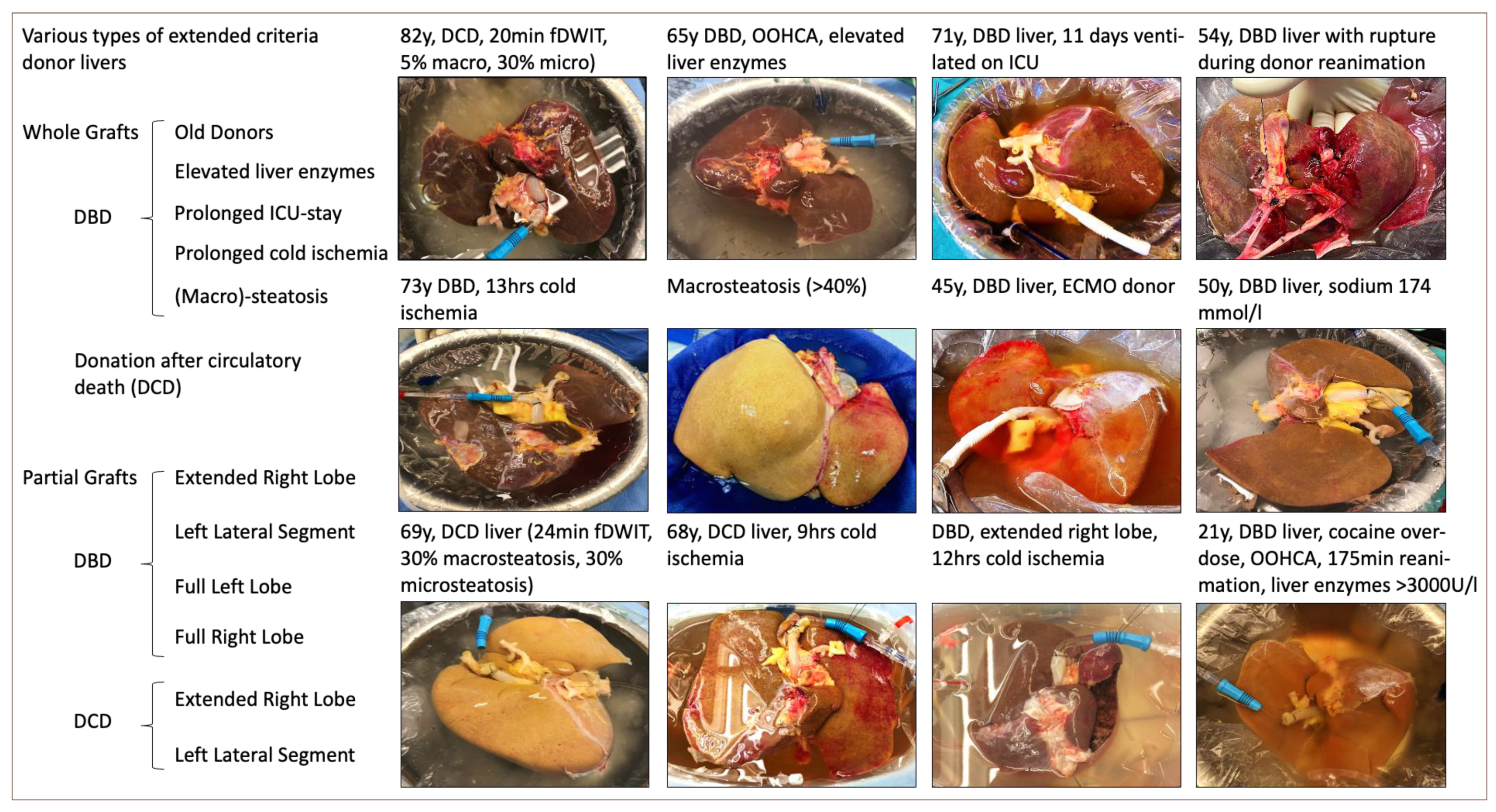
2. Types of Extended Criteria Donor Livers and Associated Risk Factors
2.1. A General Overview
2.2. Donation after Circulatory Death (DCD) Livers
2.3. Donor Age
2.4. Cold Ischemia
2.5. Steatosis
2.6. The Role of Other Donor Risk Factors
2.7. Partial Grafts from Deceased Donors
| Parameter | ECD Categories: Eurotransplant/EASL [13] | Criteria Specification | ILTS Consensus Criteria 2020: Controlled DCD [68] | Criteria Specification | Split Criteria (ELTR Criteria) [102] | Criteria Specification |
|---|---|---|---|---|---|---|
| Functional donor risk factors | Donor age > 65 y | Donor age >70 y [29], >80 y [50], >90 y [55,56] | Donor age ≤ 60 y | Donor age > 60, >70 | Donor age ≤ 40 y (10–40 y) | ≤50 y, variable in different counties [102], 18–40 y [29,101] |
| Donor BMI > 30 kg/m2 | Donor BMI ≤ 30 kg/m2 | Explore BMI > 30 kg/m2 donor with experienced donor surgery team [68] | Donor BMI ≤ 30 kg/m2 | Donor BMI < 28 kg/m2 [29,101] | ||
| Donor ALT > 105 U/L, Donor AST > 90 U/L (“>3-times normal”) | Higher transaminases (>1000 U/L) when down-trend is confirmed and liver maintained [95,97,98] | Downwards-trend maintained function, individual decision [68] | Donor AST and ALT <5-fold normal values | Donor AST and ALT <3-fold normal values [101] | ||
| Donor serum sodium > 165 mmol/L | Donor serum sodium > 180 mmol/L | Donor serum sodium > 160 mmol/L | Selective use of donors with sodium > 160 mmol/L | |||
| Donor serum bilirubin > 3 mg/dL | ||||||
| Donor ICU stay with ventilation > 7 days | BTS guidelines suggest ≤ 5 days donor ICU stay [38] | Donor ICU stay with ventilation ≤ 5 days | ||||
| Liver steatosis > 40% | Macrosteatosis > 30% [29] | Macrosteatosis ≤ 30% | No relevant steatosis | |||
| DCD donor [13] | Only DBD | DCD livers very selectively | ||||
| Cold ischemia time > 14 h [13] * | >10.5 h [13] * | Cold ischemic time ≤ 8 h | Keep as short as possible | Cold ischemia time ≤ 8 h (>8 h increase risk for ERL [101]) | 6 h for urgent and <6 kg recipient weight [102] | |
| Donor cardiac arrest [13] National share [29] 90 th percentile of DRI [29] | Donor body weight > 40 kg (Italy) > 50 kg BTS, [101] | |||||
| Single vasopressor [29,101] | ||||||
| Recipent risk factors | Lab MELD ≤ 25 points | Selective use in recipients with lab MELD > 25 points [68] | Age, weight, status, availability of LDLT graft, waiting time are among additional factors of impact | |||
| Selective use in NASH recipients | ||||||
| No known complex PVT, selective use for re-TPL or acute liver failure or combined liver-kidney transplant | No specific recipient age cut-off, high or low, selective use in pediatric candidates [68] | |||||
| Donor risk factors, not relevant for graft function | Positive hepatitis serology [13] | Consider donor infections and malignancy according to WHO/EASL guidelines | ||||
| History of extrahepatic malignancy [13] |
3. The Role of Recipient Risk Factors
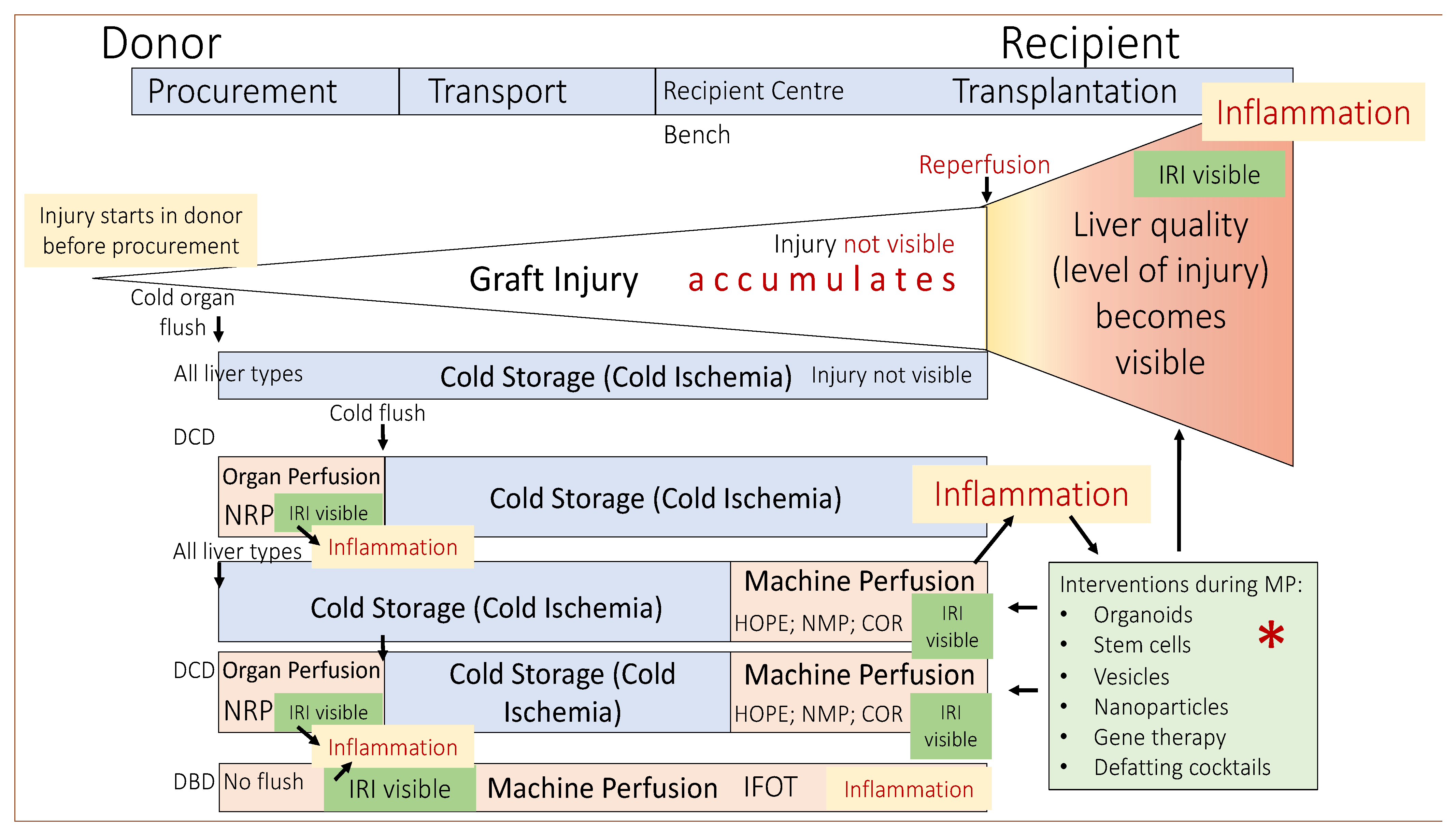
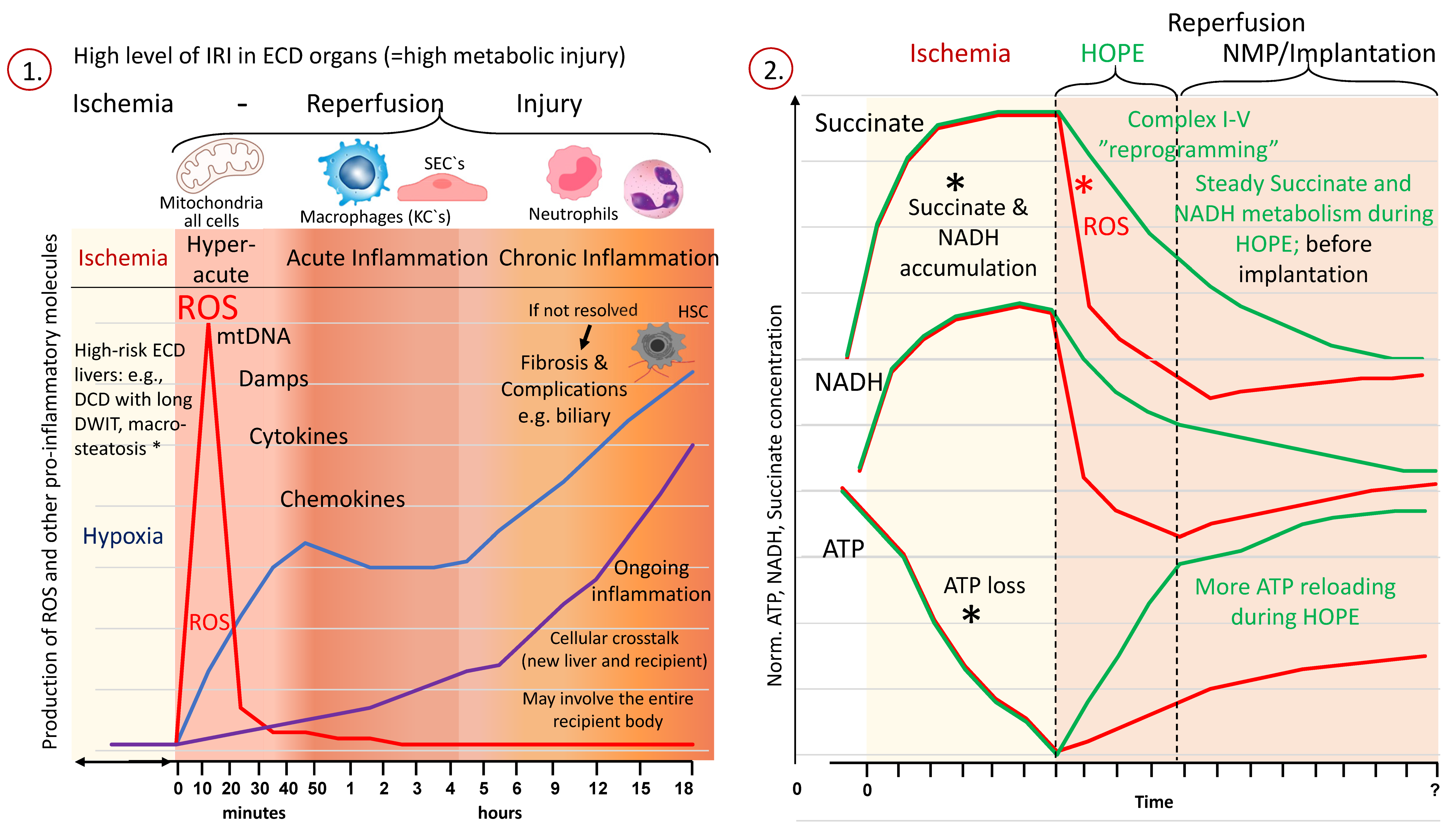
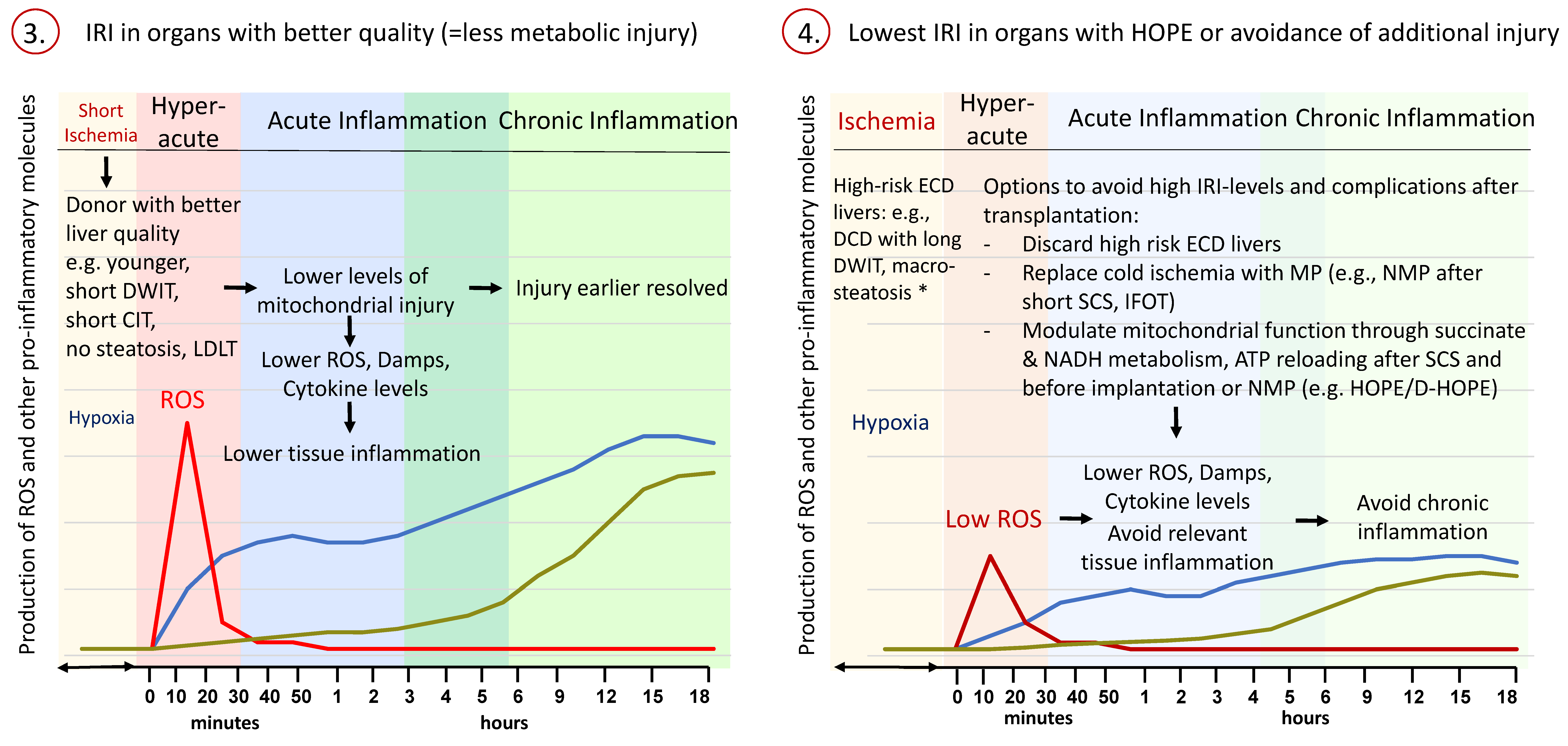
4. Mechanisms of Ischemia-Reperfusion Injury with the Use of ECD Livers
5. The Impact of Dynamic Organ Perfusion Strategies
5.1. Concepts of Normothermic Organ Perfusion
5.2. Hypothermic Perfusion Organ Perfusion
| Study Type | Reference | Number and Type of Livers | Criteria to Define ECD Livers | Type and Duration of DWIT (Min) | NRP-Duration (Min) | CIT after NRP (h) | Discard Rate | Follow-Up (Months) | Main Findings | Discussion |
|---|---|---|---|---|---|---|---|---|---|---|
| Case-control cohort study, retrospective (Level of Evidence IV) | Schurink et al. [120] | 25 DCD NRP vs. 49 DCD SCS vs. 81 DBD SCS | ET-DRI: 3.1 (2.97–3.21) vs. 2.19 (1.9–2.42) vs. 1.69 (1.49–2.01), UK-DCD Risk: 9 (8–12) vs. 6 (5–9) | NRP vs. SCS: fDWIT 29 (range 26–33) vs. 24 (19–28) | 120 (range 110–128) | 5.7 (range 4.9–6.45) | 5 discarded after NRP | NRP: 23 (range: 14–28) | DCD NRP vs. DCD SCS vs. DBD: additional D-HOPE in 5 (25%) vs. 19 (39%) vs. 3 (4%). 1-year graft survival: 90% vs. 82% vs. 86%, LoHS 13 (10–18) vs. 14 (11–20) vs. 17 (12–27); IC 11% vs. 18% vs. 7% | Low case number, heterogenous cohort, pilot |
| Rodriguez et al. [154] | 39 DCD NRP vs. 78 DBD SCS | - | fDWIT 13.1 (6–26), tDWIT 19.23 (10–38) | 90–120 | NRP DCD vs. DBD 5.0 vs. 5.2 | - | mean 22 | NRP vs. DBD: PNF 0% vs. 3.8%, EAD 34.2% vs. 19.2%, biliary complications early 5.1% vs. 3.8%, late 7.6% vs. 12%, IC 0% versus 1.2%, LoHS 14.7 (1–51) vs. 13.7 (7–57) | No cold storage DCD control | |
| Hessheimer et al. [119] | 545 DCD NRP vs. 258 DCD SCS | UK DCD risk: 27 (5%) vs. 15 (5.8%) futile | NRP vs. SCS: fDWIT 12 (9–16) vs. 14 (11–20); tDWIT (18 (13–23) vs. 22 (17–26) | 111 (81–126) | NRP vs. SCS: 5.3 (4.5–6.3) vs. 5.6 (4.7–6.5) | - | median 31 | NRP vs. SCS: PNF 16 (3%) vs. 15 (6%); HAT 22 (4%) vs. 19 (7%); biliary complications 63 (12%) vs. 75 (29%); ITBL 6 (1%) vs. 24 (9%), graft loss 77 (14%) vs. 88 (34%) | Large cohort, retrospective, short DWIT | |
| Ruiz et al. [155] | 100 DCD NRP vs. 200 DBD SCS | UK DCD risk: 3 (3%) futile | fDWIT: 10 (IQR: 8.5–12.2) | 121 (IQR: 118–128) | NRP vs. SCS: 4.6 (4–5.2) vs. 4.4 (3.8–5.7) | - | Median 36 (20–48.3) | similar results, same EAD rate and enzyme release, 3-year graft survival 92% vs. 87% | Retrospective matched, short cold storage, short DWIT | |
| Muñoz et al. [156] | 23 DCD NRP vs. 22 DCD SCS | - | NRP vs. SCS: tDWIT: 23.7 vs. 23.1; fDWIT: 14.4 vs. 15.8 | 90–120 | NRP vs. SCS: 4.7 vs. 4.7 | - | NRP 14.4 vs. SCS 34.8 | NRP vs. SCS: EAD rate 30.5% vs. 68.1%; overall biliary complications 4.3% vs. 22.7%; IC rates 0–5% vs. 13.6%, re-transplantation 0% vs. 9.1% | shorter follow up in NRP group: severe complications may develop later, small case load, short DWIT | |
| Savier et al. [122] | 50 DCD NRP vs. 100 DBD SCS | National DCD guidelines * | fDWIT: 22 (IQR: 20–26.8); Asystolic DWIT: 17 (IQR: 14–22.3); | Median 190 (IQR: 151–223) | DCD NRP: 5.8 (5–6.7), DBD SCS: 6.3 (5.4–7.3) | - | ≥24 | DCD NRP vs. DBD SCS: EAD 18% vs. 32%; AKI 26% vs. 33%; 12% graft loss within 2 years (HCC recurrence) vs. 3%; overall biliary complications 16% vs. 17% | Retrospective matched study, transparent presentation of utilization | |
| Hessheimer et al. [115] | 95 DCD NRP vs. 117 DCD SCS | - | NRP vs. SCS: tDWIT: median 20 vs. 21, fDWIT: median 14 vs. 13 | 120 (range 79–136) | NRP vs. SCS: 5.3 (4.4–6.1) vs. 5.7 (4.8–6.4) | - | ≥12 (median 20) | NRP vs. SCS: EAD 22% vs. 27%, same rate of PNF and HAT, overall biliary complications 8% vs. 31%, IC 2% vs. 13%, graft loss 12% vs. 24% | Retrospective, large cumulative cohort | |
| Watson et al. [157] | 43 DCD NRP vs. 187 DCD SCS | DRI: 1.8 (1.7–2.4) vs. 2.5 (2–2.9) | NRP vs. SCS: tDWIT: 30 (26–36) vs. 27 (22–32); asystolic DWIT: 16 (13–20) vs. 13 (11–16) | Median 123 (IQR: 103–130) | 6.4 (5–8.4) vs. 7.4 (6.6–8.2) | - | ≥3 | NRP vs. SCS: 0% graft loss due to IC vs. 6%; similar graft survival but shorter follow up in NRP group | Despite shorter follow-up (NRP), survival comparisons calculated |
| Study Type (Level) | Reference | Number and Type of Livers | Criteria to Define ECD Livers | Type and Duration of DWIT (Min) | CIT before NMP (h) | CIT in Control | NMP-Duration (h) | Discard Rate | Follow-Up (Months) | Main Findings | Discussion |
|---|---|---|---|---|---|---|---|---|---|---|---|
| RCT (II) | Markmann et al. [125] | DBD/ECD/DCD livers: 151 NMP vs. 142 SCS | Age > 40 y, CIT > 6 h, DCD (inclusion: donor age < 55 y, macrosteatosis < 40%) | not available | 2.9 ± 1.53 | 5.6 ± 1.5 | 4.6 ± 1.96 | Discarded: 2 NMP, 5 SCS | 6 | NMP: histologically less IRI (p = 0.004), reduction of IC (p = 0.02), EAD (p = 0.01) | heterogenous population: DBD, ECD, DCD, no report of DWIT, focus on EAD as endpoint (not powered for) |
| Nasralla et al. [124] | 55 DCD NMP vs. 34 DCD SCS; 87 DBD NMP vs. 80 DBD SCS | 55/34 DCD livers, cold storage; ET DRI: 1.7 (1.47–2.07) vs. 1.71 (1.5–2.01) | fDWIT NMP: 21 (IQR: 17–25), SCS: 16 (IQR: 10–20) | 2.1 (1.8–2.4) | 7.8 (6.3–9.6) | 9.1 (6.2–11.8) DBD: 9.9; DCD 8.8 | 64/334 excluded (19.1%), 48/270 discarded (17.8%) | 12 | Lower liver enzyme release after NMP (primary endpoint), no differences in biliary complications or graft survival | High exclusion/discard rate; control group with higher injury, no report on perfusate transaminases, NMP replacing SCS | |
| Prospective, matched case control (III) | Fodor et al. [158] | 59 DBD/DCD with NMP; matched with 59 SCS | ET DRI: 1.78 (0.51) vs. 1.85 (0.72) | not available | 6 | 7 | * estimated duration of NMP: 15 (total preservation time: 21) | 16/75 (20%) discarded after NMP | ≥3 | NMP vs. SCS: patient and graft survival 81% vs. 82%. Same rate of major complications, lower biliary complications with NMP (p = 0.047) | short follow-up, retrospective, high rate of biliary complications in DBD SCS control group |
| Mergental et al. [126] | 10 DCD NMP vs. 12 DBD NMP vs. 44 SCS (matched controls) | Overall US DRI: 2.1 (1.9–3) | fDWIT 22.5 (IQR:19.0–35.0) | DCD 6.9 (5.9–7.7); DBD 8.5 (6.8–12) | not available | 9.8 (7.5–11.8) | 9/31 (29%) not transplanted; 3/25 (12%) discarded after NMP | 12 | DCD/DBD vs. SCS: EAD: 7 (31.8%) vs. 4 (9.1%); NAS: 4 (18.2%, 3/4 DCDs) vs. 1 (2.3%) (4/22; 3 DCD); 80% of DCD recipients had PRS | Prospective study with retrospective matched control, heterogenous risk profile | |
| Case-control cohort study, retrospective (IV) | Ceresa et al. [159] | 23 DBD/8 DCD with SCS-NMP vs. 104 only NMP from [125] | DRI 1.87 (range 1.06–3.2) vs. 1.45 (0.78–6.35) | SCS-NMP vs. only NMP: fDWIT 16 (range: 12–28) vs. 20 (range:10–35) | 6 ± 1.3 | No nonperfused control | SCS-NMP 8.4 ± 4 vs. only NMP 12 ± 4.2 | 20/51 (39%) excluded | 12 | Comparable outcomes among SCS-NMP vs. only NMP (EAD, PRS, hospital/ITU stay); similar rate of major complications; 1 y graft survival: 84% vs. 94%; | Standard risk DBD and DCD grafts, 8 DCD only |
| Quintini et al. [160] | 21 discarded DCD/DBD with NMP: 6 discarded; 15 transplanted | Macrosteatosis >30%, combined up to 60%; hypertransaminase | fDWIT 21 (±10) | 5.4 ± 1.1; DBD: 4.8 ± 1.4; DCD: 5.5 ± 1.2 | No control | 6.7 ± 2.1; DBD: 7.1 ± 0.9; DCD: 7.7 ± 2.9 | 6/21 (29%) discarded | 2–14 | Hospital stay: DBD 9.5 ± 4.4 vs. DCD 19.5 ± 10.2; 1 × IC, 7/15 EAD; |
| Protocol. | Reference | Number and Type of Livers | Criteria to Define ECD Livers | Type and Duration of DWIT (min) | CIT after NRP/before MP (h) | Perfusion (h) | Discard Rate | Follow-Up (Months) | Main Findings | Discussion |
|---|---|---|---|---|---|---|---|---|---|---|
| NRP + SCS vs. SCS + NMP vs. SCS alone | Gaurav et al. [123] | 69 DCD NRP vs. 67 DCD NMP vs. 97 DCD SCS | DRI: 2.2 (1.8–2.5) vs. 2.5 (2–2.9) vs. 2.5 (2–3), UK DCD Risk: 9 vs. 3 vs. 1 futile | NRP vs. NMP vs. SCS: fDWIT 19 (15–24), 15 (12–18), 15 (11–18); tDWIT: 29 (23–33), 26 (22–31), 26 (22–31) | NRP vs. NMP vs. SCS: 6.7 (5.7–7.9) vs. 6.6 (5.8–7.4) vs. 7.2 (6.6–7.9) | NRP vs. NMP: 2.2 (2–2.4) vs. 7.7 (5.5–9.5) | - | median 38 | NRP vs. NMP vs. SCS: PNF 0% vs. 1.5% vs. 5%, EAD 14% vs. 11% vs. 21%. Total biliary complication: 22% vs. 37% vs. 42%; IC: 6% vs. 19% vs. 25%; HAT 1% vs. 8% vs. 8%. LoHS 15 (13–23) vs. 19 (13–29) vs. 18 (15–30) | Large cohort, NRP vs. NMP vs. SCS, relevant clinical endpoint, not randomized |
| NRP + SCS vs. SCS + NMP | Mohkam et al. [127] | 157 DCD NRP vs. 34 DCD NMP | DRI: 1.98 (1.68–2.43) vs. 2.13 (1.9–2.42) | NRP vs. NMP: tDWI: 31 vs. 25; asystolic DWIT 18 vs. 12 | NRP: 5.8 NMP: 2.3 | NRP: 3.1, NMP: 8.8 | - | 23 | NRP vs. NMP: 1 PNF, 1 hep vein thrombosis, hyperacute rejection, 2 HAT in NRP, 1 Cava thrombosis, HAT, graft infraction in NMP; 11.8% vs. 20.6% biliary complication (ns), anastomotic strictures 8.8 vs17.6%, NAS 1.5 vs2.9%; LoHS 14 (8–17) vs. 16 (13–20) | no control, short cold storage prior to NMP |
| NRP + HOPE/D-HOPE vs. SCS alone | Patrono et al. [42] | 20 DCD with NRP + D-HOPE vs. 40 DBD SCS | - | fDWIT 43 (IQR 35–46) | DCD vs. DBD 4.4 (3.8–4.9 vs. 7 (6.3–8.5) | NRP 4.1 (3.7–4.5), D-HOPE 3.4 (2.4–4.6) | none | DCD 15.5 m (12–27), DBD 40 (21–56) | DCD vs. DBD: EAD 1% vs. 28%; patient-survival 100% vs. 95%, graft-survival 90% vs. 95%. Biliary strictures anastomotic 22% vs. 15%, NAS 18% vs. 10% | Single centre, retrospective, matched, high donor risk (DWIT) |
| De Carlis et al. [41] | 37 DCD with NRP + D-HOPE matched with 37 SCS | UK DCD risk: 24 (Italy) vs. 4 (UK) futile | NRP/D-HOPE vs. NRP/SCS: fDWIT 40 (IQR: 30–80) vs. 18 (IQR: 10–44) | NRP/D-HOPE: 6.9 (5.5–11); NRP/SCS: 6.5 (4–9.7) | NRP: 4.2 (0.9–7.7); D-HOPE: 2 (0.7–6.3) | 86.6% transplanted after NRP | ≥12 | Despite longer DWIT DCD with NRP + D-HOPE showed less biliary complications and better graft survival as DCD with SCS alone | High donor risk, prolonged DWIT, shorter follow up in perfusion group; retrospective | |
| NRP + HOPE vs. NRP + SCS | Maroni et al. [161] | 36 DCD total; 19 NRP + HOPE vs. 17 NRP + SCS | UK DCD risk: 11 (range 6–16) vs. 7 (range: 3–12) | NRP + HOPE vs. NRP + SCS: tDWIT 56.65 ± 20.4 vs. 39.1 ± 21.6; fDWIT: 41.9 ± 12.5 vs. 25.5 ± 3.7; asystolic DWIT: 30.5 ± 7.7 vs. 20.5 ± 4.1 | NRP + HOPE: 7.9 ± 1.4, NRP + SCS: 6.5 ± 2.9 | NRP + HOPE: NRP: 3.8 ± 1.1, HOPE: 2.5 ± 1.1; NRP + SCS: NRP: 3.3 ± 0.8 | - | Median 24 | Italian DCD with NRP + HOPE had 0% IC compared to NRP/SCS (France) 12.5%; additional HOPE after NRP plus SCS improves outcomes | Higher risk in Italy, retrospective, matched cohorts |
| NRP + HOPE vs. SCS + HOPE | Dondossola er al. [162] | 28 DBD (ECD) DHOPE/HOPE vs. 22 DCD: NRP + DHOPE/HOPE | ECD definition: Vodkin et al. [163] | tDWIT 54 (IQR: 40–66) fDWIT 40 (33.5–51) | DBD (ECD) vs. DCD: 9.7 (7.8–11.1) vs. 8.3 (7.0–9.4) | NRP: 4 (3–5) DBD (ECD) vs. DCD: HOPE: 2.7 (1.6–3.5) vs. 3 (1.5–4) | - | 17 (IQR: 10–26) | 1 PNF (DCD), 5 EAD each group, 3 biliary complications (1 leak, 2 stenosis) (DBD), 1 ITBL (DCD), CIT > 9 h with prolonged hospital stay, higher rates of EAD, worse complications | Two centers, two DHOPE and HOPE, 7% have 6 months FU |
| NRP + SCS vs. SCS + HOPE | Muller et al. [153] | DCD: 132 NRP vs. 93 HOPE | US DRI: 2.01 (1.75–2.31) vs. 2.47 (2.08–2.8); UK DCD risk: 12 (9.1%) vs. 42 (45.2%) futile | tDWIT 31 (26–36) vs. 35 (30–39); fDWIT 22 (19–26) vs. 31 (26–35), p < 0.001 | 5.7 (4.7–6.6) vs. 4 (3.1–5) | NRP: 3.1 (2.7–3.5); HOPE: 2.2 (1.8–2.8) | - | 20 (9–25) vs. 28 (15–248) | NRP vs. HOPE: No differences in LoHS, PNF, IC, art. complications. biliary complication 23 (17.4%) vs. 32 (34.4%), p = 0.004; AS 14 (10.6%) vs. 24 (25.8%), p = 0.003 | More donor risk in HOPE cohort, retrospective matched |
| SCS + COR | Van Leeuwen et al. [152] | DCD: 54 COR: 24 transplanted (12 HBOC vs. 22 RBC) | ET-DRI: 2.91 (2.6–3.16) vs. 3.12 (2.63–3.38) | Transplanted vs. discarded: fDWIT 29 (25–35) vs. 32 (26–35) | 4.5 (4.1–4.9) vs. 4.8 (4.5–5.8) | 1 D-HOPE, 1 COR, ≥2.5 NMP | 20 discarded | HBOC: 38 (34–41); RBC: 17 (13–25) | HBOC vs. RBC: similar results for patient and graft survival and complications | Heterogenous cohort with different perfusates, retrospective |
| Van Leeuwen et al. [164] | DCD: 11 COR vs. 36 DBD/SCS vs. 24 DCD/SCS | ET-DRI: 2.81 (2.6–2.9) vs. 1.75 (1.48–1.9) vs. 2.34 (2.14–2.49) | COR vs. DCD: tDWIT: 32 (25–33) vs. 28 (23–33); asystolic DWIT: 16 (14–16); 16 (14–20) | DCD/COR: 4.6 (4–4.9); DCD/SCS: 7.4 (6.3–8.2); DBD/SCS: 6.8 (5.9–7.9) | 1 D-HOPE + 1 COR + ≥ 2.5 NMP total NMP 6.7–9) | - | Median 12 m (8–22 m) | 9% IC after COR compared to 18% in DCD/SCS control group, higher rate of AS after COR (27% vs. 18% in DCD/SCS control | Retrospective, unperfused DCD with higher risk, longer SCS in control group |
| Study Type (Level) | Reference | Number and Type of Livers | Criteria to Define ECD Livers | Type and Duration of DWIT (min) | CIT before HOPE/D-HOPE (h) | CIT in Control (h) | Duration of HOPE/D-HOPE (h) | Discard Rate | Follow-Up (Months) | Main Findings | Discussion |
|---|---|---|---|---|---|---|---|---|---|---|---|
| RCT (II) | Ravaioli et al. [139] | 55 DBD (ECD) per arm (HOPE vs. SCS) | UNOS criteria for ECD; US DRI 1.85 (1.72–1.9) vs. 1.77 (1.55–1.9) | No DCD livers | 4.3 (3.6–5.4) | 7 (6–7.5) | 2.4 (2–3.1) | 54/55 and 52/55 achieved primary endpoint | 15.7 | HOPE vs. SCS: EAD: 13% vs. 35%, p = 0.007; Re-TPL: 0% vs. 11%, p = 0.03; biliary/vascular complictions similar, HOPE with lower rate of acute/chronic rejection and cardiovascular events; graft failure higher in SCS (p = 0.03) | Power analysis done for combined study with livers and kidneys, endpoint EAD |
| Van Rijn et al. [137] | 78 DCD per arm (D-HOPE vs. SCS) | US DRI: 2.12 (1.84–2.38) vs. 2.12 (1.86–2.42) | D-HOPE vs. SCS: tDWIT 29 (IQR: 22–33) vs. 27 (IQR: 21–35); Asystolic DWIT: 11 (IQR: 8–13) vs. 11 (IQR: 8–15) | 6.2 (5.9–6.9) | 6.8 (5.9–8) | 2.2 (2–2.5) | 156/160 achieved primary endpoint | 6 | D-HOPE significantly reduces IC rates (p = 0.03) and the number of required interventions, less EAD, less acute rejections | Follow-up only 6 months | |
| Czigany et al. [138] | 23 DBD (ECD) per arm (HOPE vs. SCS) | German medical chamber *; ET-DRI: 2.05 (1.88–2.2) | No DCD livers | 6.3 (5.2–7.8) | 8.4 (7.8–9.7) | 2.4 (1.7–3.4) | no drop out | 12 | HOPE treatment reduced peak ALT levels (p = 0.03), ICU (p = 0.045)/hospital stay (p = 0.002), major complications (p = 0.036), CCI (p = 0.021), costs (p = 0.016) | Study was not powered for complications | |
| Case-control cohort study, retrospective (IV) | Patrono et al. [141] | DBD (ECD): 121 D-HOPE vs. 723 SCS | - | No DCD livers | 5.8 (5.3–6.65) | 7.3 (6.5–8.2) | 2.3 (1.9–3) | - | D-HOPE 22, SCS 47.3 | D-HOPE with EAD reduction (p = 0.024), CCI (p = 0.003), lower IC severity in D-HOPE (p = 0.007). Subgroup with elderly donors: same results | Retrospective mached, ECD-DBD grafts |
| Rayar et al. [165] | DBD (ECD), DCD: 25 HOPE vs. 69 SCS | Age > 65 y, BMI > 30 kg/m2, ICU stay > 7 d, Na+ > 155 mmol/L, ALT/AST > 3 x normal, macrosteatosis >30% | not available | 8.8 (range: 6.3–13.7) | 9.3 (range: 3.5–12) | 1.95 (range: 1.3–4.2) | - | 12 | HOPE with lower recipient ALT, shorter ICU/ hospital stays, HOPE vs. SCS: AS 8% (n = 2/25) vs. 10.1% (7/69); Leaks 0% (n = 0/25) vs. 1.4% (1/69; ischemic), no cost difference | Retrospective, matched, DBD and DCD livers mixed | |
| Schlegel et al. [142] | 50 DCD HOPE vs. 50 DCD SCS vs. 50 DBD SCS | UK DCD risk score | DCD HOPE vs. DCD SCS: tDWIT: 36 (IQR: 31–40) vs. 25.5 (IQR: 21–31); fDWIT: 31 (IQR: 27–36) vs. 17 (IQR: 15–19); Asystolic DWIT: 19 (IQR: 17–21) vs. 12.5 (10–15) | 4.4 (3.7–5.2) | DCD-SCS: 4.7 (4.3–5.3) DBD-SCS: 5 (4–5) | 2 (1.6–2.4) | - | 60 | DCD HOPE vs. DCD SCS vs. DBD SCS: AS 24% (n = 12/50) vs. 18% (n = 9/50) vs8% (4); 1 biliary leak each group (2%), IC 8% (n = 4/50) with 0% graft loss vs. 22% (n = 11/50) with 10% (n = 1/69) graft loss; DCD HOPE with less PNF, HAT and IC | Retrospective matched cohort study | |
| Patrono et al. [166] | DBD (ECD): 25 HOPE vs. 50 SCS | Age > 80 y, BMI > 30 kg/m2, CIT > 10 h; DRI 2.09 (0.52) vs. 2.15 (0.42) | No DCD livers | 5.2 ± 0.9 | 6.5 ± 1.2 | 3.1 ± 0.8 | - | 6 | HOPE: lower rate of PRS, AKI grade 2–3 and EAD. HOPE vs. SCS: biliary complications 16% vs. 12% | Retrospective atched cohort study, ECD-DBD grafts | |
| Rossignol et al. [103] | 40 split liver TPL: 8 HOPE splits vs. 12 SCS | Standard split criteria in France | No DCD livers | Adults: 7.2 (6.6–8.5); Pediatric 8.2 (7.8–8.6) | Adults: 8.9 (7.5–10); Pediatric: 9.1 (8.6–9.5) | Adult: 2.6 (2.1–2.8); Pediatric: 1.6 (1.4–2.1) | - | 7.5 | similar outcome with low complication rate, 1 graft/patient loss in the pediatric SCS-group |
6. Remaining Challenges and Future Perspectives
Author Contributions
Funding
Institutional Review Board Statement
Informed Consent Statement
Data Availability Statement
Acknowledgments
Conflicts of Interest
References
- Starzl, T.E.; Groth, C.G.; Brettschneider, L.; Moon, J.B.; Fulginiti, V.A.; Cotton, E.K.; Porter, K.A. Extended survival in 3 cases of ortho-topic homotransplantation of the human liver. Surgery 1968, 63, 549–563. [Google Scholar] [PubMed]
- Starzl, T.E.; Groth, C.G.; Brettschneider, L.; Penn, I.; Fulginiti, V.A.; Moon, J.B.; Blanchard, H.; Martin, A.J., Jr.; Porter, K.A. Orthotopic homotransplantation of the human liver. Ann. Surg. 1968, 168, 392–415. [Google Scholar] [CrossRef]
- Brettschneider, L.; Daloze, P.M.; Huguet, C.; Porter, K.A.; Groth, C.G.; Kashiwagi, N.; Hutchison, D.E.; Starzl, T.E. The use of combined preservation techniques for extended storage of orthotopic liver homografts. Surg. Gynecol. Obstet. 1968, 126, 263–274. [Google Scholar] [CrossRef] [PubMed][Green Version]
- Ad Hoc Committee. A definition of irreversible coma. Report of the Ad Hoc Committee of the Harvard Medical School to Examine the Definition of Brain Death. JAMA 1968, 205, 337–340. [Google Scholar] [CrossRef]
- DeVita, M.A.; Snyder, J.V.; Grenvik, A. History of organ donation by patients with cardiac death. Kennedy Inst. Ethic J. 1993, 3, 113–129. [Google Scholar] [CrossRef]
- Giorgakis, E.; Khorsandi, S.E.; Mathur, A.K.; Burdine, L.; Jassem, W.; Heaton, N. Comparable graft survival is achievable with the usage of donation after circulatory death liver grafts from donors at or above 70 years of age: A long-term UK national analysis. Am. J. Transplant. 2020, 21, 2200–2210. [Google Scholar] [CrossRef]
- Croome, K.P.; Mathur, A.K.; Lee, D.D.; Moss, A.A.; Rosen, C.B.; Heimbach, J.K.; Taner, C.B. Outcomes of Donation After Circulatory Death Liver Grafts From Donors 50 Years or Older: A Multicenter Analysis. Transplantation 2018, 102, 1108–1114. [Google Scholar] [CrossRef]
- Marcon, F.; Schlegel, A.; Bartlett, D.C.; Kalisvaart, M.; Bishop, D.; Mergental, H.; Roberts, K.J.; Mirza, D.F.; Isaac, J.; Muiesan, P.; et al. Utilisation of declined liver grafts yields comparable transplant outcomes and previous decline should not be a deterrent to graft use. Transplantation 2018, 102, e211–e218. [Google Scholar] [CrossRef]
- Busuttil, R.W. The utility of marginal donors in liver transplantation. Liver Transplant. 2003, 9, 651–663. [Google Scholar] [CrossRef] [PubMed]
- Avolio, A.; Nardo, B.; Agnes, S.; Montalti, R.; Pepe, G.; Cavallari, A.; Castagneto, M. The Mismatch Choice in Liver Transplantation: A Suggestion for the Selection of the Recipient in Relation to the Characteristics of the Donor. Transplant. Proc. 2005, 37, 2584–2586. [Google Scholar] [CrossRef] [PubMed]
- Schlegel, A.; Kalisvaart, M.; Scalera, I.; Laing, R.W.; Mergental, H.; Mirza, D.F.; Perera, T.; Isaac, J.; Dutkowski, P.; Muiesan, P. The UK DCD Risk Score: A new proposal to define futility in donation-after-circulatory-death liver transplantation. J. Hepatol. 2018, 68, 456–464. [Google Scholar] [CrossRef]
- Dutkowski, P.; Oberkofler, C.E.; Slankamenac, K.; Puhan, M.A.; Schadde, E.; Müllhaupt, B.; Geier, A.; Clavien, P.A. Are there better guidelines for allocation in liver transplantation? A novel score targeting justice and utility in the model for end-stage liver disease era? Ann. Surg. 2011, 254, 745–754. [Google Scholar] [CrossRef]
- Kahn, J.; Pregartner, G.; Avian, A.; Kniepeiss, D.; Müller, H.; Schemmer, P. The Graz Liver Allocation Strategy—Impact of Extended Criteria Grafts on Outcome Considering Immunological Aspects. Front. Immunol. 2020, 11, 1584. [Google Scholar] [CrossRef] [PubMed]
- Foster, R.; Zimmerman, M.; Trotter, J.F. Expanding Donor Options: Marginal, Living, and Split Donors. Clin. Liver Dis. 2007, 11, 417–429. [Google Scholar] [CrossRef] [PubMed]
- Dutkowski, P.; Schlegel, A.; Slankamenac, K.; Oberkofler, C.E.; Adam, R.; Burroughs, A.K.; Schadde, E.; Müllhaupt, B.; Clavien, P.-A. The Use of Fatty Liver Grafts in Modern Allocation Systems. Ann. Surg. 2012, 256, 861–869. [Google Scholar] [CrossRef]
- Monbaliu, D.; Pirenne, J.; Talbot, D. Liver transplantation using Donation after Cardiac Death donors. J. Hepatol. 2011, 56, 474–485. [Google Scholar] [CrossRef]
- Schlegel, A.; Porte, R.J.; Dutkowski, P. Protective mechanisms and current clinical evidence of hypothermic oxygenated machine perfusion (HOPE) in preventing post-transplant cholangiopathy. J. Hepatol. 2022, 76, 1330–1347. [Google Scholar] [CrossRef] [PubMed]
- Chouchani, E.T.; Pell, V.R.; Gaude, E.; Aksentijević, D.; Sundier, S.Y.; Robb, E.L.; Logan, A.; Nadtochiy, S.M.; Ord, E.N.J.; Smith, A.C.; et al. Ischaemic accumulation of succinate controls reperfusion injury through mitochondrial ROS. Nature 2014, 515, 431–435. [Google Scholar] [CrossRef] [PubMed]
- van Golen, R.F.; van Gulik, T.M.; Heger, M. Mechanistic overview of reactive species-induced degradation of the endothelial glycocalyx during hepatic ischemia/reperfusion injury. Free Radic. Biol. Med. 2012, 52, 1382–1402. [Google Scholar] [CrossRef]
- Da Silva, R.X.S.; Weber, A.; Dutkowski, P.; Clavien, P. Machine perfusion in liver transplantation. Hepatology, 2022; Online ahead of print. [Google Scholar] [CrossRef] [PubMed]
- Panconesi, R.; Carvalho, M.F.; Mueller, M.; Meierhofer, D.; Dutkowski, P.; Muiesan, P.; Schlegel, A. Viability Assessment in Liver Transplantation—What Is the Impact of Dynamic Organ Preservation? Biomedicines 2021, 9, 161. [Google Scholar] [CrossRef]
- Brüggenwirth, I.M.A.; de Meijer, V.E.; Porte, R.J.; Martins, P.N. Viability criteria assessment during liver machine perfusion. Nat. Biotechnol. 2020, 38, 1260–1262. [Google Scholar] [CrossRef] [PubMed]
- Watson, C.J.E.; Jochmans, I. From “Gut Feeling” to Objectivity: Machine Preservation of the Liver as a Tool to Assess Organ Viability. Curr. Transplant. Rep. 2018, 5, 72–81. [Google Scholar] [CrossRef] [PubMed]
- NEW Release: 7th Edition Guide to the Quality and Safety of Organs for Transplantation—European Directorate for the Quality of Medicines & HealthCare n.d. Available online: https://www.edqm.eu/en/-/new-release-7th-edition-guide-to-the-quality-and-safety-of-organs-for-transplantation (accessed on 1 August 2022).
- White, S.L.; Rawlinson, W.; Boan, P.; Sheppeard, V.; Wong, G.; Waller, K.; Opdam, H.; Kaldor, J.; Fink, M.; Verran, D.; et al. Infectious Disease Transmission in Solid Organ Transplantation: Donor Evaluation, Recipient Risk, and Outcomes of Transmission. Transplant. Direct 2019, 5, e416. [Google Scholar] [CrossRef] [PubMed]
- Domínguez-Gil, B.; Moench, K.; Watson, C.; Serrano, M.T.; Hibi, T.; Asencio, J.M.; Van Rosmalen, M.; Detry, O.; Heimbach, J.; Durand, F. Prevention and Management of Donor-transmitted Cancer After Liver Transplantation: Guidelines From the ILTS-SETH Consensus Conference. Transplantation 2021, 106, e12–e29. [Google Scholar] [CrossRef]
- European Association for the Study of the Liver. EASL Clinical Practice Guidelines: Liver transplantation. J. Hepatol. 2016, 64, 433–485. [Google Scholar] [CrossRef]
- Zhang, T.; Dunson, J.; Kanwal, F.; Galvan, N.T.N.; Vierling, J.M.; O’Mahony, C.; Goss, J.A.; Rana, A. Trends in Outcomes for Marginal Allografts in Liver Transplant. JAMA Surg. 2020, 155, 926. [Google Scholar] [CrossRef]
- Goff, C.; Zhang, T.; McDonald, M.; Anand, A.; Galvan, N.T.N.; Kanwal, F.; Cholankeril, G.; Hernaez, R.; Goss, J.A.; Rana, A. Marginal allografts in liver transplantation have a limited impact on length of stay. Clin. Transplant. 2021, 36, e14544. [Google Scholar] [CrossRef] [PubMed]
- Angrisani, M.; Colasanti, M.; Meniconi, R.; Ferretti, S.; Guglielmo, N.; Sandri, G.B.L.; Mariano, G.; Berardi, G.; Usai, S.; Ettorre, G.M. Transplantation of a Severely Traumatized Liver During the COVID-19 Pandemic: A Case Report and Review of the Literature. Exp. Clin. Transplant. 2021, 19, 1232–1237. [Google Scholar] [CrossRef]
- Casavilla, A.; Ramirez, C.; Shapiro, R.; Nghiem, D.; Miracle, K.; Bronsther, O.; Randhawa, P.; Broznick, B.; Fung, J.J.; Starzl, T. Experience With Liver And Kidney Allografts From Non-Heart-Beating Donors. Transplantation 1995, 59, 197–203. [Google Scholar] [CrossRef] [PubMed]
- Panconesi, R.; Carvalho, M.F.; Muiesan, P.; Dutkowski, P.; Schlegel, A. Liver perfusion strategies: What is best and do ischemia times still matter? Curr. Opin. Organ Transplant. 2022, 27, 285–299. [Google Scholar] [CrossRef]
- Lomero, M.; Gardiner, D.; Coll, E.; Haase-Kromwijk, B.; Procaccio, F.; Immer, F.; Gabbasova, L.; Antoine, C.; Jushinskis, J.; Lynch, N.; et al. Donation after circulatory death today: An updated overview of the European landscape. Transpl. Int. 2019, 33, 76–88. [Google Scholar] [CrossRef] [PubMed]
- Muller, X.; Marcon, F.; Sapisochin, G.; Marquez, M.; Dondero, F.; Rayar, M.; Doyle, M.M.B.; Callans, L.; Li, J.; Nowak, G.; et al. Defining Benchmarks in Liver Transplantation: A Multicenter Outcome Analysis Determining Best Achievable Results. Ann. Surg. 2018, 267, 419–425. [Google Scholar] [CrossRef] [PubMed]
- Schlegel, A.; van Reeven, M.; Croome, K.; Parente, A.; Dolcet, A.; Widmer, J.; Meurisse, N.; De Carlis, R.; Hessheimer, A.; Jochmans, I.; et al. A multicentre outcome analysis to define global benchmarks for donation after circulatory death liver transplantation. J. Hepatol. 2021, 76, 371–382. [Google Scholar] [CrossRef] [PubMed]
- Taner, C.B.; Bulatao, I.G.; Perry, D.K.; Sibulesky, L.; Willingham, D.L.; Kramer, D.J.; Nguyen, J.H. Asystole to cross-clamp period predicts development of biliary complications in liver transplantation using donation after cardiac death donors. Transpl. Int. 2012, 25, 838–846. [Google Scholar] [CrossRef]
- Kalisvaart, M.; de Haan, J.E.; Polak, W.G.; Ijzermans, J.N.M.; Gommers, D.; Metselaar, H.J.; de Jonge, J. Onset of Donor Warm Ischemia Time in Donation After Circulatory Death Liver Transplantation: Hypotension or Hypoxia? Liver Transplant. 2018, 24, 1001–1010. [Google Scholar] [CrossRef]
- Andrews, P.A.; Burnapp, L.; Manas, D. Summary of the British Transplantation Society Guidelines for Transplantation from Donors After Deceased Circulatory Death. Transplantation 2014, 97, 265–270. [Google Scholar] [CrossRef] [PubMed]
- Schlegel, A.; Panconesi, R.; Muiesan, P. Outcomes in DCD Liver Transplantation. Donation after Circulatory Death (DCD) Liver Transplantation; Springer: Cham, Switzerland, 2020; pp. 137–160. [Google Scholar] [CrossRef]
- Kalisvaart, M.; Croome, K.P.; Hernandez-Alejandro, R.; Pirenne, J.; Cortés-Cerisuelo, M.; Miñambres, E.; Abt, P.L. Donor Warm Ischemia Time in DCD Liver Transplantation—Working Group Report From the ILTS DCD, Liver Preservation, and Machine Perfusion Consensus Conference. Transplantation 2021, 105, 1156–1164. [Google Scholar] [CrossRef] [PubMed]
- De Carlis, R.; Schlegel, A.; Frassoni, S.; Olivieri, T.; Ravaioli, M.; Camagni, S.; Patrono, D.; Bassi, D.; Pagano, D.; Di Sandro, S.; et al. How to Preserve Liver Grafts From Circulatory Death With Long Warm Ischemia? A Retrospective Italian Cohort Study With Normothermic Regional Perfusion and Hypothermic Oxygenated Perfusion. Transplantation 2021, 105, 2385–2396. [Google Scholar] [CrossRef]
- Patrono, D.; Zanierato, M.; Vergano, M.; Magaton, C.; Diale, E.; Rizza, G.; Catalano, S.; Mirabella, S.; Cocchis, D.; Potenza, R.; et al. Normothermic Regional Perfusion and Hypothermic Oxygenated Machine Perfusion for Livers Donated After Controlled Circulatory Death With Prolonged Warm Ischemia Time: A Matched Comparison With Livers From Brain-Dead Donors. Transpl. Int. 2022, 35, 10390. [Google Scholar] [CrossRef]
- Dasari, B.V.; Schlegel, A.; Mergental, H.; Perera, M.T.P. The use of old donors in liver transplantation. Best Pract. Res. Clin. Gastroenterol. 2017, 31, 211–217. [Google Scholar] [CrossRef]
- Fiel, M.I.; Deniz, K.; Elmali, F.; Schiano, T.D. Increasing hepatic arteriole wall thickness and decreased luminal diameter occur with increasing age in normal livers. J. Hepatol. 2011, 55, 582–586. [Google Scholar] [CrossRef]
- Hunt, N.J.; Kang, S.W.; Lockwood, G.P.; Le Couteur, D.G.; Cogger, V.C. Hallmarks of Aging in the Liver. Comput. Struct. Biotechnol. J. 2019, 17, 1151–1161. [Google Scholar] [CrossRef]
- Hoare, M.; Das, T.; Alexander, G. Ageing, telomeres, senescence, and liver injury. J. Hepatol. 2010, 53, 950–961. [Google Scholar] [CrossRef]
- Mourmoura, E.; Leguen, M.; Dubouchaud, H.; Couturier, K.; Vitiello, D.; Lafond, J.-L.; Richardson, M.; Leverve, X.; Demaison, L. Middle age aggravates myocardial ischemia through surprising upholding of complex II activity, oxidative stress, and reduced coronary perfusion. AGE 2010, 33, 321–336. [Google Scholar] [CrossRef]
- Poulose, N. Aging and Injury: Alterations in Cellular Energetics and Organ Function. Aging Dis. 2014, 5, 101–108. [Google Scholar] [CrossRef]
- Bertuzzo, V.R.; Cescon, M.; Odaldi, F.; Di Laudo, M.; Cucchetti, A.; Ravaioli, M.; Del Gaudio, M.; Ercolani, G.; D’Errico, A.; Pinna, A.D. Actual Risk of Using Very Aged Donors for Unselected Liver Transplant Candidates. Ann. Surg. 2017, 265, 388–396. [Google Scholar] [CrossRef] [PubMed]
- Nardo, B.; Masetti, M.; Urbani, L.; Caraceni, P.; Montalti, R.; Filipponi, F.; Mosca, F.; Martinelli, G.; Bernardi, M.; Pinna, A.D.; et al. Liver Transplantation from Donors Aged 80 Years and Over: Pushing the Limit. Am. J. Transplant. 2004, 4, 1139–1147. [Google Scholar] [CrossRef] [PubMed]
- Wang, W.; Liu, Z.; Qian, J.; Xu, J.; Que, S.; Zhuang, L.; Geng, L.; Zhou, L.; Zheng, S. Systematic Assessment of Safety Threshold for Donor Age in Cadaveric Liver Transplantation. Front. Med. 2021, 8, 543–551. [Google Scholar] [CrossRef] [PubMed]
- Haugen, C.E.; Holscher, C.M.; Luo, X.; Bowring, M.G.; Orandi, B.J.; Thomas, A.; Garonzik-Wang, J.; Massie, A.B.; Philosophe, B.; McAdams-DeMarco, M.; et al. Assessment of Trends in Transplantation of Liver Grafts From Older Donors and Outcomes in Recipients of Liver Grafts From Older Donors, 2003-2016. JAMA Surg. 2019, 154, 441–449. [Google Scholar] [CrossRef]
- Haugen, C.E.; Bowring, M.G.; Holscher, C.M.; Jackson, K.R.; Garonzik-Wang, J.; Cameron, A.M.; Philosophe, B.; McAdams-DeMarco, M.; Segev, D.L. Survival benefit of accepting livers from deceased donors over 70 years old. Am. J. Transplant. 2019, 19, 2020–2028. [Google Scholar] [CrossRef] [PubMed]
- Detre, M.K.M.; Lombardero, M.; Belle, S.; Beringer, K.; Breen, T.; Daily, O.P.; Ascher, N.L. Influence of donor age on graft survival after liver transplantation-united network for organ sharing registry. Liver Transplant. Surg. 1995, 1, 311–319. [Google Scholar] [CrossRef] [PubMed]
- Maestro, O.C.; Alonso, I.J.; Quinto, A.M.; Municio, A.M.; Pulido, J.C.; García-Sesma, A.; Jiménez-Romero, C. Expanding donor age in liver transplantation using liver grafts from nonagenarian donors. Clin. Transplant. 2022, 36, e14684. [Google Scholar] [CrossRef] [PubMed]
- Ghinolfi, D.; De Simone, P.; Tincani, G.; Pezzati, D.; Filipponi, F. Beyond the Limit: Approaching Systematic Use of Nonagenarian Donors in Liver Transplantation. Transplantation 2016, 100, e37–e38. [Google Scholar] [CrossRef]
- Domagala, P.; Takagi, K.; Ijzermans, J.N.; Polak, W.G. Grafts from selected deceased donors over 80 years old can safely expand the number of liver transplants: A systematic review and meta-analysis. Transplant. Rev. 2019, 33, 209–218. [Google Scholar] [CrossRef]
- Stewart, Z.A.; Locke, J.E.; Segev, D.L.; Dagher, N.N.; Singer, A.L.; Montgomery, R.A.; Cameron, A.M. Increased risk of graft loss from hepatic artery thrombosis after liver transplantation with older donors. Liver Transplant. 2009, 15, 1688–1695. [Google Scholar] [CrossRef]
- Braat, A.E.; Blok, J.J.; Putter, H.; Adam, R.; Burroughs, A.K.; Rahmel, A.O.; Porte, R.J.; Rogiers, X.; Ringers, J. The eurotransplant donor risk index in liver transplantation: ET-DRI. Am. J. Transplant. 2012, 12, 2789–2796. [Google Scholar] [CrossRef]
- Feng, S.; Goodrich, N.; Bragg-Gresham, J.; Dykstra, D.; Punch, J.; DebRoy, M.; Greenstein, S.; Merion, R. Characteristics Associated with Liver Graft Failure: The Concept of a Donor Risk Index. Am. J. Transplant. 2006, 6, 783–790. [Google Scholar] [CrossRef]
- Lozanovski, V.J.; Khajeh, E.; Fonouni, H.; Pfeiffenberger, J.; von Haken, R.; Brenner, T.; Mieth, M.; Schirmacher, P.; Michalski, C.W.; Weiss, K.H.; et al. The Impact of Major Extended Donor Criteria on Graft Failure and Patient Mortality after Liver Transplantation. Langenbecks Arch. Surg. 2018, 403, 719–731. [Google Scholar] [CrossRef]
- Martins, P.N.; Chang, S.; Mahadevapa, B.; Martins, A.-B.; Sheiner, P. Liver grafts from selected older donors do not have significantly more ischaemia reperfusion injury. HPB 2011, 13, 212–220. [Google Scholar] [CrossRef]
- Ghinolfi, D.; Lai, Q.; Pezzati, D.; De Simone, P.; Rreka, E.; Filipponi, F. Use of Elderly Donors in Liver Transplantation: A Paired-match Analysis at a Single Center. Ann. Surg. 2018, 268, 325–331. [Google Scholar] [CrossRef] [PubMed]
- Gao, Q.; Mulvihill, M.S.; Scheuermann, U.; Davis, R.P.; Yerxa, J.; Yerokun, B.A.; Hartwig, M.G.; Sudan, D.L.; Knechtle, S.J.; Barbas, A.S. Improvement in Liver Transplant Outcomes from Older Donors: A US National Analysis. Ann. Surg. 2019, 270, 333–339. [Google Scholar] [CrossRef]
- Pratschke, S.; Bender, A.; Boesch, F.; Andrassy, J.; van Rosmalen, M.; Samuel, U.; Rogiers, X.; Meiser, B.; Küchenhoff, H.; Driesslein, D.; et al. Association between donor age and risk of graft failure after liver transplantation: An analysis of the Eurotransplant database. Transpl. Int. 2018, 32, 270–279. [Google Scholar] [CrossRef] [PubMed]
- Capri, M.; Olivieri, F.; Lanzarini, C.; Remondini, D.; Borelli, V.; Lazzarini, R.; Graciotti, L.; Albertini, M.C.; Bellavista, E.; Santoro, A.; et al. Identification of miR-31-5p, miR-141-3p, miR-200c-3p, and GLT1 as human liver aging markers sensitive to donor-recipient age-mismatch in transplants. Aging Cell 2016, 16, 262–272. [Google Scholar] [CrossRef]
- Schlegel, A.; Scalera, I.; Perera, M.T.P.R.; Kalisvaart, M.; Mergental, H.; Mirza, D.F.; Isaac, J.; Muiesan, P. Impact of donor age in donation after circulatory death liver transplantation: Is the cutoff “60” still of relevance? Liver Transplant. 2018, 24, 352–362. [Google Scholar] [CrossRef] [PubMed]
- Schlegel, A.; Foley, D.P.; Savier, E.; Carvalho, M.F.; De Carlis, L.; Heaton, N.; Taner, C.B. Recommendations for Donor and Recipient Selection and Risk Prediction: Working Group Report From the ILTS Consensus Conference in DCD Liver Transplantation. Transplantation 2021, 105, 1892–1903. [Google Scholar] [CrossRef]
- Mihaylov, P.; Mangus, R.; Ekser, B.; Cabrales, A.; Timsina, L.; Fridell, J.; Lacerda, M.; Ghabril, M.; Nephew, L.; Chalasani, N.; et al. Expanding the Donor Pool With the Use of Extended Criteria Donation After Circulatory Death Livers. Liver Transplant. 2019, 25, 1198–1208. [Google Scholar] [CrossRef]
- Kumar, S.; Miller, C.M.; Hashimoto, K.; Quintini, C.; Kumar, A.; Balci, N.C.; Pinna, A.D. Liver Transplantation in the United Arab Emirates From Deceased and Living Donors: Initial 2-Year Experience. Transplantation 2021, 105, 1881–1883. [Google Scholar] [CrossRef]
- Croome, K.P.; Mathur, A.K.; Mao, S.; Aqel, B.; Piatt, J.; Senada, P.; Heimbach, J.K.; Moss, A.; Rosen, C.B.; Taner, C.B. Perioperative and long-term outcomes of utilizing donation after circulatory death liver grafts with macrosteatosis: A multicenter analysis. Am. J. Transplant. 2020, 20, 2449–2456. [Google Scholar] [CrossRef] [PubMed]
- Kron, P.; Schlegel, A.; Mancina, L.; Clavien, P.-A.; Dutkowski, P. Hypothermic oxygenated perfusion (HOPE) for fatty liver grafts in rats and humans. J. Hepatol. 2018, 68, 82–91. [Google Scholar] [CrossRef] [PubMed]
- Fukumori, T.; Ohkohchi, N.; Tsukamoto, S.; Satomi, S. Why is fatty liver unsuitable for transplantation? Deterioration of mitochondrial ATP synthesis and sinusoidal structure during cold preservation of a liver with steatosis. Transplant. Proc. 1997, 29, 412–415. [Google Scholar] [CrossRef]
- Baccarani, U.; Isola, M.; Adani, G.L.; Avellini, C.; Lorenzin, D.; Rossetto, A.; Currò, G.; Comuzzi, C.; Toniutto, P.; Risaliti, A.; et al. Steatosis of the hepatic graft as a risk factor for post-transplant biliary complications. Clin. Transplant. 2009, 24, 631–635. [Google Scholar] [CrossRef] [PubMed]
- Gaba, R.C.; Knuttinen, M.G.; Brodsky, T.R.; Palestrant, S.; Omene, B.O.; Owens, C.A.; Bui, J.T. Hepatic steatosis: Correlation of bmi, CT fat measurements and liver density, and biopsy results. Diagn. Interv. Radiol. 2011, 18, 282–287. [Google Scholar] [CrossRef] [PubMed]
- Selzner, M.; Clavien, P.-A. Fatty Liver in Liver Transplantation and Surgery. Semin. Liver Dis. 2001, 21, 105–114. [Google Scholar] [CrossRef]
- El-Badry, A.M.; Breitenstein, S.; Jochum, W.; Washington, K.; Paradis, V.; Rubbia-Brandt, L.; Puhan, M.A.; Slankamenac, K.; Graf, R.; Clavien, P.-A. Assessment of hepatic steatosis by expert pathologists: The end of a gold standard. Ann. Surg. 2009, 250, 691–697. [Google Scholar] [CrossRef]
- Raptis, D.A.; Fischer, M.A.; Graf, R.; Nanz, D.; Weber, A.; Moritz, W.; Tian, Y.; Oberkofler, C.E.; Clavien, P.-A. MRI: The new reference standard in quantifying hepatic steatosis? Gut 2011, 61, 117–127. [Google Scholar] [CrossRef]
- Rajamani, A.S.; Rammohan, A.; Sai, V.R.; Rela, M. Non-invasive real-time assessment of hepatic macrovesicular steatosis in liver donors: Hypothesis, design and proof-of-concept study. World J. Hepatol. 2021, 13, 1208–1214. [Google Scholar] [CrossRef]
- Croome, K.P.; Lee, D.D.; Croome, S.; Nakhleh, R.E.; Senada, P.A.S.; Livingston, D.; Yataco, M.; Taner, C.B. Does Donor Allograft Microsteatosis Matter? Comparison of Outcomes in Liver Transplantation With a Propensity-Matched Cohort. Liver Transplant. 2019, 25, 1533–1540. [Google Scholar] [CrossRef]
- Posner, A.; Sultan, S.T.; Zaghloul, N.A.; Twaddell, W.S.; Bruno, D.A.; Hanish, S.I.; Hutson, W.R.; Hebert, L.; Barth, R.; LaMattina, J.C. Resolution of donor non-alcoholic fatty liver disease following liver transplantation. Clin. Transplant. 2017, 31, e13032. [Google Scholar] [CrossRef]
- McCormack, L.; Dutkowski, P.; El-Badry, A.M.; Clavien, P.-A. Liver transplantation using fatty livers: Always feasible? J. Hepatol. 2011, 54, 1055–1062. [Google Scholar] [CrossRef]
- Croome, K.P.; Lee, D.D.; Croome, S.; Chadha, R.; Livingston, D.; Abader, P.; Keaveny, A.P.; Taner, C.B. The impact of postreperfusion syndrome during liver transplantation using livers with significant macrosteatosis. Am. J. Transplant. 2019, 19, 2550–2559. [Google Scholar] [CrossRef]
- Bardallo, R.G.; Company-Marin, I.; Folch-Puy, E.; Roselló-Catafau, J.; Panisello-Rosello, A.; Carbonell, T. PEG35 and Glutathione Improve Mitochondrial Function and Reduce Oxidative Stress in Cold Fatty Liver Graft Preservation. Antioxidants 2022, 11, 158. [Google Scholar] [CrossRef] [PubMed]
- Doyle, M.M.; Collins, K.; Vachharajani, N.; Lowell, J.A.; Shenoy, S.; Nalbantoglu, I.; Byrnes, K.; Garonzik-Wang, J.; Wellen, J.; Lin, Y.; et al. Outcomes Using Grafts from Donors after Cardiac Death. J. Am. Coll. Surg. 2015, 221, 142–152. [Google Scholar] [CrossRef]
- Selck, F.W.; Grossman, E.B.; Ratner, L.E.; Renz, J.F. Utilization, outcomes, and retransplantation of liver allografts from donation after cardiac death: Implications for further expansion of the deceased-donor pool. Ann. Surg. 2008, 248, 599–607. [Google Scholar] [CrossRef]
- Foley, D.P.; Fernandez, L.A.; Leverson, G.; Anderson, M.; Mezrich, J.; Sollinger, H.W.; D’Alessandro, A. Biliary complications after liver transplantation from donation after cardiac death donors: An analysis of risk factors and long-term outcomes from a single center. Ann. Surg. 2011, 253, 817–825. [Google Scholar] [CrossRef] [PubMed]
- Kalisvaart, M.; Schlegel, A.; Trivedi, P.J.; Roberts, K.; Mirza, D.F.; Perera, T.; Isaac, J.I.; Ferguson, J.; De Jonge, J.; Muiesan, P. Chronic Kidney Disease After Liver Transplantation: Impact of Extended Criteria Grafts. Liver Transplant. 2019, 25, 922–933. [Google Scholar] [CrossRef] [PubMed]
- Kalisvaart, M.; Schlegel, A.; Umbro, I.; de Haan, J.E.; Polak, W.G.; Ijzermans, J.N.; Mirza, D.F.; Perera, M.P.; Isaac, J.R.; Ferguson, J.; et al. The AKI Prediction Score: A new prediction model for acute kidney injury after liver transplantation. HPB 2019, 21, 1707–1717. [Google Scholar] [CrossRef]
- Giorgakis, E.; Khorsandi, S.E.; Jassem, W.; Heaton, N. Minimization of Ischemic Cholangiopathy in Donation After Cardiac Death Liver Transplantation: Is It Thrombolytic Therapy or Warm Ischemic Time Stringency and Donor Bile Duct Flush? Am. J. Transplant. 2017, 18, 274–275. [Google Scholar] [CrossRef]
- Boteon, A.P.C.S.; Schlegel, A.; Kalisvaart, M.; Boteon, Y.; Abradelo, M.; Mergental, H.; Roberts, J.K.; Mirza, D.F.; Perera, M.T.P.R.; Isaac, J.R.; et al. Retrieval Practice or Overall Donor and Recipient Risk: What Impacts on Outcomes After Donation After Circulatory Death Liver Transplantation in the United Kingdom? Liver Transplant. 2019, 25, 545–558. [Google Scholar] [CrossRef] [PubMed]
- Farid, S.G.; Attia, M.S.; Vijayanand, D.; Upasani, V.; Barlow, A.D.; Willis, S.; Hidalgo, E.; Ahmad, N. Impact of Donor Hepatectomy Time During Organ Procurement in Donation After Circulatory Death Liver Transplantation: The United Kingdom Experience. Transplantation 2019, 103, e79–e88. [Google Scholar] [CrossRef] [PubMed]
- Sotiropoulos, G.; Lang, H.; Saner, F.; Beckebaum, S.; Wandelt, M.; Molmenti, E.; Nadalin, S.; Treckmann, J.; Bockhorn, M.; Fouzas, I.; et al. Long-Term Results After Liver Transplantation With “Livers That Nobody Wants” Within Eurotransplant: A Center’s Experience. Transplant. Proc. 2008, 40, 3196–3197. [Google Scholar] [CrossRef] [PubMed]
- Cheetham, O.V.; Thomas, M.J.; Hadfield, J.; O’Higgins, F.; Mitchell, C.; Rooney, K.D. Rates of organ donation in a UK tertiary cardiac arrest centre following out-of-hospital cardiac arrest. Resuscitation 2016, 101, 41–43. [Google Scholar] [CrossRef]
- Mangus, R.S.; Schroering, J.R.; Fridell, J.A.; Kubal, C.A. Impact of Donor Pre-Procurement Cardiac Arrest (PPCA) on Clinical Outcomes in Liver Transplantation. Ann. Transplant. 2018, 23, 808–814. [Google Scholar] [CrossRef] [PubMed]
- Sun, K.; Liu, Z.-S.; Sun, Q. Role of mitochondria in cell apoptosis during hepatic ischemia-reperfusion injury and protective effect of ischemic postconditioning. World J. Gastroenterol. 2004, 10, 1934–1938. [Google Scholar] [CrossRef]
- Champigneulle, B.; Geri, G.; Bougouin, W.; Dumas, F.; Arnaout, M.; Zafrani, L.; Pène, F.; Charpentier, J.; Mira, J.; Cariou, A. Hypoxic hepatitis after out-of-hospital cardiac arrest: Incidence, determinants and prognosis. Resuscitation 2016, 103, 60–65. [Google Scholar] [CrossRef] [PubMed]
- Kaltenbach, M.G.; Harhay, M.O.; Abt, P.L.; Goldberg, D.S. Trends in deceased donor liver enzymes prior to transplant: The impact on graft selection and outcomes. Am. J. Transplant. 2019, 20, 213–219. [Google Scholar] [CrossRef] [PubMed]
- Cortes-Cerisuelo, M.; Schlegel, A. Donor Selection in DCD Liver Transplantation. Donation after Circulatory Death (DCD) Liver Transplantation; Springer: Cham, Switzerland, 2020; pp. 87–112. [Google Scholar] [CrossRef]
- Kamei, H.; Komagome, M.; Kurata, N.; Ogiso, S.; Onishi, Y.; Hara, T.; Takatsuki, M.; Eguchi, S.; Ogura, Y. Brain death organ donor supported by a left ventricular assist device showing unexpected congestive liver fibrosis: A case report. Int. J. Surg. Case Rep. 2018, 47, 57–60. [Google Scholar] [CrossRef]
- Angelico, R.; Trapani, S.; Spada, M.; Colledan, M.; De Goyet, J.V.; Salizzoni, M.; De Carlis, L.; Andorno, E.; Gruttadauria, S.; Ettorre, G.M.; et al. A national mandatory-split liver policy: A report from the Italian experience. Am. J. Transplant. 2019, 19, 2029–2043. [Google Scholar] [CrossRef]
- Angelico, R.; Nardi, A.; Adam, R.; Nadalin, S.; Polak, W.G.; Karam, V.; Troisi, R.I.; Muiesan, P.; the European Liver and Intestine Transplant Association (ELITA). Outcomes of left split graft transplantation in Europe: Report from the European Liver Transplant Registry. Transpl. Int. 2018, 31, 739–750. [Google Scholar] [CrossRef]
- Rossignol, G.; Muller, X.; Hervieu, V.; Collardeau-Frachon, S.; Breton, A.; Boulanger, N.; Lesurtel, M.; Dubois, R.; Mohkam, K.; Mabrut, J. Liver transplantation of partial grafts after ex situ splitting during hypothermic oxygenated perfusion—The HOPE–Split pilot study. Liver Transplant. 2022. [Google Scholar] [CrossRef] [PubMed]
- Briceño, J.; Ciria, R.; de la Mata, M. Donor-recipient matching: Myths and realities. J. Hepatol. 2012, 58, 811–820. [Google Scholar] [CrossRef] [PubMed]
- Asrani, S.K.; Saracino, G.; O’Leary, J.G.; Gonzales, S.; Kim, P.T.; McKenna, G.J.; Klintmalm, G.; Trotter, J. Recipient characteristics and morbidity and mortality after liver transplantation. J. Hepatol. 2018, 69, 43–50. [Google Scholar] [CrossRef] [PubMed]
- Targher, G.; Marra, F.; Marchesini, G. Increased risk of cardiovascular disease in non-alcoholic fatty liver disease: Causal effect or epiphenomenon? Diabetologia 2008, 51, 1947–1953. [Google Scholar] [CrossRef] [PubMed]
- Saeb-Parsy, K.; Martin, J.L.; Summers, D.M.; Watson, C.J.; Krieg, T.; Murphy, M.P. Mitochondria as Therapeutic Targets in Transplantation. Trends Mol. Med. 2020, 27, 185–198. [Google Scholar] [CrossRef]
- Martin, J.L.; Costa, A.S.; Gruszczyk, A.V.; Beach, T.E.; Allen, F.M.; Prag, H.A.; Hinchy, E.C.; Mahbubani, K.; Hamed, M.; Tronci, L.; et al. Succinate accumulation drives ischaemia-reperfusion injury during organ transplantation. Nat. Metab. 2019, 1, 966–974. [Google Scholar] [CrossRef]
- Stepanova, A.; Shurubor, Y.; Valsecchi, F.; Manfredi, G.; Galkin, A. Differential susceptibility of mitochondrial complex II to inhibition by oxaloacetate in brain and heart. Biochim. Biophys. Acta 2016, 1857, 1561–1568. [Google Scholar] [CrossRef]
- Schlegel, A.; Muller, X.; Mueller, M.; Stepanova, A.; Kron, P.; de Rougemont, O.; Muiesan, P.; Clavien, P.-A.; Galkin, A.; Meierhofer, D.; et al. Hypothermic oxygenated perfusion protects from mitochondrial injury before liver transplantation. eBioMedicine 2020, 60, 103014. [Google Scholar] [CrossRef]
- Ghaidan, H.; Stenlo, M.; Niroomand, A.; Mittendorfer, M.; Hirdman, G.; Gvazava, N.; Edström, D.; Silva, I.A.N.; Broberg, E.; Hallgren, O.; et al. Reduction of primary graft dysfunction using cytokine adsorption during organ preservation and after lung transplantation. Nat. Commun. 2022, 13, 4713. [Google Scholar] [CrossRef]
- Baroni, S.; Marudi, A.; Rinaldi, S.; Ghedini, S.; Magistri, P.; Guerrini, G.P.; Olivieri, T.; Dallai, C.; Talamonti, M.; Maccieri, J.; et al. Cytokine mass balance levels in donation after circulatory death donors using hemoadsorption: Case series report. Int. J. Artif. Organs 2022, 45, 642–646. [Google Scholar] [CrossRef]
- Lascaris, B.; de Meijer, V.E.; Porte, R.J. Normothermic liver machine perfusion as a dynamic platform for regenerative purposes: What does the future have in store for us? J. Hepatol. 2022. [Google Scholar] [CrossRef]
- Abele, D.; Heise, K.; Pörtner, H.O.; Puntarulo, S. Temperature-dependence of mitochondrial function and production of reactive oxygen species in the intertidal mud clam Mya arenaria. J. Exp. Biol. 2002, 205, 1831–1841. [Google Scholar] [CrossRef] [PubMed]
- Hessheimer, A.J.; Coll, E.; Torres, F.; Ruíz, P.; Gastaca, M.; Rivas, J.I.; Gómez, M.; Sánchez, B.; Santoyo, J.; Ramírez, P.; et al. Normothermic regional perfusion vs. super-rapid recovery in controlled donation after circulatory death liver transplantation. J. Hepatol. 2018, 70, 658–665. [Google Scholar] [CrossRef] [PubMed]
- Jochmans, I.; Hessheimer, A.J.; Neyrinck, A.P.; Paredes, D.; Bellini, M.I.; Dark, J.H.; Kimenai, H.J.; Pengel, L.H.; Watson, C.J. Consensus statement on normothermic regional perfusion in donation after circulatory death: Report from the European Society for Organ Transplantation’s Transplant Learning Journey. Transpl. Int. 2021, 34, 2019–2030. [Google Scholar] [CrossRef]
- Hessheimer, A.J.; Gastaca, M.; Miñambres, E.; Colmenero, J.; Fondevila, C.; Briceño, J.; Caralt, M.; de la Rosa, G.; Aguilar, J.L.F.; Fundora, Y.; et al. Donation after circulatory death liver transplantation: Consensus statements from the Spanish Liver Transplantation Society. Transpl. Int. 2020, 33, 902–916. [Google Scholar] [CrossRef] [PubMed]
- Barbier, L.; Guillem, T.; Savier, E.; Scatton, O.; Dondero, F.; Larbi, A.S.; Bucur, P.; Sulpice, L.; Robin, F.; Goumard, C.; et al. Impact of the duration of normothermic regional perfusion on the results of liver transplant from controlled circulatory death donors: A retrospective, multicentric study. Clin. Transplant. 2021, 36, e14536. [Google Scholar] [CrossRef]
- Hessheimer, A.J.; Rosa, G.; Gastaca, M.; Ruíz, P.; Otero, A.; Gómez, M.; Alconchel, F.; Ramírez, P.; Bosca, A.; López-Andújar, R.; et al. Abdominal normothermic regional perfusion in controlled donation after circulatory determination of death liver transplantation: Outcomes and risk factors for graft loss. Am. J. Transplant. 2021, 22, 1169–1181. [Google Scholar] [CrossRef]
- Schurink, I.J.; van de Leemkolk, F.E.M.; Fondevila, C.; De Carlis, R.; Savier, E.; Oniscu, G.C.; Huurman, V.A.L.; de Jonge, J. Donor eligibility criteria and liver graft acceptance criteria during normothermic regional perfusion: A systematic review. Liver Transplant. 2022. [Google Scholar] [CrossRef]
- Antoine, C.; Jasseron, C.; Dondero, F.; Savier, E.; The French National Steering Committee of Donors After Circulatory Death. Liver Transplantation from Controlled Donors after Circulatory Death Using Normothermic Regional Perfusion: An Initial French Experience. Liver Transplant. 2020, 26, 1516–1521. [Google Scholar] [CrossRef]
- Savier, E.; Lim, C.; Rayar, M.; Orlando, F.; Boudjema, K.; Mohkam, K.; Lesurtel, M.; Mabrut, J.Y.; Pittau, G.; Begdadi, N.; et al. Favorable Outcomes of Liver Transplantation from Controlled Circulatory Death Donors Using Normothermic Regional Perfusion Compared to Brain Death Donors. Transplantation 2020, 104, 1943–1951. [Google Scholar] [CrossRef]
- Gaurav, R.; Butler, A.J.; Kosmoliaptsis, V.; Mumford, L.; Fear, C.; Swift, L.; Fedotovs, A.; Upponi, S.; Khwaja, S.; Richards, J.; et al. Liver Transplantation Outcomes from Controlled Circulatory Death Donors: SCS vs. in situ NRP vs. ex situ NMP. Ann. Surg. 2022, 275, 1156–1164. [Google Scholar] [CrossRef]
- Nasralla, D.; Coussios, C.C.; Mergental, H.; Akhtar, M.Z.; Butler, A.J.; Ceresa, C.D.; Chiocchia, V.; Dutton, S.J.; García-Valdecasas, J.C.; Heaton, N.; et al. A randomized trial of normothermic preservation in liver transplantation. Nature 2018, 557, 50–56. [Google Scholar] [CrossRef] [PubMed]
- Markmann, J.F.; Abouljoud, M.S.; Ghobrial, R.M.; Bhati, C.S.; Pelletier, S.J.; Lu, A.D.; Ottmann, S.; Klair, T.; Eymard, C.; Roll, G.R.; et al. Impact of Portable Normothermic Blood-Based Machine Perfusion on Outcomes of Liver Transplant: The OCS Liver PROTECT Randomized Clinical Trial. JAMA Surg. 2022, 157, 189. [Google Scholar] [CrossRef] [PubMed]
- Mergental, H.; Laing, R.W.; Kirkham, A.J.; Perera, M.T.P.R.; Boteon, Y.L.; Attard, J.; Barton, D.; Curbishley, S.; Wilkhu, M.; Neil, D.A.H.; et al. Transplantation of discarded livers following viability testing with normothermic machine perfusion. Nat. Commun. 2020, 11, 2939. [Google Scholar] [CrossRef] [PubMed]
- Mohkam, K.; Nasralla, D.; Mergental, H.; Muller, X.; Butler, A.; Jassem, W.; Imber, C.; Monbaliu, D.; Perera, M.T.P.; Laing, R.W.; et al. In-situ normothermic regional perfusion versus ex-situ normothermic machine perfusion in liver transplantation from donation after circulatory death. Liver Transplant. 2022. [Google Scholar] [CrossRef]
- Raigani, S.; Carroll, C.; Griffith, S.; Pendexter, C.; Rosales, I.; Deirawan, H.; Beydoun, R.; Yarmush, M.; Uygun, K.; Yeh, H. Improvement of steatotic rat liver function with a defatting cocktail during ex situ normothermic machine perfusion is not directly related to liver fat content. PLoS ONE 2020, 15, e0232886. [Google Scholar] [CrossRef]
- Eshmuminov, D.; Becker, D.; Borrego, L.B.; Hefti, M.; Schuler, M.J.; Hagedorn, C.; Muller, X.; Mueller, M.; Onder, C.; Graf, R.; et al. An integrated perfusion machine preserves injured human livers for 1 week. Nat. Biotechnol. 2020, 38, 189–198. [Google Scholar] [CrossRef]
- De Carlis, R.; Lauterio, A.; Centonze, L.; Buscemi, V.; Schlegel, A.; Muiesan, P.; De Carlis, L.; Carraro, A.; Ghinolfi, D.; De Simone, P.; et al. Current practice of normothermic regional perfusion and machine perfusion in donation after circulatory death liver transplants in Italy. Updat. Surg. 2022, 74, 501–510. [Google Scholar] [CrossRef]
- Brüggenwirth, I.M.; van Leeuwen, O.B.; Müller, M.; Dutkowski, P.; Monbaliu, D.; Martins, P.N.; Porte, R.J.; de Meijer, V.E. The importance of adequate oxygenation during hypothermic machine perfusion. JHEP Rep. 2020, 3, 100194. [Google Scholar] [CrossRef]
- Schlegel, A.; Kron, P.; Graf, R.; Dutkowski, P.; Clavien, P.-A. Warm vs. cold perfusion techniques to rescue rodent liver grafts. J. Hepatol. 2014, 61, 1267–1275. [Google Scholar] [CrossRef]
- Wyss, R.K.; Carmona, N.M.; Arnold, M.; Segiser, A.; Mueller, M.; Dutkowski, P.; Carrel, T.P.; Longnus, S.L. Hypothermic, oxygenated perfusion (HOPE) provides cardioprotection via succinate oxidation prior to normothermic perfusion in a rat model of donation after circulatory death (DCD). Am. J. Transplant. 2020, 21, 1003–1011. [Google Scholar] [CrossRef]
- Darius, T.; Vergauwen, M.; Smith, T.; Gerin, I.; Joris, V.; Mueller, M.; Aydin, S.; Muller, X.; Schlegel, A.; Nath, J.; et al. Brief O 2 uploading during continuous hypothermic machine perfusion is simple yet effective oxygenation method to improve initial kidney function in a porcine autotransplant model. Am. J. Transplant. 2020, 20, 2030–2043. [Google Scholar] [CrossRef] [PubMed]
- Nakajima, D.; Chen, F.; Okita, K.; Motoyama, H.; Hijiya, K.; Ohsumi, A.; Sakamoto, J.; Yamada, T.; Sato, M.; Aoyama, A.; et al. Reconditioning Lungs Donated After Cardiac Death Using Short-Term Hypothermic Machine Perfusion. Transplantation 2012, 94, 999–1004. [Google Scholar] [CrossRef] [PubMed]
- Prudhomme, T.; Kervella, D.; Ogbemudia, A.E.; Gauttier, V.; Le Bas-Bernardet, S.; Minault, D.; Hervouet, J.; Cantarovich, D.; Karam, G.; Renaudin, K.; et al. Successful pancreas allotransplantations after hypothermic machine perfusion in a novel diabetic porcine model: A controlled study. Transpl. Int. 2020, 34, 353–364. [Google Scholar] [CrossRef]
- van Rijn, R.; Schurink, I.J.; de Vries, Y.; Berg, A.P.V.D.; Cerisuelo, M.C.; Murad, S.D.; Erdmann, J.I.; Gilbo, N.; de Haas, R.J.; Heaton, N.; et al. Hypothermic Machine Perfusion in Liver Transplantation—A Randomized Trial. N. Engl. J. Med. 2021, 384, 1391–1401. [Google Scholar] [CrossRef] [PubMed]
- Czigany, Z.; Pratschke, J.; Froněk, J.; Guba, M.; Schöning, W.; Raptis, D.A.; Andrassy, J.; Kramer, M.; Strnad, P.; Tolba, R.H.; et al. Hypothermic Oxygenated Machine Perfusion (HOPE) Reduces Early Allograft Injury and Improves Post-Transplant Outcomes in Extended Criteria Donation (ECD) Liver Transplantation from Donation After Brain Death (DBD): Results from a Multicenter Randomized Con. Ann. Surg. 2021, 274, 705–712. [Google Scholar] [CrossRef]
- Ravaioli, M.; Germinario, G.; Dajti, G.; Sessa, M.; Vasuri, F.; Siniscalchi, A.; Morelli, M.C.; Serenari, M.; Del Gaudio, M.; Zanfi, C.; et al. Hypothermic oxygenated perfusion in extended criteria donor liver transplantation—A randomized clinical trial. Am. J. Transplant. 2022. [Google Scholar] [CrossRef]
- Patrono, D.; Catalano, G.; Rizza, G.; Lavorato, N.; Berchialla, P.; Gambella, A.; Caropreso, P.; Mengozzi, G.; Romagnoli, R. Perfusate Analysis during Dual Hypothermic Oxygenated Machine Perfusion of Liver Grafts: Correlations with Donor Factors and Early Outcomes. Transplantation 2020, 104, 1929–1942. [Google Scholar] [CrossRef]
- Patrono, D.; Cussa, D.; Sciannameo, V.; Montanari, E.; Panconesi, R.; Berchialla, P.; Lepore, M.; Gambella, A.; Rizza, G.; Catalano, G.; et al. Outcome of liver transplantation with grafts from brain-dead donors treated with dual hypothermic oxygenated machine perfusion, with particular reference to elderly donors. Am. J. Transplant. 2022, 22, 1382–1395. [Google Scholar] [CrossRef]
- Schlegel, A.; Muller, X.; Kalisvaart, M.; Muellhaupt, B.; Perera, M.T.P.; Isaac, J.R.; Clavien, P.-A.; Muiesan, P.; Dutkowski, P. Outcomes of liver transplantations from donation after circulatory death (DCD) treated by hypothermic oxygenated perfusion (HOPE) before implantation. J. Hepatol. 2018, 70, 50–57. [Google Scholar] [CrossRef]
- Brüggenwirth, I.M.; Lantinga, V.; Rayar, M.; Berg, A.P.V.D.; Blokzijl, H.; Reyntjens, K.M.E.M.; Porte, R.J.; de Meijer, V. Prolonged dual hypothermic oxygenated machine preservation (DHOPE-PRO) in liver transplantation: Study protocol for a stage 2, prospective, dual-arm, safety and feasibility clinical trial. BMJ Open Gastroenterol. 2022, 9, e000842. [Google Scholar] [CrossRef]
- Brüggenwirth, I.M.A.; Mueller, M.; Lantinga, V.A.; Camagni, S.; De Carlis, R.; De Carlis, L.; Colledan, M.; Dondossola, D.; Drefs, M.; Eden, J.; et al. Prolonged preservation by hypothermic machine perfusion facilitates logistics in liver transplantation: A European observational cohort study. Am. J. Transplant. 2022, 22, 1842–1851. [Google Scholar] [CrossRef] [PubMed]
- Boteon, A.P.C.S.; Schlegel, A.; Carvalho, M.F.; Boteon, Y.L. Hypothermic oxygenated machine perfusion as a tool to facilitate liver transplantation in the acute-on-chronic liver failure scenario. Liver Transplant. 2022. [Google Scholar] [CrossRef] [PubMed]
- Pavicevic, S.; Uluk, D.; Reichelt, S.; Fikatas, P.; Globke, B.; Raschzok, N.; Schmelzle, M.; Öllinger, R.; Schöning, W.; Eurich, D.; et al. Hypothermic oxygenated machine perfusion for extended criteria donor allografts: Preliminary experience with extended organ preservation times in the setting of organ reallocation. Artif. Organs 2021, 46, 306–311. [Google Scholar] [CrossRef] [PubMed]
- Thorne, A.M.; Lantinga, V.; Bodewes, S.; de Kleine, R.H.J.; Nijkamp, M.W.; Sprakel, J.; Hartog, H.; Polak, W.G.; Porte, R.J.; de Meijer, V.E. Ex Situ Dual Hypothermic Oxygenated Machine Perfusion for Human Split Liver Transplantation. Transplant. Direct 2021, 7, e666. [Google Scholar] [CrossRef] [PubMed]
- Oldhafer, F.; Cammann, S.; Beetz, O.; Pfister, E.; Junge, N.; Laue, T.; Richter, N.; Lurje, G.; Klempnauer, J.; Baumann, U.; et al. Hypothermic Oxygenated Perfusion for Transplantation of a Pediatric Extended Right Lobe: Report of the First Case. Transplantation 2022, 106, e322–e323. [Google Scholar] [CrossRef]
- Spada, M.; Angelico, R.; Grimaldi, C.; Francalanci, P.; Saffioti, M.C.; Rigamonti, A.; Pariante, R.; Bianchi, R.; Vici, C.D.; Candusso, M.; et al. The New Horizon of Split-Liver Transplantation: Ex Situ Liver Splitting During Hypothermic Oxygenated Machine Perfusion. Liver Transplant. 2020, 26, 1363–1367. [Google Scholar] [CrossRef]
- Rossignol, G.; Muller, X.; Mohkam, K.; Dubois, R.; Lesurtel, M.; Mabrut, J. Full left/full right liver graft ex situ split during hypothermic oxygenated perfusion. Pediatr. Transplant. 2022, 26, e14284. [Google Scholar] [CrossRef]
- Watson, C.J.E.; Gaurav, R.; Fear, C.; Swift, L.; Selves, L.; Ceresa, C.D.L.; Upponi, S.S.; Brais, R.; Allison, M.; Vacdonald-Wallis, C.; et al. Predicting early allograft function after normo-thermic machine perfusion. Transplantation, 2022; in press. [Google Scholar] [CrossRef]
- van Leeuwen, O.B.; Bodewes, S.B.; Lantinga, V.A.; Haring, M.P.; Thorne, A.M.; Brüggenwirth, I.M.; Berg, A.P.V.D.; de Boer, M.T.; de Jong, I.E.; de Kleine, R.H.; et al. Sequential hypothermic and normothermic machine perfusion enables safe transplantation of high-risk donor livers. Am. J. Transplant. 2022, 22, 1658–1670. [Google Scholar] [CrossRef]
- Muller, X.; Schlegel, A.; Kron, P.; Eshmuminov, D.; Würdinger, M.; Meierhofer, D.; Clavien, P.-A.; Dutkowski, P. Novel Real-time Prediction of Liver Graft Function During Hypothermic Oxygenated Machine Perfusion Before Liver Transplantation. Ann. Surg. 2019, 270, 783–790. [Google Scholar] [CrossRef]
- Rodriguez, R.P.; Perez, B.S.; Daga, J.A.P.; Diaz, F.J.L.; Aguilar, J.L.F.; Muñoz, M.A.S.; Casado, M.C.M.; Narvaez, J.M.A.; Santoyo, J.S. Outcome of Liver Transplants Using Donors After Cardiac Death With Normothermic Regional Perfusion. Transplant. Proc. 2021, 54, 37–40. [Google Scholar] [CrossRef] [PubMed]
- Ruiz, P.; Valdivieso, A.; Palomares, I.; Prieto, M.; Ventoso, A.; Salvador, P.; Senosiain, M.; Fernandez, J.R.; Testillano, M.; Bustamante, F.J.; et al. Similar Results in Liver Transplantation From Controlled Donation After Circulatory Death Donors With Normothermic Regional Perfusion and Donation After Brain Death Donors: A Case-Matched Single-Center Study. Liver Transplant. 2021, 27, 1747–1757. [Google Scholar] [CrossRef] [PubMed]
- Muñoz, D.C.; Pérez, B.S.; Martínez, M.P.; Díaz, F.J.L.; Aguilar, J.L.F.; Daga, J.A.P.; Santoyo, J. Does Normothermic Regional Perfusion Improve the Results of Donation After Circulatory Death Liver Transplantation? Transplant. Proc. 2020, 52, 1477–1480. [Google Scholar] [CrossRef] [PubMed]
- Watson, C.J.E.; Hunt, F.; Messer, S.; Currie, I.; Large, S.; Sutherland, A.; Crick, K.; Wigmore, S.J.; Fear, C.; Cornateanu, S.; et al. In situ normothermic perfusion of livers in controlled circulatory death donation may prevent ischemic cholangiopathy and improve graft survival. Am. J. Transplant. 2018, 19, 1745–1758. [Google Scholar] [CrossRef] [PubMed]
- Fodor, M.; Cardini, B.; Peter, W.; Weissenbacher, A.; Oberhuber, R.; Hautz, T.; Otarashvili, G.; Margreiter, C.; Maglione, M.; Resch, T.; et al. Static cold storage compared with normothermic machine perfusion of the liver and effect on ischaemic-type biliary lesions after transplantation: A propensity score-matched study. Br. J. Surg. 2021, 108, 1082–1089. [Google Scholar] [CrossRef] [PubMed]
- Ceresa, C.D.L.; Nasralla, D.; Watson, C.J.E.; Butler, A.J.; Coussios, C.C.; Crick, K.; Hodson, L.; Imber, C.; Jassem, W.; Knight, S.R.; et al. Transient Cold Storage Prior to Normothermic Liver Perfusion May Facilitate Adoption of a Novel Technology. Liver Transplant. 2019, 25, 1503–1513. [Google Scholar] [CrossRef]
- Quintini, C.; Del Prete, L.; Simioni, A.; Del Angel, L.; Uso, T.D.; D’Amico, G.; Hashimoto, K.; Aucejo, F.; Fujiki, M.; Eghtesad, B.; et al. Transplantation of declined livers after normothermic perfusion. Surgery 2022, 171, 747–756. [Google Scholar] [CrossRef]
- Maroni, L.; Musa, N.; Ravaioli, M.; Dondossola, D.E.; Germinario, G.; Sulpice, L.; Cescon, M.; Rossi, G.E.; Boudjema, K. Normothermic with or without hypothermic oxygenated perfusion for DCD before liver transplantation: European multicentric experience. Clin. Transplant. 2021, 35, e14448. [Google Scholar] [CrossRef] [PubMed]
- Dondossola, D.; Ravaioli, M.; Lonati, C.; Maroni, L.; Pini, A.; Accardo, C.; Germinario, G.; Antonelli, B.; Odaldi, F.; Zanella, A.; et al. The Role of Ex Situ Hypothermic Oxygenated Machine Perfusion and Cold Preservation Time in Extended Criteria Donation After Circulatory Death and Donation After Brain Death. Liver Transplant. 2021, 27, 1130–1143. [Google Scholar] [CrossRef]
- Vodkin, I.; Kuo, A. Extended Criteria Donors in Liver Transplantation. Clin. Liver Dis. 2017, 21, 289–301. [Google Scholar] [CrossRef]
- van Leeuwen, O.; De Vries, Y.; Fujiyoshi, M.; Nijsten, M.W.N.; Ubbink, R.; Pelgrim, G.J.; Werner, M.J.M.; Reyntjens, K.M.E.M.; Berg, A.P.V.D.; De Boer, M.T.; et al. Transplantation of High-Risk Donor Livers After Ex Situ Resuscitation and Assessment Using Combined Hypo- and Normothermic Machine Perfusion: A Prospective Clinical Trial. Ann. Surg. 2019, 270, 906–914. [Google Scholar] [CrossRef]
- Rayar, M.; Beaurepaire, J.; Bajeux, E.; Hamonic, S.; Renard, T.; Locher, C.; Desfourneaux, V.; Merdrignac, A.; Bergeat, D.; Lakehal, M.; et al. Hypothermic Oxygenated Perfusion Improves Extended Criteria Donor Liver Graft Function and Reduces Duration of Hospitalization Without Extra Cost: The PERPHO Study. Liver Transplant. 2020, 27, 349–362. [Google Scholar] [CrossRef] [PubMed]
- Patrono, D.; Surra, A.; Catalano, G.; Rizza, G.; Berchialla, P.; Martini, S.; Tandoi, F.; Lupo, F.; Mirabella, S.; Stratta, C.; et al. Hypothermic Oxygenated Machine Perfusion of Liver Grafts from Brain-Dead Donors. Sci. Rep. 2019, 9, 9337. [Google Scholar] [CrossRef] [PubMed]
- Karangwa, S.A.; Dutkowski, P.; Fontes, P.; Friend, P.J.; Guarrera, J.V.; Markmann, J.F.; Mergental, H.; Minor, T.; Quintini, C.; Selzner, M.; et al. Machine Perfusion of Donor Livers for Transplantation: A Proposal for Standardized Nomenclature and Reporting Guidelines. Am. J. Transplant. 2016, 16, 2932–2942. [Google Scholar] [CrossRef] [PubMed]
Publisher’s Note: MDPI stays neutral with regard to jurisdictional claims in published maps and institutional affiliations. |
© 2022 by the authors. Licensee MDPI, Basel, Switzerland. This article is an open access article distributed under the terms and conditions of the Creative Commons Attribution (CC BY) license (https://creativecommons.org/licenses/by/4.0/).
Share and Cite
Widmer, J.; Eden, J.; Carvalho, M.F.; Dutkowski, P.; Schlegel, A. Machine Perfusion for Extended Criteria Donor Livers: What Challenges Remain? J. Clin. Med. 2022, 11, 5218. https://doi.org/10.3390/jcm11175218
Widmer J, Eden J, Carvalho MF, Dutkowski P, Schlegel A. Machine Perfusion for Extended Criteria Donor Livers: What Challenges Remain? Journal of Clinical Medicine. 2022; 11(17):5218. https://doi.org/10.3390/jcm11175218
Chicago/Turabian StyleWidmer, Jeannette, Janina Eden, Mauricio Flores Carvalho, Philipp Dutkowski, and Andrea Schlegel. 2022. "Machine Perfusion for Extended Criteria Donor Livers: What Challenges Remain?" Journal of Clinical Medicine 11, no. 17: 5218. https://doi.org/10.3390/jcm11175218
APA StyleWidmer, J., Eden, J., Carvalho, M. F., Dutkowski, P., & Schlegel, A. (2022). Machine Perfusion for Extended Criteria Donor Livers: What Challenges Remain? Journal of Clinical Medicine, 11(17), 5218. https://doi.org/10.3390/jcm11175218


