

Neuroprotective Effects of Intermittent Theta Burst Stimulation in Parkinson’s Disease (NET-PD): A Study Protocol for a Delayed-Start Randomized Double-Blind Sham-Controlled Trial

 ,
,
Abstract
:1. Introduction
2. Methods/Design
2.1. Study Aims
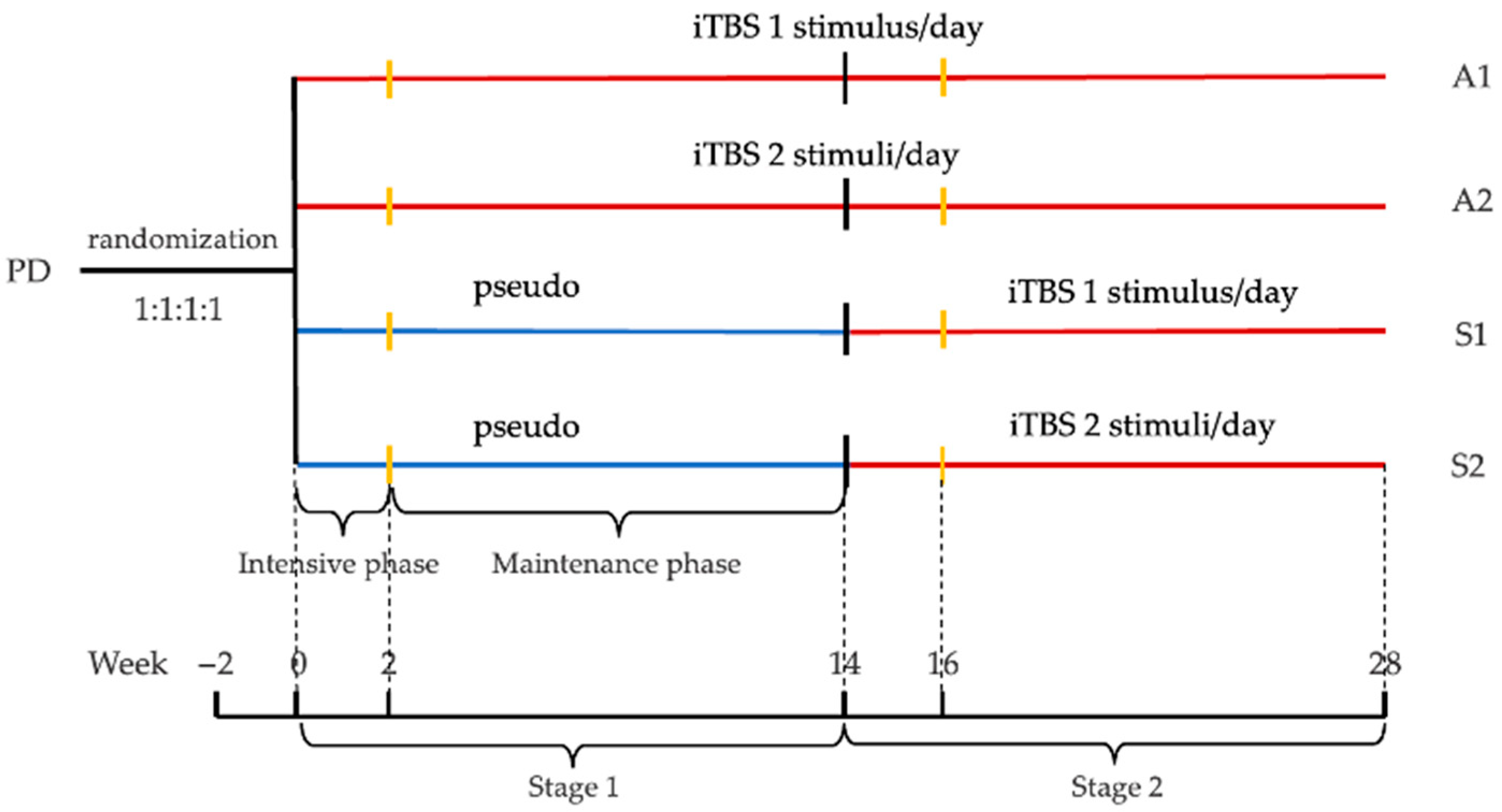
2.2. Study Design
2.3. Inclusion Criteria
- PD diagnosed according to the revised clinical diagnostic criteria of the Movement Disorder Society (MDS) International (2015 version);
- Aged 50–80, age of diagnostic ≥50, male or female;
- Hoehn–Yahr stage ≤4;
- With or without levodopa, maintaining medication stability during the study period;
- Good compliance with the long-term intervention and follow-up.
2.4. Exclusion Criteria
- Presence of any of the features that rules out PD (e.g., unequivocal cerebellar abnormalities, downward vertical supranuclear gaze palsy, selective slowing of downward vertical saccades etc.);
- Patients with severe mental illness or neurological disorders (e.g., epilepsy, cerebrovascular accidents, etc.) or a history of traumatic brain injury or brain surgery;
- Patients with significant cognitive impairment (MMSE <24) or inability to complete questionnaires independently;
- Previously treated with TMS, deep brain stimulation (DBS) or spinal cord stimulation (SCS);
- Have any physical illness that can precipitate epilepsy or intracranial hypertension, including cardiovascular and respiratory disease;
- Have human implantable materials such as intracranial stents, pacemakers, coronary stents, cochlear implants, etc.;
- Are currently taking other investigational drugs or participating in other clinical trials;
- Any other condition that the investigator deems unsuitable for this study.
2.5. Withdrawal Criteria
- Poor compliance to the protocol;
- Occurrence of an intolerable adverse event;
- Patients decline further treatment or follow-up;
- Loss of visits due to unavoidable situations, such as death;
- The investigators terminate the subject’s continued participation after review.
2.6. Endpoint Criteria
2.7. Recruitment and Screening
2.8. Randomization and Blinding
2.9. iTBS Intervention and Follow-Up
2.10. Paired Pulse TMS (ppTMS) Strategy
2.11. Neuroimaging
2.12. Electroencephalogram (EEG) and Electromyographic (EMG)
2.13. Blood Test
2.14. Baseline Assessments and Follow-Up Evaluations
2.15. Outcome Measures
2.16. Study Hypothesis
2.17. Sample Size and Statistical Analysis
3. Results
4. Discussion
Author Contributions
Funding
Institutional Review Board Statement
Informed Consent Statement
Acknowledgments
Conflicts of Interest
References
- Bloem, B.R.; Okun, M.S.; Klein, C. Parkinson’s disease. Lancet 2021, 397, 2284–2303. [Google Scholar] [CrossRef]
- de Lau, L.M.; Breteler, M.M. Epidemiology of Parkinson’s disease. Lancet Neurol. 2006, 5, 525–535. [Google Scholar] [CrossRef]
- Alves, G.; Forsaa, E.B.; Pedersen, K.F.; Dreetz Gjerstad, M.; Larsen, J.P. Epidemiology of Parkinson’s disease. J. Neurol. 2008, 255 (Suppl. S5), 18–32. [Google Scholar] [CrossRef]
- Armstrong, M.J.; Okun, M.S. Diagnosis and Treatment of Parkinson Disease: A Review. JAMA 2020, 323, 548–560. [Google Scholar] [CrossRef]
- Ahlskog, J.E.; Uitti, R.J. Rasagiline, Parkinson neuroprotection, and delayed-start trials: Still no satisfaction? Neurology 2010, 74, 1143–1148. [Google Scholar] [CrossRef]
- Chen, K.S.; Chen, R. Invasive and Noninvasive Brain Stimulation in Parkinson’s Disease: Clinical Effects and Future Perspectives. Clin. Pharmacol. Ther. 2019, 106, 763–775. [Google Scholar] [CrossRef]
- Chou, Y.H.; Hickey, P.T.; Sundman, M.; Song, A.W.; Chen, N.K. Effects of repetitive transcranial magnetic stimulation on motor symptoms in Parkinson disease: A systematic review and meta-analysis. JAMA Neurol. 2015, 72, 432–440. [Google Scholar] [CrossRef]
- Chung, C.L.; Mak, M.K. Effect of Repetitive Transcranial Magnetic Stimulation on Physical Function and Motor Signs in Parkinson’s Disease: A Systematic Review and Meta-Analysis. Brain Stimul. 2016, 9, 475–487. [Google Scholar] [CrossRef]
- Speer, A.M.; Kimbrell, T.A.; Wassermann, E.M.; Repella, J.D.; Willis, M.W.; Herscovitch, P.; Post, R.M. Opposite effects of high and low frequency rTMS on regional brain activity in depressed patients. Biol. Psychiatry 2000, 48, 1133–1141. [Google Scholar] [CrossRef]
- Elahi, B.; Elahi, B.; Chen, R. Effect of transcranial magnetic stimulation on Parkinson motor function--systematic review of controlled clinical trials. Mov. Disord. 2009, 24, 357–363. [Google Scholar] [CrossRef]
- Huang, Y.Z.; Edwards, M.J.; Rounis, E.; Bhatia, K.P.; Rothwell, J.C. Theta burst stimulation of the human motor cortex. Neuron 2005, 45, 201–206. [Google Scholar] [CrossRef] [PubMed] [Green Version]
- Hsieh, T.H.; Huang, Y.Z.; Rotenberg, A.; Pascual-Leone, A.; Chiang, Y.H.; Wang, J.Y.; Chen, J.J. Functional Dopaminergic Neurons in Substantia Nigra are Required for Transcranial Magnetic Stimulation-Induced Motor Plasticity. Cereb. Cortex. 2015, 25, 1806–1814. [Google Scholar] [CrossRef] [PubMed] [Green Version]
- Ghiglieri, V.; Pendolino, V.; Sgobio, C.; Bagetta, V.; Picconi, B.; Calabresi, P. Θ-burst stimulation and striatal plasticity in experimental parkinsonism. Exp. Neurol. 2012, 236, 395–398. [Google Scholar] [CrossRef] [PubMed]
- Natale, G.; Pignataro, A.; Marino, G.; Campanelli, F.; Calabrese, V.; Cardinale, A.; Pelucchi, S.; Marcello, E.; Gardoni, F.; Viscomi, M.T.; et al. Transcranial Magnetic Stimulation Exerts “Rejuvenation” Effects on Corticostriatal Synapses after Partial Dopamine Depletion. Mov. Disord. 2021, 36, 2254–2263. [Google Scholar] [CrossRef] [PubMed]
- Lee, J.Y.; Kim, H.S.; Kim, S.H.; Kim, H.S.; Cho, B.P. Combination of Human Mesenchymal Stem Cells and Repetitive Transcranial Magnetic Stimulation Enhances Neurological Recovery of 6-Hydroxydopamine Model of Parkinsonian’s Disease. Tissue Eng. Regen. Med. 2020, 17, 67–80. [Google Scholar] [CrossRef]
- Degardin, A.; Devos, D.; Defebvre, L.; Destée, A.; Plomhause, L.; Derambure, P.; Devanne, H. Effect of intermittent theta-burst stimulation on akinesia and sensorimotor integration in patients with Parkinson’s disease. Eur. J. Neurosci. 2012, 36, 2669–2678. [Google Scholar] [CrossRef]
- Stocchi, F.; Olanow, C.W. Obstacles to the development of a neuroprotective therapy for Parkinson’s disease. Mov. Disord. 2013, 28, 3–7. [Google Scholar] [CrossRef]
- Athauda, D.; Foltynie, T. The ongoing pursuit of neuroprotective therapies in Parkinson disease. Nat. Rev. Neurol 2015, 11, 25–40. [Google Scholar] [CrossRef]
- D’Agostino, R.B. The delayed-start study design. N. Engl. J. Med. 2009, 361, 1304–1306. [Google Scholar] [CrossRef]
- Clarke, C.E. Are delayed-start design trials to show neuroprotection in Parkinson’s disease fundamentally flawed? Mov. Disord. 2008, 23, 784–789. [Google Scholar] [CrossRef]
- Olanow, C.W.; Hauser, R.A.; Jankovic, J.; Langston, W.; Lang, A.; Poewe, W.; Tolosa, E.; Stocchi, F.; Melamed, E.; Eyal, E.; et al. A randomized, double-blind, placebo-controlled, delayed start study to assess rasagiline as a disease modifying therapy in Parkinson’s disease (the ADAGIO study): Rationale, design, and baseline characteristics. Mov. Disord. 2008, 23, 2194–2201. [Google Scholar] [CrossRef] [PubMed]
- Schapira, A.H.; McDermott, M.P.; Barone, P.; Comella, C.L.; Albrecht, S.; Hsu, H.H.; Massey, D.H.; Mizuno, Y.; Poewe, W.; Rascol, O.; et al. Pramipexole in patients with early Parkinson’s disease (PROUD): A randomised delayed-start trial. Lancet Neurol. 2013, 12, 747–755. [Google Scholar] [CrossRef] [Green Version]
- Group, P.S. A controlled, randomized, delayed-start study of rasagiline in early Parkinson disease. Arch. Neurol. 2004, 61, 561–566. [Google Scholar] [CrossRef]
- Benninger, D.H.; Berman, B.D.; Houdayer, E.; Pal, N.; Luckenbaugh, D.A.; Schneider, L.; Miranda, S.; Hallett, M. Intermittent theta-burst transcranial magnetic stimulation for treatment of Parkinson disease. Neurology 2011, 76, 601–609. [Google Scholar] [CrossRef] [Green Version]
- Cheng, B.; Zhu, T.; Zhao, W.; Sun, L.; Shen, Y.; Xiao, W.; Zhang, S. Effect of Theta Burst Stimulation-Patterned rTMS on Motor and Nonmotor Dysfunction of Parkinson’s Disease: A Systematic Review and Metaanalysis. Front. Neurol. 2021, 12, 762100. [Google Scholar] [CrossRef]
- Vanbellingen, T.; Wapp, M.; Stegmayer, K.; Bertschi, M.; Abela, E.; Kübel, S.; Nyffeler, T.; Müri, R.; Walther, S.; Nef, T.; et al. Theta burst stimulation over premotor cortex in Parkinson’s disease: An explorative study on manual dexterity. J. Neural. Transm 2016, 123, 1387–1393. [Google Scholar] [CrossRef]
- Rossini, P.M.; Berardelli, A.; Deuschl, G.; Hallett, M.; Maertens de Noordhout, A.M.; Paulus, W.; Pauri, F. Applications of magnetic cortical stimulation. The International Federation of Clinical Neurophysiology. Electroencephalogr. Clin. Neurophysiol. Suppl. 1999, 52, 171–185. [Google Scholar]
- Lynch, G.; Kramár, E.A.; Babayan, A.H.; Rumbaugh, G.; Gall, C.M. Differences between synaptic plasticity thresholds result in new timing rules for maximizing long-term potentiation. Neuropharmacology 2013, 64, 27–36. [Google Scholar] [CrossRef] [Green Version]
- Massé-Alarie, H.; Elgueta Cancino, E.; Schneider, C.; Hodges, P. Paired-Pulse TMS and Fine-Wire Recordings Reveal Short-Interval Intracortical Inhibition and Facilitation of Deep Multifidus Muscle Fascicles. PLoS ONE 2016, 11, e0159391. [Google Scholar] [CrossRef] [Green Version]
- Premoli, I.; Király, J.; Müller-Dahlhaus, F.; Zipser, C.M.; Rossini, P.; Zrenner, C.; Ziemann, U.; Belardinelli, P. Short-interval and long-interval intracortical inhibition of TMS-evoked EEG potentials. Brain Stimul. 2018, 11, 818–827. [Google Scholar] [CrossRef] [Green Version]
- Valls-Solé, J.; Pascual-Leone, A.; Wassermann, E.M.; Hallett, M. Human motor evoked responses to paired transcranial magnetic stimuli. Electroencephalogr. Clin. Neurophysiol. 1992, 85, 355–364. [Google Scholar] [CrossRef]
- Kujirai, T.; Caramia, M.D.; Rothwell, J.C.; Day, B.L.; Thompson, P.D.; Ferbert, A.; Wroe, S.; Asselman, P.; Marsden, C.D. Corticocortical inhibition in human motor cortex. J. Physiol. 1993, 471, 501–519. [Google Scholar] [CrossRef] [PubMed]
- Rothwell, J.C. Techniques and mechanisms of action of transcranial stimulation of the human motor cortex. J. Neurosci. Methods 1997, 74, 113–122. [Google Scholar] [CrossRef]
- Du, X.; Summerfelt, A.; Chiappelli, J.; Holcomb, H.H.; Hong, L.E. Individualized brain inhibition and excitation profile in response to paired-pulse TMS. J. Mot. Behav. 2014, 46, 39–48. [Google Scholar] [CrossRef] [Green Version]
- Nakamura, H.; Kitagawa, H.; Kawaguchi, Y.; Tsuji, H. Intracortical facilitation and inhibition after transcranial magnetic stimulation in conscious humans. J. Physiol. 1997, 498 Pt 3, 817–823. [Google Scholar] [CrossRef] [PubMed]
- Tauro, B.J.; Greening, D.W.; Mathias, R.A.; Ji, H.; Mathivanan, S.; Scott, A.M.; Simpson, R.J. Comparison of ultracentrifugation, density gradient separation, and immunoaffinity capture methods for isolating human colon cancer cell line LIM1863-derived exosomes. Methods 2012, 56, 293–304. [Google Scholar] [CrossRef]
- Zhao, A.; Li, Y.; Niu, M.; Li, G.; Luo, N.; Zhou, L.; Kang, W.; Liu, J. SNCA Hypomethylation in Rapid Eye Movement Sleep Behavior Disorder Is a Potential Biomarker for Parkinson’s Disease. J. Parkinsons Dis. 2020, 10, 1023–1031. [Google Scholar] [CrossRef]
- Shi, M.; Liu, C.; Cook, T.J.; Bullock, K.M.; Zhao, Y.; Ginghina, C.; Li, Y.; Aro, P.; Dator, R.; He, C.; et al. Plasma exosomal α-synuclein is likely CNS-derived and increased in Parkinson’s disease. Acta Neuropathol. 2014, 128, 639–650. [Google Scholar] [CrossRef]
- Verschuur, C.V.M.; Suwijn, S.R.; Boel, J.A.; Post, B.; Bloem, B.R.; van Hilten, J.J.; van Laar, T.; Tissingh, G.; Munts, A.G.; Deuschl, G.; et al. Randomized Delayed-Start Trial of Levodopa in Parkinson’s Disease. N. Engl. J. Med. 2019, 380, 315–324. [Google Scholar] [CrossRef]
- Lang, S.; Gan, L.S.; Yoon, E.J.; Hanganu, A.; Kibreab, M.; Cheetham, J.; Hammer, T.; Kathol, I.; Sarna, J.; Martino, D.; et al. Theta-Burst Stimulation for Cognitive Enhancement in Parkinson’s Disease With Mild Cognitive Impairment: A Randomized, Double-Blind, Sham-Controlled Trial. Front. Neurol. 2020, 11, 584374. [Google Scholar] [CrossRef]
- Olanow, C.W.; Rascol, O.; Hauser, R.; Feigin, P.D.; Jankovic, J.; Lang, A.; Langston, W.; Melamed, E.; Poewe, W.; Stocchi, F.; et al. A double-blind, delayed-start trial of rasagiline in Parkinson’s disease. N. Engl J. Med. 2009, 361, 1268–1278. [Google Scholar] [CrossRef] [PubMed] [Green Version]
- Di Lazzaro, V.; Pilato, F.; Dileone, M.; Profice, P.; Oliviero, A.; Mazzone, P.; Insola, A.; Ranieri, F.; Meglio, M.; Tonali, P.A.; et al. The physiological basis of the effects of intermittent theta burst stimulation of the human motor cortex. J. Physiol. 2008, 586, 3871–3879. [Google Scholar] [CrossRef] [PubMed]


| Screening | Baseline | Visit 1 | Visit 2 | Visit 3 | Visit 4 | |
|---|---|---|---|---|---|---|
| −2 Weeks | 0 | 2 Weeks | 14 Weeks | 16 Weeks | 28 Weeks | |
| Written informed consent | × | |||||
| Inclusion/exclusion criteria | × | |||||
| Randomization | × | |||||
| Basic demographic | × | |||||
| Hoehn–Yahr stage | × | × | × | × | × | |
| UPDRS | × | × | × | × | × | |
| BBS | × | × | × | |||
| HAMD | × | × | × | |||
| HAMA | × | × | × | |||
| MMSE | × | × | × | |||
| MoCA | × | × | × | |||
| PDQ-39 | × | × | × | |||
| PSQI | × | × | × | |||
| SS-16 | × | × | × | |||
| SCOPA-AUT | × | × | × | |||
| Wexner | × | × | × | |||
| MRI | × | × | × | × | ||
| EEG | × | × | × | |||
| EMG | × | × | × | × | × | |
| Blood test | × | × | × | × | ||
| Compliancy | × | × | × | |||
| Medication usage | × | × | × | × | × | × |
| Side effects | × | × | × | × |
Publisher’s Note: MDPI stays neutral with regard to jurisdictional claims in published maps and institutional affiliations. |
© 2022 by the authors. Licensee MDPI, Basel, Switzerland. This article is an open access article distributed under the terms and conditions of the Creative Commons Attribution (CC BY) license (https://creativecommons.org/licenses/by/4.0/).
Share and Cite
Li, P.; Luo, N.; Sun, S.; Li, Y.; Shen, D.; Zhu, X.; Zhou, L.; Zhou, H.; Liu, J. Neuroprotective Effects of Intermittent Theta Burst Stimulation in Parkinson’s Disease (NET-PD): A Study Protocol for a Delayed-Start Randomized Double-Blind Sham-Controlled Trial. J. Clin. Med. 2022, 11, 4972. https://doi.org/10.3390/jcm11174972
Li P, Luo N, Sun S, Li Y, Shen D, Zhu X, Zhou L, Zhou H, Liu J. Neuroprotective Effects of Intermittent Theta Burst Stimulation in Parkinson’s Disease (NET-PD): A Study Protocol for a Delayed-Start Randomized Double-Blind Sham-Controlled Trial. Journal of Clinical Medicine. 2022; 11(17):4972. https://doi.org/10.3390/jcm11174972
Chicago/Turabian StyleLi, Puyu, Ningdi Luo, Sainan Sun, Yuanyuan Li, Dingding Shen, Xue Zhu, Liche Zhou, Haiyan Zhou, and Jun Liu. 2022. "Neuroprotective Effects of Intermittent Theta Burst Stimulation in Parkinson’s Disease (NET-PD): A Study Protocol for a Delayed-Start Randomized Double-Blind Sham-Controlled Trial" Journal of Clinical Medicine 11, no. 17: 4972. https://doi.org/10.3390/jcm11174972
APA StyleLi, P., Luo, N., Sun, S., Li, Y., Shen, D., Zhu, X., Zhou, L., Zhou, H., & Liu, J. (2022). Neuroprotective Effects of Intermittent Theta Burst Stimulation in Parkinson’s Disease (NET-PD): A Study Protocol for a Delayed-Start Randomized Double-Blind Sham-Controlled Trial. Journal of Clinical Medicine, 11(17), 4972. https://doi.org/10.3390/jcm11174972