
Diabetes Affects the Relationship between Heart Rate Variability and Arterial Stiffness in a Gender-Specific Manner

 ,
,
Abstract
:1. Introduction
2. Methods
2.1. Study Population
2.2. Assessment of Pulse Wave Velocity (PWV)
2.3. HRV Parameters
- −
- power spectral density at the high-frequency (HF) range (0.15–0.4 Hz);
- −
- power spectral density at the low-frequency (LF) range (0.04–0.15 Hz);
- −
- the LF/HF ratio.
2.4. Statistical Analysis
3. Results
4. Discussion
Author Contributions
Funding
Institutional Review Board Statement
Informed Consent Statement
Data Availability Statement
Conflicts of Interest
References
- Scuteri, A.; Benetos, A.; Sierra, C.; Coca, A.; Chicherio, C.; Frisoni, G.B.; Gasecki, D.; Hering, D.; Lovic, D.; Manios, E.; et al. Routine assessment of cognitive function in older patients with hypertension seen by primary care physicians: Why and how-a decision-making support from the working group on ‘hypertension and the brain’ of the European Society of Hypertension and from the European Geriatric Medicine Society. J. Hypertens. 2021, 39, 90–100. [Google Scholar]
- Vos, T.; Flaxman, A.D.; Naghavi, M.; Lozano, R.; Michaud, C.; Ezzati, M.; Shibuya, K.; Salomon, J.A.; Abdalla, S.; Aboyans, V.; et al. Years lived with disability (YLDs) for 1160 sequelae of 289 diseases and injuries 1990–2010: A systematic analysis for the Global Burden of Disease Study 2010. Lancet 2012, 380, 2163–2196. [Google Scholar] [CrossRef]
- Calimport, S.R.G.; Bentley, B.L.; Stewart, C.E.; Pawelec, G.; Scuteri, A.; Vinciguerra, M.; Slack, C.; Chen, D.; Harries, L.W.; Marchant, G.; et al. To help aging populations, classify organismal senescence. Science 2019, 366, 576–578. [Google Scholar] [CrossRef] [PubMed] [Green Version]
- Cunha, P.G.; Cotter, J.; Oliveira, P.; Vila, I.; Boutouyrie, P.; Laurent, S.; Nilsson, P.M.; Scuteri, A.; Sousa, N. Pulse wave velocity distribution in a cohort study: From arterial stiffness to early vascular aging. J. Hypertens. 2015, 33, 1438–1445. [Google Scholar] [CrossRef] [PubMed] [Green Version]
- Nilsson, P.M.; Laurent, S.; Cunha, P.; Olsen, M.H.; Rietzschel, E.; Franco, O.; Ryliškytė, L.; Strazhesko, I.; Vlachopoulos, C.; Chen, C.-H.; et al. For the MARE Consortium. Characteristics of Healthy Vascular Ageing (HVA) in pooled population-based cohort studies: The global MARE consortium. J. Hypertens. 2018, 36, 2340–2349. [Google Scholar] [CrossRef]
- Scuteri, A.; Morrell, C.H.; Orru’, M.; AlGhatrif, M.; Saba, P.S.; Terracciano, A.; Ferreli, L.A.P.; Loi, F.; Marongiu, M.; Pilia, M.G.; et al. Gender specific profiles of white coat and masked hypertension impacts on arterial structure and function in the SardiNIA study. Int. J. Cardiol. 2016, 217, 92–98. [Google Scholar] [CrossRef] [Green Version]
- Scuteri, A.; Rovella, V.; Fegatelli, D.A.; Tesauro, M.; Gabriele, M.; Di Daniele, N. An operational definition of SHATS (Systemic Hemodynamic Atherosclerotic Syndrome): Role of arterial stiffness and blood pressure variability in elderly hypertensive subjects. Int. J. Cardiol. 2018, 263, 132–137. [Google Scholar] [CrossRef]
- Vlachopoulos, C.; Aznaouridis, K.; Stefanadis, C. Prediction of Cardiovascular Events and All-Cause Mortality with Arterial Stiffness: A Systematic Review and Meta-Analysis. J. Am. Coll. Cardiol. 2010, 55, 1318–1327. [Google Scholar] [CrossRef] [Green Version]
- Scuteri, A.; Wang, H. Pulse Wave Velocity as a Marker of Cognitive Impairment in the Elderly. J. Alzheimer’s Dis. 2014, 42 (Suppl. 4), S401–S410. [Google Scholar] [CrossRef]
- Chang, P.-Y.; Wang, I.-T.; Chiang, C.-E.; Chen, C.-H.; Yeh, W.-Y.; Henderson, V.W.; Tsai, Y.-W.; Cheng, H.-M. Vascular complications of diabetes: Natural history and corresponding risks of dementia in a national cohort of adults with diabetes. Geol. Rundsch. 2021, 58, 859–867. [Google Scholar] [CrossRef]
- Stehouwer, C.D.A.; Henry, R.M.A.; Ferreira, I. Arterial stiffness in diabetes and the metabolic syndrome: A pathway to cardiovascular disease. Diabetologia 2008, 51, 527–539. [Google Scholar] [CrossRef] [PubMed]
- Thayer, J.F.; Yamamoto, S.S.; Brosschot, J.F. The relationship of autonomic imbalance, heart rate variability and cardiovascular disease risk factors. Int. J. Cardiol. 2010, 141, 122–131. [Google Scholar] [CrossRef] [PubMed]
- Task Force of the European Society of Cardiology and the North American Society of Pacing and Electrophysiology. Heart rate variability standards of measurement, physiological interpretation and clinical use. Circulation 1996, 93, 1043–1065. [Google Scholar] [CrossRef] [Green Version]
- Fisher, J.P.; Young, C.N.; Fadel, P.J. Central sympathetic overactivity: Maladies and mechanisms. Auton. Neurosci. 2009, 148, 5–15. [Google Scholar] [CrossRef] [PubMed] [Green Version]
- Yu, Y.; Hu, L.; Xu, Y.; Wu, S.; Chen, Y.; Zou, W.; Zhang, M.; Wang, Y.; Gu, Y. Impact of blood glucose control on sympathetic and vagus nerve functional status in patients with type 2 diabetes mellitus. Geol. Rundsch. 2019, 57, 141–150. [Google Scholar] [CrossRef] [Green Version]
- Shah, A.S.; El Ghormli, L.; Vajravelu, M.E.; Bacha, F.; Farrell, R.M.; Gidding, S.S.; Levitt Katz, L.E.; Tryggestad, J.B.; White, N.H.; Urbina, E.M. Heart Rate Variability and Cardiac Autonomic Dysfunction: Prevalence, Risk Factors, and Relationship to Arterial Stiffness in the Treatment Options for Type 2 Diabetes in Adolescents and Youth (TODAY) Study. Diabetes Care 2019, 42, 2143–2150. [Google Scholar] [CrossRef] [Green Version]
- Millasseau, S.C.; Stewart, A.D.; Patel, S.J.; Redwood, S.R.; Chowieñczyk, P.J. Evaluation of carotid-femoral pulse wave velocity: Influence of timing algorithm and heart rate. Hypertension 2005, 45, 222–226. [Google Scholar] [CrossRef] [Green Version]
- Chorepsima, S.; Eleftheriadou, I.; Tentolouris, A.; Moyssakis, I.; Protogerou, A.; Kokkinos, A.; Sfikakis, P.P.; Tentolouris, N. Pulse wave velocity and cardiac autonomic function in type 2 diabetes mellitus. BMC Endocr. Disord. 2017, 17, 1–8. [Google Scholar] [CrossRef]
- Tentolouris, N.; Liatis, S.; Moyssakis, I.; Tsapogas, P.; Psallas, M.; Diakoumopoulou, E.; Voteas, V.; Katsilambros, N. Aortic distensibility is reduced in subjects with type 2 diabetes and cardiac autonomic neuropathy. Eur. J. Clin. Investig. 2003, 33, 1075–1083. [Google Scholar] [CrossRef]
- Shah, A.S.; Isom, S.; D’Agostino, J.R.; Dolan, L.M.; Dabelea, D.; Imperatore, G.; Mottl, A.; Lustigova, E.; Pihoker, C.; Marcovina, S.; et al. Longitudinal Changes in Arterial Stiffness and Heart Rate Variability in Youth-Onset Type 1 Versus Type 2 Diabetes: The SEARCH for Diabetes in Youth Study. Diabetes Care 2022, 45, 1647–1656. [Google Scholar] [CrossRef]
- Shah, A.S.; El Ghormli, L.; Gidding, S.S.; Hughan, K.S.; Levitt Katz, L.E.; Koren, D.; Tryggestad, J.B.; Bacha, F.; Braffett, B.H.; Arslanian, S.; et al. Longitudinal changes in vascular stiffness and heart rate variability among young adults with youth-onset type 2 diabetes: Results from the follow-up observational treatment options for type 2 diabetes in adolescents and youth (TODAY) study. Acta Diabetol. 2022, 59, 197–205. [Google Scholar] [PubMed]
- Sharman, J.E.; O’Brien, E.; Alpert, B.; Schutte, A.E.; Delles, C.; Olsen, M.H.; Asmar, R.; Atkins, N.; Barbosa, E.; Calhoun, D.; et al. Lancet Commission on Hypertension Group. Lancet commission on hypertension group position statement on the global improvement of accuracy standards for devices that measure blood pressure. J. Hypertens. 2020, 38, 21–29. [Google Scholar] [CrossRef] [PubMed]
- Tolea, M.I.; Costa, P.; Terracciano, A.; Griswold, M.; Simonsick, E.M.; Najjar, S.S.; Scuteri, A.; Deiana, B.; Orrù, M.; Masala, M.; et al. Sex-Specific Correlates of Walking Speed in a Wide Age-Ranged Population. J. Gerontol. Ser. B Psychol. Sci. Soc. Sci. 2010, 65B, 174–184. [Google Scholar] [CrossRef] [PubMed]
- Koenig, J.; Thayer, J.F. Sex differences in healthy human heart rate variability: A meta-analysis. Neurosci. Biobehav. Rev. 2016, 64, 288–310. [Google Scholar] [CrossRef]
- Rannelli, L.A.; MacRae, J.; Mann, M.C.; Ramesh, S.; Hemmelgarn, B.R.; Rabi, D.; Sola, D.Y.; Ahmed, S.B. Sex differences in associations between insulin resistance, heart rate variability, and arterial stiffness in healthy women and men: A physiology study. Can. J. Physiol. Pharmacol. 2017, 95, 349–355. [Google Scholar] [CrossRef]
- Zuo, J.; Chao, H.; Tang, B.; Avolio, A.; Schlaich, M.; Nolde, J.; Adji, A.; Carnagarin, R. Female Gender Is Associated with Higher Susceptibility of Weight Induced Arterial Stiffening and Rise in Blood Pressure. J. Clin. Med. 2021, 10, 3479. [Google Scholar] [CrossRef]
- Gómez-Sánchez, L.; Gómez-Sánchez, M.; Rodríguez-Sánchez, E.; Patino-Alonso, C.; Alonso-Dominguez, R.; Sanchez-Aguadero, N.; Lugones-Sánchez, C.; Llamas-Ramos, I.; García-Ortiz, L.; Gómez-Marcos, M.A.; et al. Relationship of Different Anthropometric Indices with Vascular Ageing in an Adult Population without Cardiovascular Disease—EVA Study. J. Clin. Med. 2022, 11, 2671. [Google Scholar] [CrossRef]
- Palatini, P.; Julius, S. The role of cardiac autonomic function in hypertension and cardio-vascular disease. Curr. Hypertens. 2009, 11, 199–205. [Google Scholar] [CrossRef]
- Sacha, J. Interaction between Heart Rate and Heart Rate Variability. Ann. Noninvasive Electrocardiol. 2014, 19, 207–216. [Google Scholar] [CrossRef]
- Dart, A.M.; Du, X.J.; Kingwell, B.A. Gender, sex hormones and autonomicnervous control of the cardiovascular system. Cardiovasc. Res. 2002, 53, 678–687. [Google Scholar] [CrossRef]

| Women (185) | Men (237) | Gender Difference p | ||
|---|---|---|---|---|
| Age (years) | 61.0 (20.0) | 60.0 (27) | 62.5 (19) | 0.05 |
| Women (%) | 43.8 | --- | -- | |
| Hypertension (%) | 62.1 | 51.9 | 70.0 | 0.001 |
| Diabetes mellitus (%) | 30.8 | 26.0 | 34.6 | 0.06 |
| BMI (Kg/m2) | 27.1 (7.1) | 26.8 (8.3) | 27.5 (6.4) | 0.62 |
| SBP (mmHg) | 137.0 (23.0) | 134.0 (23.0) | 139.0 (19.0) | 0.01 |
| DBP (mmHg) | 77.0 (14.0) | 75.0 (16.0) | 79.0 (14.0) | 0.001 |
| HR (bpm) | 67.0 (16.0) | 67.0 (16.0) | 68.0 (16.0) | 0.86 |
| pNN50 | 2.0 (10.8) | 2.2 (12.5) | 2.0 (10.0) | 0.75 |
| rmSDD | 24.6 (27.1) | 25.0 (25.9) | 24.5 (26.1) | 0.72 |
| LF | 727.0 (259.0) | 788.0 (288.0) | 734.0 (234.0) | 0.23 |
| HF | 273.0 (258.0) | 292.0 (288.0) | 266.0 (234.0) | 0.23 |
| LF/HF ratio | 2.7 (2.5) | 2.4 (2.6) | 2.8 (2.5) | 0.23 |
| PWV (m/s) | 9.3 (3.1) | 8.9 (2.6) | 9.8 (3.4) | 0.0001 |
| Antihypertensive medication | 51.6 | 42.4 | 60.1 | 0.01 |
| Antidiabetic medication | 27.8 | 24.3 | 30.3 | 0.05 |
| Lipid-lowering therapy | 14.7 | 11 | 17.6 | 0.05 |
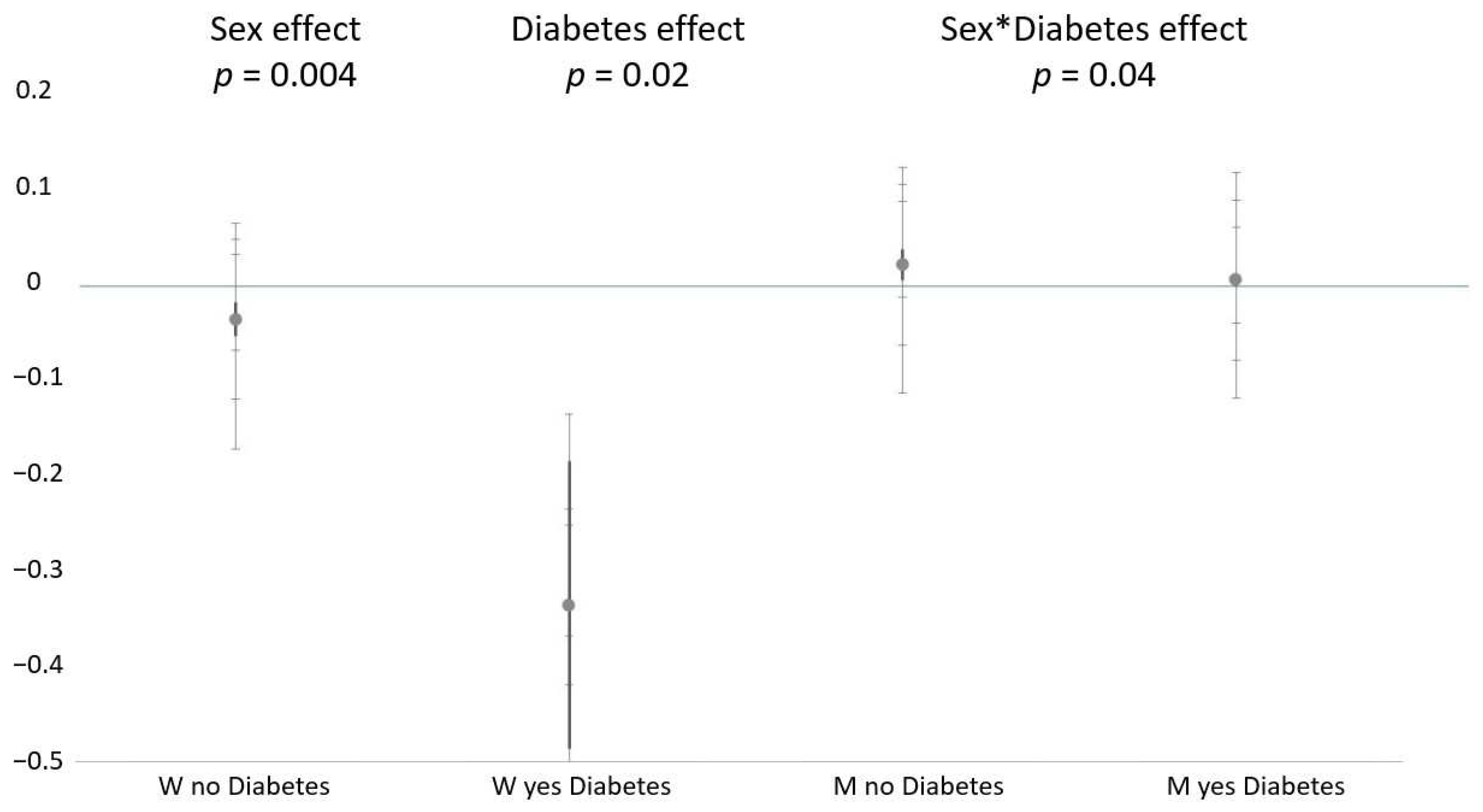
| Women NO Diabetes (n = 136) | Women YES Diabetes (n = 49) | Men NO Diabetes (n = 155) | Men YES Diabetes (n = 82) | |||||
|---|---|---|---|---|---|---|---|---|
| Age | 3.72 ± 0.97 | 0.0002 | 12.22 ± 4.89 | 0.02 | 9.32 ± 1.38 | 0.0001 | 16.78 ± 3.76 | 0.0001 |
| SBP | 5.72 ± 1.13 | 0.0001 | 7.48 ± 2.82 | 0.02 | 5.24 ± 1.55 | 0.001 | 5.87 ± 3.15 | 0.05 |
| LF | −0.039 ± 0.018 | 0.05 | −0.337 ± 0.150 | 0.03 | 0.018 ± 0.016 | 0.28 | 0.002 ± 0.005 | 0.72 |
| Model R2 | 0.484 | 0.489 | 0.479 | 0.325 | ||||
Publisher’s Note: MDPI stays neutral with regard to jurisdictional claims in published maps and institutional affiliations. |
© 2022 by the authors. Licensee MDPI, Basel, Switzerland. This article is an open access article distributed under the terms and conditions of the Creative Commons Attribution (CC BY) license (https://creativecommons.org/licenses/by/4.0/).
Share and Cite
Serra, C.; Sestu, A.; Murru, V.; Greco, G.; Vacca, M.; Scuteri, A. Diabetes Affects the Relationship between Heart Rate Variability and Arterial Stiffness in a Gender-Specific Manner. J. Clin. Med. 2022, 11, 4937. https://doi.org/10.3390/jcm11174937
Serra C, Sestu A, Murru V, Greco G, Vacca M, Scuteri A. Diabetes Affects the Relationship between Heart Rate Variability and Arterial Stiffness in a Gender-Specific Manner. Journal of Clinical Medicine. 2022; 11(17):4937. https://doi.org/10.3390/jcm11174937
Chicago/Turabian StyleSerra, Carla, Alessandro Sestu, Veronica Murru, Giulia Greco, Matteo Vacca, and Angelo Scuteri. 2022. "Diabetes Affects the Relationship between Heart Rate Variability and Arterial Stiffness in a Gender-Specific Manner" Journal of Clinical Medicine 11, no. 17: 4937. https://doi.org/10.3390/jcm11174937
APA StyleSerra, C., Sestu, A., Murru, V., Greco, G., Vacca, M., & Scuteri, A. (2022). Diabetes Affects the Relationship between Heart Rate Variability and Arterial Stiffness in a Gender-Specific Manner. Journal of Clinical Medicine, 11(17), 4937. https://doi.org/10.3390/jcm11174937