Review

Chemically Induced Colitis-Associated Cancer Models in Rodents for Pharmacological Modulation: A Systematic Review

1
H&TRC—Health and Technology Research Center, ESTeSL—Escola Superior de Tecnologia da Saúde de Lisboa, Instituto Politécnico de Lisboa, 1990-096 Lisbon, Portugal
2
iMed.ULisboa, Faculdade de Farmácia, Universidade de Lisboa, 1649-003 Lisbon, Portugal
3
Joaquim Chaves Saúde, Joaquim Chaves Laboratório de Análises Clínicas, Miraflores, 1495-069 Algés, Portugal
*
Author to whom correspondence should be addressed.
J. Clin. Med. 2022, 11(10), 2739; https://doi.org/10.3390/jcm11102739
Submission received: 10 February 2022 / Revised: 29 April 2022 / Accepted: 10 May 2022 / Published: 12 May 2022
(This article belongs to the Section Gastroenterology & Hepatopancreatobiliary Medicine)

Abstract

:
Animal models for colitis-associated colorectal cancer (CACC) represent an important tool to explore the mechanistic basis of cancer-related inflammation, providing important evidence that several inflammatory mediators play specific roles in the initiation and perpetuation of colitis and CACC. Although several original articles have been published describing the CACC model in rodents, there is no consensus about the induction method. This review aims to identify, summarize, compare, and discuss the chemical methods for the induction of CACC through the PRISMA methodology. Methods: We searched MEDLINE via the Pubmed platform for studies published through March 2021, using a highly sensitive search expression. The inclusion criteria were only original articles, articles where a chemically-induced animal model of CACC is described, preclinical studies in vivo with rodents, and articles published in English. Results: Chemically inducible models typically begin with the administration of a carcinogenic compound (as azoxymethane (AOM) or 1,2-dimethylhydrazine (DMH)), and inflammation is caused by repeated cycles of colitis-inducing agents (such as 2,4,6-trinitrobenzenesulfonic acid (TNBS) or dextran sulfate sodium (DSS)). The strains mostly used are C57BL/6 and Balb/c with 5–6 weeks. To characterize the preclinical model, the parameters more used include body weight, stool consistency and morbidity, inflammatory biomarkers such as tumor necrosis factor (TNF)-α, interleukin (IL)-6 and IL-1β, angiogenesis markers such as proliferating cell nuclear antigen (PCNA), marker of proliferation Ki-67, and caspase 3, the presence of ulcers, thickness or hyperemia in the colon, and histological evaluation of inflammation. Conclusion: The AOM administration seems to be important to the CACC induction method, since the carcinogenic effect is achieved with just one administration. DSS has been the more used inflammatory agent; however, the TNBS contribution should be more studied, since it allows a reliable, robust, and a highly reproducible animal model of intestinal inflammation.

1. Introduction

Colorectal cancer represents the third most diagnosed form of cancer, and it is a key cause of cancer mortality worldwide [1]. Indeed, this type of cancer is characterized by being responsible for 10% of all annually diagnosed cancer, and it is the world’s fourth most deadly cancer with, approximately 900,000 deaths annually [2]. Additionally, over 1.9 million new colorectal cancer cases and 935,173 deaths were predictable in 2020 [1,3], with more than half of the cases occurring in developed countries [4]. The disease prognosis is strongly related to the diagnosis stage; however, currently, there is a 5-year survival rate estimated at 66.1% for all stages of the disease [5,6].
Carcinogenesis of colon cancer departs from normal mucosa; however, through non-well-established steps, it tends to develop and leads to malignancy. It is a dynamic and multi-phase disease, which develops slowly over years and progresses through cytologically distinct benign and malignant states, from single crypt lesions through adenoma, to malignant carcinoma with possible metastasis [7]. In colorectal cancer, factors such as intestinal injury, oxidative stress, and chronic inflammation contribute to an alteration of the intestinal homeostasis [8,9,10].
Colorectal cancer pathogenesis could evolve from two different molecular pathways, sporadic and colitis-associated colorectal cancer. CACC results from defects in the cell cycle, even though different factors can be responsible for the neoplastic changes. Particularly, proinflammatory pathways, especially the NF-κB, IL-6/STAT3, COX-2/PGE2, and IL-23/Th17 signaling pathways, are dysregulated and consequently, they have a crucial role in the pathogenesis of CACC. The increased regulation of antiapoptotic proteins and the higher proliferation of epithelial cells, as well as new blood vessels, are essential in the tumor initiation, growth, and progression [7,8,9,10].
Inflammatory bowel disease (IBD) is characterized by a chronic inflammation of the large bowel including ulcerative colitis and Crohn’s disease and has been associated in several epidemiological studies with the spontaneous development of colorectal neoplasia, which is described as CACC. CACC is responsible for, approximately, 90% of the cases of CRC, which is allied with a possible hereditary influence [11,12,13,14,15]. Patients with IBD face an increased lifetime risk of developing CACC and a worse prognosis in comparison to healthy individuals [9,16]. Additionally, colon cancer is associated with significant morbidity and mortality up to 15% in IBD patients [17,18]. The risk for the development of CACC is closely associated with the extent of colonic involvement, duration of colitis, and severity of active inflammation [19,20,21]. The immune signaling pathways contribute to the pathogenesis of colitis and CAAC; however, only a limited number of studies were performed to understand the exact mechanisms underlying the link between chronic colitis and the development of CACC [9,22].
The treatment for CACC is personalized, taking into account the patient’s needs and tumor’s specificities [23]. In general, surgery is the mainstream curative treatment for CACC combined with radiotherapy and/or chemotherapy. However, several side effects are noticed due to the treatment toxicity. Side effects from chemotherapy for colon cancer include nausea, vomiting, loss of appetite, hair loss, mouth sores, diarrhea, and rashes. It also lowers white blood cell counts, so there is usually an increased risk of infection as well as low blood platelet counts resulting in bleeding or bruising. These treatments decrease life quality, and the survival rate depends primarily on the stage of the disease. Stage I has a 90% survival rate in 5 years against a 10% survival rate in stage IV in the same time spectrum [2,11,24].
Actually, numerous efforts have been made to find a new effective method to regulate undesirable immune responses during the autoimmune reaction [19]. The development of preclinical studies allows the evaluation of other therapeutic alternatives, knowledge of the pathogenesis, and possible future treatments, which are essential to improving the pharmacological approaches in the treatment of CACC [25]. Indeed, animal models represent an important tool to explore the mechanistic basis of cancer-related inflammation, where the induction of CACC can be perceived and manipulated by researchers [25,26]. Additionally, it also has an immense potential to provide important evidence about the inflammatory mediator’s roles in the initiation and perpetuation of IBD and CACC as well as in the development of new therapeutic approaches and their mechanism of action [7,25,26].
There are different types of animal models of CACC; however, chemically induced models are the greatest commonly used [25,27,28]. Such models are characterized by starting with the administration of a carcinogenic compound followed by repeated cycles of proinflammatory agents, which are toxic to colonic cells and generate an exacerbated inflammatory response [20,21]. This experimental intestinal carcinogenesis model should represent characteristics observed in human disease where tumors develop quickly and express biological modifications similar to those found in humans as well as mimic the disease progression from inflammation through dysplasia to carcinoma [29,30].
Currently, there is no agreement in the literature concerning the induction method taking into account several parameters, such as doses and/or concentrations of the carcinogenic and proinflammatory agents, frequency of administrations, gender, age, and strains of the mice utilized. Thus, our research group purposes to identify, summarize, compare, and discuss different protocols for the induction of CACC through the PRISMA methodology. The challenge of the present systematic review is to promote a synthesis of the information available in the literature, which can be an important tool for future research on new possible pharmacological approaches for the treatment of CACC, contributing to a more effective and safe treatment.

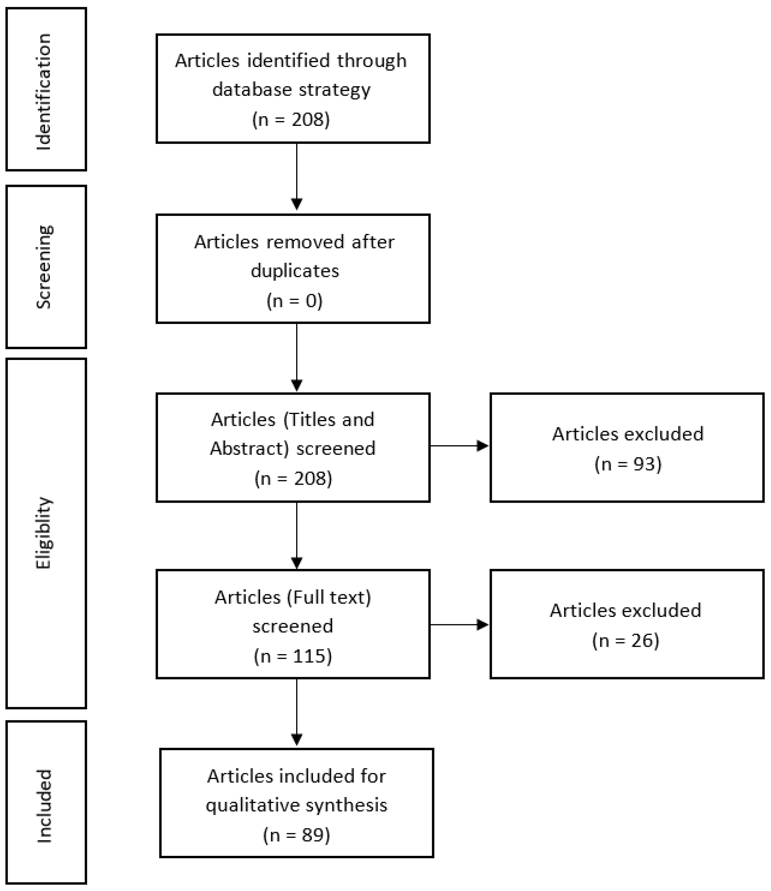
2. Materials and Methods

2.1. Search Strategy

Following the establishment of a review protocol based on PRISMA methodology, the electronic database MEDLINE via the PubMed platform was searched from initiation up to March 2021 for all studies with a chemically induced animal model of CACC in mice. The search strategy was initiated with the insertion of keywords in the MeSH Database in order to find MeSH terms. Posteriorly, a combination of the keywords was carried out, and the final search expression was: (“Mice”[Mesh] OR Mice[tiab] OR Mice OR Mouse) AND (“Animal Experimentation”[Mesh] OR “Animal Experimentation”[tiab] OR “Animal Experimentation” OR “Preclinical studies”[tiab] OR “Preclinical studies” OR “Non-clinical studies”[tiab] OR “Non-clinical studies” OR “animal model”[tiab] OR “animal model” OR “disease models, animal” [Mesh] OR “disease model”[tiab] OR “disease model” OR “disease models”[tiab] OR “disease models” OR “disease animal model”[tiab] OR “disease animal model” OR “disease animal models”[tiab] OR “disease animal models”)) AND (“colitis-associated neoplasms”[Mesh] OR “colitis-associated neoplasms”[tiab] OR “colitis-associated neoplasms” OR “colitis-associated colorectal cancer”[tiab] OR “colitis-associated colorectal cancer” OR “colitis-associated cancer”[tiab] OR “colitis-associated cancer”). The results of the literature search are outlined in Figure 1.

2.2. Selection of Studies

In order to select the articles after the search expression was performed, there were several inclusion criteria taken into account, such as: (1) only original articles; (2) articles where a chemically induced animal model of CACC is described; (3) preclinical studies in vivo with rodents; and (4) articles published in English. Additionally, the research group also selected exclusion criteria, such as: (1) review articles; (2) short communications; (3) case reports, and (4) expert opinions. Throughout the process of selecting the studies, the exclusion started with the analysis of the abstracts, and then, the full texts of the remaining articles were retrieved and reviewed. In case of disagreements between the reviewers and the absence of consensus, a third element was included to make the final decision.

2.3. Data Extraction

The data of interest were independently extracted by both reviewers into a Microsoft Excel spreadsheet (Windows 10 edition; Microsoft Corporation, Lisbon, Portugal). As well as in the process of selecting the articles, the possible disagreements between the reviewers were resolved by mutual consensus or by the inclusion of a third element to make the final decision. The information of interest extracted from each study was as follows: pro-inflammatory reagent-related parameters (number of administrations, frequency of administration, doses, volume, concentrations, and vehicles used), procarcinogen reagent-related parameters (number of administrations and doses), mice-related parameters (strain, gender, and animal age), model characterization (clinical signs and symptoms, biochemical markers and inflammatory and angiogenesis biomarkers, macroscopic evaluation, and histological evaluation), authors and year of publication.

2.4. Articles Eligibility

Related to our article’s eligibility, studies using a carcinogenic chemical combined with a proinflammatory substance were selected, promoting the development of tumors with an inflammatory response associated. The parameters such as dosage, timing, and frequency of administration were all included, since the objective is to compare and evaluate their influence in the induction method. We excluded the utilization of only a procarcinogen agent without a proinflammatory chemical, which mimics the development of cancer in the colon without an inflammatory response associated. Studies with genetically modified strains of mice were also excluded because tumors develop in the colon without the action of chemicals. Finally, studies with transplanted tumors were excluded, too.
After performing the search strategy, the retrieved articles were exported from the MEDLINE database to a Systematic Reviews Web Application (Rayyan QCRI), and the titles and abstracts were analyzed by two independent reviewers in order to select the relevant and potentially eligible studies. Then, after selecting all the articles, the same two independent reviewers assessed the full text of each one and decided whether the article was eligible or not, considering the inclusion and exclusion criteria. In these two steps, a third element was included in case of discrepancies between the two reviewers in order to provide a final decision. After the selection of the eligible studies, the same two independent reviewers extracted the relevant data present in those and inserted them in a customized data extraction document. The data of interest were extracted from the text, graphs, and/or tables present in the chosen articles. In case of discrepancies and an absence of a consensus at the end of the task, a third element was included in order to make a final decision.
To evaluate the internal validity of the selected studies and the methodological quality, through the analysis of the potential risk of bias present, we used SYRCLE’s risk of bias tool. Throughout the process, several key points will be formulated to be aware of the analysis of each study and attributed a punctuation, which in the end culminated in a final score for each article.
Our animal systematic review was posteriorly submitted to PROSPERO, which is an international prospective register of systematic reviews.
Animal care was in accordance with the internationally accepted principles for laboratory animal use and care, Directive 2010/63/EU.
Thus, this review aims to identify, summarize, and analyze different chemical methods for inducing colitis-associated cancer in mice for that we propose to compare and debate some important parameters, such as proinflammatory reagent-related parameters, procarcinogen reagent-related parameters, mice-related parameters, and the model characterization.

3. Results

The electronic database allowed identifying 208 publications in total after the application of the search expression. The publications were then screened according to the inclusion and exclusion criteria (Figure 1). No duplicates were identified in the abstract analysis; however, 93 original articles were excluded, since they were published more than 5 years ago. Of these, 115 published articles appeared to be pertinent to the study question and were saved for extra assessment. From the 115 papers remaining, 25 were not aligned with the purpose of the work and were excluded as ineligible based on the inclusion criteria described previously. The reasons for the excluded articles were: the article corresponds to a protocol (n = 4); letter (n = 1); short communication (n = 1); review (n = 2); systematic review (n = 1); method of induction that does not use only chemicals (n = 10); tumour induction at other organs (n = 1); articles where a chemically induced animal model of CACC was not described (n = 3), and the article was written in Chinese (n = 2). Thus, 89 original articles were included in the qualitative analysis, since all of these studies have described a chemically induced animal model of CACC in rodents (Table 1).
Preclinical studies in vivo, particularly animal model studies, mimic the pathogenesis of CACC disease in humans and allow testing new pharmacological approaches, and they are vital for knowing the underlying pathogenesis and for conceivable upcoming treatments [31,32]. There are several types of animal models of CACC: chemically induced models; transplantation models, those that express intestinal inflammation spontaneously, those in which intestinal inflammation can be induced by specific immunological methods, the genetically engineered models by gene knockout, knockin, or transgenic methods, and the last includes adaptative transfer models [33,34]. The chemically induced models are studied in the greatest detail so far for CRC. The CACC models are appropriated to develop and test novel therapeutic strategies for the treatment of the disease. The knowledge of molecular pathways involved in CACC may provide opportunities for innovative therapeutic strategies for the treatment of CACC in the future. The exact involvement of genetic susceptibility, microenvironment, and immune reactivity remains unclear; therefore, the prevention and therapy of CACC are challenging [35]. There is interest in the use and study of more than one animal model, since differences between models may reflect the different subgroups of patients with IBD. The most used chemicals to induce colitis models are 2,4,6-trinitrobenzenesulfonic acid (TNBS), which promotes a Th1 response, resembling CD, and dextran sulfate sodium (DSS), which promotes a Th2 response, resembling UC [36,37]. DSS-induced colitis and TNBS-induced colitis models are the most widely used to induce IBD since they symptomatically, morphologically, and histopathologically resemble human IBD and allow the development and test of novel therapeutic strategies [38,39,40].
DSS is easy to use and briefly obtain results resembling UC in humans [41,42]. To cause inflammation in rats or mice, DSS protocol uses DSS added to drinking water; then, acute or chronic colitis model experiments can be conducted only by altering the concentrations of the administered substance as well as the number of cycles of supply of the chemical agent. The severity of colitis caused by DSS depends on the dose, duration of administration, and animal strain [38,39] as well as the manufacturer and molecular weight of DSS, gender, and animals raising environment such as germ-free or specific pathogen-free environments [43,44].
The TNBS model is an easily induced, rapid, reliable, robust, and highly reproducible animal model of intestinal inflammation. The induction of the disease occurs quickly and appears 4 to 7 days after intrarectal administration of the TNBS hapten reagent, gradually progressing into a chronic pattern during at most approximately about 8 weeks [45,46,47]. Protocols of the chronic TNBS-induced colitis model are not standardized concerning the dose of TNBS, the depth of TNBS administration, the animal strain, and the time point for model evaluation [48].
The CACC animal model requires first the administration of a procarcinogen compound AOM or DMH. AOM is used to enhance the formation of colorectal tumors. AOM is transported to the liver and is metabolized by cytochrome P450 to methylazocymethanol, which is a highly reactive alkylating species that induces nucleotide transitions, the active agent which is then secreted with bile into colonic epithelium, inducing mutagenesis. DMH is also a compound used experimentally to induce tumors in animal models of carcinogenesis, since it induces carcinogenesis through deregulation of the cell cycle, acting as a DNA methylating agent [49,50].
This experimental animal model of CACC must be capable of providing an intestinal carcinogenesis model, where tumors develop over the short term and express biological modifications similar to those found in humans. The two-step tumor model of CACC mimics the progression of CACC development in humans from inflammation through dysplasia to carcinoma [48,49,50].
However, a consensus in the CACC procedures model is not achieved, resulting in the absence of a standardized protocol for the development of the disease. Parameters such as the doses and concentration of proinflammatory agent, procarcinogen agent concentration, the animal strain, and the time point for model evaluation remain indefinite, creating a deficiency in obtaining a reproducible model [26,50]. Since there are several accessible CACC models, the main concern of the researchers is having all of the variables, previously referred, to in deliberation for future application in the preclinical testing to achieve the greatest possible results [35].

3.1. Pro-Inflammatory Reagent Related Parameters

3.1.1. Number of TNBS Administrations

TNBS induces an acute and chronic form of colitis dependent on the dose and frequency of administration, reacting with some amino acid groups on the intestinal mucosa and bacterial proteins of the colon and rendering them immunogenic. This model is based on increased permeability of the membrane that occurs in IBD, which facilitates the entry of a luminal antigen that is not adequately eliminated by the immune system, the haptenization [140,141]. A large part of the articles consider that the chronic colitis only can be induced by more than one administration; however, our data only find one article with TNBS, with one single administration, indicating a bigger use of DSS in CACC mouse models in the last 5 years, allowing the opening of a new window of knowledge with the use of TNBS as a disease inducer. The main advantages of this model include a simple and low-cost protocol and reproducible colonic damage, short experiment duration, enduring damage accompanied by inflammatory cell infiltration, and ulcers. The know-how of our research group in the development of the chronic mouse model of colitis as well as the literature defending those repeated administrations of TNBS are preferred, resulting in a local Th1 response that has the characteristics of Crohn’s disease. Other authors refer to a dose-escalating or repeated enemas of TNBS as a possible strategy to achieve the induction of chronic colitis, but never by oral feeding, since this will endorse significant oral tolerance [142,143]. Built from the knowledge of our research group in the development and validation of a chronic mouse model of colitis, TNBS-induced chronic colitis should be developed in 4 weeks, providing a chronic intestinal inflammation model. Accordingly, the acute transmural damage became maximal from 3 days to 1 week after instillation and resolved within 2 weeks; however, if multiple TNBS administrations are used, the colonic inflammation can gradually progress, lasting for about 8 weeks [34,144,145]. In addition, the disease severity and clinical course may be altered with the use of a TNBS hapten suboptimal reagent [48].

3.1.2. TNBS Dose

To generate chronic colitis, the optimization of TNBS concentrations is important. The dose to induce colitis oscillates due to several key factors, including genetic background, gender, age, body weight, as well as sterility conditions of the animal facility and strain. According to the literature, adjusting the respective doses of TNBS may bring about a spectrum of disease, from acute to chronic. While high dosages of TNBS lead to massive colitis, necrosis, colon perforation, and consequently an acute mortality rate due to massive colitis, lower dosage may be inefficient in the induction of colitis, causing short-lasting, weak, or even completely absent disease activity [32,45,146]. Relatively to TNBS dose, our data refer to a 2.5 mg single dose; however, based on previous studies about chronic colitis from our research group, doses ranges can vary from 0.3 to 5.0 mg per mouse (for an average body weight in each mouse of 20 mg) to induce chronic colitis [30]. All experimental studies should be performed in a selected area exclusively for the colitis induction with TNBS, and the sterility conditions of the animal accommodations should be assured, as they have a large impact on disease outcome. In addition, the cohabitation of the experimental mice with other strains of mice or pathogens may modify the immune response and consequently the expected results [37].

3.1.3. TNBS Volume

As well as the dose, the volume of administration is a crucial parameter to evaluate by the investigators before administering any substance to an animal. The recommended volumes of administration are described in guidelines, considering the route of administration, the toxicity of the administrated substance, and the size of the rodent. Inappropriate volumes of solution can shock the animal. Rectal administration is an enteral administration made directly in the gastrointestinal tract that can be performed using soft small-gauge flexible tubing with a dosing syringe attached to the end. In the mouse, the injection volume limit on rectal administration is 500 µL [147].
In this review, the authors used enemas with 150 µL, which are in agreement with the literature. According to our experience with rectal administration in mice, the injected volume varies between 50 and 500 µL; nevertheless, the risk of leakage is higher for volumes above 100 µL [30]. However, there is no consensus about any recommend ideal volume for rectal administration in mice. To prevent colonic reflux, the mice should be post-maintained in the Trendelenburg position after the rectal administration, since a lack of practice in the technique, presence of feces in the colon, anatomical positioning of the descending colon, and injection rate of the volume to be administered can contribute to the rectal reflux of TNBS and consequently promote deficiencies in the induction method or increase the variability in animals’ disease.
These findings suggest that there is no defined volume. It is desirable always to use reduced volume to the same dose in order to ensure the complete absorption and retention of the entire solution to reduce the commitment of a correct validation model.

3.1.4. TNBS Vehicle

The range known of ethanol concentrations used in the literature varies between 10 and 80%; however, most studies use ethanol between 45% and 55% as a TNBS vehicle, accordingly to the optimal dosage of ethanol described as 30% to 50% [41,45]. The ethanol permeabilizes the epithelial layer that separates the luminal contents of the colon from the cells of the mucosal immune system, allowing the penetration of TNBS in the bowel wall, the ethanol at 50% is the most recommended to disrupt the intestinal barrier and enable the translocation of the TNBS into the submucosal layer. Still, some authors use lower concentrations of ethanol in order to avoid ethanol interference in inducing damage to the intestinal epithelium, but there is no described evidence of the effects of 50% ethanol in colon lesions in the TNBS colitis model. In our previous chronic colitis model, the findings corroborate the same [148,149,150,151,152,153]. The use of ethanol is only required to break the intestinal barrier, increasing its permeability [25,30,154,155].

3.1.5. Number of DSS Administrations

DSS-induced colitis is a reproducible model that morphologically and symptomatically resembles UC in humans. DSS acts as a toxin to colonic epithelium originating epithelial cell injury, the disruption of the intestinal epithelial monolayer lining outcomes in colonic inflammation, resulting in the entrance of antigens and luminal bacteria in the bowel mucosa, permitting the exacerbation of the inflammation and spread of the intestinal contents into the tissue [141]. The commonly used protocol for DSS-induced colitis in mice is to add DSS to drinking water in a dose range of 2–10% by repeated exposure administering in three to five cycles punctuated with recovery periods. The addition of DSS to drinking water, modifying the concentration of DSS, and the frequency of administration permit obtaining a very reproducible acute or chronic and relapsing model of intestinal colonic inflammation as well as a useful model for a better understanding of the innate immune mechanisms of UC [144,145]. The severity of the DSS-induced colitis model depends on the dose, duration of administration, and animal strain. However, the DSS model presents some disadvantages, such as the cost and the possible variations in disease severity, taking into account the presence of impurities in the DSS preparation or the quantity consumed by each mouse. In addition, the disease is characterized by progressive crypt dropout, suggesting a direct effect of DSS on the epithelial cells as opposed to lamina propria cells as suggested in human IBD [144,145,146].
Attending our studied papers, we observe an extensive number of papers using DSS as an inducer of colitis in the studied animal models (n = 89) just like a wide range of administration patterns. The administration design varies according to the different authors from one DSS cycle to four DSS cycles of 5 or 7 days followed by a recovering period. The majority of our papers indicate three DSS cycles as preferable (n = 41) contrarily to one DSS cycle (n = 7) or four DSS cycles (n = 3), leading us to believe that fewer DSS cycles should not be sufficient to establish the disease or even develop a chronic pattern, and similarly, plentiful DSS cycles could be prejudicial to the animal, taking into account his life span and posteriorly lead to death.

3.1.6. DSS Dose

DSS is usually administered in a dose range of 2–10% for 5–10 days to induce acute inflammation following a single continuous exposure. By prolonging DSS administration, acute colitis may be extrapolated to chronic colitis by repeated exposure administered in three to five cycles interrupted with recovery periods [144,145,146].
In our review, we could find dose variation between 1% and 3% of DSS dose. The great part of the analyzed papers maintains the initial dose during the treatment; however, some authors refer to the use of increasing doses during the treatment to obtain a pre-sensitization effect, which permits reducing the mortality rate with higher doses, and, according to the authors, obtaining a more reliable chronicity animal model. The most applied dose is 2% DSS observed in thirty-nine of our studied articles (n = 39); however, we observe a great fraction of the articles using 3% DSS in the animal models (n = 15). Meanwhile, only two articles refer to the use of 1% of DSS as enough to develop a chronic model of colitis to induce CACC (n = 2). However, DSS promotes a generalized inflammation in the whole intestine, including the colon and rectum, which allows us to assume TNBS as a better chemical inducer in an animal model of CACC.

3.2. Procarcinogen Reagent Related Parameters

3.2.1. Number of AOM Administrations

A procarcinogen is a compound that is not itself carcinogenic but undergoes metabolic activation in the body to yield a carcinogen. AOM is a metabolite of the procarcinogenic 1,2-dimethylhydrazine (DMH). The mutagenic agent AOM initially needs metabolic activation to form DNA-reactive products. Firstly, in the liver cytochrome, P-450 isoform hydroxylates AOM to the stable methylazoxymethanol glycoside (MAM), which is then transported to the colon where it finally promotes DNA damage. In our review, the articles are completely elucidative with an overwhelming majority of the papers using AOM as a procarcinogen (n = 89). As described in the literature, AOM on par with 1,2-dimethylhydrazine (DMH) are the most used procarcinogens in mice models. DMH, a metabolic precursor of MAM, was used in several early studies to induce tumors in rats [11,13,17]. Repetitive treatment with this methylating agent was reported to produce colon tumors in rodents that exhibit many of the pathological features associated with the human disease [19,20,21,22]. Thus, DMH has provided cancer researchers with a reproducible experimental system for studying forms of CACC. However, AOM offers advantages over DMH, including enhanced potency and greater stability in dosing solution [16,18,22]. The number of procarcinogen administrations was also targeted by our review; the majority of the articles indicate preferably the use of only one single administration of AOM (n = 56), others use two administrations (n = 5), and the last author uses five administrations of the procarcinogen AOM (n = 1). The AOM administration normally is performed one week before the following treatment. The induction as well as the number of administrations has a crucial role in the AOM absorption by the animal organism and consequent carcinogenesis model success. The data demonstrate a preference for the intraperitoneal administration of AOM (n = 84) against one paper that describes the administration intravenously; besides this, intrarectal administration is well described in the literature. However, the performance of carcinogen administration should be preferably intraperitoneal.

3.2.2. AOM Dose

A wide range of azoxymethane concentrations can be identified from our selected articles; the lowest dose observed was 3 mg/kg, and the highest dose was 20 mg/kg. Nevertheless, we observed a consensus in the majority of the CACC inductions with an optimal concentration of 10 mg/kg (n = 53). In the literature, some authors perform a dose–response study with AOM and different doses of a proinflammatory agent, suggesting that severe types of inflammation and nitrosative stress were caused by high doses of the proinflammatory agent. Thus, the tumor-promoting effect is dose-dependent, and the effect corresponds to the degree of inflammation and nitrosative stress, which is assessed in this study by an increased variety of cell types (neoplastic, cryptal, and endothelial cells, as with infiltrative mononuclear cells) within the colonic mucosa [22].

3.3. Rodent-Related Parameters

3.3.1. Strain

In the animal models, the susceptibility to develop the studied disease varies with strains; hence, it is important to choose the correct animal model strain. Preclinical studies of experimental colitis have been developed in different animal species such as rats, mice, pigs, rabbits, nonhuman primates, and dogs [156,157]. The majority of the animal models using mice demonstrate susceptibility to disease development at a rate of 90% [45]. Originally, SJL/J mice were described as the mouse strain with higher susceptibility for the induction of colitis [145]; actually, this fact remains well accepted in actual scientific data [32,158]. Mainly in DSS animal models, C3H/HeJ and Balb/c mice strains are pronounced as more susceptible [146]. However, strains such as BALB/C and C57BL/6 are frequently used with success for the development of colitis with different inflammatory agents. The comparison between strains demonstrates a relative resistance between Balb/c mice and C57BL/6, and SJL/J, which can be mitigated with the use of pre-sensitization; the data are not shown in our review. According to our data, the most frequently used strains to induce chronic colitis were C57BL/6 (n = 59) and Balb/c mice (n = 16). It is important to ponder that the inconsistency between mice strains requires also the optimization of the proinflammatory reagent concentration [41], as we have mentioned before.

3.3.2. Gender

Relatively to animal gender, our analyzed studies allow us to understand that there is no tendency toward gender. There are several articles referring to the use of males and females in the same protocol (n = 11). A great percentage of the papers use males (n = 39) against an inferior number using females (n = 17). However, some articles do not even refer to gender (n = 15). There is no agreement about this topic in the literature, since some studies argue for a more exacerbated disease in males, and on the other hand, some authors studied particularly the hormonal involvement in the disease model progression, concluding no association between the gender and the disease evolution. Particularly, in DSS animal models, gender can influence the severity and susceptibility of exposure, since some authors describe males as more susceptible to developing the disease [144]. Nevertheless, the literature indicates that both males and females can develop an animal model with the same clinical characteristics [45]. Thus, it seems that there is no variation in the results about the gender.

3.3.3. Age

According to the literature, animal age represents a significant parameter in animal model studies, since it is directly related to the animal susceptibility to the disease and the consequent mortality rate. The data from our investigated articles demonstrate a large range of ages, specifically between four and fourteen weeks. However, some studies do not mention the animal age (n = 10). Scheiffele and Fuss described colitis induction with animals at 5 to 6 weeks of age, because younger animals have a greater success rate of induction; otherwise, animals up to 4 weeks of age suffer an increased mortality rate [45]. Instead of age, other articles define the weight instead the age of the animal or even combine both. The used average weight is around 20 g (data not shown), which conforms to the weight of an adult mouse [147]. Regarding this parameter, it is being considered that colitis induction should be performed in animals between 18 and 20 g of body weight. Nevertheless, the importance retained from the data is the preferential use of adult mice in preclinical studies.

3.4. Model Characterization

The evaluated papers are coherent in the majority of parameters analyzed. The authors evaluate clinical signs and symptoms, biochemical markers, and observed macroscopic lesions, and then make a histological assessment of the colon samples. Still, different parameters are investigated in different papers considering the preclinical models proposed. Summarily, we describe each one of them below.

3.4.1. Clinical Signs and Symptoms

During the experimental development, the animals were observed daily, and we monitored clinical signs throughout the evaluation of different parameters, such as body weight, morbidity, stool consistency, and anus appearance. Regarding scrutinized articles, we expected alterations of intestinal motility characterized by diarrhea or soft stools, edema of the anus, and moderate morbidity, accompanied by a general deterioration in their appearance. The studied groups presented a decrease in body weight, demonstrating that sick mice became weaker, with progressive weight loss and subsequently increased mortality. These are in agreement with other authors who describe that the animals develop visible signs of disease and decrease in the activity level [33,45]. Although the clinical signs and symptoms appear to be fewer sensitive parameters (especially body weight and morbidity), almost all of the observed studies with this preclinical model have shown the necessity of monitoring them (n = 63). However, in addition to the aforementioned signs/symptoms regularly evaluated, it is suggested that the principles of the Rat/Mouse Grimace Scale should always be monitored daily, as they allow for better monitoring of pain and, thus, a better perception/assessment of model evolution. Some criteria are the closure of the orbital area, the protuberance of the nose when the animal is not under active exploration, the contraction of the cheek muscle, and the position of the ears and whiskers.

3.4.2. Biochemical Markers

The severity and the occurrence of pathology can be detected or measured in blood or tissues through biochemical markers. From our collected data, we find a significant percentage of articles using the determination of biochemical markers, since it allows a precise parameters quantification in blood to determine the severity of colitis [159,160,161]. (n = 16). In the literature, we can find that the analysis of serum is conducted in order to evaluate several parameters, such as alkaline phosphatase (ALP) and extra-intestinal manifestations, urea, creatinine and alanine aminotransferase (ALT), aspartate aminotransferase (AST), albumin as well as additional analyses, such as fecal hemoglobin. ALP is expressed by the intestinal epithelium; it has an important role in mucosal defense and will be determined as a marker of intestinal homeostasis. Extra-intestinal manifestations are evaluated as representative of external and consequent manifestations of the inflammation. All of them are evaluated spectrophotometrically. Urea, creatinine, and alanine aminotransferase (ALT) are biomarkers not significantly evaluated by the articles studied in this review (n = 2). However, based on the know-how of our research group in the anterior developed mouse model of TNBS-induced colitis, these parameters allow the evaluation of the extra-intestinal influence of our induction model. These biochemical markers, non-related directly to the intestine, are representative of external and consequent manifestations of the inflammation. Urea and creatinine are determined as markers of renal function, and ALT is determined to be a marker of hepatic function. Consistent with the literature, the higher serum levels represent extraintestinal manifestations and secondary effects involved with almost every organ system, and some of the most frequently involved organs are the liver and kidney [162,163,164,165]. TNBS-induced colitis is therefore expected to show a significant change in renal and hepatic functions compared to the control groups, which is characterized by increased levels of these markers in serum. Feces are collected from all groups in order to measure the fecal hemoglobin. Fecal hemoglobin is evaluated using a quantitative method by immunoturbidimetry as an index of hemorrhagic focus. Our research group used fecal hemoglobin, an extremely sensitive parameter, in the acute colitis model. The determination of fecal hemoglobin allows the diagnosis and evaluation of various colorectal diseases once it determines the intensity of the hemorrhagic focus in the damage of colonic tissue [162,163,164,165]. In this sense, we expect to have high values of fecal hemoglobin in the colitis groups as opposed to the control group, where we suppose that they present residual fecal hemoglobin concentrations. Fecal hemoglobin is determined in our data in ten articles (n = 10).

3.4.3. Pro-Inflammatory Markers

In the analyzed articles, several distinct inflammatory biomarkers are described as interferon (IFN)-γ (n = 13), tumor necrosis factor (TNF)-α (n = 38), myeloperoxidase (MPO) (n = 10), cyclo-oxygenase-2 (COX-2) (n = 21), interleukin (IL)-6 (n = 42), IL-12 (n = 4), IL-1β (n = 21), and IL-10 (n = 18). Cytokines are molecules involved in signal emission between cells during the triggering of immune responses and are crucial for fighting infections and other immune responses. The proinflammatory cytokines, IFN-γ, TNF-α, IL-6, IL-12, IL-1β, and the anti-inflammatory cytokine, IL-10, are the most used as biomarkers and should be measured in the colon with a spectrophotometer. Proinflammatory cytokines work by promoting the inflammatory process, ensuring that reactions occur and consequently the initial insult is eliminated. In immune responses, IFN-γ, TNF-α, IL-6, IL-12, and IL-1β are proinflammatory cytokines released after triggering the inflammatory process. The increased values of these proinflammatory cytokines are related to IBD pathogenesis once they are augmented in colitic tissue [166,167]. TNF-α is a proinflammatory cytokine produced during the innate immune response of IBD. TNF-α is associated with the pathogenesis of colitis since is increased in inflamed colon tissue. Anti-inflammatory cytokines act as a brake on this process, preventing an exacerbated response and possibly producing undesirable effects of the inflammation itself and the healing process [166,168]. The presence of anti-inflammatory cytokines, such as IL-10, suggests a decreased serum value in different data, which is consistent with the expected hypothesis that the immune system in the presence of a chronic inflammatory insult tends to dispel the disease, balance values of biochemical markers of inflammation, and consequently re-establish homeostasis [169,170]. IL-10 plays a central role in the mucosal immune system by inhibiting proinflammatory cytokine synthesis and antigen presentation, and at the same time, it relieves intestinal inflammation [171]. COX-2 is another biomarker widely found in our data; it is an enzyme responsible for the phenomena of inflammation and production of prostaglandins, which is largely studied in the literature of the inflammatory process. MPO is a peroxidase enzyme most abundantly expressed in neutrophil granulocytes. The MPO activity is indirectly related to neutrophil infiltration in the inflamed colon. In the presence of inflammation, the MPO enzyme is released from the colonic mucosa, allowing a direct correlation of its release with the values in the systemic circulation [94]. The specific inflammation markers studied in the colitic mouse models represent and prove the consistency of the induction method used as well as confirm the occurrence of inflammation in the intestine caused by colonic damage.

3.4.4. Carcinogenesis Markers

Angiogenesis is essential for tumor growth and metastatic spread. Therefore, angiogenic factors are important targets of anti-tumor therapy. Samples collected from colonic tissue should be used for spectrophotometric measurement in a spectrophotometer to determine tumor markers levels [155]. Markers such as vascular endothelial growth factor A (VEGF-A), granulocyte colony-stimulating factor (G-CSF), chemokine (C-X-C motif) ligand 1 (CXCL1), β- catenin, proliferating cell nuclear antigen (PCNA), a marker of proliferation Ki-67, caspase 3 and epidermal growth factor (EGF) are found in the literature as well as are present in our collected data, and they are important as representative of tumor progression. VEGF-A is a signal protein produced by many cells that stimulates the formation of new blood vessels and angiogenesis (n = 7). GCSF is a cytokine that stimulates the production of granulocytes and stem cells (n = 8). CXCL1 has a role in angiogenesis and thus has been shown to act in the process of tumor progression (n = 10). β-catenin plays a role in the most diverse pathways of cell signaling, acting mainly as a transcription factor, which mainly highlights its essential signaling in developmental biology and as a protein involved in cell adhesion (n = 11). Mutations and super expression are associated with various types of cancer, including pulmonary, breast, ovarian, endometrial and hepatocellular, and colorectal carcinomas. Proliferating cell nuclear antigen (PCNA) was originally identified as an antigen that is expressed in the nuclei of cells during the DNA synthesis phase of the cell cycle (n = 16). Ki-67 is an excellent marker to determine the growth fraction of a given cell population (n = 21). The fraction of Ki-67-positive tumor cells is often correlated with the clinical course of cancer. Antibodies against PCNA or monoclonal antibody termed Ki-67 can be used for grading different neoplasms. They can be of diagnostic and prognostic value. The imaging of the nuclear distribution of PCNA can be used to distinguish between the early, mid, and late S phases of the cell cycle. The caspase 3 protein by his side plays a central role in the execution phase of cell apoptosis (n = 13). Lastly, EGF is important for cancer cell proliferation, angiogenesis, and metastasis in many types of cancer [155,172,173]. There is a broad angiogenesis marker that represents key molecules in angiogenesis and vasculogenesis; however, its molecular mechanisms of action remain incompletely understood with no discussion about its importance in the evaluation of the disease progression.

3.4.5. Macroscopic Evaluation

Relatively to the macroscopic evaluation, colons must be observed macroscopically and scored to gross morphology according to the Morris method, and several parameters can be analyzed in the necropsied colon, such as weight and length of the colon, wall thickness, hyperemia, ulceration, and adhesions. The macroscopic evaluation, although unspecific, represents an important tool in the disease characterization, as present in our data (n = 29). In inflammation, the gut wall tends to increase in the thickness, and the macroscopic observation of the bowel also demonstrates ulcerations and hyperemia, which are parameters analyzed in data that are consistent with those expected by other authors and the presence of disease. Macroscopic score evaluation of the colon was based on descriptions by Morris et al. (1989) [145]. Colon length (n = 28) is evaluated in our studies as a picture of intestinal damage and measured as a marker of tissue integrity, which is determined using a measuring scale; the literature points to a reduction in colonic length after TNBS treatment, indicating a reduction in the colon length after the disease installation [174,175,176,177].
The number, incidence, size, and distribution of tumors (n = 79) were the most scrutinized parameters observed macroscopically, which represents the capability of the induction method in developing polyps as well as the location in the gut and dimensions.

3.4.6. Histological Evaluation

Histopathology is carried out by two independent histopathologists blinded to the treatment groups. The colon samples are fixed, processed routinely for paraffin embedding, sectioned, and stained with hematoxylin and eosin. The assessment of colitis-associated cancer is scored based on the sum of the inflammation score and carcinogenesis score of colons. The histopathological score of lesions is partially scored with some parameters: namely, the presence of tissue loss/necrosis, the severity of the mucosal epithelial lesion, inflammation, the percentage of intestine affected in any manner, and the percentage of intestine affected by the most severe lesion. The articles show histological evaluation as an important parameter to be considered, since it is present in several examined papers (n = 30). The histologic analysis method allows a qualitative evaluation of the analyzed sample, since it determines the impact in colon tissue, the change in intestinal permeability, and the damage in the colon tissue as well as the inflammation severity in the intestine [178,179,180,181].

4. Conclusions

In CACC research, preclinical studies in vivo are pillars for understanding the pathogenesis and possible future treatments. Murine models have an immense potential, where the induction of CACC can be witnessed and manipulated by researchers [24]. The results from animal models research may provide insight into potential therapeutic approaches to ameliorate the inflammation and minimize the morbidity and mortality associated with CACC. However, variability in the results in preclinical studies, due to several conditions such as type of induction method, administered doses, and treatment period, makes the translation of the data for clinical practice difficult. Careful attention may be required to translate animal studies to clinical settings by ensuring that both safety and efficacy can be modeled [2].
Based on this review study, the previous AOM administration seems important to the CACC induction method; however, just one administration is necessary to stimulate the carcinogenic effect. DSS has been the more used inflammatory agent to promote chronic colitis. However, more studies with the TNBS contribution are suggested, since this inflammatory agent allows a reliable, robust, and highly reproducible animal model of intestinal inflammation.
The strains mostly used are Balb/c and C57BL/6 with 5–6 weeks with males or females. The most used parameters to characterize this preclinical model include clinical signs and symptoms (body weight, stool consistency, and morbidity), the concentration of inflammatory biomarkers (IFN-γ, MPO, TNF-α, IL-6, and IL-10), the concentration of angiogenesis markers (VEGF-A, G-CSF, CXCL1, β-catenine, PCNA, Ki-67, caspase 3 and EGF), macroscopic evaluation of the colon (ulcers, thickness, and hyperemia) and histological evaluation of the colon. Since the two-step tumor model of CACC mimics the progression of CACC development in humans from inflammation through dysplasia to carcinoma, this systematic review allows us to better understand the different methods to induce a CACC model. Considering that there are several protocols published for inducing this disease in animals through the use of chemicals with the absence of a standard process, this systematic review summarizes and analyzes the different chemical procedures existent and the underlying evidence. In this sense, this systematic review provides a clearer vision about the use of chemicals in the development of CAC in animal models, which can be useful for the scientific community in terms of the formulation of a protocol based on the information present in the final version of this study.

Author Contributions

R.M. and J.E. carried out the systematic review. R.M. wrote the manuscript. J.E. and I.S. supported the writing of manuscript. J.R., R.P. and V.M. conceived the original idea and V.M. supervised the project. All authors have read and agreed to the published version of the manuscript.

Funding

This research was funded by Polytechnic Institute of Lisbon, grant referenceIPL/2021/PharmCAC_ESTeSL.

Institutional Review Board Statement

Not applicable.

Informed Consent Statement

Not applicable.

Data Availability Statement

Not applicable.

Acknowledgments

H&TRC authors gratefully acknowledge the FCT/MCTES national support through the UIDB/05608/2020 and UIDP/05608/2020.

Conflicts of Interest

The authors declare no conflict of interest.

References

  1. Bray, F.; Ferlay, J.; Soerjomataram, I.; Siegel, R.L.; Torre, L.A.; Jemal, A. Global cancer statistics 2018: GLOBOCAN estimates of incidence and mortality worldwide for 36 cancers in 185 countries. CA Cancer J. Clin. 2018, 68, 394–424. [Google Scholar] [CrossRef] [PubMed] [Green Version]
  2. Dekker, E.; Tanis, P.J.; Vleugels, J.L.A.; Kasi, P.M.; Wallace, M.B. Colorectal cancer. Lancet 2019, 394, 1467–1480. [Google Scholar] [CrossRef]
  3. IARC International Agency for Research on Cancer. Cancer Fact Sheets. Available online: http://gco.iarc.fr/today/fact-sheets-cancers (accessed on 3 June 2021).
  4. Brody, H. Colorectal cancer. Nature 2015, 521, S1. [Google Scholar] [CrossRef] [PubMed]
  5. Siegel, R.; Desantis, C.; Jemal, A. Colorectal cancer statistics, 2014. CA Cancer J. Clin. 2014, 64, 104–117. [Google Scholar] [CrossRef] [PubMed]
  6. Tommelein, J.; Verset, L.; Boterberg, T.; Demetter, P.; Bracke, M.; De Wever, O. Cancer-Associated Fibroblasts Connect Metastasis-Promoting Communication in Colorectal Cancer. Front. Oncol. 2015, 5, 63. [Google Scholar] [CrossRef] [PubMed] [Green Version]
  7. Grizzi, F.; Bianchi, P.; Malesci, A.; Laghi, L. Prognostic value of innate and adaptive immunity in colorectal cancer. World J. Gastroenterol. 2013, 19, 174–184. [Google Scholar] [CrossRef] [PubMed]
  8. Beaugerie, L.; Itzkowitz, S.H. Cancers Complicating Inflammatory Bowel Disease. N. Engl. J. Med. 2015, 372, 1441–1452. [Google Scholar] [CrossRef]
  9. Pandey, A.; Shen, C.; Man, S.M. Inflammasomes in colitis and colorectal cancer. Yale J. Biol. Med. 2019, 92, 481–498. [Google Scholar]
  10. Saxena, A.; Baliga, M.S.; Ponemone, V.; Kaur, K.; Larsen, B.; Fletcher, E.; Greene, J.; Fayad, R. Mucus and adiponectin deficiency: Role in chronic inflammation-induced colon cancer. Int. J. Colorectal Dis. 2013, 28, 1267–1279. [Google Scholar] [CrossRef]
  11. Weitz, J.; Koch, M.; Debus, J.; Höhler, T.; Galle, P.R.; Büchler, M.W. Colorectal cancer. Lancet 2005, 365, 153–165. [Google Scholar] [CrossRef]
  12. Kuipers, E.J.; Grady, W.M.; Lieberman, D.; Seufferlein, T.; Sung, J.J.; Boelens, P.G.; van de Velde, C.J.; Watanabe, T. Colorectal cancer. Nat. Rev. Dis. Primers 2015, 1, 15065. [Google Scholar] [CrossRef] [PubMed] [Green Version]
  13. De-Souza, A.S.C.; Costa-Casagrande, T.A. Animal Models for Colorectal cancer. ABCD Arq. Bras. Cir. Dig. 2018, 31, e1369. [Google Scholar] [CrossRef] [PubMed]
  14. Artis, D. Epithelial-cell recognition of commensal bacteria and maintenance of immune homeostasis in the gut. Nat. Rev. Immunol. 2008, 8, 411–420. [Google Scholar] [CrossRef] [PubMed]
  15. Saxena, A.; Chumanevich, A.; Fletcher, E.; Larsen, B.; Lattwein, K.; Kaur, K.; Fayad, R. Adiponectin deficiency: Role in chronic inflammation induced colon cancer. Biochim. Biophys. Acta (BBA)-Mol. Basis Dis. 2012, 1822, 527–536. [Google Scholar] [CrossRef] [Green Version]
  16. Kim, E.R.; Chang, D.K. Colorectal cancer in inflammatory bowel disease: The risk, pathogenesis, prevention and diagnosis. World J. Gastroenterol. 2014, 20, 9872–9881. [Google Scholar] [CrossRef] [PubMed]
  17. Munkholm, P. Review article: The incidence and prevalence of colorectal cancer in inflammatory bowel disease. Aliment. Pharmacol. Ther. 2003, 18, 1–5. [Google Scholar] [CrossRef]
  18. Derikx, L.A.; Nissen, L.H.; Smits, L.J.; Shen, B.; Hoentjen, F. Risk of Neoplasia After Colectomy in Patients With Inflammatory Bowel Disease: A Systematic Review and Meta-analysis. Clin. Gastroenterol. Hepatol. 2015, 14, 798–806.e20. [Google Scholar] [CrossRef] [Green Version]
  19. Itzkowitz, S.H.; Harpaz, N. Diagnosis and management of dysplasia in patients with inflammatory bowel diseases. Gastroenterology 2004, 126, 1634–1648. [Google Scholar] [CrossRef]
  20. Rutter, M.D.; Saunders, B.P.; Wilkinson, K.H.; Rumbles, S.; Schofield, G.; Kamm, M.A.; Williams, C.B.; Price, A.B.; Talbot, I.C.; Forbes, A. Thirty-Year Analysis of a Colonoscopic Surveillance Program for Neoplasia in Ulcerative Colitis. Gastroenterology 2006, 130, 1030–1038. [Google Scholar] [CrossRef]
  21. Eaden, J.A.; Abrams, K.R.; Mayberry, J.F. The risk of colorectal cancer in ulcerative colitis: A meta-analysis. Gut 2001, 48, 526–535. [Google Scholar] [CrossRef] [Green Version]
  22. Man, S.M. Inflammasomes in the gastrointestinal tract: Infection, cancer and gut microbiota homeostasis. Nat. Rev. Gastroenterol. Hepatol. 2018, 15, 721–737. [Google Scholar] [CrossRef] [PubMed]
  23. Modest, D.P.; Pant, S.; Sartore-Bianchi, A. Treatment sequencing in metastatic colorectal cancer. Eur. J. Cancer 2019, 109, 70–83. [Google Scholar] [CrossRef] [PubMed]
  24. Mittal, V.K.; Bhullar, J.S.; Jayant, K. Animal models of human colorectal cancer: Current status, uses and limitations. World J. Gastroenterol. 2015, 21, 11854–11861. [Google Scholar] [CrossRef] [PubMed] [Green Version]
  25. Kanneganti, M.; Mino-Kenudson, M.; Mizoguchi, E. Animal Models of Colitis-Associated Carcinogenesis. J. Biomed. Biotechnol. 2011, 2011, 342637. [Google Scholar] [CrossRef] [Green Version]
  26. Danese, S.; Mantovani, A. Inflammatory bowel disease and intestinal cancer: A paradigm of the Yin–Yang interplay between inflammation and cancer. Oncogene 2010, 29, 3313–3323. [Google Scholar] [CrossRef] [PubMed] [Green Version]
  27. Leystra, A.A.; Clapper, M.L. Gut Microbiota Influences Experimental Outcomes in Mouse Models of Colorectal Cancer. Genes 2019, 10, 900. [Google Scholar] [CrossRef] [PubMed] [Green Version]
  28. Bürtin, F.; Mullins, C.; Linnebacher, M. Mouse models of colorectal cancer: Past, present and future perspectives. World J. Gastroenterol. 2020, 26, 1394–1426. [Google Scholar] [CrossRef]
  29. Parang, B.; Barrett, C.W.; Williams, C.S. AOM/DSS Model of Colitis-Associated Cancer. In Gastrointestinal Physiology and Diseases; Humana Press: New York, NY, USA, 2016; Volume 1422, pp. 297–307. [Google Scholar] [CrossRef] [Green Version]
  30. Silva, I.; Pinto, R.; Mateus, V. Preclinical Study in Vivo for New Pharmacological Approaches in Inflammatory Bowel Disease: A Systematic Review of Chronic Model of TNBS-Induced Colitis. J. Clin. Med. 2019, 8, 1574. [Google Scholar] [CrossRef] [Green Version]
  31. Randhawa, P.K.; Singh, K.; Singh, N.; Jaggi, A.S. A Review on Chemical-Induced Inflammatory Bowel Disease Models in Rodents. Korean J. Physiol. Pharmacol. 2014, 18, 279–288. [Google Scholar] [CrossRef] [Green Version]
  32. Wirtz, S.; Neurath, M.F. Mouse models of inflammatory bowel disease. Adv. Drug Deliv. Rev. 2007, 59, 1073–1083. [Google Scholar] [CrossRef]
  33. Bang, B.; Lichtenberger, L.M. Methods of Inducing Inflammatory Bowel Disease in Mice. Curr. Protoc. Pharmacol. 2016, 72, 1–42. [Google Scholar] [CrossRef] [PubMed]
  34. Qin, H.-Y.; Wu, J.C.Y.; Tong, X.-D.; Sung, J.J.Y.; Xu, H.-X.; Bian, Z.-X. Systematic review of animal models of post-infectious/post-inflammatory irritable bowel syndrome. J. Gastroenterol. 2010, 46, 164–174. [Google Scholar] [CrossRef] [PubMed]
  35. Fu, S.K.; Lawrance, I.C. Animal Models of IBD-Associated CRC and Colorectal Cancer Tumorigenesis. Clin. Med. Insights Ther. 2015, 7, CMT-S18489. [Google Scholar] [CrossRef]
  36. Abbas, A.K.; Murphy, K.M.; Sher, A. Functional diversity of helper T lymphocytes. Nature 1996, 383, 787–793, Erratum in J. Clin. Med. 2019, 8, 1574. [Google Scholar] [CrossRef]
  37. Antoniou, E.; Margonis, G.A.; Angelou, A.; Pikouli, A.; Argiri, P.; Karavokyros, I.; Papalois, A.; Pikoulis, E. The TNBS-induced colitis animal model: An overview. Ann. Med. Surg. 2016, 11, 9–15. [Google Scholar] [CrossRef]
  38. Bramhall, M.; Flórez-Vargas, O.; Stevens, R.; Brass, A.; Cruickshank, S. Quality of Methods Reporting in Animal Models of Colitis. Inflamm. Bowel Dis. 2015, 21, 1248–1259. [Google Scholar] [CrossRef] [Green Version]
  39. Ohkusa, T. Production of experimental ulcerative colitis in hamsters by dextran sulfate sodium and changes in intestinal microflora. Jpn. J. Gastroenterol. 1985, 82, 1327–1336. [Google Scholar]
  40. Okayasu, I.; Hatakeyama, S.; Yamada, M.; Ohkusa, T.; Inagaki, Y.; Nakaya, R. A novel method in the induction of reliable experimental acute and chronic ulcerative colitis in mice. Gastroenterology 1990, 98, 694–702. [Google Scholar] [CrossRef]
  41. Gadaleta, R.M.; Garcia-Irigoyen, O.; Moschetta, A. Exploration of Inflammatory Bowel Disease in Mice: Chemically Induced Murine Models of Inflammatory Bowel Disease (IBD). Curr. Protoc. Mouse Biol. 2017, 7, 13–28. [Google Scholar] [CrossRef]
  42. Thakur, B.K.; Saha, P.; Banik, G.; Saha, D.R.; Grover, S.; Batish, V.K.; Das, S. Live and heat-killed probiotic Lactobacillus casei Lbs2 protects from experimental colitis through Toll-like receptor 2-dependent induction of T-regulatory response. Int. Immunopharmacol. 2016, 36, 39–50. [Google Scholar] [CrossRef]
  43. Perše, M.; Cerar, A. Dextran Sodium Sulphate Colitis Mouse Model: Traps and Tricks. J. Biomed. Biotechnol. 2012, 2012, 718617. [Google Scholar] [CrossRef] [Green Version]
  44. Mizoguchi, E.; Nguyen, D.; Low, D. Animal models of ulcerative colitis and their application in drug research. Drug Des. Dev. Ther. 2013, 7, 1341–1357. [Google Scholar] [CrossRef] [Green Version]
  45. Scheiffele, F.; Fuss, I.J. Induction of TNBS Colitis in Mice. Curr. Protoc. Immunol. 2002, 49, 1–14. [Google Scholar] [CrossRef]
  46. Aljahdali, N.; Gadonna-Widehem, P.; Delayre-Orthez, C.; Marier, D.; Garnier, B.; Carbonero, F.; Anton, P.M. Repeated Oral Exposure to N ε-Carboxymethyllysine, a Maillard Reaction Product, Alleviates Gut Microbiota Dysbiosis in Colitic Mice. Am. J. Dig. Dis. 2017, 62, 3370–3384. [Google Scholar] [CrossRef]
  47. Linden, D.R.; Chen, J.-X.; Gershon, M.D.; Sharkey, K.A.; Mawe, G.M. Serotonin availability is increased in mucosa of guinea pigs with TNBS-induced colitis. Am. J. Physiol. Liver Physiol. 2003, 285, G207–G216. [Google Scholar] [CrossRef] [Green Version]
  48. Linden, D.R.; Foley, K.F.; McQuoid, C.; Simpson, J.; Sharkey, K.A.; Mawe, G.M. Serotonin transporter function and expression are reduced in mice with TNBS-induced colitis. Neurogastroenterol. Motil. 2005, 17, 565–574. [Google Scholar] [CrossRef]
  49. Chen, J.; Pitmon, E.; Wang, K. Microbiome, Inflammation and Colorectal Cancer. Semin. Immunol. 2017, 32, 43–53. [Google Scholar] [CrossRef] [PubMed]
  50. Huang, W.; du Sert, N.P.; Vollert, J.; Rice, A.S.C. General Principles of Preclinical Study Design. Handb. Exp. Pharmacol. 2019, 257, 55–69. [Google Scholar] [CrossRef] [Green Version]
  51. Walter, L.; Pujada, A.; Bhatnagar, N.; Bialkowska, A.B.; Yang, V.W.; Laroui, H.; Garg, P. Epithelial derived-matrix metalloproteinase (MMP9) exhibits a novel defensive role of tumor suppressor in colitis associated cancer by activating MMP9-Notch1-ARF-p53 axis. Oncotarget 2017, 8, 364–378. [Google Scholar] [CrossRef] [PubMed] [Green Version]
  52. Xiao, Y.; Dai, X.; Li, K.; Gui, G.; Liu, J.; Yang, H. Clostridium butyricum partially regulates the development of colitis-associated cancer through miR-200c. Cell. Mol. Biol. 2017, 63, 59–66. [Google Scholar] [CrossRef] [PubMed] [Green Version]
  53. Bai, H.; Wang, J.; Phan, C.U.; Chen, Q.; Hu, X.; Shao, G.; Zhou, J.; Lai, L.; Tang, G. Cyclodextrin-based host-guest complexes loaded with regorafenib for colorectal cancer treatment. Nat. Commun. 2021, 12, 759. [Google Scholar] [CrossRef] [PubMed]
  54. Zhao, Q.; Bi, Y.; Zhong, J.; Ren, Z.; Liu, Y.; Jia, J.; Yu, M.; Tan, Y.; Zhang, Q.; Yu, X. Pristimerin suppresses colorectal cancer through inhibiting inflammatory responses and Wnt/β-catenin signaling. Toxicol. Appl. Pharmacol. 2020, 386, 114813. [Google Scholar] [CrossRef] [PubMed]
  55. Wang, Q.; Wang, K.; Wu, W.; Lv, L.; Bian, X.; Yang, L.; Wang, Q.; Li, Y.; Ye, J.; Fang, D.; et al. Administration of Bifidobacterium bifidum CGMCC 15068 modulates gut microbiota and metabolome in azoxymethane (AOM)/dextran sulphate sodium (DSS)-induced colitis-associated colon cancer (CAC) in mice. Appl. Microbiol. Biotechnol. 2020, 104, 5915–5928. [Google Scholar] [CrossRef] [PubMed]
  56. Li, J.; Su, X.; Dai, L.; Chen, N.; Fang, C.; Dong, Z.; Fu, J.; Yu, Y.; Wang, W.; Zhang, H.; et al. Temporal DNA methylation pattern and targeted therapy in colitis-associated cancer. Carcinogenesis 2020, 41, 235–244. [Google Scholar] [CrossRef]
  57. Josa, V.; Ferenczi, S.; Szalai, R.; Fuder, E.; Kuti, D.; Horvath, K.; Hegedus, N.; Kovacs, T.; Bagamery, G.; Juhasz, B.; et al. Thrombocytosis and Effects of IL-6 Knock-Out in a Colitis-Associated Cancer Model. Int. J. Mol. Sci. 2020, 21, 6218. [Google Scholar] [CrossRef]
  58. Levi-Galibov, O.; Lavon, H.; Wassermann-Dozorets, R.; Pevsner-Fischer, M.; Mayer, S.; Wershof, E.; Stein, Y.; Brown, L.E.; Zhang, W.; Friedman, G.; et al. Heat Shock Factor 1-dependent extracellular matrix remodeling mediates the transition from chronic intestinal inflammation to colon cancer. Nat. Commun. 2020, 11, 6245. [Google Scholar] [CrossRef]
  59. Chartier, L.C.; Hebart, M.L.; Howarth, G.S.; Whittaker, A.L.; Mashtoub, S. Affective state determination in a mouse model of colitis-associated colorectal cancer. PLoS ONE 2020, 15, e0228413. [Google Scholar] [CrossRef] [Green Version]
  60. Chartier, L.C.; Howarth, G.S.; Mashtoub, S. Chemotherapy-induced mucositis development in a murine model of colitis-associated colorectal cancer. Scand. J. Gastroenterol. 2020, 55, 47–54. [Google Scholar] [CrossRef]
  61. Chen, L.; Chen, M.-Y.; Shao, L.; Zhang, W.; Rao, T.; Zhou, H.-H.; Huang, W.-H. Panax notoginseng saponins prevent colitis-associated colorectal cancer development: The role of gut microbiota. Chin. J. Nat. Med. 2020, 18, 500–507. [Google Scholar] [CrossRef]
  62. Hwang, S.; Lee, C.G.; Jo, M.; Park, C.O.; Gwon, S.-Y.; Hwang, S.; Yi, H.C.; Lee, S.-Y.; Eom, Y.-B.; Karim, B.; et al. Enterotoxigenic Bacteroides fragilis infection exacerbates tumorigenesis in AOM/DSS mouse model. Int. J. Med Sci. 2020, 17, 145–152. [Google Scholar] [CrossRef] [Green Version]
  63. Li, X.; Liu, Y.; Wang, Y.; Li, X.; Liu, X.; Guo, M.; Tan, Y.; Qin, X.; Wang, X.; Jiang, M. Sucralose Promotes Colitis-Associated Colorectal Cancer Risk in a Murine Model Along With Changes in Microbiota. Front. Oncol. 2020, 10, 710. [Google Scholar] [CrossRef] [PubMed]
  64. Lin, L.; Wang, D.; Qu, S.; Zhao, H.; Lin, Y. miR-370-3p Alleviates Ulcerative Colitis-Related Colorectal Cancer in Mice Through Inhibiting the Inflammatory Response and Epithelial-Mesenchymal Transition. Drug Des. Dev. Ther. 2020, 14, 1127–1141. [Google Scholar] [CrossRef] [PubMed] [Green Version]
  65. Jiang, F.; Liu, M.; Wang, H.; Shi, G.; Chen, B.; Chen, T.; Yuan, X.; Zhu, P.; Zhou, J.; Wang, Q.; et al. Wu Mei Wan attenuates CAC by regulating gut microbiota and the NF-kB/IL6-STAT3 signaling pathway. Biomed. Pharmacother. 2020, 125, 109982. [Google Scholar] [CrossRef]
  66. Oh, N.S.; Joung, J.Y.; Lee, J.Y.; Kim, Y.J.; Kim, Y.; Kim, S.H. A synbiotic combination of Lactobacillus gasseri 505 and Cudrania tricuspidata leaf extract prevents hepatic toxicity induced by colorectal cancer in mice. J. Dairy Sci. 2020, 103, 2947–2955. [Google Scholar] [CrossRef] [PubMed]
  67. Angelou, A.; Papalois, A.E.; Antoniou, E.; Wang, J.; Amini, N.; Pikouli, A.; Andreatos, N.; Buettner, S.; Munir, M.; Theodoropoulos, G.; et al. The Interplay Between Innate Immunity (TLR-4) and sCD40L in the Context of an Animal Model of Colitis-associated Cancer. Anticancer Res. 2020, 40, 5457–5462. [Google Scholar] [CrossRef] [PubMed]
  68. Matsumoto, K.; Deguchi, A.; Motoyoshi, A.; Morita, A.; Maebashi, U.; Nakamoto, T.; Kawanishi, S.; Sueyoshi, M.; Nishimura, K.; Takata, K.; et al. Role of transient receptor potential vanilloid subtype 4 in the regulation of azoymethane/dextran sulphate sodium-induced colitis-associated cancer in mice. Eur. J. Pharmacol. 2020, 867, 172853. [Google Scholar] [CrossRef]
  69. Yang, M.; Zhang, F.; Yang, C.; Wang, L.; Sung, J.; Garg, P.; Zhang, M.; Merlin, D. Oral Targeted Delivery by Nanoparticles Enhances Efficacy of an Hsp90 Inhibitor by Reducing Systemic Exposure in Murine Models of Colitis and Colitis-Associated Cancer. J. Crohns Colitis 2020, 14, 130–141. [Google Scholar] [CrossRef]
  70. Ganzleben, I.; Hohmann, M.; Grünberg, A.; Gonzales-Menezes, J.; Vieth, M.; Liebing, E.; Günther, C.; Thonn, V.; Beß, D.; Becker, C.; et al. Topical application of Chlorin e6-PVP (Ce6-PVP) for improved endoscopic detection of neoplastic lesions in a murine colitis-associated cancer model. Sci. Rep. 2020, 10, 13129. [Google Scholar] [CrossRef]
  71. Li, Z.-W.; Sun, B.; Gong, T.; Guo, S.; Zhang, J.; Wang, J.; Sugawara, A.; Jiang, M.; Yan, J.; Gurary, A.; et al. GNAI1 and GNAI3 Reduce Colitis-Associated Tumorigenesis in Mice by Blocking IL6 Signaling and Down-regulating Expression of GNAI2. Gastroenterology 2019, 156, 2297–2312. [Google Scholar] [CrossRef]
  72. Hua, F.; Shang, S.; Yang, Y.-W.; Zhang, H.-Z.; Xu, T.-L.; Yu, J.-J.; Zhou, D.-D.; Cui, B.; Li, K.; Lv, X.-X.; et al. TRIB3 Interacts With β-Catenin and TCF4 to Increase Stem Cell Features of Colorectal Cancer Stem Cells and Tumorigenesis. Gastroenterology 2019, 156, 708–721.e15. [Google Scholar] [CrossRef] [Green Version]
  73. Zhu, W.; Miyata, N.; Winter, M.G.; Arenales, A.; Hughes, E.R.; Spiga, L.; Kim, J.; Sifuentes-Dominguez, L.; Starokadomskyy, P.; Gopal, P.; et al. Editing of the gut microbiota reduces carcinogenesis in mouse models of colitis-associated colorectal cancer. J. Exp. Med. 2019, 216, 2378–2393. [Google Scholar] [CrossRef] [PubMed]
  74. Xu, Z.-S.; Zhang, H.-X.; Li, W.-W.; Ran, Y.; Liu, T.-T.; Xiong, M.-G.; Li, Q.-L.; Wang, S.-Y.; Wu, M.; Shu, H.-B.; et al. FAM64A positively regulates STAT3 activity to promote Th17 differentiation and colitis-associated carcinogenesis. Proc. Natl. Acad. Sci. USA 2019, 116, 10447–10452. [Google Scholar] [CrossRef] [PubMed] [Green Version]
  75. Wang, S.-Q.; Cui, S.-X.; Qu, X.-J. Metformin inhibited colitis and colitis-associated cancer (CAC) through protecting mitochondrial structures of colorectal epithelial cells in mice. Cancer Biol. Ther. 2019, 20, 338–348. [Google Scholar] [CrossRef] [PubMed] [Green Version]
  76. Xu, Y.-L.; Ding, C.-L.; Qian, C.-L.; Qi, Z.-T.; Wang, W. Retinoid acid induced 16 deficiency aggravates colitis and colitis-associated tumorigenesis in mice. Cell Death Dis. 2019, 10, 958. [Google Scholar] [CrossRef]
  77. Lv, Z.; Wang, Z.; Luo, L.; Chen, Y.; Han, G.; Wang, R.; Xiao, H.; Li, X.; Hou, C.; Feng, J.; et al. Spliceosome protein Eftud2 promotes colitis-associated tumorigenesis by modulating inflammatory response of macrophage. Mucosal Immunol. 2019, 12, 1164–1173. [Google Scholar] [CrossRef]
  78. Jeyakumar, T.; Fodil, N.; Van Der Kraak, L.; Meunier, C.; Cayrol, R.; McGregor, K.; Langlais, D.; Greenwood, C.M.T.; Beauchemin, N.; Gros, P. Inactivation of Interferon Regulatory Factor 1 Causes Susceptibility to Colitis-Associated Colorectal Cancer. Sci. Rep. 2019, 9, 18897. [Google Scholar] [CrossRef] [Green Version]
  79. Kim, Y.; Lee, Y.-S.; Kang, S.W.; Kim, S.; Kim, T.-Y.; Lee, S.-H.; Hwang, S.W.; Kim, J.; Na Kim, E.; Ju, J.-S.; et al. Loss of PKM2 in Lgr5+ intestinal stem cells promotes colitis-associated colorectal cancer. Sci. Rep. 2019, 9, 6212. [Google Scholar] [CrossRef] [Green Version]
  80. Liu, Y.; Zhao, J.; Zhao, Y.; Zong, S.; Tian, Y.; Chen, S.; Li, M.; Liu, H.; Zhang, Q.; Jing, X.; et al. Therapeutic effects of lentinan on inflammatory bowel disease and colitis-associated cancer. J. Cell. Mol. Med. 2019, 23, 750–760. [Google Scholar] [CrossRef]
  81. Deng, F.; He, S.; Cui, S.; Shi, Y.; Tan, Y.; Li, Z.; Huang, C.; Liu, D.; Zhi, F.; Peng, L. A Molecular Targeted Immunotherapeutic Strategy for Ulcerative Colitis via Dual-targeting Nanoparticles Delivering miR-146b to Intestinal Macrophages. J. Crohns Colitis 2019, 13, 482–494. [Google Scholar] [CrossRef]
  82. Han, W.; Xie, B.; Li, Y.; Shi, L.; Wan, J.; Chen, X.; Wang, H. Orally Deliverable Nanotherapeutics for the Synergistic Treatment of Colitis-Associated Colorectal Cancer. Theranostics 2019, 9, 7458–7473. [Google Scholar] [CrossRef]
  83. Kanehara, K.; Ohnuma, S.; Kanazawa, Y.; Sato, K.; Kokubo, S.; Suzuki, H.; Karasawa, H.; Suzuki, T.; Suzuki, C.; Naitoh, T.; et al. The indole compound MA-35 attenuates tumorigenesis in an inflammation-induced colon cancer model. Sci. Rep. 2019, 9, 12739. [Google Scholar] [CrossRef] [PubMed] [Green Version]
  84. Capuano, A.; Pivetta, E.; Sartori, G.; Bosisio, G.; Favero, A.; Cover, E.; Andreuzzi, E.; Colombatti, A.; Cannizzaro, R.; Scanziani, E.; et al. Abrogation of EMILIN1-β1 integrin interaction promotes experimental colitis and colon carcinogenesis. Matrix Biol. 2019, 83, 97–115. [Google Scholar] [CrossRef]
  85. Lee, J.G.; Eun, C.S.; Jo, S.V.; Lee, A.-R.; Park, C.H.; Han, D.S. The impact of gut microbiota manipulation with antibiotics on colon tumorigenesis in a murine model. PLoS ONE 2019, 14, e0226907. [Google Scholar] [CrossRef] [PubMed]
  86. Sun, X.; Ng, T.T.; Sham, K.W.; Zhang, L.; Chan, M.T.; Wu, W.K.; Cheng, C.H. Bufalin, a Traditional Chinese Medicine Compound, Prevents Tumor Formation in Two Murine Models of Colorectal Cancer. Cancer Prev. Res. 2019, 12, 653–666. [Google Scholar] [CrossRef] [PubMed] [Green Version]
  87. Gröschel, C.; Prinz-Wohlgenannt, M.; Mesteri, I.; George, S.K.; Trawnicek, L.; Heiden, D.; Aggarwal, A.; Tennakoon, S.; Baumgartner, M.; Gasche, C.; et al. Switching to a Healthy Diet Prevents the Detrimental Effects of Western Diet in a Colitis-Associated Colorectal Cancer Model. Nutrients 2019, 12, 45. [Google Scholar] [CrossRef] [Green Version]
  88. Fujita, M.; Ito-Fujita, Y.; Iyoda, T.; Sasada, M.; Okada, Y.; Ishibashi, K.; Osawa, T.; Kodama, H.; Fukai, F.; Suzuki, H. Peptide TNIIIA2 Derived from Tenascin-C Contributes to Malignant Progression in Colitis-Associated Colorectal Cancer via β1-Integrin Activation in Fibroblasts. Int. J. Mol. Sci. 2019, 20, 2752. [Google Scholar] [CrossRef] [Green Version]
  89. Wu, C.; Ouyang, M.; Guo, Q.; Jia, J.; Liu, R.; Jiang, Y.; Wu, M.; Shen, S. Changes in the intestinal microecology induced by bacillus subtilis inhibit the occurrence of ulcerative colitis and associated cancers: A study on the mechanisms. Am. J. Cancer Res. 2019, 9, 872–886. [Google Scholar]
  90. Wunderlich, C.M.; Ackermann, P.J.; Ostermann, A.L.; Adams-Quack, P.; Vogt, M.C.; Tran, M.L.; Nikolajev, A.; Waisman, A.; Garbers, C.; Theurich, S.; et al. Obesity exacerbates colitis-associated cancer via IL-6-regulated macrophage polarisation and CCL-20/CCR-6-mediated lymphocyte recruitment. Nat. Commun. 2018, 9, 1646. [Google Scholar] [CrossRef]
  91. Lee, Y.K.; Mehrabian, P.; Boyajian, S.; Wu, W.-L.; Selicha, J.; Vonderfecht, S.; Mazmanian, S.K. The Protective Role of Bacteroides fragilis in a Murine Model of Colitis-Associated Colorectal Cancer. mSphere 2018, 3, e00587-18. [Google Scholar] [CrossRef] [Green Version]
  92. Wang, Q.; Gao, X.; Yu, T.; Yuan, L.; Dai, J.; Wang, W.; Chen, G.; Jiao, C.; Zhou, W.; Huang, Q.; et al. REGγ Controls Hippo Signaling and Reciprocal NF-κB–YAP Regulation to Promote Colon Cancer. Clin. Cancer Res. 2018, 24, 2015–2025. [Google Scholar] [CrossRef] [Green Version]
  93. Deng, F.; Peng, L.; Li, Z.; Tan, G.; Liang, E.; Chen, S.; Zhao, X.; Zhi, F. YAP triggers the Wnt/β-catenin signalling pathway and promotes enterocyte self-renewal, regeneration and tumorigenesis after DSS-induced injury. Cell Death Dis. 2018, 9, 153. [Google Scholar] [CrossRef] [PubMed]
  94. Liu, L.Q.; Nie, S.P.; Shen, M.Y.; Hu, J.L.; Yu, Q.; Gong, D.; Xie, M.Y. Tea Polysaccharides Inhibit Colitis-Associated Colorectal Cancer via Interleukin-6/STAT3 Pathway. J. Agric. Food Chem. 2018, 66, 4384–4393. [Google Scholar] [CrossRef] [PubMed]
  95. Servais, L.; Wéra, O.; Epoh, J.D.; Delierneux, C.; Bouznad, N.; Rahmouni, S.; Mazzucchelli, G.; Baiwir, D.; Delvenne, P.; Lancellotti, P.; et al. Platelets contribute to the initiation of colitis-associated cancer by promoting immunosuppression. J. Thromb. Haemost. 2018, 16, 762–777. [Google Scholar] [CrossRef] [PubMed]
  96. Song, H.; Wang, W.; Shen, B.; Jia, H.; Hou, Z.; Chen, P.; Sun, Y. Pretreatment with probiotic Bifico ameliorates colitis-associated cancer in mice: Transcriptome and gut flora profiling. Cancer Sci. 2018, 109, 666–677. [Google Scholar] [CrossRef] [Green Version]
  97. Liu, L.-Q.; Li, H.-S.; Nie, S.-P.; Shen, M.-Y.; Hu, J.-L.; Xie, M.-Y. Tea Polysaccharide Prevents Colitis-Associated Carcinogenesis in Mice by Inhibiting the Proliferation and Invasion of Tumor Cells. Int. J. Mol. Sci. 2018, 19, 506. [Google Scholar] [CrossRef] [Green Version]
  98. Chang, Y.-Y.; Yu, L.C.-H.; Yu, I.-S.; Jhuang, Y.-L.; Huang, W.-J.; Yang, C.-Y.; Jeng, Y.-M. Deletion of cadherin-17 enhances intestinal permeability and susceptibility to intestinal tumour formation. J. Pathol. 2018, 246, 289–299. [Google Scholar] [CrossRef]
  99. Ma, J.; Yang, Y.; Fu, Y.; Guo, F.; Zhang, X.; Xiao, S.; Zhu, W.; Huang, Z.; Zhang, J.; Chen, J. PIAS3-mediated feedback loops promote chronic colitis-associated malignant transformation. Theranostics 2018, 8, 3022–3037. [Google Scholar] [CrossRef]
  100. Niu, W.; Wu, Z.; Wang, J.; Zhang, H.; Jia, W.; Yang, M.; Luo, Y.; Zhang, X. Tumor Necrosis Factor Ligand-Related Molecule 1A Regulates the Occurrence of Colitis-Associated Colorectal Cancer. Am. J. Dig. Dis. 2018, 63, 2341–2350. [Google Scholar] [CrossRef]
  101. Amerizadeh, F.; Rezaei, N.; Rahmani, F.; Hassanian, S.M.; Moradi-Marjaneh, R.; Fiuji, H.; Boroumand, N.; Nosrati-Tirkani, A.; Ghayour-Mobarhan, M.; Ferns, G.A.; et al. Crocin synergistically enhances the antiproliferative activity of 5-flurouracil through Wnt/PI3K pathway in a mouse model of colitis-associated colorectal cancer. J. Cell Biochem. 2018, 119, 10250–10261. [Google Scholar] [CrossRef]
  102. Zhu, Y.; Zhou, J.; Feng, Y.; Chen, L.; Zhang, L.; Yang, F.; Zha, H.; Wang, X.; Han, X.; Shu, C.; et al. Control of Intestinal Inflammation, Colitis-Associated Tumorigenesis, and Macrophage Polarization by Fibrinogen-Like Protein 2. Front. Immunol. 2018, 9, 87. [Google Scholar] [CrossRef] [Green Version]
  103. Dubé, P.E.; Liu, C.Y.; Girish, N.; Washington, M.K.; Polk, D.B. Pharmacological activation of epidermal growth factor receptor signaling inhibits colitis-associated cancer in mice. Sci. Rep. 2018, 8, 9119. [Google Scholar] [CrossRef] [PubMed]
  104. Sharp, S.; Malizia, R.A.; Walrath, T.; D’Souza, S.S.; Booth, C.J.; Kartchner, B.J.; Lee, E.C.; Stain, S.C.; O’Connor, W. DNA damage response genes mark the early transition from colitis to neoplasia in colitis-associated colon cancer. Gene 2018, 677, 299–307. [Google Scholar] [CrossRef] [PubMed]
  105. Wang, F.; Song, Z.-Y.; Qu, X.-J.; Li, F.; Zhang, L.; Li, W.-B.; Cui, S.-X. M10, a novel derivative of Myricetin, prevents ulcerative colitis and colorectal tumor through attenuating robust endoplasmic reticulum stress. Carcinogenesis 2018, 39, 889–899. [Google Scholar] [CrossRef] [PubMed]
  106. Yuan, G.; Chen, T.; Zhang, H.; Cao, Q.; Qiu, Y.; Que, B.; Peng, S.; Chen, M.; Ji, W. Comprehensive analysis of differential circular RNA expression in a mouse model of colitis-induced colon carcinoma. Mol. Carcinog. 2018, 57, 1825–1834. [Google Scholar] [CrossRef] [PubMed]
  107. Masuda, J.; Umemura, C.; Yokozawa, M.; Yamauchi, K.; Seko, T.; Yamashita, M.; Yamashita, Y. Dietary Supplementation of Selenoneine-Containing Tuna Dark Muscle Extract Effectively Reduces Pathology of Experimental Colorectal Cancers in Mice. Nutrients 2018, 10, 1380. [Google Scholar] [CrossRef] [Green Version]
  108. Liu, X.; Wei, W.; Li, X.; Shen, P.; Ju, D.; Wang, Z.; Zhang, R.; Yang, F.; Chen, C.; Cao, K.; et al. BMI1 and MEL18 Promote Colitis-Associated Cancer in Mice via REG3B and STAT3. Gastroenterology 2017, 153, 1607–1620. [Google Scholar] [CrossRef] [Green Version]
  109. Viennois, E.; Merlin, D.; Gewirtz, A.T.; Chassaing, B. Dietary Emulsifier–Induced Low-Grade Inflammation Promotes Colon Carcinogenesis. Cancer Res. 2017, 77, 27–40. [Google Scholar] [CrossRef] [Green Version]
  110. Menghini, P.; Di Martino, L.; Lopetuso, L.R.; Corridoni, D.; Webster, J.C.; Xin, W.; Arseneau, K.O.; Lam, M.; Pizarro, T.T.; Cominelli, F. A novel model of colitis-associated cancer in SAMP1/YitFc mice with Crohn’s disease-like ileitis. PLoS ONE 2017, 12, e0174121. [Google Scholar] [CrossRef]
  111. Lai, C.S.; Yang, G.; Li, S.; Lee, P.S.; Wang, B.N.; Chung, M.C.; Nagabhushanam, K.; Ho, C.T.; Pan, M.H. 3′-Hydroxypterostilbene Suppresses Colitis-Associated Tumorigenesis by Inhibition of IL-6/STAT3 Signaling in Mice. J. Agric. Food Chem. 2017, 65, 9655–9664. [Google Scholar] [CrossRef]
  112. Zhu, H.; Xu, W.-Y.; Hu, Z.; Zhang, H.; Shen, Y.; Lu, S.; Wei, C.; Wang, Z.-G. RNA virus receptor Rig-I monitors gut microbiota and inhibits colitis-associated colorectal cancer. J. Exp. Clin. Cancer Res. 2017, 36, 2. [Google Scholar] [CrossRef] [Green Version]
  113. Hennenberg, E.M.; Eyking, A.; Reis, H.; Cario, E. MDR1A deficiency restrains tumor growth in murine colitis-associated carcinogenesis. PLoS ONE 2017, 12, e0180834. [Google Scholar] [CrossRef] [PubMed] [Green Version]
  114. Liu, W.; Guo, W.; Shen, L.; Chen, Z.; Luo, Q.; Luo, X.; Feng, G.; Shu, Y.; Gu, Y.; Xu, Q.; et al. T lymphocyte SHP2-deficiency triggers anti-tumor immunity to inhibit colitis-associated cancer in mice. Oncotarget 2017, 8, 7586–7597. [Google Scholar] [CrossRef] [PubMed] [Green Version]
  115. Lu, T.; Dang, S.; Zhu, R.; Wang, Y.; Nie, Z.; Hong, T.; Zhang, W. Adamts18 deficiency promotes colon carcinogenesis by enhancing β-catenin and p38MAPK/ERK1/2 signaling in the mouse model of AOM/DSS-induced colitis-associated colorectal cancer. Oncotarget 2017, 8, 18979–18990. [Google Scholar] [CrossRef] [PubMed] [Green Version]
  116. Rothschild, D.E.; Zhang, Y.; Diao, N.; Lee, C.K.; Chen, K.; Caswell, C.C.; Slade, D.J.; Helm, R.F.; LeRoith, T.; Li, L.; et al. Enhanced Mucosal Defense and Reduced Tumor Burden in Mice with the Compromised Negative Regulator IRAK-M. eBioMedicine 2017, 15, 36–47. [Google Scholar] [CrossRef] [Green Version]
  117. Liu, Z.; Bai, J.; Zhang, L.; Lou, F.; Ke, F.; Cai, W.; Wang, H. Conditional knockout of microRNA-31 promotes the development of colitis associated cancer. Biochem. Biophys. Res. Commun. 2017, 490, 62–68. [Google Scholar] [CrossRef]
  118. Marelli, G.; Erreni, M.; Anselmo, A.; Taverniti, V.; Guglielmetti, S.; Mantovani, A.; Allavena, P. Heme-oxygenase-1 Production by Intestinal CX3CR1+ Macrophages Helps to Resolve Inflammation and Prevents Carcinogenesis. Cancer Res. 2017, 77, 4472–4485. [Google Scholar] [CrossRef] [Green Version]
  119. Wei, T.-T.; Lin, Y.-T.; Tseng, R.-Y.; Shun, C.-T.; Lin, Y.-C.; Wu, M.-S.; Fang, J.-M.; Chen, C.-C. Prevention of Colitis and Colitis-Associated Colorectal Cancer by a Novel Polypharmacological Histone Deacetylase Inhibitor. Clin. Cancer Res. 2016, 22, 4158–4169. [Google Scholar] [CrossRef] [Green Version]
  120. Giner, E.; Recio, M.C.; Ríos, J.L.; Cerdá-Nicolás, J.M.; Giner, R.M. Chemopreventive effect of oleuropein in colitis-associated colorectal cancer in c57bl/6 mice. Mol. Nutr. Food Res. 2016, 60, 242–255. [Google Scholar] [CrossRef]
  121. Das, S.; Rachagani, S.; Sheinin, Y.; Smith, L.M.; Gurumurthy, C.B.; Roy, H.K.; Batra, S.K. Mice deficient in Muc4 are resistant to experimental colitis and colitis-associated colorectal cancer. Oncogene 2016, 35, 2645–2654. [Google Scholar] [CrossRef] [Green Version]
  122. Wang, K.; Xu, R.; Snider, A.J.; Schrandt, J.; Li, Y.; Bialkowska, A.B.; Li, M.; Zhou, J.; Hannun, Y.A.; Obeid, L.M.; et al. Alkaline ceramidase 3 deficiency aggravates colitis and colitis-associated tumorigenesis in mice by hyperactivating the innate immune system. Cell Death Dis. 2016, 7, e2124. [Google Scholar] [CrossRef] [Green Version]
  123. Wu, M.; Wu, Y.; Deng, B.; Li, J.; Cao, H.; Qu, Y.; Qian, X.; Zhong, G. Isoliquiritigenin decreases the incidence of colitis-associated colorectal cancer by modulating the intestinal microbiota. Oncotarget 2016, 7, 85318–85331. [Google Scholar] [CrossRef] [PubMed] [Green Version]
  124. Di Martino, L.; Dave, M.; Menghini, P.; Xin, W.; Arseneau, K.O.; Pizarro, T.T.; Cominelli, F. Protective Role for TWEAK/Fn14 in Regulating Acute Intestinal Inflammation and Colitis-Associated Tumorigenesis. Cancer Res. 2016, 76, 6533–6542. [Google Scholar] [CrossRef] [PubMed] [Green Version]
  125. Chen, G.; Yang, Y.; Hu, C.; Cheng, X.; Xu, Y.; Cai, X.; Wang, M.; Yang, C.S.; Cao, P. Protective effects of Huangqin Decoction against ulcerative colitis and associated cancer in mice. Oncotarget 2016, 7, 61643–61655. [Google Scholar] [CrossRef] [PubMed] [Green Version]
  126. Mattaveewong, T.; Wongkrasant, P.; Chanchai, S.; Pichyangkura, R.; Chatsudthipong, V.; Muanprasat, C. Chitosan oligosaccharide suppresses tumor progression in a mouse model of colitis-associated colorectal cancer through AMPK activation and suppression of NF-κB and mTOR signaling. Carbohydr. Polym. 2016, 145, 30–36. [Google Scholar] [CrossRef] [PubMed]
  127. Urrutia-Ortega, I.M.; Garduño-Balderas, L.G.; Delgado-Buenrostro, N.L.; Freyre-Fonseca, V.; Flores-Flores, J.O.; González-Robles, A.; Pedraza-Chaverri, J.; Hernández-Pando, R.; Rodríguez-Sosa, M.; León-Cabrera, S.; et al. Food-grade titanium dioxide exposure exacerbates tumor formation in colitis associated cancer model. Food Chem. Toxicol. 2016, 93, 20–31. [Google Scholar] [CrossRef] [PubMed]
  128. Lee, S.M.; Kim, N.; Son, H.J.; Park, J.H.; Nam, R.H.; Ham, M.H.; Choi, D.; Sohn, S.H.; Shin, E.; Hwang, Y.-J.; et al. The Effect of Sex on the Azoxymethane/Dextran Sulfate Sodium-treated Mice Model of Colon Cancer. J. Cancer Prev. 2016, 21, 271–278. [Google Scholar] [CrossRef] [PubMed] [Green Version]
  129. Xie, L.; Jiang, F.C.; Zhang, L.M.; He, W.T.; Liu, J.H.; Li, M.Q.; Zhang, X.; Xing, S.; Guo, H.; Zhou, P. Targeting of MyD88 Homodimerization by Novel Synthetic Inhibitor TJ-M2010-5 in Preventing Colitis-Associated Colorectal Cancer. J. Natl. Cancer Inst. 2016, 108, djv364. [Google Scholar] [CrossRef] [Green Version]
  130. Kangwan, N.; Kim, Y.-J.; Han, Y.-M.; Jeong, M.; Park, J.-M.; Hahm, K.-B. Concerted actions of ameliorated colitis, aberrant crypt foci inhibition and 15-hydroxyprostaglandin dehydrogenase induction by sonic hedgehog inhibitor led to prevention of colitis-associated cancer. Int. J. Cancer 2016, 138, 1482–1493. [Google Scholar] [CrossRef] [Green Version]
  131. Koblansky, A.A.; Truax, A.D.; Liu, R.; Montgomery, S.A.; Ding, S.; Wilson, J.E.; Brickey, W.J.; Mühlbauer, M.; McFadden, R.-M.T.; Hu, P.; et al. The Innate Immune Receptor NLRX1 Functions as a Tumor Suppressor by Reducing Colon Tumorigenesis and Key Tumor-Promoting Signals. Cell Rep. 2016, 14, 2562–2575. [Google Scholar] [CrossRef] [Green Version]
  132. Sharma, D.; Malik, A.; Guy, C.S.; Karki, R.; Vogel, P.; Kanneganti, T.-D. Pyrin Inflammasome Regulates Tight Junction Integrity to Restrict Colitis and Tumorigenesis. Gastroenterology 2018, 154, 948–964.e8. [Google Scholar] [CrossRef]
  133. Fung, K.Y.; Putoczki, T. In Vivo Models of Inflammatory Bowel Disease and Colitis-Associated Cancer. In Inflammation and Cancer. Methods in Molecular Biology; Humana Press: New York, NY, USA, 2018; Volume 1725, pp. 3–13. [Google Scholar] [CrossRef]
  134. Olesch, C.; Sirait-Fischer, E.; Berkefeld, M.; Fink, A.F.; Susen, R.M.; Ritter, B.; Michels, B.E.; Steinhilber, D.; Greten, F.R.; Savai, R.; et al. S1PR4 ablation reduces tumor growth and improves chemotherapy via CD8+ T cell expansion. J. Clin. Investig. 2020, 130, 5461–5476. [Google Scholar] [CrossRef] [PubMed]
  135. Lin, Y.; Li, B.; Yang, X.; Liu, T.; Shi, T.; Deng, B.; Zhang, Y.; Jia, L.; Jiang, Z.; He, R. Non-hematopoietic STAT6 induces epithelial tight junction dysfunction and promotes intestinal inflammation and tumorigenesis. Mucosal Immunol. 2019, 12, 1304–1315. [Google Scholar] [CrossRef] [PubMed]
  136. Zhao, H.; Pan, W.M.; Zhang, H.H.; Song, Y.; Chen, J.; Xiang, Y.; Gu, B.; Li, S.Z.; Du, R.L.; Zhang, X.D. Cancer testis antigen 55 deficiency attenuates colitis-associated colorectal cancer by inhibiting NF-κB signaling. Cell Death Dis. 2019, 10, 304. [Google Scholar] [CrossRef] [Green Version]
  137. Flood, B.; Manils, J.; Nulty, C.; Flis, E.; Kenealy, S.; Barber, G.; Fay, J.; Mills, K.; Kay, E.W.; Creagh, E.M. Caspase-11 regulates the tumour suppressor function of STAT1 in a murine model of colitis-associated carcinogenesis. Oncogene 2019, 38, 2658–2674. [Google Scholar] [CrossRef] [PubMed] [Green Version]
  138. Amini-Khoei, H.; Momeny, M.; Abdollahi, A.; Dehpour, A.R.; Amiri, S.; Haj-Mirzaian, A.; Tavangar, S.M.; Ghaffari, S.H.; Rahimian, R.; Mehr, S.E. Tropisetron suppresses colitis-associated cancer in a mouse model in the remission stage. Int. Immunopharmacol. 2016, 36, 9–16. [Google Scholar] [CrossRef] [PubMed]
  139. Hammad, A.; Zheng, Z.-H.; Gao, Y.; Namani, A.; Shi, H.-F.; Tang, X. Identification of novel Nrf2 target genes as prognostic biomarkers in colitis-associated colorectal cancer in Nrf2-deficient mice. Life Sci. 2019, 238, 116968. [Google Scholar] [CrossRef] [PubMed]
  140. Patil, M.V.K.; Kandhare, A.D.; Bhise, S.D. Anti-inflammatory effect of daucus carota root on experimental colitis in rats. Int. J. Pharm. Pharm. Sci. 2012, 4, 337–343. [Google Scholar]
  141. Jurjus, A.R.; Khoury, N.N.; Reimund, J.M. Animal models of inflammatory bowel disease. J. Pharmacol. Toxicol. Methods 2004, 50, 81–92. [Google Scholar] [CrossRef]
  142. Elson, C.O.; Beagley, K.W.; Sharmanov, A.T.; Fujihashi, K.; Kiyono, H.; Tennyson, G.S.; Cong, Y.; Black, C.A.; Ridwan, B.W.; McGhee, J.R. Hapten-induced model of murine inflammatory bowel disease: Mucosa immune responses and protection by tolerance. J. Immunol. 1996, 157, 2174–2185. [Google Scholar]
  143. Radi, Z.A.; Heuvelman, D.M.; Masferrer, J.L.; Benson, E.L. Pharmacologic evaluation of sulfasalazine, FTY720, and anti-IL-12/23p40 in a TNBS-induced crohn’s disease model. Dig. Dis. Sci. 2011, 56, 2283–2291. [Google Scholar] [CrossRef]
  144. Lamb, K.; Zhong, F.; Gebhart, G.F.; Bielefeldt, K. Experimental colitis in mice and sensitization of converging visceral and somatic afferent pathways. Am. J. Physiol. Gastrointest. Liver Physiol. 2006, 290, G451–G457. [Google Scholar] [CrossRef] [PubMed] [Green Version]
  145. Morris, G.P.; Beck, P.L.; Herridge, M.S.; Depew, W.T.; Szewczuk, M.R.; Wallace, J.L. Hapten-induced model of chronic inflammation and ulceration in the rat colon. Gastroenterology 1989, 96, 795–803. [Google Scholar] [CrossRef]
  146. Wirtz, S.; Popp, V.; Kindermann, M.; Gerlach, K.; Weigmann, B.; Fichtner-Feigl, S.; Neurath, M.F. Chemically induced mouse models of acute and chronic intestinal inflammation. Nat. Protoc. 2017, 12, 1295–1309. [Google Scholar] [CrossRef]
  147. Institute for Laboratory Animal Research. Guide for the Care and Use of Laboratory Animals, 8th ed.; National Academies Press: Washington, DC, USA, 2011; pp. 105–133. [Google Scholar]
  148. Mateus, V.; Rocha, J.; Alves, P.; Mota-Filipe, H.; Sepodes, B.; Pinto, R.M.A. Anti-Inflammatory Effect of Erythropoietin in the TNBS-induced Colitis. Basic Clin. Pharmacol. Toxicol. 2016, 120, 138–145. [Google Scholar] [CrossRef] [PubMed]
  149. Mateus, V.; Rocha, J.; Alves, P.; Mota-Filipe, H.; Sepodes, B.; Pinto, R. Thiadiazolidinone-8 Ameliorates Inflammation Associated with Experimental Colitis in Mice. Pharmacology 2017, 101, 35–42. [Google Scholar] [CrossRef] [PubMed]
  150. Mateus, V.; Rocha, J.; Mota-Filipe, H.; Sepodes, B.; Pinto, R. Hemin reduces inflammation associated with TNBS-induced colitis. Clin. Exp. Gastroenterol. 2018, 11, 325–334. [Google Scholar] [CrossRef] [PubMed] [Green Version]
  151. Rocha, J.; Eduardo-Figueira, M.; Barateiro, A.; Fernandes, A.; Brites, D.; Bronze, R.; Duarte, C.M.; Serra, A.T.; Pinto, R.; Freitas, M.; et al. Anti-Inflammatory effect of rosmarinic acid and an extract of Rosmarinus officinalis in rat models of local and systemic inflammation. Basic Clin. Pharmacol. Toxicol. 2015, 116, 398–413. [Google Scholar] [CrossRef] [PubMed]
  152. Direito, R.; Rocha, J.; Lima, A.; Gonçalves, M.M.; Duarte, M.P.; Mateus, V.; Sousa, C.; Fernandes, A.; Pinto, R.; Ferreira, R.B.; et al. Reduction of Inflammation and Colon Injury by a Spearmint Phenolic Extract in Experimental Bowel Disease in Mice. Medicines 2019, 6, 65. [Google Scholar] [CrossRef] [Green Version]
  153. Direito, R.; Lima, A.; Rocha, J.; Ferreira, R.B.; Mota, J.; Rebelo, P.; Fernandes, A.; Pinto, R.; Alves, P.; Bronze, R.; et al. Dyospiros kaki phenolics inhibit colitis and colon cancer cell proliferation, but not gelatinase activities. J. Nutr. Biochem. 2017, 46, 100–108. [Google Scholar] [CrossRef]
  154. Hua, H.; Kong, Q.; Zhang, H.; Wang, J.; Luo, T.; Jiang, Y. Targeting mTOR for cancer therapy. J. Hematol. Oncol. 2019, 12, 71. [Google Scholar] [CrossRef]
  155. Narayanankutty, A. PI3K/Akt/mTOR Pathway as a Therapeutic Target for Colorectal Cancer: A Review of Preclinical and Clinical Evidence. Curr. Drug Targets 2019, 20, 1217–1226. [Google Scholar] [CrossRef] [PubMed]
  156. Holst, I.; Kleinschmidt, S.; Nolte, I.; Hewicker-Trautwein, M. Expression of Inducible Nitric Oxide, Nitrotyrosine and Manganese Superoxide Dismutase in Dogs with Inflammatory Bowel Disease. J. Comp. Pathol. 2012, 146, 76. [Google Scholar] [CrossRef]
  157. Mansfield, K.G.; Lin, K.C.; Xia, D.; Newman, J.V.; Schauer, D.B.; MacKey, J.; Lackner, A.A.; Carville, A. Enteropathogenic Escherichia coli and ulcerative colitis in cotton-top tamarins (Saguinus oedipus). J. Infect. Dis. 2001, 184, 803–807. [Google Scholar] [CrossRef] [Green Version]
  158. Padua, D.; Vu, J.P.; Germano, P.M.; Pisegna, J.R. The Role of Neuropeptides in Mouse Models of Colitis. J. Mol. Neurosci. 2015, 59, 203–210. [Google Scholar] [CrossRef] [PubMed] [Green Version]
  159. Corica, D.; Romano, C. Renal Involvement in Inflammatory Bowel Diseases. J. Crohns Colitis 2015, 10, 226–235. [Google Scholar] [CrossRef] [PubMed]
  160. Oikonomou, K.; Kapsoritakis, A.; Eleftheriadis, T.; Stefanidis, I.; Potamianos, S. Renal manifestations and complications of inflammatory bowel disease. Inflamm. Bowel Dis. 2011, 17, 1034–1045. [Google Scholar] [CrossRef] [PubMed]
  161. Rojas-Feria, M.; Castro, M.; Suárez, E.; Ampuero, J.; Romero-Gómez, M. Hepatobiliary manifestations in inflammatory bowel disease: The gut, the drugs and the liver. World J. Gastroenterol. 2013, 19, 7327–7340. [Google Scholar] [CrossRef] [PubMed]
  162. Hirata, I.; Hoshimoto, M.; Saito, O.; Kayazawa, M.; Nishikawa, T.; Murano, M.; Toshina, K.; Wang, F.-Y.; Matsuse, R. Usefulness of fecal lactoferrin and hemoglobin in diagnosis of colorectal diseases. World J. Gastroenterol. 2007, 13, 1569–1574. [Google Scholar] [CrossRef] [Green Version]
  163. Jagtap, A.G.; Niphadkar, P.V.; Phadke, A.S. Protective effect of aqueous extract of Bombax malabaricum DC on experimental models of inflammatory bowel disease in rats and mice. Indian J. Exp. Biol. 2011, 49, 343–351. [Google Scholar]
  164. Mooiweer, E.; Fidder, H.H.; Siersema, P.D.; Laheij, R.J.F.; Oldenburg, B. Fecal Hemoglobin and Calprotectin Are Equally Effective in Identifying Patients with Inflammatory Bowel Disease with Active Endoscopic Inflammation. Inflamm. Bowel Dis. 2014, 20, 307–314. [Google Scholar] [CrossRef]
  165. Yarur, A.J.; Czul, F.; Levy, C. Hepatobiliary Manifestations of Inflammatory Bowel Disease. Inflamm. Bowel Dis. 2014, 20, 1655–1667. [Google Scholar] [CrossRef] [PubMed]
  166. Carty, E.; De Brabander, M.; Feakins, R.M.; Rampton, D.S. Measurement of in vivo rectal mucosal cytokine and eicosanoid production in ulcerative colitis using filter paper. Gut 2000, 46, 487–492. [Google Scholar] [CrossRef] [PubMed] [Green Version]
  167. Nairz, M.; Schroll, A.; Moschen, A.; Sonnweber, T.; Theurl, M.; Theurl, I.; Taub, N.; Jamnig, C.; Neurather, D.; Huber, L.A.; et al. Erythropoietin contrastingly affects bacterial infection and experimental colitis by inhibiting nuclear factor-kB-inducible immune pathways. Immunity 2011, 34, 61–74. [Google Scholar] [CrossRef] [PubMed] [Green Version]
  168. Műzes, G.; Molnár, B.; Tulassay, Z.; Sipos, F. Changes of the cytokine profile in inflammatory bowel diseases. World J. Gastroenterol. 2012, 18, 5848–5861. [Google Scholar] [CrossRef] [Green Version]
  169. Coquerelle, C.; Oldenhove, G.; Acolty, V.; Denoeud, J.; Vansanten, G.; Verdebout, J.-M.; Mellor, A.; Bluestone, J.A.; Moser, M. Anti-CTLA-4 treatment induces IL-10-producing ICOS+ regulatory T cells displaying IDO-dependent anti-inflammatory properties in a mouse model of colitis. Gut 2009, 58, 1363–1373. [Google Scholar] [CrossRef] [Green Version]
  170. Yang, X.; Meng, S.; Jiang, H.; Chen, T.; Wu, W. Exosomes derived from interleukin-10-treated dendritic cells can inhibit trinitrobenzene sulfonic acid-induced rat colitis. Scand. J. Gastroenterol. 2010, 45, 1168–1177. [Google Scholar] [CrossRef]
  171. Xiong, J.; Lin, Y.-H.; Bi, L.-H.; Wang, J.-D.; Bai, Y.; Liu, S.-D. Effects of Interleukin-4 or Interleukin-10 gene therapy on trinitrobenzenesulfonic acid-induced murine colitis. BMC Gastroenterol. 2013, 13, 165. [Google Scholar] [CrossRef] [Green Version]
  172. Brenner, H.; Kloor, M.; Pox, C.P. Colorectal Cancer. Lancet 2014, 383, 1490–1502. [Google Scholar] [CrossRef]
  173. Cooper, H.S.; Murthy, S.; Kido, K.; Yoshitake, H.; Flanigan, A. Dysplasia and cancer in the dextran sulfate sodium mouse colitis model. Relevance to colitis-associated neoplasia in the human: A study of histopathology, B-catenin and p53 expression and the role of inflammation. Carcinogenesis 2000, 21, 757–768. [Google Scholar] [CrossRef] [Green Version]
  174. Nayak, S.B.; George, B.; Mishra, S. Abnormal Length and Position of the Sigmoid Colon and Its Clinical Significance. Kathmandu Univ. Med. J. 2014, 10, 95–97. [Google Scholar] [CrossRef] [Green Version]
  175. Marion-Letellier, R.; Bohn, P.; Modzelewski, R.; Vera, P.; Aziz, M.; Guerin, C.; Savoye, G.; Savoye-Collet, C. SPECT-computed tomography in rats with TNBS-induced colitis: A first step toward functional imaging. World J. Gastroenterol. 2017, 23, 216–223. [Google Scholar] [CrossRef] [PubMed]
  176. Melchior, C.; Loeuillard, E.; Marion-Letellier, R.; Nicol, L.; Mulder, P.; Guerin, C.; Bôle-Feysot, C.; Aziz, M.; Déchelotte, P.; Vera, P.; et al. Magnetic Resonance Colonography for Fibrosis Assessment in Rats with Chronic Colitis. PLoS ONE 2014, 9, e100921. [Google Scholar] [CrossRef] [PubMed]
  177. Alex, P.; Zachos, N.C.; Nguyen, T.; Gonzales, L.; Chen, T.E.; Conklin, L.S.; Centola, M.; Li, X. Distinct Cytokine Patterns Identified from Multiplex Profiles of Murine DSS and TNBS-Induced Colitis. Inflamm. Bowel Dis. 2009, 15, 341–352. [Google Scholar] [CrossRef]
  178. Bennink, R.J.; van Montfrans, C.; de Jonge, W.J.; Bruin, K.; van Deventer, S.J.; te Velde, A.A. Imaging of intestinal lymphocyte homing by means of pinhole SPECT in a TNBS colitis mouse model. Nucl. Med. Biol. 2004, 31, 93–101. [Google Scholar] [CrossRef]
  179. He, Q.; Li, X.; Liu, C.; Su, L.; Xia, Z.; Li, X.; Li, Y.; Li, L.; Yan, T.; Feng, Q.; et al. Dysbiosis of the fecal microbiota in the TNBS-induced Crohn’s disease mouse model. Appl. Microbiol. Biotechnol. 2016, 100, 4485–4494. [Google Scholar] [CrossRef] [PubMed]
  180. Neurath, M.F. Animal Models of Inflammatory Bowel Diseases: Illuminating the Pathogenesis of Colitis, Ileitis and Cancer. Dig. Dis. 2012, 30, 91–94. [Google Scholar] [CrossRef] [Green Version]
  181. Pawar, P.; Gilda, S.; Sharma, S.; Jagtap, S.; Paradkar, A.; Mahadik, K.; Ranjekar, P.; Harsulkar, A. Rectal gel application of Withania somnifera root extract expounds anti-inflammatory and muco-restorative activity in TNBS-induced Inflammatory Bowel Disease. BMC Complement. Altern. Med. 2011, 11, 34–39. [Google Scholar] [CrossRef] [Green Version]
Figure 1. PRISMA flow diagram showing results of the literature search.
Jcm 11 02739 g001
Table 1. Summarized outcomes of induction methods for CACC models.
Procarcinogen Reagent-Related
Parameters—AOM
Pro-Inflammatory Reagent-Related ParametersRodent-Related
Parameters
Model
Characterization
Reference
Number of
Administrations
Dose
(mg/Kg)
Number of TNBS
Administrations
TNBS Dose (mg)TNBS Volume (µL)TNBS
Vehicle
Number of DSS Administrations (Cycles)DSS Dose (%)StrainGenderAge (Weeks)
------33C57BL/6M; F10CSS; M; H[51]
11012.5150EtOH--C57BL/6-8CSS; BM; M[52]
3–20----1–41-3C57BL/6; FVB/Ant and IL-6; BALB/c; A/J; FVB/NJ; B6:129; ICR; SAMP; AKRM; F4–12CSS; BM; M; H[53,54,55,56,57,58,59,60,61,62,63,64,65,66,67,68,69,70,71,72,73,74,75,76,77,78,79,80,81,82,83,84,85,86,87,88,89,90,91,92,93,94,95,96,97,98,99,100,101,102,103,104,105,106,107,108,109,110,111,112,113,114,115,116,117,118,119,120,121,122,123,124,125,126,127,128,129,130,131,132,133,134]
25–12.5----2–30.5–2C57BL/6; STAT6; Balb/cM; F5–14CSS; BM; M; H[135,136,137,138,139]
510----31.70C57BL/6F6CSS; BM; M[140]
Legend: CSS: Clinical signs and symptoms (e.g., body weight, mortality, morbidity, stool consistency, number of tumors); BM: Biochemical markers (e.g., TNF-α, TGF-β, IL-6,10,12; IL-1β, IFN-γ, MPO, CD4+ lymphocytes); M: Macroscopic evaluation (e.g., ulcers, thickness, hyperemia, colon weight, and length); H: Histological evaluation (e.g., inflammation); F—Female; M—Male.
Publisher’s Note: MDPI stays neutral with regard to jurisdictional claims in published maps and institutional affiliations.

