
Laser Fiber Displacement Velocity during Tm-Fiber and Ho:YAG Laser Lithotripsy: Introducing the Concept of Optimal Displacement Velocity

 ,
, 
Abstract
:1. Introduction
2. Materials and Methods
2.1. Superpulsed Thulium Fiber Laser and Holmium:YAG Generators
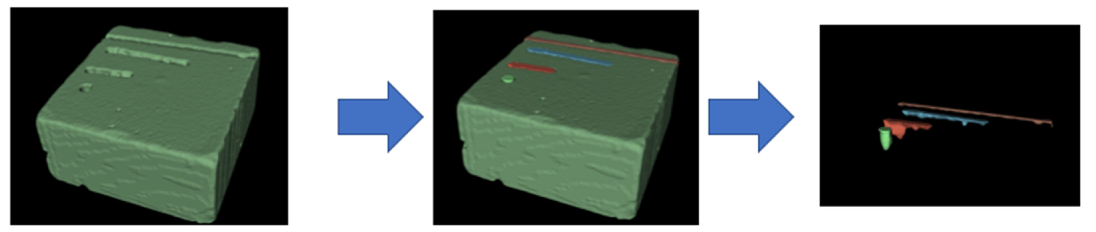
2.2. Stone Phantoms
2.3. Experimental Setup
2.4. Statistical Analysis
3. Results
3.1. Optimal Displacement Velocity Experiments
3.2. Comparative Static and Dynamic Ablated Volumes
4. Discussion
4.1. Optimal Displacement Velocity
4.1.1. Difference between Tm-Fiber and Ho:YAG Lasers
4.1.2. From In Vitro to Clinical Practice
4.2. Comparative Ablative Efficiency
4.3. Strengths and Limitations
5. Conclusions
Author Contributions
Funding
Institutional Review Board Statement
Informed Consent Statement
Data Availability Statement
Acknowledgments
Conflicts of Interest
References
- Türk, C.; Petřík, A.; Sarica, K.; Seitz, C.; Skolarikos, A.; Straub, M. EAU Guidelines on Interventional Treatment for Urolithiasis. Eur. Urol. 2016, 69, 475–482. [Google Scholar] [CrossRef]
- Johnson, D.E.; Cromeens, D.M.; Price, R.E. Use of the holmium:YAG laser in urology. Lasers Surg. Med. 1992, 12, 353–363. [Google Scholar] [CrossRef] [PubMed]
- Traxer, O.; Keller, E.X. Thulium fiber laser: The new player for kidney stone treatment? A comparison with Holmium:YAG laser. World J. Urol. 2020, 38, 1883–1894. [Google Scholar] [CrossRef] [Green Version]
- Panthier, F.; Doizi, S.; Lapouge, P.; Chaussain, C.; Kogane, N.; Berthe, L. Comparison of the ablation rates, fissures and fragments produced with 150 µm and 272 µm laser fibers with superpulsed thulium fiber laser: An in vitro study. World J. Urol. 2021, 39, 1683–1691. [Google Scholar] [CrossRef] [PubMed]
- Panthier, F.; Doizi, S.; Gorny, C.; Berthe, L.; Traxer, O. Impact of Laser Fiber Diameter and Irrigation Fluids on Induced Bubble Stream Dynamics with Thulium Fiber Laser: An In Vitro Study. J. Endourol. 2021, 35, 1883–1890. [Google Scholar] [CrossRef] [PubMed]
- Panthier, F.; Doizi, S.; Corrales, M.; Traxer, O. Pulsed lasers and endocorporeal laser lithotripsy Lasers pulses et lithotritie laser endocorporelle. Progrès Urol. 2020, 31, 451–457. [Google Scholar] [CrossRef]
- Panthier, F.; Traxer, O.; Yonneau, L.; Lebret, T.; Berthe, L.; Illoul, L. Evaluation of a free 3D software for kidney stones’ surgical planning: «kidney stone calculator» a pilot study. World J. Urol. 2021, 39, 3607–3614. [Google Scholar] [CrossRef] [PubMed]
- Talso, M.; Emiliani, E.; Haddad, M.; Berthe, L.; Baghdadi, M.; Montanari, E. Laser Fiber and Flexible Ureterorenoscopy: The Safety Distance Concept. J. Endourol. 2016, 30, 1269–1274. [Google Scholar] [CrossRef] [Green Version]
- Ventimiglia, E.; Pauchard, F.; Gorgen, A.R.H.; Panthier, F.; Doizi, S.; Traxer, O. How do we assess the efficacy of Ho:YAG low-power laser lithotripsy for the treatment of upper tract urinary stones? Introducing the Joules/mm3 and laser activity concepts. World J. Urol. 2020, 39, 891–896. [Google Scholar] [CrossRef]
- Somani, B.K.; Ploumidis, A.; Pappas, A.; Doizi, S.; Babawale, O.; Dragos, L. Pictorial review of tips and tricks for ureteroscopy and stone treatment: An essential guide for urologists from PETRA research consortium. Transl. Androl. Urol. 2019, 8 (Suppl. S4), S371. [Google Scholar] [CrossRef]
- Doizi, S.; Keller, E.X.; De Coninck, V.; Traxer, O. Dusting technique for lithotripsy: What does it mean? Nat. Rev. Urol. 2018, 15, 653–654. [Google Scholar] [CrossRef]
- Keller, E.X.; De Coninck, V.; Doizi, S.; Daudon, M.; Traxer, O. What is the exact definition of stone dust? An in vitro evaluation. World J. Urol. 2020, 39, 187–194. [Google Scholar] [CrossRef] [PubMed]
- Esch, E.; Simmons, W.N.; Sankin, G.; Cocks, H.F.; Preminger, G.M.; Zhong, P. A simple method for fabricating artificial kidney stones of different physical properties. Urol. Res. Août. 2010, 38, 315–319. [Google Scholar] [CrossRef] [Green Version]
- Fedorov, A.; Beichel, R.; Kalpathy-Cramer, J.; Finet, J.; Fillion-Robin, J.-C.; Pujol, S. 3D Slicer as an image computing platform for the Quantitative Imaging Network. Magn. Reson. Imaging 2012, 30, 1323–1341. [Google Scholar] [CrossRef] [Green Version]
- Panthier, F.; Ventimiglia, E.; Berthe, L.; Chaussain, C.; Daudon, M.; Doizi, S. How much energy do we need to ablate 1 mm3 of stone during Ho:YAG laser lithotripsy? An in vitro study. World J. Urol. 2020, 38, 2945–2953. [Google Scholar] [CrossRef]
- Aldoukhi, A.H.; Roberts, W.W.; Hall, T.L.; Ghani, K.R. Watch Your Distance: The Role of Laser Fiber Working Distance on Fragmentation When Altering Pulse Width or Modulation. J. Endourol. Févr. 2019, 33, 120–126. [Google Scholar] [CrossRef]
- Aldoukhi, A.H.; Roberts, W.W.; Hall, T.L.; Teichman, J.M.H.; Ghani, K.R. Understanding the Popcorn Effect During Holmium Laser Lithotripsy for Dusting. Urology 2018, 122, 52–57. [Google Scholar] [CrossRef] [PubMed]
- Liu, M.; Peng, Y.; Wang, Z.; Li, L.; Ming, S.; Fang, Z. Ablation Efficiency of a Novel Thulium Fiber Laser: An In Vitro Study on Laser Setting and Fiber Usage. J. Endourol. Août. 2021, 35, 1211–1216. [Google Scholar] [CrossRef]
- Taratkin, M.; Laukhtina, E.; Singla, N.; Tarasov, A.; Alekseeva, T.; Enikeev, M. How Lasers Ablate Stones: In Vitro Study of Laser Lithotripsy (Ho:YAG and Tm-Fiber Lasers) in Different Environments. J. Endourol. 2021, 35, 931–936. [Google Scholar] [CrossRef] [PubMed]
- Taratkin, M.; Kovalenko, A.; Laukhtina, E.; Paramonova, N.; Spivak, L.; Wachtendorf, L.J. Ex vivo study of Ho:YAG and thulium fiber lasers for soft tissue surgery: Which laser for which case? Lasers Med. Sci. 2021, 1–6. [Google Scholar] [CrossRef] [PubMed]
- Ventimiglia, E.; Doizi, S.; Kovalenko, A.; Andreeva, V.; Traxer, O. Effect of temporal pulse shape on urinary stone phantom retropulsion rate and ablation efficiency using Holmium:YAG and Super Pulse Thulium Fiber lasers. BJU Int. 2020, 126, 159–167. [Google Scholar] [CrossRef] [PubMed]
- Roger, C.; Abid, N.; Dubourg, L.; Auvergnon, C.; Lemoine, S.; Machon, C. Composition of urinary calculi: Lessons from a French epidemiologic retrospective study. Progrès Urol. 2020, 30, 339–345. [Google Scholar] [CrossRef] [PubMed]
- Corrales, M.; Traxer, O. Initial clinical experience with the new thulium fiber laser: First 50 cases. World J. Urol. 2021, 39, 3945–3950. [Google Scholar] [CrossRef] [PubMed]
- Panthier, F.; Doizi, S.; Illoul, L.; Berthe, L.; Traxer, O. Developing Free Three-Dimensional Software for Surgical Planning for Kidney Stones: Volume is Better than Diameter. Eur. Urol. Focus. Available online: http://www.sciencedirect.com/science/article/pii/S2405456920301619 (accessed on 24 June 2020). [CrossRef]
- Keller, E.X.; de Coninck, V.; Audouin, M.; Doizi, S.; Bazin, D.; Daudon, M. Fragments and dust after Holmium laser lithotripsy with or without «Moses technology»: How are they different? J. Biophotonics. 2019, 12, e201800227. [Google Scholar] [CrossRef]
- Aldoukhi, A.H.; Ghani, K.R. Reply to: Letter-to-the-editor: Understanding the Popcorn Effect During Holmium Laser Lithotripsy for Dusting. Urology 2019, 127, 135–136. [Google Scholar] [CrossRef]
- Ventimiglia, E.; Traxer, O. Is Very High Power/Frequency Really Necessary During Laser Lithotripsy? RE: Understanding the Popcorn Effect during Holmium Laser Lithotripsy for Dusting. Urology 2019, 127, 135. [Google Scholar] [CrossRef]
- Ventimiglia, E.; Pauchard, F.; Quadrini, F.; Sindhubodee, S.; Kamkoum, H.; Jiménez Godínez, A. High- and low-power laser lithotripsy achieve similar results: A systematic review and meta-analysis of available clinical series. J. Endourol. 2021, 35, 1146–1152. [Google Scholar] [CrossRef]


| Laser Source | Lithotripsy Mode | Optimal Displacement Velocity (mm/s, Ablated Volume (mm3)) |
|---|---|---|
| Tm-Fiber | Fine Dusting 1 (0.05 J-300 Hz) | 5 mm/s (1.97 mm3) |
| Fine Dusting 2 (0.15 J-100 Hz) | 10 mm/s (4.66 mm3) | |
| Dusting (0.5 J-30 Hz) | 5 mm/s (5.34 mm3) | |
| Fragmentation (1 J-15 Hz) | 5 mm/s (6.8 mm3) | |
| Ho:YAG | Dusting (0.5 J-30 Hz) | 5 mm/s (1.24 mm3) |
| Fragmentation (1 J-15 Hz) | 10 mm/s (2.02 mm3) |
| Lithotripsy Mode | Laser Fiber Displacement Velocity (mm/s) | Ablation Volume (mm3) | ||
|---|---|---|---|---|
| Tm-Fiber | Ho:YAG | p-Value | ||
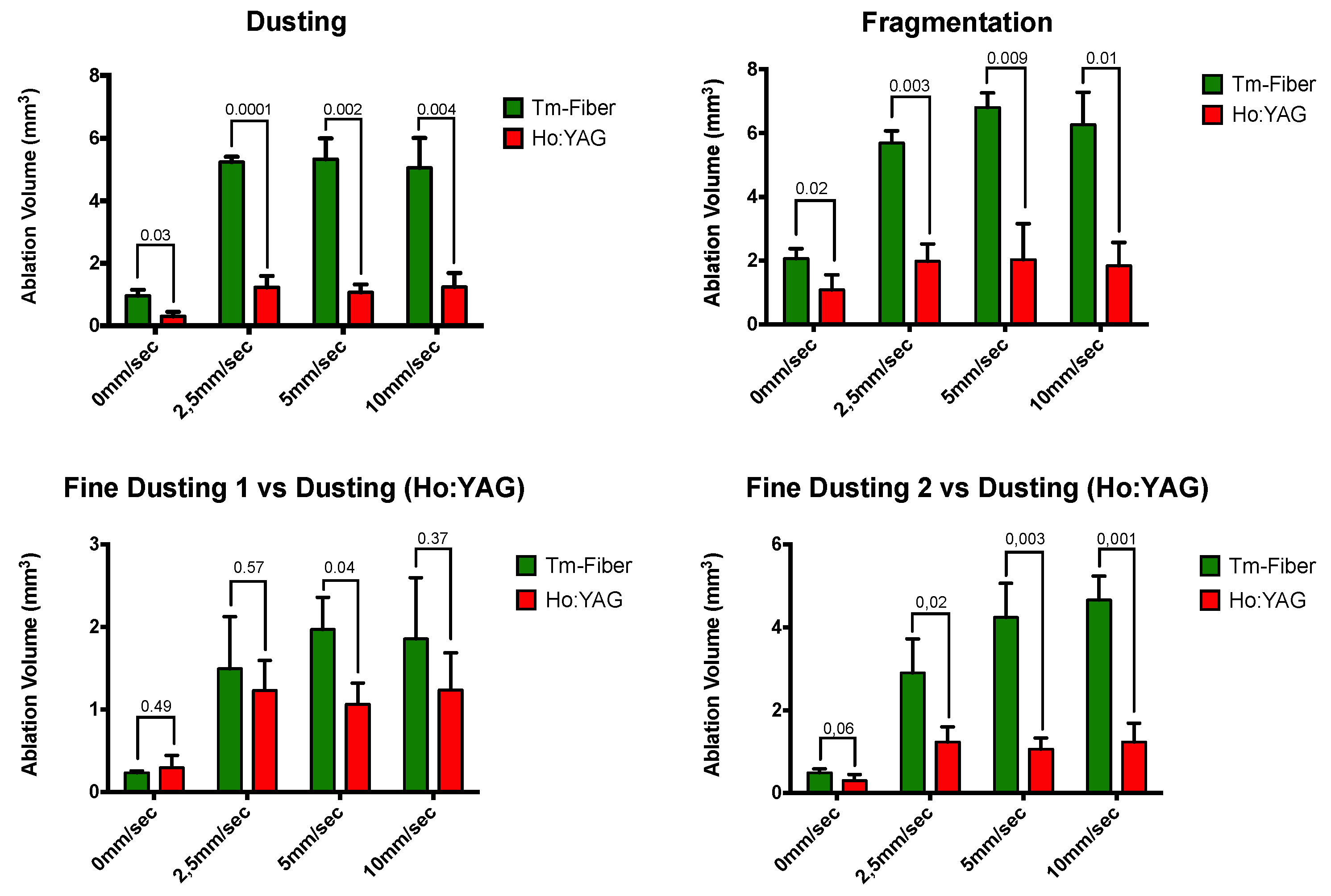
| Dusting | 0 | 0.96 ± 0.19 | 0.3 ± 0.15 | 0.003 |
| 2.5 | 5.24 ± 0.17 | 1.23 ± 0.36 | 0.0001 | |
| 5 | 5.34 ± 0.66 | 1.07 ± 0.26 | 0.002 | |
| 10 | 5.05 ± 0.95 | 1.24 ± 0.45 | 0.004 | |
| Fragmentation | 0 | 2.06 ± 0.31 | 1.09 ± 0.46 | 0.02 |
| 2.5 | 5.69 ± 0.38 | 1.98 ± 0.54 | 0.003 | |
| 5 | 6.8 ± 0.45 | 2.02 ± 1.14 | 0.009 | |
| 10 | 6.27 ± 1 | 1.84 ± 0.72 | 0.01 | |
| Lithotripsy Mode | Laser Fiber Displacement Velocity (mm/s) | Ablation Volume (mm3) | ||
| Tm-Fiber (0.05 J-300 Hz) | Ho:YAG (0.5 J-20 Hz) | p-Value | ||
| Fine Dusting 1 (Tm-Fiber) vs. Dusting (Ho:YAG) | 0 | 0.24 ± 0.02 | 0.3 ± 0.15 | 0.49 |
| 2.5 | 1.49 ± 0.63 | 1.23 ± 0.36 | 0.57 | |
| 5 | 1.97 ± 0.39 | 1.07 ± 0.26 | 0.04 | |
| 10 | 1.86 ± 0.74 | 1.24 ± 0.45 | 0.37 | |
| Lithotripsy Mode | Laser Fiber Displacement Velocity (mm/s) | Ablation Volume (mm3) | ||
| Tm-Fiber (0.15 J-100 Hz) | Ho:YAG (0.5 J-20 Hz) | p-Value | ||
| Fine Dusting 2 (Tm-Fiber) vs. Dusting (Ho:YAG) | 0 | 0.49 ± 0.09 | 0.3 ± 0.15 | 0.06 |
| 2.5 | 2.9 ± 0.82 | 1.23 ± 0.36 | 0.02 | |
| 5 | 4.24 ± 0.82 | 1.07 ± 0.26 | 0.003 | |
| 10 | 4.66 ± 0.57 | 1.24 ± 0.45 | 0.001 | |
| Lithotripsy Mode | Ablation Volume (mm3) | ||
|---|---|---|---|
| Tm-Fiber | Ho:YAG | p-Value | |
| Fine Dusting 1 (Tm-Fiber) vs. Dusting (Ho:YAG) | 1.97 ± 0.39 | 1.24 ± 0.45 | 0.18 |
| Fine dusting 2 (Tm-Fiber) vs. Dusting (Ho:YAG) | 4.66 ± 0.57 | 1.24 ± 0.45 | 0.001 |
| Dusting | 5.34 ± 0.66 | 1.24 ± 0.45 | 0.001 |
| Fragmentation | 6.8 ± 0.45 | 2.02 ± 1.14 | 0.009 |
Publisher’s Note: MDPI stays neutral with regard to jurisdictional claims in published maps and institutional affiliations. |
© 2021 by the authors. Licensee MDPI, Basel, Switzerland. This article is an open access article distributed under the terms and conditions of the Creative Commons Attribution (CC BY) license (https://creativecommons.org/licenses/by/4.0/).
Share and Cite
Panthier, F.; Germain, T.; Gorny, C.; Berthe, L.; Doizi, S.; Traxer, O. Laser Fiber Displacement Velocity during Tm-Fiber and Ho:YAG Laser Lithotripsy: Introducing the Concept of Optimal Displacement Velocity. J. Clin. Med. 2022, 11, 181. https://doi.org/10.3390/jcm11010181
Panthier F, Germain T, Gorny C, Berthe L, Doizi S, Traxer O. Laser Fiber Displacement Velocity during Tm-Fiber and Ho:YAG Laser Lithotripsy: Introducing the Concept of Optimal Displacement Velocity. Journal of Clinical Medicine. 2022; 11(1):181. https://doi.org/10.3390/jcm11010181
Chicago/Turabian StylePanthier, Frederic, Thibault Germain, Cyril Gorny, Laurent Berthe, Steeve Doizi, and Olivier Traxer. 2022. "Laser Fiber Displacement Velocity during Tm-Fiber and Ho:YAG Laser Lithotripsy: Introducing the Concept of Optimal Displacement Velocity" Journal of Clinical Medicine 11, no. 1: 181. https://doi.org/10.3390/jcm11010181
APA StylePanthier, F., Germain, T., Gorny, C., Berthe, L., Doizi, S., & Traxer, O. (2022). Laser Fiber Displacement Velocity during Tm-Fiber and Ho:YAG Laser Lithotripsy: Introducing the Concept of Optimal Displacement Velocity. Journal of Clinical Medicine, 11(1), 181. https://doi.org/10.3390/jcm11010181