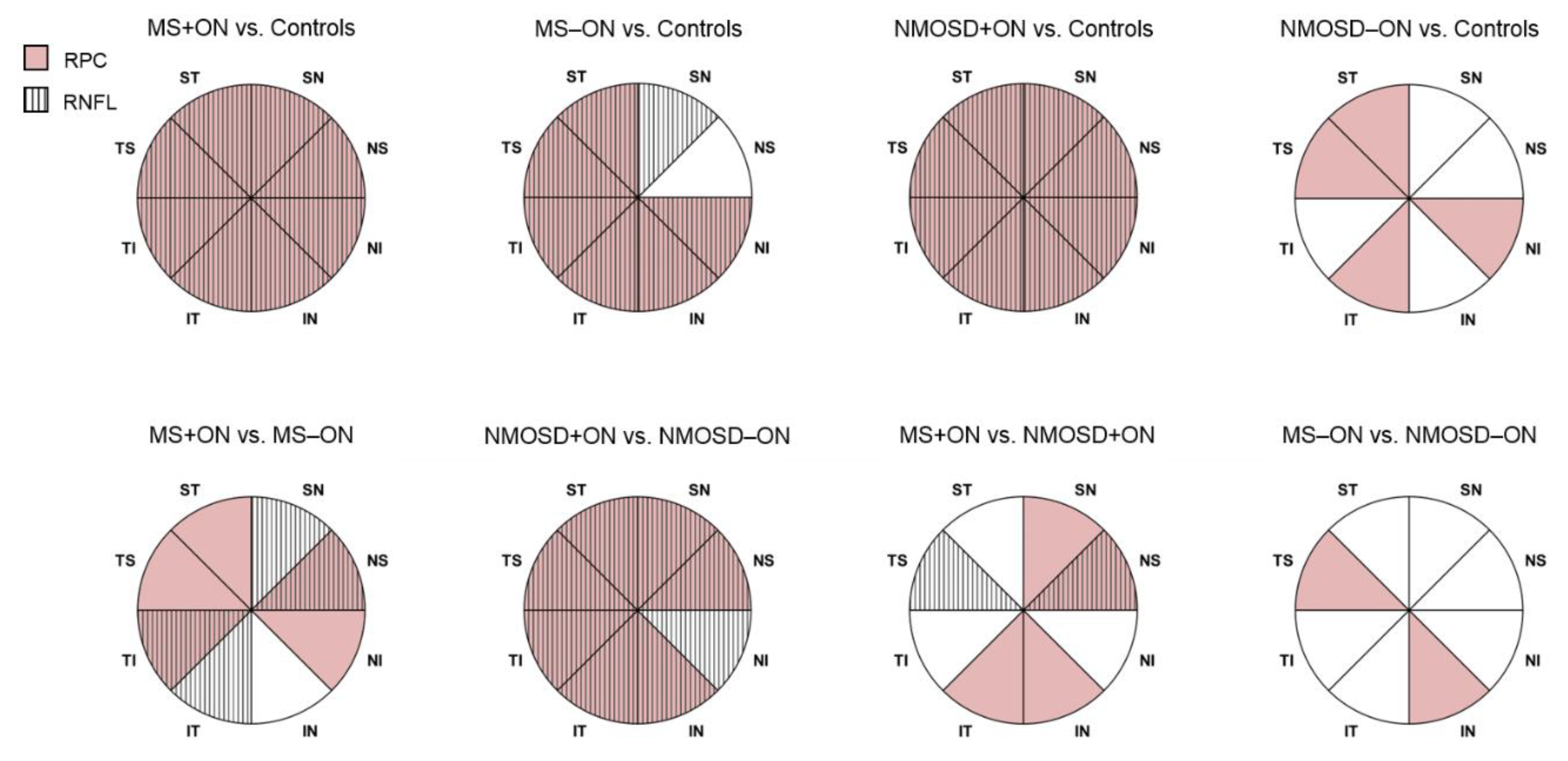
Optical Coherence Tomography Angiography of Peripapillary Vessel Density in Multiple Sclerosis and Neuromyelitis Optica Spectrum Disorder: A Comparative Study
Abstract
1. Introduction
2. Materials and Methods
2.1. Ethical Approval
2.2. Study Participants
2.3. SD-OCT
2.4. OCT Angiography
2.5. Statistical Analysis
3. Results
3.1. Study Population
3.2. SD-OCT
3.3. OCTA
3.4. Association of OCTA and SD-OCT
4. Discussion
Author Contributions
Funding
Institutional Review Board Statement
Informed Consent Statement
Data Availability Statement
Acknowledgments
Conflicts of Interest
References
- Filippi, M.; Bar-Or, A.; Piehl, F.; Preziosa, P.; Solari, A.; Vukusic, S.; Rocca, M.A. Multiple sclerosis. Nat. Rev. Dis. Primers 2018, 4, 43. [Google Scholar] [CrossRef]
- Jasiak-Zatonska, M.; Kalinowska-Lyszczarz, A.; Michalak, S.; Kozubski, W. The immunology of neuromyelitis optica-current knowledge, clinical implications, controversies and future perspectives. Int. J. Mol. Sci. 2016, 17, 273. [Google Scholar] [CrossRef]
- Jarius, S.; Wildemann, B.; Paul, F. Neuromyelitis optica: Clinical features, immunopathogenesis and treatment. Clin. Exp. Immunol. 2014, 176, 149–164. [Google Scholar] [CrossRef]
- Kawachi, I. Clinical characteristics of autoimmune optic neuritis. Clin. Exp. Neuroimmunol. 2017, 8, 8–16. [Google Scholar] [CrossRef]
- de Seze, J. Inflammatory optic neuritis: From multiple sclerosis to neuromyelitis optica. Neuroophthalmology 2013, 37, 141–145. [Google Scholar] [CrossRef]
- Srikajon, J.; Siritho, S.; Ngamsombat, C.; Prayoonwiwat, N.; Chirapapaisan, N.; Siriraj Neuroimmunology Research Group. Differences in clinical features between optic neuritis in neuromyelitis optica spectrum disorders and in multiple sclerosis. Mult. Scler. J. Exp. Transl. Clin. 2018, 4, 2055217318791196. [Google Scholar] [CrossRef]
- Hokari, M.; Yokoseki, A.; Arakawa, M.; Saji, E.; Yanagawa, K.; Yanagimura, F.; Toyoshima, Y.; Okamoto, K.; Ueki, S.; Hatase, T.; et al. Clinicopathological features in anterior visual pathway in neuromyelitis optica. Ann. Neurol. 2016, 79, 605–624. [Google Scholar] [CrossRef]
- Naismith, R.T.; Tutlam, N.T.; Xu, J.; Klawiter, E.C.; Shepherd, J.; Trinkaus, K.; Song, S.K.; Cross, A.H. Optical coherence tomography differs in neuromyelitis optica compared with multiple sclerosis. Neurology 2009, 72, 1077–1082. [Google Scholar] [CrossRef]
- Campbell, J.P.; Zhang, M.; Hwang, T.S.; Bailey, S.T.; Wilson, D.J.; Jia, Y.; Huang, D. Detailed vascular anatomy of the human retina by projection-resolved optical coherence tomography angiography. Sci. Rep. 2017, 7, 42201. [Google Scholar] [CrossRef]
- Cennamo, G.; Carotenuto, A.; Montorio, D.; Petracca, M.; Moccia, M.; Melenzane, A.; Tranfa, F.; Lamberti, A.; Spiezia, A.L.; Servillo, G.; et al. Peripapillary Vessel Density as Early Biomarker in Multiple Sclerosis. Front. Neurol. 2020, 11, 542. [Google Scholar] [CrossRef]
- Spain, R.I.; Liu, L.; Zhang, X.; Jia, Y.; Tan, O.; Bourdette, D.; Huang, D. Optical coherence tomography angiography enhances the detection of optic nerve damage in multiple sclerosis. Br. J. Ophthalmol. 2018, 102, 520–524. [Google Scholar] [CrossRef]
- Wang, X.; Jia, Y.; Spain, R.; Potsaid, B.; Liu, J.J.; Baumann, B.; Hornegger, J.; Fujimoto, J.G.; Wu, Q.; Huang, D. Optical coherence tomography angiography of optic nerve head and parafovea in multiple sclerosis. Br. J. Ophthalmol. 2014, 98, 1368–1373. [Google Scholar] [CrossRef] [PubMed]
- Huang, Y.; Zhou, L.; ZhangBao, J.; Cai, T.; Wang, B.; Li, X.; Wang, L.; Lu, C.; Zhao, C.; Lu, J.; et al. Peripapillary and parafoveal vascular network assessment by optical coherence tomography angiography in aquaporin-4 antibody-positive neuromyelitis optica spectrum disorders. Br. J. Ophthalmol. 2019, 103, 789–796. [Google Scholar] [CrossRef] [PubMed]
- Chen, Y.; Shi, C.; Zhou, L.; Huang, S.; Shen, M.; He, Z. The detection of retina microvascular density in subclinical aquaporin-4 antibody seropositive neuromyelitis optica spectrum disorders. Front. Neurol. 2020, 11, 35. [Google Scholar] [CrossRef]
- Thompson, A.J.; Banwell, B.L.; Barkhof, F.; Carroll, W.M.; Coetzee, T.; Comi, G.; Correale, J.; Fazekas, F.; Filippi, M.; Freedman, M.S.; et al. Diagnosis of multiple sclerosis: 2017 revisions of the McDonald criteria. Lancet Neurol. 2018, 17, 162–173. [Google Scholar] [CrossRef]
- Wingerchuk, D.M.; Banwell, B.; Bennett, J.L.; Cabre, P.; Carroll, W.; Chitnis, T.; de Seze, J.; Fujihara, K.; Greenberg, B.; Jacob, A.; et al. International consensus diagnostic criteria for neuromyelitis optica spectrum disorders. Neurology 2015, 85, 177–189. [Google Scholar] [CrossRef]
- Huang, D.; Jia, Y.; Gao, S.S.; Lumbroso, B.; Rispoli, M. Optical coherence tomography angiography using the Optovue device. Dev. Ophthalmol. 2016, 56, 6–12. [Google Scholar] [CrossRef]
- Jia, Y.; Tan, O.; Tokayer, J.; Potsaid, B.; Wang, Y.; Liu, J.J.; Kraus, M.F.; Subhash, H.; Fujimoto, J.G.; Hornegger, J.; et al. Split-spectrum amplitude-decorrelation angiography with optical coherence tomography. Opt. Express. 2012, 20, 4710–4725. [Google Scholar] [CrossRef]
- Ferrara, D.; Waheed, N.K.; Duker, J.S. Investigating the choriocapillaris and choroidal vasculature with new optical coherence tomography technologies. Prog. Retin. Eye Res. 2016, 52, 130–155. [Google Scholar] [CrossRef]
- Green, A.J.; Cree, B.A. Distinctive retinal nerve fibre layer and vascular changes in neuromyelitis optica following optic neuritis. J. Neurol. Neurosurg. Psychiatry 2009, 80, 1002–1005. [Google Scholar] [CrossRef]
- Verkman, A.S.; Ruiz-Ederra, J.; Levin, M.H. Functions of aquaporins in the eye. Prog. Retin. Eye Res. 2008, 27, 420–433. [Google Scholar] [CrossRef] [PubMed]
- Mandler, R.N.; Davis, L.E.; Jeffery, D.R.; Kornfeld, M. Devic’s neuromyelitis optica: A clinicopathological study of 8 patients. Ann. Neurol. 1993, 34, 162–168. [Google Scholar] [CrossRef] [PubMed]
- Lucchinetti, C.F.; Guo, Y.; Popescu, B.F.; Fujihara, K.; Itoyama, Y.; Misu, T. The pathology of an autoimmune astrocytopathy: Lessons learned from neuromyelitis optica. Brain Pathol. 2014, 24, 83–97. [Google Scholar] [CrossRef] [PubMed]
- Outteryck, O.; Majed, B.; Defoort-Dhellemmes, S.; Vermersch, P.; Zéphir, H. A comparative optical coherence tomography study in neuromyelitis optica spectrum disorder and multiple sclerosis. Mult. Scler. 2015, 21, 1781–1793. [Google Scholar] [CrossRef]
- Nakamura, M.; Nakazawa, T.; Doi, H.; Hariya, T.; Omodaka, K.; Misu, T.; Takahashi, T.; Fujihara, K.; Nishida, K. Early high-dose intravenous methylprednisolone is effective in preserving retinal nerve fiber layer thickness in patients with neuromyelitis optica. Graefes Arch. Clin. Exp. Ophthalmol. 2010, 248, 1777–1785. [Google Scholar] [CrossRef]
- FitzGibbon, T.; Taylor, S.F. Mean retinal ganglion cell axon diameter varies with location in the human retina. Jpn. J. Ophthalmol. 2012, 56, 631–637. [Google Scholar] [CrossRef]
- Evangelou, N.; Konz, D.; Esiri, M.M.; Smith, S.; Palace, J.; Matthews, P.M. Size-selective neuronal changes in the anterior optic pathways suggest a differential susceptibility to injury in multiple sclerosis. Brain 2001, 124, 1813–1820. [Google Scholar] [CrossRef]
- Petzold, A.; de Boer, J.F.; Schippling, S.; Vermersch, P.; Kardon, R.; Green, A.; Calabresi, P.A.; Polman, C. Optical coherence tomography in multiple sclerosis: A systematic review and meta-analysis. Lancet Neurol. 2010, 9, 921–932. [Google Scholar] [CrossRef]


| MS | NMOSD | Controls | |
|---|---|---|---|
| Number of subjects | 40 | 13 | 20 |
| Number of eyes enrolled ON eyes Non-ON eyes | 75 30 45 | 20 9 11 | 40 - 40 |
| Age (years), mean ± SD | 35.15 ± 7.47 | 42.08 ± 10.23 | 37.90 ± 11.47 |
| Sex (female/male) | 32/8 | 11/2 | 17/3 |
| Disease duration (years), median (min–max) | 8 (3–32) | 9 (1–33) | - |
| BCVA of enrolled eyes (logMAR), median (min–max) | 0.00 (0.00–0.20) | 0.00 (0.00–2.30) | 0.00 (0.00–0.00) |
| MS+ON Mean ± SD | MS−ON Mean ± SD | NMOSD+ON Mean ± SD | NMOSD−ON Mean ± SD | Controls Mean ± SD | |
|---|---|---|---|---|---|
| RNFL (μm) | |||||
| average | 85.57 ± 11.39 | 91.00 ± 10.48 | 73.89 ± 15.93 | 99.73 ± 12.26 | 101.60 ± 7.46 |
| ST | 119.17 ± 15.78 | 125.98 ± 16.85 | 101.78 ± 24.30 | 134.64 ± 19.09 | 137.90 ± 13.81 |
| SN | 93.43 ± 15.75 | 100.18 ± 15.95 | 79.00 ± 23.90 | 111.00 ± 18.73 | 108.93 ± 13.36 |
| NS | 72.33 ± 10.49 | 79.89 ± 14.43 | 59.67 ± 11.98 | 81.55 ± 12.64 | 84.83 ± 14.26 |
| NI | 66.27 ± 12.35 | 70.16 ± 10.99 | 59.00 ± 13.93 | 77.45 ± 15.01 | 75.50 ± 9.19 |
| IN | 100.97 ± 17.83 | 104.29 ± 13.14 | 89.11 ± 28.53 | 116.64 ± 20.22 | 116.60 ± 14.14 |
| IT | 115.77 ± 21.78 | 126.87 ± 17.91 | 102.33 ± 24.35 | 133.18 ± 20.03 | 138.38 ± 12.73 |
| TI | 51.23 ± 12.34 | 58.98 ± 10.58 | 46.33 ± 10.31 | 63.00 ± 8.93 | 68.05 ± 8.28 |
| TS | 65.50 ± 18.23 | 70.96 ± 13.19 | 55.33 ± 12.43 | 79.82 ± 12.49 | 83.18 ± 8.84 |
| RPC (%) | |||||
| average | 46.76 ± 4.96 | 49.87 ± 3.10 | 40.80 ± 8.99 | 50.60 ± 1.71 | 52.97 ± 2.51 |
| ST | 50.85 ± 7.44 | 54.56 ± 4.53 | 43.53 ± 11.62 | 53.68 ± 2.73 | 57.40 ± 2.59 |
| SN | 46.52 ± 6.60 | 48.78 ± 5.02 | 36.81 ± 11.10 | 49.19 ± 3.02 | 49.98 ± 3.98 |
| NS | 45.64 ± 5.59 | 48.30 ± 4.05 | 38.79 ± 8.76 | 47.69 ± 2.58 | 49.23 ± 3.78 |
| NI | 43.41 ± 6.27 | 46.50 ± 4.17 | 40.61 ± 7.90 | 45.63 ± 4.08 | 49.09 ± 3.45 |
| IN | 48.24 ± 6.15 | 48.98 ± 4.22 | 39.09 ± 112.00 | 51.14 ± 2.90 | 52.80 ± 4.40 |
| IT | 54.16 ± 6.50 | 56.14 ± 4.40 | 43.46 ± 12.45 | 56.26 ± 3.33 | 59.99 ± 3.39 |
| TI | 41.99 ± 6.85 | 46.82 ± 4.85 | 41.56 ± 6.93 | 50.20 ± 4.93 | 52.59 ± 3.97 |
| TS | 46.14 ± 7.35 | 51.44 ± 4.31 | 43.76 ± 7.33 | 53.68 ± 3.67 | 56.10 ± 3.63 |
| MS+ON vs. Controls | MS−ON vs. Controls | NMOSD+ON vs. Controls | NMOSD−ON vs. Controls | |||||
|---|---|---|---|---|---|---|---|---|
| RNFL (μm) | β (SE) | p-Value | β (SE) | p-Value | β (SE) | p-Value | β (SE) | p-Value |
| average | −16.033 (2.665) | <0.001 | −9.400 (2.446) | <0.001 | −27.711 (5.779) | <0.001 | −1.873 (4.555) | 0.681 |
| ST | −18.733 (4.089) | <0.001 | −11.922 (3.980) | 0.003 | −36.122 (9.103) | <0.001 | −3.264 (6.714) | 0.627 |
| SN | −15.492 (4.120) | <0.001 | −8.747 (3.708) | 0.018 | −29.925 (8.244) | <0.001 | 2.075 (6.690) | 0.756 |
| NS | −12.492 (3.669) | <0.001 | −4.936 (3.894) | 0.205 | −25.158 (4.889) | <0.001 | −3.280 (5.447) | 0.547 |
| NI | −9.233 (2.998) | 0.002 | −5.344 (2.637) | 0.043 | −16.500 (5.520) | 0.003 | 1.955 (5.641) | 0.729 |
| IN | −15.633 (4.494) | <0.001 | −12.311 (3.723) | <0.001 | −27.489 (11.277) | 0.015 | 0.036 (8.288) | 0.996 |
| IT | −22.608 (5.000) | <0.001 | −11.508 (4.111) | 0.005 | −36.042 (9.291) | <0.001 | −5.193 (7.943) | 0.513 |
| TI | −16.817 (2.935) | <0.001 | −9.072 (2.482) | <0.001 | −21.717 (3.461) | <0.001 | −5.050 (3.405) | 0.138 |
| TS | −17.675 (4.184) | <0.001 | −12.219 (2.931) | <0.001 | −27.842 (2.632) | <0.001 | −3.357 (4.436) | 0.449 |
| MS+ON vs. MS−ON | NMOSD+ON vs. NMOSD−ON | NMOSD+ON vs. MS+ON | NMOSD−ON vs. MS−ON | |||||
| RNFL (μm) | β (SE) | p-Value | β (SE) | p-Value | β (SE) | p-Value | β (SE) | p-Value |
| average | −6.633 (2.460) | 0.007 | −25.838 (7.069) | <0.001 | −11.678 (5.948) | 0.050 | 7.527 (4.649) | 0.105 |
| ST | −6.811 (3.641) | 0.061 | −32.859 (10.421) | 0.002 | −17.389 (9.207) | 0.059 | 8.659 (6.791) | 0.202 |
| SN | −6.744 (3.279) | 0.040 | −32.000 (9.711) | <0.001 | −14.433 (8.402) | 0.086 | 10.822 (6.646) | 0.103 |
| NS | −7.556 (2.925) | 0.010 | −21.879 (6.099) | <0.001 | −12.667 (4.363) | 0.004 | 1.657 (5.148) | 0.748 |
| NI | −3.889 (2.788) | 0.163 | −18.455 (7.410) | 0.013 | −7.267 (5.661) | 0.199 | 7.299 (5.600) | 0.192 |
| IN | −3.322 (3.748) | 0.375 | −27.525 (13.220) | 0.037 | −11.856 (11.398) | 0.298 | 12.347 (8.069) | 0.126 |
| IT | −11.100 (4.832) | 0.022 | −30.848 (11.551) | 0.008 | −13.433 (9.911) | 0.175 | 6.315 (8.180) | 0.440 |
| TI | −7.744 (2.728) | 0.005 | −16.667 (4.563) | <0.001 | −4.900 (3.915) | 0.211 | 4.022 (3.534) | 0.255 |
| TS | −5.456 (4.108) | 0.184 | −24.485 (4.613) | <0.001 | −10.167 (4.248) | 0.017 | 8.863 (4.678) | 0.058 |
| MS+ON vs. Controls | MS−ON vs. Controls | NMOSD+ON vs. Controls | NMOSD−ON vs. Controls | |||||
|---|---|---|---|---|---|---|---|---|
| RPC (%) | β (SE) | p-Value | β (SE) | p-Value | β (SE) | p-Value | β (SE) | p-Value |
| average | −6.209 (1.042) | <0.001 | −3.104 (0.711) | <0.001 | −12.172 (3.272) | <0.001 | −2.372 (0.739) | 0.001 |
| ST | −6.548 (1.576) | <0.001 | −2.839 (0.859) | <0.001 | −13.862 (4.407) | 0.002 | −3.713 (0.925) | <0.001 |
| SN | −3.466 (1.458) | 0.017 | −1.207 (1.116) | 0.279 | −13.171 (3.701) | <0.001 | −0.792 (1.235) | 0.521 |
| NS | −3.587 (1.223) | 0.003 | −0.925 (1.014) | 0.361 | −10.439 (2.991) | <0.001 | −1.537 (1.089) | 0.158 |
| NI | −5.668 (1.334) | <0.001 | −2.585 (0.967) | 0.007 | −8.474 (3.227) | 0.009 | −3.458 (1.587) | 0.029 |
| IN | −4.557 (1.392) | 0.001 | −3.820 (1.050) | <0.001 | −13.709 (3.694) | <0.001 | −1.661 (1.132) | 0.142 |
| IT | −5.824 (1.403) | <0.001 | −3.843 (1.001) | <0.001 | −16.532 (4.808) | <0.001 | −3.724 (1.251) | 0.003 |
| TI | −10.603 (1.600) | <0.001 | −5.770 (1.031) | <0.001 | −11.034 (2.691) | <0.001 | −2.390 (1.859) | 0.198 |
| TS | −9.955 (1.700) | <0.001 | −4.653 (0.939) | <0.001 | −12.339 (2.626) | <0.001 | −2.413 (1.158) | 0.037 |
| MS+ON vs. MS−ON | NMOSD+ON vs. NMOSD−ON | NMOSD+ON vs. MS+ON | NMOSD−ON vs. MS−ON | |||||
| RPC (%) | β(SE) | p-Value | β(SE) | p-Value | β(SE) | p-Value | β(SE) | p-Value |
| average | −3.106 (0.906) | <0.001 | −9.800 (3.266) | 0.003 | −5.963 (3.364) | 0.076 | 0.731 (0.756) | 0.333 |
| ST | −3.709 (1.551) | 0.017 | −10.148 (4.356) | 0.020 | −7.313 (4.628) | 0.114 | −0.874 (1.055) | 0.408 |
| SN | −2.259 (1.367) | 0.098 | −12.380 (3.761) | <0.001 | −9.706 (3.854) | 0.012 | 0.415 (1.342) | 0.757 |
| NS | −2.662 (1.092) | 0.015 | −8.902 (2.960) | 0.003 | −6.851 (3.055) | 0.025 | −0.611 (1.051) | 0.561 |
| NI | −3.083 (1.173) | 0.009 | −5.016 (3.440) | 0.145 | −2.806 (3.362) | 0.404 | −0.873 (1.601) | 0.586 |
| IN | −0.738 (1.128) | 0.513 | −12.047 (3.670) | 0.001 | −9.151 (3.780) | 0.015 | 2.159 (1.044) | 0.039 |
| IT | −1.981 (1.402) | 0.158 | −12.808 (4.659) | 0.006 | −10.708 (4.925) | 0.030 | 0.119 (1.317) | 0.928 |
| TI | −4.833 (1.444) | <0.001 | −8.644 (3.206) | 0.007 | −0.431 (2.976) | 0.885 | 3.380 (1.890) | 0.074 |
| TS | −5.302 (1.508) | <0.001 | −9.926 (2.757) | <0.001 | −2.384 (2.967) | 0.422 | 2.240 (1.116) | 0.045 |
Publisher’s Note: MDPI stays neutral with regard to jurisdictional claims in published maps and institutional affiliations. |
© 2021 by the authors. Licensee MDPI, Basel, Switzerland. This article is an open access article distributed under the terms and conditions of the Creative Commons Attribution (CC BY) license (http://creativecommons.org/licenses/by/4.0/).
Share and Cite
Rogaczewska, M.; Michalak, S.; Stopa, M. Optical Coherence Tomography Angiography of Peripapillary Vessel Density in Multiple Sclerosis and Neuromyelitis Optica Spectrum Disorder: A Comparative Study. J. Clin. Med. 2021, 10, 609. https://doi.org/10.3390/jcm10040609
Rogaczewska M, Michalak S, Stopa M. Optical Coherence Tomography Angiography of Peripapillary Vessel Density in Multiple Sclerosis and Neuromyelitis Optica Spectrum Disorder: A Comparative Study. Journal of Clinical Medicine. 2021; 10(4):609. https://doi.org/10.3390/jcm10040609
Chicago/Turabian StyleRogaczewska, Małgorzata, Sławomir Michalak, and Marcin Stopa. 2021. "Optical Coherence Tomography Angiography of Peripapillary Vessel Density in Multiple Sclerosis and Neuromyelitis Optica Spectrum Disorder: A Comparative Study" Journal of Clinical Medicine 10, no. 4: 609. https://doi.org/10.3390/jcm10040609
APA StyleRogaczewska, M., Michalak, S., & Stopa, M. (2021). Optical Coherence Tomography Angiography of Peripapillary Vessel Density in Multiple Sclerosis and Neuromyelitis Optica Spectrum Disorder: A Comparative Study. Journal of Clinical Medicine, 10(4), 609. https://doi.org/10.3390/jcm10040609


