Background Factors Affecting Visual Acuity at Initial Visit in Eyes with Central Retinal Vein Occlusion: Multicenter Study in Japan
Abstract
1. Introduction
2. Materials and Methods
2.1. Study Design and Approval
2.2. Subjects
2.3. Best-Corrected Visual Acuity and Central Macular Thickness
2.4. Classification of Ischemic Status
2.5. Statistical Analyses
3. Results
3.1. Demographic Information
3.2. Background Factors Affecting Visual Acuity at Initial Visit
3.3. Relationship between Age and BCVA at Initial Visit to Hospital
3.4. Comparisons of Visual Acuity and Central Macular Thickness for Right and Left Eyes
4. Discussion
Author Contributions
Funding
Institutional Review Board Statement
Informed Consent Statement
Data Availability Statement
Acknowledgments
Conflicts of Interest
References
- Hayreh, S.S. So-called “central retinal vein occlusion”: I. Pathogenesis, terminology, clinical features. Ophthalmologica 1976, 172, 1–13. [Google Scholar] [CrossRef]
- Green, W.R.; Chan, C.C.; Hutchins, G.M.; Terry, J.M. Central retinal vein occlusion: A prospective histopathologic study of 29 eyes in 28 cases. Trans. Am. Ophthalmol. Soc. 1981, 79, 371–422. [Google Scholar] [CrossRef] [PubMed]
- Prisco, D.; Marcucci, R. Retinal vein thrombosis: Risk factors, pathogenesis and therapeutic approach. Pathophysiol. Haemost. Thromb. 2002, 32, 308–311. [Google Scholar] [CrossRef] [PubMed]
- Rehak, M.; Wiedemann, P. Retinal vein thrombosis: Pathogenesis and management. J. Thromb. Haemost. 2010, 8, 1886–1894. [Google Scholar] [CrossRef]
- Rogers, S.; McIntosh, R.L.; Cheung, N.; Lim, L.; Wang, J.J.; Mitchell, P.; Kowalski, J.W.; Nguyen, H.; Wong, T.Y.; International Eye Disease Consortium. The prevalence of retinal vein occlusion: Pooled data from population studies from the United States, Europe, Asia, and Australia. Ophthalmology 2010, 117, 313–319. [Google Scholar] [CrossRef] [PubMed]
- Ponto, K.A.; Elbaz, H.; Peto, T.; Laubert-Reh, D.; Binder, H.; Wild, P.S.; Lackner, K.; Pfeiffer, N.; Mirshahi, A. Prevalence and risk factors of retinal vein occlusion: The Gutenberg Health Study. J. Thromb. Haemost. 2015, 13, 1254–1263. [Google Scholar] [CrossRef]
- Song, P.; Xu, Y.; Zha, M.; Zhang, Y.; Rudan, I. Global epidemiology of retinal vein occlusion: A systematic review and meta-analysis of prevalence, incidence, and risk factors. J. Glob. Health 2019, 9, 010427. [Google Scholar] [CrossRef]
- The Central Vein Occlusion Study Group N Report. A randomized clinical trial of early panretinal photocoagulation for ischemic central vein occlusion. Ophthalmology 1995, 102, 1434–1444. [Google Scholar] [CrossRef]
- Ip, M.S.; Scott, I.U.; VanVeldhuisen, P.C.; Oden, N.L.; Blodi, B.A.; Fisher, M.; Singerman, L.J.; Tolentino, M.; Chan, C.K.; Gonzalez, V.H.; et al. A randomized trial comparing the efficacy and safety of intravitreal triamcinolone with observation to treat vision loss associated with macular edema secondary to central retinal vein occlusion: The Standard Care vs Corticosteroid for Retinal Vein Occlusion (SCORE) study report 5. Arch. Ophthalmol. 2009, 127, 1101–1114. [Google Scholar] [PubMed]
- Haller, J.A.; Bandello, F.; Belfort, R., Jr.; Blumenkranz, M.S.; Gillies, M.; Heier, J.; Loewenstein, A.; Yoon, Y.H.; Jiao, J.; Li, X.Y.; et al. Dexamethasone intravitreal implant in patients with macular edema related to branch or central retinal vein occlusion twelve-month study results. Ophthalmology 2011, 118, 2453–2460. [Google Scholar] [CrossRef]
- Campochiaro, P.A.; Brown, D.M.; Awh, C.C.; Lee, S.Y.; Gray, S.; Saroj, N.; Murahashi, W.Y.; Rubio, R.G. Sustained benefits from ranibizumab for macular edema following central retinal vein occlusion: Twelve-month outcomes of a phase III study. Ophthalmology 2011, 118, 2041–2049. [Google Scholar] [CrossRef]
- Heier, J.S.; Clark, W.; Boyer, D.S.; Brown, D.M.; Vitti, R.; Berliner, A.J.; Kazmi, H.; Ma, Y.; Stemper, B.; Zeitz, O.; et al. Intravitreal aflibercept injection for macular edema due to central retinal vein occlusion: Two-year results from the COPERNICUS study. Ophthalmology 2014, 121, 1414–1420. [Google Scholar] [CrossRef]
- Callizo, J.; Ziemssen, F.; Bertelmann, T.; Feltgen, N.; Vögeler, J.; Koch, M.; Eter, N.; Liakopoulos, S.; Schmitz-Valckenberg, S.; Spital, G. Real-world data: Ranibizumab treatment for retinal vein occlusion in the OCEAN Study. Clin. Ophthalmol. 2019, 13, 2167–2179. [Google Scholar] [CrossRef]
- Costa, J.V.; Moura-Coelho, N.; Abreu, A.C.; Neves, P.; Ornelas, M.; Furtado, M.J. Macular edema secondary to retinal vein occlusion in a real-life setting: A multicenter, nationwide, 3-year follow-up study. Graefes. Arch. Clin. Exp. Ophthalmol. 2021, 259, 343–350. [Google Scholar] [CrossRef] [PubMed]
- Ciulla, T.; Pollack, J.S.; Williams, D.F. Visual acuity outcomes and anti-VEGF therapy intensity in macular oedema due to retinal vein occlusion: A real-world analysis of 15,613 patient eyes. Br. J. Ophthalmol. 2020. Online ahead of print. [Google Scholar] [CrossRef] [PubMed]
- Glacet-Bernard, A.; Coscas, G.; Chabanel, A.; Zourdani, A.; Lelong, F.; Samama, M.M. Prognostic factors for retinal vein occlusion: Prospective study of 175 cases. Ophthalmology 1996, 103, 551–560. [Google Scholar] [CrossRef]
- The Central Vein Occlusion Study Group. Natural history and clinical management of central retinal vein occlusion. Arch. Ophthalmol. 1997, 115, 486–491. [Google Scholar] [CrossRef] [PubMed]
- Hayreh, S.S.; Podhajsky, P.A.; Zimmerman, M.B. Natural history of visual outcome in central retinal vein occlusion. Ophthalmology 2011, 118, 119–133. [Google Scholar] [CrossRef] [PubMed]
- Nagasato, D.; Muraoka, Y.; Osaka, R.; Iida-Miwa, Y.; Mitamura, Y.; Tabuchi, H.; Kadomoto, S.; Murakami, T.; Ooto, S.; Suzuma, K.; et al. Factors associated with extremely poor visual outcomes in patients with central retinal vein occlusion. Sci. Rep. 2020, 10, 19667. [Google Scholar] [CrossRef] [PubMed]
- Sen, P.; Gurudas, S.; Ramu, J.; Patrao, N.; Chandra, S.; Rasheed, R.; Nicholson, L.; Peto, T.; Sivaprasad, S.; Hykin, P. Predictors of visual acuity outcomes after anti-vascular endothelial growth factor treatment for macular edema secondary to central retinal vein occlusion. Ophthalmol. Retina 2021. Online ahead of print. [Google Scholar] [CrossRef] [PubMed]
- Chylack, L.T., Jr.; Leske, M.C.; McCarthy, D.; Khu, P.; Kashiwagi, T.; Sperduto, R. Lens opacities classification system II (LOCS II). Arch. Ophthalmol. 1989, 107, 991–997. [Google Scholar] [CrossRef]
- Schulze-Bonsel, K.; Feltgen, N.; Burau, H.; Hansen, L.; Bach, M. Visual acuities “hand motion” and “counting fingers” can be quantified with the freiburg visual acuity test. Investig. Ophthalmol. Vis. Sci. 2006, 47, 1236–1240. [Google Scholar] [CrossRef]
- Hayreh, S.S.; Klugman, M.R.; Beri, M.; Kimura, A.E.; Podhajsky, P. Differentiation of ischemic from non-ischemic central retinal vein occlusion during the early acute phase. Graefes. Arch. Clin. Exp. Ophthalmol. 1990, 228, 201–217. [Google Scholar] [CrossRef] [PubMed]
- Brown, D.M.; Wykoff, C.C.; Wong, T.P.; Mariani, A.F.; Croft, D.E.; Schuetzle, K.L.; RAVE Study Group. Ranibizumab in preproliferative (ischemic) central retinal vein occlusion: The rubeosis anti-VEGF (RAVE) trial. Retina 2014, 34, 1728–1735. [Google Scholar] [CrossRef] [PubMed]
- Khayat, M.; Williams, M.; Lois, N. Ischemic retinal vein occlusion: Characterizing the more severe spectrum of retinal vein occlusion. Surv. Ophthalmol. 2018, 63, 816–850. [Google Scholar] [CrossRef] [PubMed]
- Eleftheriadou, M.; Nicholson, L.; D’Alonzo, G.; Addison, P.K.F. Real-life evidence for using a treat-and-extend injection regime for patients with central retinal vein occlusion. Ophthalmol. Ther. 2019, 8, 289–296. [Google Scholar] [CrossRef] [PubMed]
- Hogg, H.D.J.; Talks, S.J.; Pearce, M.; Di Simplicio, S. Real-world visual and neovascularization outcomes from anti-VEGF in central retinal vein occlusion. Ophthalmic Epidemiol. 2021, 28, 70–76. [Google Scholar] [CrossRef] [PubMed]
- Li, Y.; Hall, N.E.; Pershing, S.; Hyman, L.; Haller, J.A.; Lee, A.Y.; Lee, C.S.; Chiang, M.; Lum, F.; Miller, J.W.; et al. Age, gender, and laterality of retinal vascular occlusion: A retrospective study from the IRIS® Registry. Ophthalmol. Retina 2021. Online ahead of print. [Google Scholar] [CrossRef] [PubMed]
- Tsugane, S. Alcohol, smoking, and obesity epidemiology in Japan. J. Gastroenterol. Hepatol. 2012, 27 (Suppl. 2), 121–126. [Google Scholar] [CrossRef] [PubMed]
- Otani, K.; Haruyama, R.; Gilmour, S. Prevalence and correlates of hypertension among Japanese adults, 1975 to 2010. Int. J. Environ. Res. Public Health 2018, 15, 1645. [Google Scholar] [CrossRef]
- Miura, K.; Nagai, M.; Ohkubo, T. Epidemiology of hypertension in Japan: Where are we now? Circ. J. 2013, 77, 2226–2231. [Google Scholar] [CrossRef] [PubMed]
- Cassidy, P.; Jones, K. A study of inter-arm blood pressure differences in primary care. J. Hum. Hypertens. 2001, 15, 519–522. [Google Scholar] [CrossRef] [PubMed][Green Version]
- Lane, D.; Beevers, M.; Barnes, N.; Bourne, J.; John, A.; Malins, S.; Beevers, D.G. Inter-arm differences in blood pressure: When are they clinically significant? J. Hypertens. 2002, 20, 1089–1095. [Google Scholar] [CrossRef] [PubMed]
- Southby, R. Some clinical observations on blood pressure and their practical application, with special reference to variation of blood pressure readings in the two arms. Med. J. Aust. 1935, 2, 569–580. [Google Scholar] [CrossRef]


| Parameter | Value | p-Value |
|---|---|---|
| Number of eyes/subjects | 517/517 | |
| Age, mean ± SD (range), years | 69.9 ± 12.2 (22 to 94) | |
| Sex | ||
| Men (%) | 296 (57.3) | |
| Women (%) | 221 (42.7) | 0.001 * |
| Affected eye | ||
| Right (%) | 240 (46.4) | |
| Left (%) | 277 (53.6) | 0.111 |
| Hypertension (%) | 334 (64.6) | |
| Diabetes mellitus (%) | 87 (16.8) | |
| Interval from symptom onset to initial visit to hospital, mean ± SD (range), in weeks | 6.4 ± 6.9 (0 to 51) | |
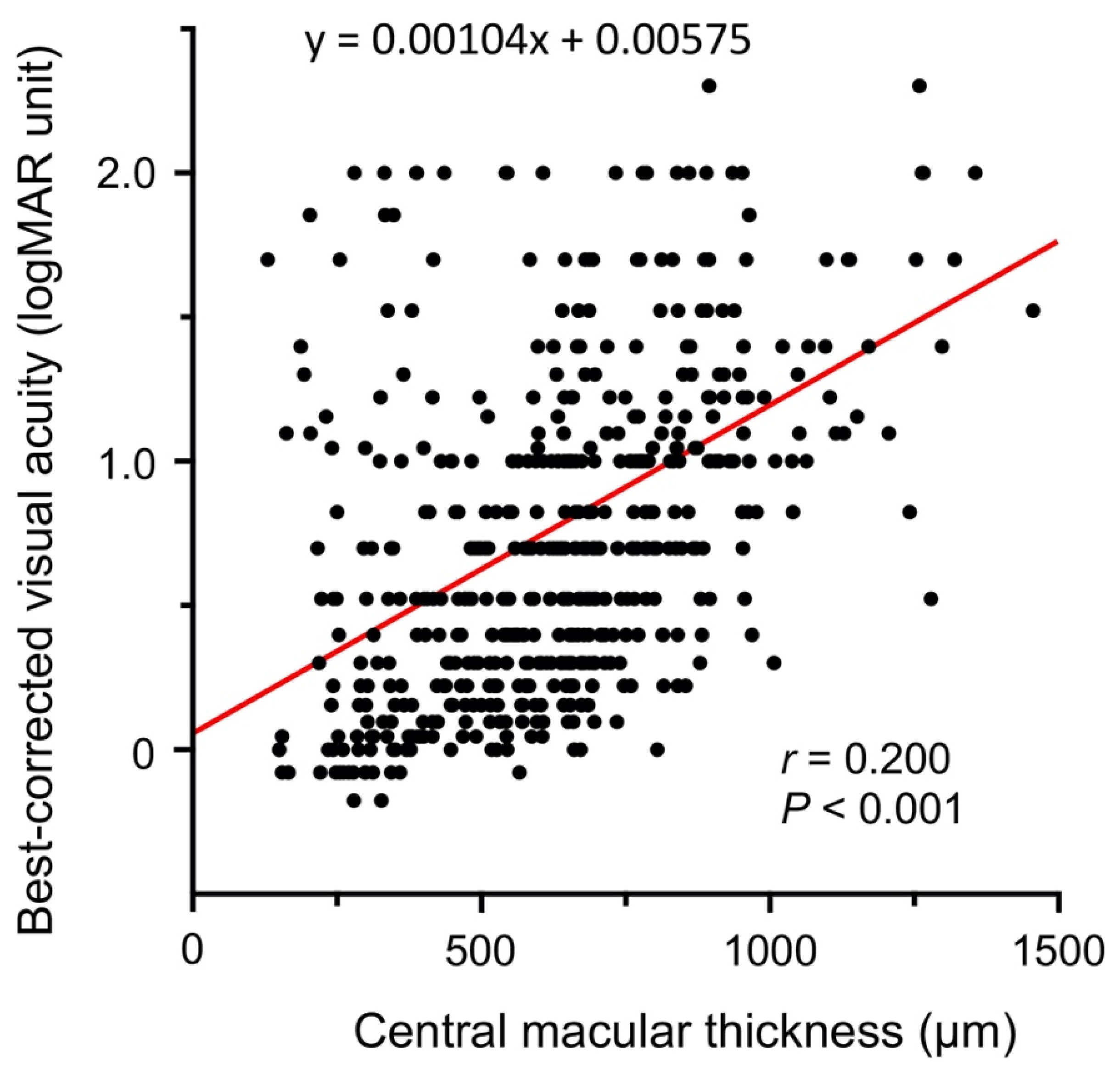
| Best-corrected visual acuity, mean ± SD (range), logMAR | 0.72 ± 0.55 (−0.18 to 2.30) | |
| Central macular thickness, mean ± SD (range), µm | 632 ± 237 (62 to 1456) | |
| Ischemic status at initial visit to hospital | ||
| Ischemic (%) | 122 (23.6) | |
| Nonischemic (%) | 377 (72.9) | |
| Unclassifiable (%) | 18 (3.5) |
| Independent Variables | Univariate Regression Analysis | Multivariate Regression Analysis | ||
|---|---|---|---|---|
| r | p-Value | β | p-Value | |
| Age (years) | 0.194 | <0.001 ** | 0.191 | <0.001 * |
| Sex (men/women) | 0.005 | 0.916 | 0.023 | 0.586 |
| Affected eye (right/left) | −0.103 | 0.019 * | −0.089 | 0.041 * |
| Hypertension | 0.035 | 0.432 | 0.013 | 0.764 |
| Diabetes mellitus | −0.043 | 0.325 | −0.045 | 0.303 |
| Interval from symptom onset to initial visit to hospital (weeks) | 0.008 | 0.848 | −0.008 | 0.849 |
| Independent Variables | β | p-Value |
|---|---|---|
| Age (years) | 0.243 | 0.016 * |
| Sex (men/women) | 0.031 | 0.741 |
| Affected eye (right/left) | −0.195 | 0.038 * |
| Hypertension | 0.002 | 0.984 |
| Diabetes mellitus | −0.122 | 0.213 |
| Interval from symptom onset to initial visit to hospital (weeks) | −0.144 | 0.155 |
Publisher’s Note: MDPI stays neutral with regard to jurisdictional claims in published maps and institutional affiliations. |
© 2021 by the authors. Licensee MDPI, Basel, Switzerland. This article is an open access article distributed under the terms and conditions of the Creative Commons Attribution (CC BY) license (https://creativecommons.org/licenses/by/4.0/).
Share and Cite
Kondo, M.; Noma, H.; Shimura, M.; Sugimoto, M.; Matsui, Y.; Kato, K.; Saishin, Y.; Ohji, M.; Ishikawa, H.; Gomi, F.; et al. Background Factors Affecting Visual Acuity at Initial Visit in Eyes with Central Retinal Vein Occlusion: Multicenter Study in Japan. J. Clin. Med. 2021, 10, 5619. https://doi.org/10.3390/jcm10235619
Kondo M, Noma H, Shimura M, Sugimoto M, Matsui Y, Kato K, Saishin Y, Ohji M, Ishikawa H, Gomi F, et al. Background Factors Affecting Visual Acuity at Initial Visit in Eyes with Central Retinal Vein Occlusion: Multicenter Study in Japan. Journal of Clinical Medicine. 2021; 10(23):5619. https://doi.org/10.3390/jcm10235619
Chicago/Turabian StyleKondo, Mineo, Hidetaka Noma, Masahiko Shimura, Masahiko Sugimoto, Yoshitsugu Matsui, Kumiko Kato, Yoshitsugu Saishin, Masahito Ohji, Hiroto Ishikawa, Fumi Gomi, and et al. 2021. "Background Factors Affecting Visual Acuity at Initial Visit in Eyes with Central Retinal Vein Occlusion: Multicenter Study in Japan" Journal of Clinical Medicine 10, no. 23: 5619. https://doi.org/10.3390/jcm10235619
APA StyleKondo, M., Noma, H., Shimura, M., Sugimoto, M., Matsui, Y., Kato, K., Saishin, Y., Ohji, M., Ishikawa, H., Gomi, F., Iwata, K., Yoshida, S., Kusuhara, S., Hirai, H., Ogata, N., Hirano, T., Murata, T., Tsuboi, K., Kamei, M., ... Group, o. b. o. J. C. R. S. (2021). Background Factors Affecting Visual Acuity at Initial Visit in Eyes with Central Retinal Vein Occlusion: Multicenter Study in Japan. Journal of Clinical Medicine, 10(23), 5619. https://doi.org/10.3390/jcm10235619


