Review

Effect of Ivabradine on Cardiac Ventricular Arrhythmias: Friend or Foe?

by
Marta Oknińska
,
Aleksandra Paterek
,
Zuzanna Zambrowska
,
Urszula Mackiewicz
and
Michał Mączewski
*
Centre of Postgraduate Medical Education, Department of Clinical Physiology, ul. Marymoncka 99/103, 01-813 Warsaw, Poland
*
Author to whom correspondence should be addressed.
J. Clin. Med. 2021, 10(20), 4732; https://doi.org/10.3390/jcm10204732
Submission received: 24 August 2021 / Revised: 24 September 2021 / Accepted: 8 October 2021 / Published: 15 October 2021
(This article belongs to the Section Cardiovascular Medicine)

Abstract

:
Life-threatening ventricular arrhythmias, such as ventricular tachycardia and ventricular fibrillation remain an ongoing clinical problem and their prevention and treatment require optimization. Conventional antiarrhythmic drugs are associated with significant proarrhythmic effects that often outweigh their benefits. Another option, the implantable cardioverter defibrillator, though clearly the primary therapy for patients at high risk of ventricular arrhythmias, is costly, invasive, and requires regular monitoring. Thus there is a clear need for new antiarrhythmic treatment strategies. Ivabradine, a heartrate-reducing agent, an inhibitor of HCN channels, may be one of such options. In this review we discuss emerging data from experimental studies that indicate new mechanism of action of this drug and further areas of investigation and potential use of ivabradine as an antiarrhythmic agent. However, clinical evidence is limited, and the jury is still out on effects of ivabradine on cardiac ventricular arrhythmias in the clinical setting.

1. Ivabradine—Brief Summary

Ivabradine is a presumably selective heartrate-reducing agent, the only inhibitor of HCN channels available for human use, currently approved for the symptomatic treatment of chronic coronary artery disease (CAD) and chronic heart failure (CHF) in patients with heart rate ≥ 70 bpm in combination with standard therapy [1,2,3].

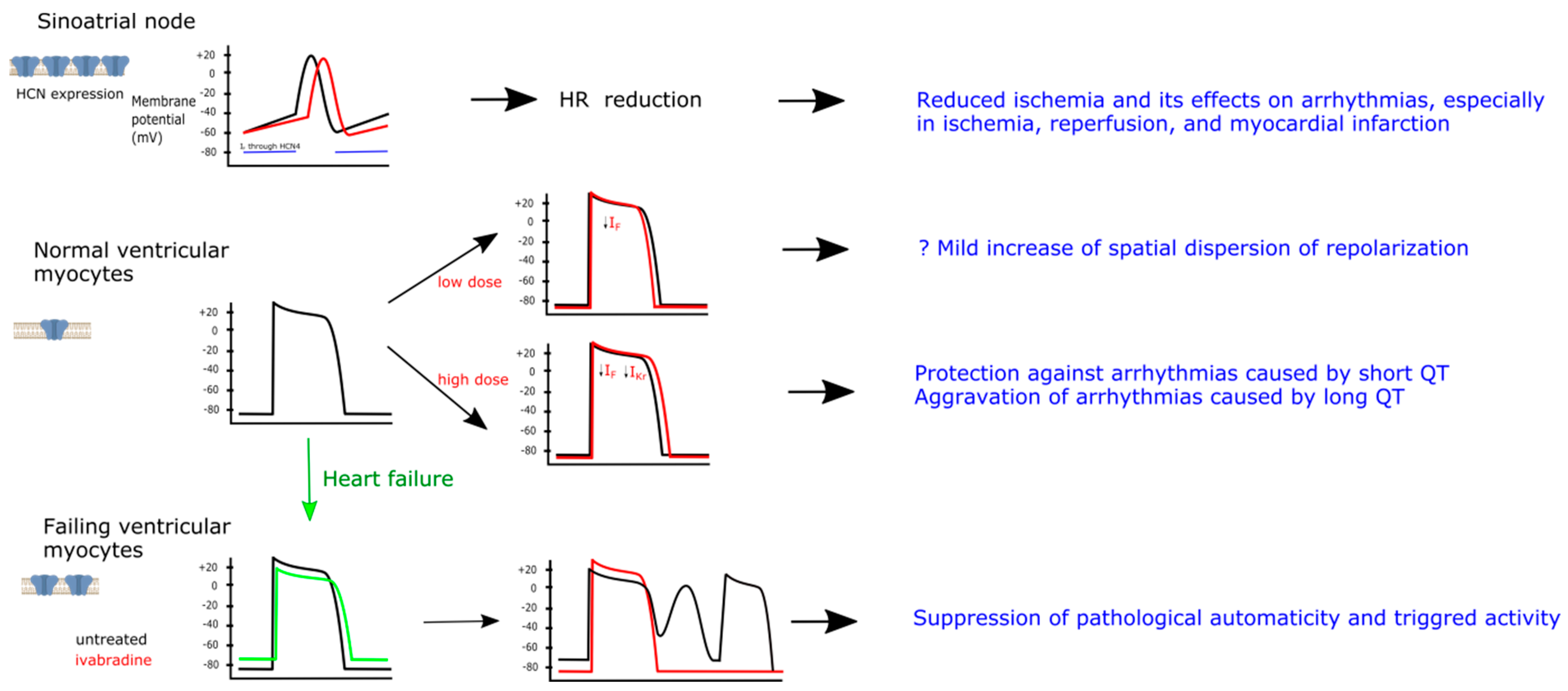
2. HCN Channel Family—A Primary Target for Ivabradine

Hyperpolarization-activated, cyclic-nucleotide-gated channels form a family of nonselective cation channels conducting mainly sodium and potassium ions through the plasma membrane and generating a current termed If (for “funny“). They have a unique biophysical behavior, i.e., they open upon hyperpolarization and result in an inward, depolarizing, predominantly sodium current and hence the name of the current, “funny” [4]; moreover, they are deactivated by depolarization. Additionally they are activated by cyclic nucleotides, cAMP and cGMP [5] and thus they are called “hyperpolarization-activated, cyclic-nucleotide-gated” (HCN) channels.
Functional HCN channels result from the assembly of four either identical or different alpha subunits. There are four different genes coding for HCN subunits (1–4) [6]. All HCN monomers (HCN1, HCN2, HCN3 and HCN4) have the same fundamental structure: contain a transmembrane core with six alpha helices (S1–S6): a voltage-sensing domain (S1–S4) and a pore region (S5–S6) carrying the ion selectivity filter as well as two cytosolic domains at the NH and COOH termini [7]. The elements conferring ion selectivity and voltage sensitivity are located in the transmembrane core and show high degree of sequence homology (80–90%) within the HCN family [8], while the NH and COOH termini vary in length and amino acidic composition. The COOH terminus harbors the cyclic-nucleotide-binding domain (CNBD), which is critical for the modulation by cyclic nucleotides [9].
HCN channels are voltage-gated channels activated at negative potentials. Their half activation voltage (V1/2) ranges from −70 mV for HCN1 to −100 mV for HCN4, while HCN2 and HCN3 fall in the middle of this range [10]. Gating kinetics are also voltage dependent and the activation time constant is inversely related to the hyperpolarization. The activation time constant is the fastest in HCN1 (25 ms), the slowest in HCN4 (200–500 ms), while HCN2 and HCN3 again lie in between [11]. Relative permeability of HCN to Na+ and K+ is 1:3/1:5 and the reversal potential is −25 to −40 mV, but due to the negative V1/2, HCN channels conduct primarily an inward Na+ current [10]. Binding of cAMP to the CNBD shifts upward the activation curve and accelerates the activation time constant, but is not sufficient to open the channels without an additional hyperpolarization of the membrane. Sensitivity to cAMP is highest in HCN2 and 4, weak in HCN1, while HCN3 exhibits no cAMP sensitivity [10].

3. HCN Channels in the Heart—Expression and Role

HCN channels are essential for normal impulse generation and conduction in the heart. In the sinoatrial node (SAN) cells they form a specialized pacemaking system involving HCN cooperation with ryanodine receptors (RyRs) of sarcoplasmic reticulum (SR) that is responsible for SAN spontaneous electrical activity [12]. HCN channels together with T- and L-type calcium channels as well as sodium-calcium exchanger (NCX) form the membrane clock, while RyRs in SR form the calcium clock. These two clocks constitute a tightly connected pacemaking system that is eventually responsible for spontaneous firing of SAN cells [13].
At maximally negative diastolic potentials (from −70 to −40 mV depending on SAN region), large number of HCN channels are open and an inward current flowing through them slowly depolarizes the membrane of SAN cells during the diastolic phase (Figure 1) [12]. Concomitantly submembrane Ca2+ wavelets appear as Ca2+ is released by RyRs from SR. This leads to NCX activation and further depolarizing membrane current. Eventually the threshold required to activate membrane L-type calcium channels is reached and a spontaneous action potential is generated. Finally Ca2+ inflow through L-type calcium channels triggers final Ca2+ release from SR [13].
In the atrioventricular node HCN channels are involved in generation of spontaneous electrical activity and conduction of stimuli from atria to ventricles [14]. cAMP-dependent regulation of HCN channel activity plays a key role in the control of SAN firing rate and consequently heart rate as well as AV node conduction by the autonomic nervous system.
HCN channels, primary site of action of ivabradine, are highly expressed in the sinoatrial node, while their expression in normal ventricular myocytes is low (left panel). In the sinoatrial node If current flowing through HCN channels is responsible for slow diastolic depolarization (indicated by a horizontal blue bar) toward the threshold required to activate calcium channels and generate a spontaneous action, thus participating in the pacemaking activity of sinoatrial node (black curve). Ivabradine inhibits If current and slows the diastolic depolarization, leading to heart rate reduction (red curve). Antiarrhythmic effects of ivabradine in the setting of ischemia are probably related to heart rate reduction. HCN channels are weakly expressed in normal ventricular myocytes. Limited evidence indicates that low doses of ivabradine, acting primarily on HCN channels, results in mild hyperpolarization and shortening of action potential duration (APD). Physiological relevance of this effect is unknown, but may include mild increase of spatial dispersion of repolarization. High doses of ivabradine can also inhibit IKr current, resulting in prolongation of APD; this could be antiarrhythmic in the setting of short QT (e.g., induced by digitalis), but proarrhythmic in the setting of long QT (e.g., induced by class III antiarrhythmic drugs). Expression of HCN channels in the ventricular myocytes is increased in heart failure. This results in diastolic cardiomyocyte depolarization and reduced amplitude of action potential overshoot. In such setting If current may be responsible for abnormal automaticity (caused by an inward current generated by overexpressed HCN channels) in the ventricular cardiomyocytes, resulting in ventricular premature complexes that may trigger ventricular arrhythmias. Ivabradine may prevent it by abolishing HCN channel overexpression and HCN channel blockade.
In the heart all the four isoforms have been detected and their expression varies according to the cardiac region, although HCN4 accounts for approximately 80% of total HCN proteins. In the adult heart the expression of HCN channels is largest in the SAN region and in the conduction system (atrioventricular node and Purkinje fibers), while their expression in the atria and ventricles is low [15]. HCN4 is the main isoform in the SAN (being responsible for 70–80% of total If current), atrioventricular node and Purkinje fibers [14], while HCN2 and HCN4 are the most abundant isoforms in the mammalian atria and ventricles and together represent more than 70–90% of the ventricular HCN channels [16], HCN3 accounting for the rest. HCN4 is also thought to play a crucial role in autonomic control of heart rate [17]. On the other hand HCN1 is almost exclusively present in SAN, being quite specific molecular marker of the latter [18].
At early embryonic stages HCN4 is abundantly expressed in the whole heart [19] and is largely responsible for If triggered automaticity of ventricular myocytes [20] as well as being involved in ventricular wall maturation at embryonic stages [21]. Toward birth, HCN4 transcription is downregulated in working-type cardiomyocytes and remains at low levels during adult stages [20].
In CHF, cardiac hypertrophy as well as in myocardial infarction (MI), expression of HCN channels in the ventricular cardiomyocytes increases. It was found in explanted human failing hearts [22,23,24], rats after MI [25,26,27], rats and mice with cardiac hypertrophy [16,28,29] and in mouse failing hearts [30,31]. Moreover, these channels are functional, since If current is also increased by twofold to threefold in both rat [22] and mouse [30] ventricular myocytes. The upregulated HCN channels exhibit their normal properties, i.e., they are activated by hyperpolarization and by β-receptor agonists [24]. Mechanism of this effect is unknown, but aldosterone has been shown to promote HCN2 and HCN4 mRNA and protein expression in cultured neonatal rat ventricular myocytes and that aldosterone blocking agents (spironolactone and eplerenone) counteract this effect by upregulating miRNA-1 expression, thus partially contributing to the posttranscriptional repression of HCN4 [32].
On the other hand, streptozocin-induced hyperglycemia resulted in downregulation of ventricular HCN2, while that in the atria was unchanged [33].
There are suggestions that HCN channels in the atria and ventricles prolong AP duration, especially in the epicardial myocytes, resulting additionally in reduction of transmural dispersion of AP duration [34]. APs recorded from epicardial myocytes of HCN3‒/‒ mice were found to be markedly shorter than those of wild-type mice because the depolarizing HCN3-mediated current component was missing. The difference was mainly due to a shortening of the late repolarization phase [34]. HCN knockout prevented hypertrophy-induced QT prolongation [16].

4. Effect of Ivabradine on Ion Channels

Ivabradine is the only HCN channel inhibitor approved for human use [35]. It blocks the HCN channel only from the intracellular side when the channels are opened by hyperpolarization [36], thus ivabradine needs an open channel to access its binding site [37] with enhanced binding upon frequent changes in the direction of ion flow [38]. When the channel is closed, the drug molecule is “trapped” in the binding site, which stabilizes the channel blockade [37]. Ivabradine blocks all four HCN isoforms with equal potency [39].
Ivabradine is a relatively selective inhibitor of HCN channels compared to other ion channels. Delpon et al. found that the hKv1.5 was blocked by the drug in a concentration-dependent way with IC50 29.0 ± 1.9 μM, therefore at doses higher than those required to block HCN channels [40]. Moreover, in the clinically relevant doses ivabradine did not affect calcium channels [41] and inhibited sodium channels at high concentrations (IC50 30 ± 3 μM) [6,41].
However, recent data indicates that ivabradine also inhibits the rapidly activating delayed rectifier potassium current (IKr) (IC50 of ≈2–6.8 μM) [41,42,43]. This current is critical for cardiac action potential repolarization and is encoded by the human ether-a-go-go-related gene (hERG). In this context, ivabradine increased action potential duration in human papillary muscles by 11% [44], presumably due to IKr blockade, although at high concentrations (10 µM, while normal ivabradine plasma concentrations in humans following oral dosing 5–20 mg are 0.03–0.13 µM [45]. Similarly it increased action potential duration in an isolated rabbit heart by 16% (at 5 µM), while lower concentrations were ineffective [46] and increased APD in fetal mouse hearts [43]. The fact that in all these studies resting membrane potential was unchanged despite prolongation of APD further corroborates that this effect was mediated by IKr inhibition [41,42,43]. Studies indicate that ivabradine at 10 μM blocks approximately 20–60% of IKr [47] and 80% of HCN4 current [43]. However, the European Medicines Agency (EMA) document library (http://www.ema.europa.eu/docs/en_GB/document_library/EPAR_scientific_.Discussion/human/000598/WC500035338.pdf; last accessed on 12 October 2021) indicates that ivabradine blocks the hERG current with an IC50 of 4.85 μM, which is also markedly higher than the reported plasma concentrations in humans. So there seems to be a consistent discrepancy between in vivo and in vitro responsiveness to ivabradine probably caused by high lipophilicity of ivabradine and its preferential accumulation in the plasma membranes, ensuring easy access to all ion channels [43].
hERG mutations or blockage by drugs resulting in reduction of IKr are principal causes of long QT syndrome predisposing individuals to ventricular arrhythmias and sudden cardiac death [48]. This raises concerns over possible proarrhythmic effects of ivabradine through QT prolongation and increased propensity to ventricular tachycardia (of torsades de pointes type), in particular in cases when cardiac repolarization reserve is reduced. This might be the case in early MI, since hypoxia was shown to downregulate hERG through upregulation of calpain mediated cleavage [49] and we indeed demonstrated that hERG abundance is reduced in post-MI rat hearts as early as 24 h after MI induction [25].
Furthermore this data suggests that coadministration of ivabradine with drugs known to prolong the QT interval should be approached with caution. Ivabradine-induced bradycardia could potentially exacerbate effects of other drugs on repolarization and there may be synergistic inhibitory effects on hERG. Indeed, there are two recent case reports documenting occurrence of torsade de pointes ventricular tachycardia in the setting of coadministration of ivabradine and azithromycin [50] or ranolazine and diltiazem [51].

5. Pathophysiology of Ventricular Arrhythmias

Pathophysiology of vast majority of ventricular arrhythmias involves (1) a trigger and (2) a substrate that propagates the trigger. The importance of a third component, the driver, is probably limited to atrial fibrillation. In specific arrhythmias these slightly vary, but the general pattern is the same.
Arrhythmic triggers involve premature complexes that are induced by triggered activity or automaticity. For example, in selected cases and in specific clinical settings, monomorphic premature ventricular complexes were found to trigger VF in 100% of cases [52].
Triggered activity can result from afterdepolarizations, i.e., voltage depolarizations (oscillations) that occur during the repolarization phase of the action potential (early afterdepolarizations, EAD) or during the diastolic phase, after completion of repolarization (delayed afterdepolarizations, DAD) [53]. If these voltage oscillations are large enough, they can trigger a full action potential. EADs are induced by reactivation of inward L-type calcium current, which is favored by prolongation of action potential duration. Alternatively spontaneous calcium release from the sarcoplasmic reticulum (SR) can activate sodium/calcium exchanger (NCX) generating inward current while exchanging one Ca2+ out for 3 Na+ ions in. DADs are induced by NCX activity and are favored by increased diastolic Ca2+ concentration and increased NCX activity. EAD and DAD are quite similar, since both are favored by cardiomyocyte Ca2+ overload and NCX overactivity and can be triggered by spontaneous SR Ca2+ release [54,55]. Beta adrenergic stimulation may favor both EADs (through phosphorylation and activation of L-type calcium channels) and DADs (through phosphorylation and inducing SR Ca2+ leak). Downregulation of sarcoplasmic reticulum Ca2+-ATPase (SERCA) has similar pro-arrhythmic effect [55].
Abnormal automaticity of working cardiac myocytes can be induced by increased extracellular K+ concentration (resulting e.g., from ischemia), reduced expression/activity of potassium channels or increased expression/activity of ion channels responsible for inward depolarizing current (e.g., HCN channels).
Arrhythmic substrates involve factors that (1) slow the intracellular conduction, (2) increase the spatial dispersion of repolarization. Slow intercellular conduction favors reentry, since it provides time for recovery of excitability [56]. Intercellular conduction depends on two factors: amplitude and upstroke velocity of cardiomyocyte action potential (a function of activation of sodium channels, SCN5A) and expression and function of gap junctions consisting of connexins that provide electrical connection between adjacent cells. On the other hand increased spatial dispersion of repolarization favors development of unidirectional block, a prerequisite of reentry.

6. Ivabradine and Ventricular Arrhythmias

6.1. Preclinical Evidence

Preclinical studies of ivabradine are summarized in Table 1. Briefly, ivabradine reduced ventricular arrhythmias (VA, both ventricular tachycardia [VT]/ventricular fibrillation [VF] and premature ventricular complexes [PVC]), arrhythmic deaths and total mortality in the rat model of non-reperfused acute MI, both when given as prophylaxis [25] and within the first minutes of ischemia [57] and was as effective as metoprolol in the latter study. Furthermore ivabradine increased VF threshold during acute myocardial ischemia in the pig [58,59], reduced VA during ischemia and reperfusion in the isolated rat heart [60]. Finally ivabradine was as effective as metoprolol in reducing VA induction rate and mortality after four weeks of administration [61] in post-MI rats and moreover, we showed that ivabradine reduced VA induced by dopamine infusion in a rat model of post-MI CHF [62]. Furthermore it almost completely abolished PVC after 3 months of administration [63] in the same model. In transgenic mice with CHF ivabradine reduced both the incidence of VT and PVC as well as isoproterenol-induced VF and improved survival [30].
Ivabradine reduced inducible VA in a Langendorff-perfused rabbit heart in the model of short-QT syndrome induced by a potassium channel opener, pinacidil [66] as well as digitalis-induced VA in the same rabbit model [67], while it was proarrhythmic in a model of long QT syndrome [68].
Last but not least, ivabradine was ineffective in a mouse model of catecholaminergic polymorphic ventricular tachycardia (CPVT) [69].

6.2. Potential Mechanisms of Ivabradine Effects

In the rat model of acute non-reperfused MI we showed that ivabradine prevented dispersion of electrical and biochemical properties between the infarct borderzone and the remote ventricular myocardium, essentially preventing early (within 45 min) ischemia-related shortening of AP and late (24 h) prolongation of AP related to downregulation of potassium channels (ERG and KVLQTI) in the infarct borderzone [25]. Despite the fact that ivabradine prevented striking differences in expression of various potassium channels (Kv4.3, KChIP2, ERG and KVLQTI) between the infarct borderzone and remote myocardium, it resulted in overall reduced expression of potassium channels, which could be responsible for QT prolongation observed at 24 h. Despite the fact that ivabradine did not affect parameters of Ca2+ handling, such as SR Ca2+ content, activity of major Ca2+ transporting proteins (NCX, sarcoplasmic Ca2+-ATPase [SERCA]), it reduced RyR sensitivity to Ca2+, potentially preventing pro-arrhythmic diastolic SR Ca2+ leak [25,57]. Moreover, in another rabbit ischemia and reperfusion study ivabradine increased FKBP12/12.6 expression and reduction of diastolic SR Ca2+ leak [70]. Another fascinating effect of ivabradine is stimulation of glycolysis and inhibition of hexosamine biosynthetic pathway in a HR-independent manner [71]. This in turn reduces O-linked N-acetylglucosamination of Ca2+-Calmodulin dependent protein kinase II and consequently reduces RyR sensitivity to Ca2+ [72]. However, ivabradine was ineffective in the model of CPVT: it did not prevent abnormal Ca2+ transients or arrhythmias resulting from abnormally increased RyR sensitivity in a transgenic mouse model of CPVT in vivo or in human induced pluripotent cells, exposed to isoproterenol [69].
Another potentially antiarrhythmic effect of ivabradine identified in our study was prevention of HCN4 overexpression in the ventricular cardiomyocytes as early as 24 h after MI induction [25], corroborating previous findings that ivabradine prevented HCN4 overexpression after 3 months of treatment in the post-MI rat [27]. However, due to the fact that in the setting of acute ischemia and reperfusion arrhythmias tend to occur early, before changes in ion channel expression could take place, [25,57], HCN4 overexpression is unlikely to play a major role in arrhythmogenesis in this setting.
In the pig model of acute ischemia [64] and ischemia and reperfusion [58,59] ivabradine increased regional blood flow, reduced the extent of myocardial ischemia, preserved myocardial energy status and prevented AP shortening [58], which was associated with protection against induced VF [58,59] and prolongation of time to onset of spontaneous VF [64]. In the latter study ivabradine was much more effective than propranolol in preventing spontaneous VF. Pacing to maintain unchanged HR essentially abolished protective effects of ivabradine, suggesting that HR reduction is the principal mechanism of its antiarrhythmic action [64]. Other experimental studies support this conclusion: we showed that ivabradine and metoprolol provided similar protection against VA induced by acute non-reperfused MI when given at doses that ensured identical HR reduction [57]. Similarly, ivabradine increased time to onset of ischemia-related conduction slowing and loss of electrical excitability and protected against development of VF during reperfusion in a model of 8-minute ischemia and reperfusion in an isolated rat heart [60]. Pacing throughout ischemia-reperfusion that completely prevented ivabradine-induced HR reduction completely abolished the protective effects of ivabradine, whereas pacing at reperfusion only partially attenuated this effect (by 40%). When ivabradine was given only at the beginning of reperfusion, it had no effect on the development of VF. The above mentioned studies strongly suggest that the principal mechanism of anti-arrhythmic effects of ivabradine in the setting of acute and chronic ischemia with/without reperfusion is prevention of ischemia itself related to HR reduction rather than specific effects on cardiac electrophysiology.
CHF is another highly proarrhythmic condition. A series of elegant studies were performed in a model of transgenic mice (dnNRSF-Tg), where CHF was induced by expression of a cardiac-specific dominant-negative form of neuron-restrictive silencer factor, an important regulator of the fetal cardiac gene program, characterized by dilated cardiomyopathy and sudden arrhythmic deaths [30,31]. In this model ivabradine prevented spontaneous PVC and VT/VF, but did not prevent pacing induced VT, indicating that it reduced the trigger rather than the substrate of ventricular arrhythmias. HCN2 and HCN4 were overexpressed and If amplitude was increased in this model. Moreover, ivabradine did not affect HR or the sympathovagal balance (as indicated by no change of HRV), again supporting the notion that reduction of an arrhythmic trigger is the principal driver of beneficial antiarrhythmic ivabradine effects and that this action is unrelated to HR reduction. Resting plasma membrane potential was slightly depolarized, which was unaffected by ivabradine, and presumably related to reduced IK1 density. Ivabradine reduced spontaneous action potentials induced by isoproterenol. However, these results need to be interpreted with caution: a model of CHF in the transgenic mice induced by reactivation of the fetal cardiac gene program is characterized by more than 60% arrhythmic mortality by week 28 of life. This model may overestimate role of HCN4 and arrhythmias in the CHF pathogenesis and mortality, respectively; its relevance for the human CHF is unknown [31]. In another studies transgenic ventricular myocytes with HCN2 overexpression exhibited ectopic activity induced by isoproterenol [30,73] and hypokalemia [74] that was reduced by ivabradine. However, HCN2 expression was increased 100-fold, while human failing ventricular myocytes demonstrate twofold to threefold increase of HCN expression. This raises questions of physiological relevance of this model.
Transgenic mice that expressed human HCN4 (hHCN4) under control of the murine cardiac troponin I gene promoter, providing moderate increase of If in cardiac myocytes to levels observed in human heart failure were used in another study [65]. It produced a phenotype of dilated cardiomyopathy, which was prevented by ivabradine. HCN4 overexpression resulted in diastolic cardiomyocyte depolarization (wild type: P50 = −61 mV vs. HCN4tg P50 = −54 mV). The amplitude of AP overshoot was diminished in transgenic cardiomyocytes, suggesting an inactivation of voltage-gated Na+-current at more depolarized resting membrane potentials. AP duration at 20 (APD20) and 50% (APD50) repolarization were shorter in transgenic cells, while APD90 did not differ between wild type and HCN4tg cardiomyocytes. This was accompanied by intracellular Ca2+ accumulation: increased diastolic Ca2+ concentration, SR Ca2+ content, and Ca2+ transients. All these abnormalities of cardiomyocyte Ca2+ handling could be blocked by a reverse mode NCX inhibitor, indicating that intracellular Na+ accumulation due to its inflow through HCN4 favors reverse mode NCX activity and cellular Ca2+ accumulation. HCN4tg cardiomyocytes were prone to afterdepolarizations resulting in trains of premature APs. However, the increased automaticity most probably originated from If augmentation rather than from spontaneous Ca2+ cycling-mediated membrane depolarization, as the NCX equilibrium was shifted toward reverse mode leading to an outward, hyperpolarizing current. This pathological automaticity was blocked by ivabradine. Unexpectedly, during telemetric recordings HCN4tg animals exhibited no sustained arrhythmias, only higher numbers of PVCs and few nonsustained VTs indicated increased arrhythmogenicity compared to wild type animals. These arrhythmias were blocked by ivabradine. This study suggests that upregulation of If in CHF may be proarrhythmic in the mechanism of increased automaticity, but this trigger alone is not sufficient to induce life threatening, clinical malignant arrhythmias [65].
As mentioned above, experimental studies suggest that HCN4 overexpression could play a role not only in arrhythmogenesis, but also in progression of CHF itself [65]. However, we have recently demonstrated that ivabradine prevented HCN4 overexpression in the rat model of chronic MI-induced HF, but unexpectedly a two-week dopamine infusion that had detrimental effects on both cardiac hemodynamic and remodeling parameters as well as induced ventricular arrhythmias, was also associated with reduced HCN4 overexpression [62]. Furthermore the degree of HCN4 overexpression in a large group of explanted human failing hearts did not correlate with severity of ventricular arrhythmias or hear failure [62].
Ivabradine was also tested in a setting of drug-induced electrophysiological changes in the rabbit heart (Table 1) [66,67,68]. A consistent pattern can be found in these studies: ivabradine prevented proarrhythmic effects of drugs that shorten QT interval (i.e., a potassium channel opener, pinacidil [66] and ouabain [67]), while exacerbated arrhythmias when combined with drugs that prolong QT interval (sotalol and veratridine [68]). What is interesting, in all these studies ivabradine only mildly increased APD or QT interval, while markedly increased effective refractory period, in particular post-repolarization refractory period, which could suggest that both IKr inhibition and HCN blockade were responsible for this effect. Since ivabradine was given at a relatively high dose (5 µM), again clinical relevance of this finding is uncertain.

6.3. Clinical Evidence

We must emphasize that there is still inadequate clinical evidence regarding ivabradine effects on cardiac ventricular arrhythmias.
For more detailed discussion of the clinical evidence the reader is referred to our previous review [75]. Briefly, three large randomized, placebo controlled clinical trials (BEAUTIFUL, SIGNIFY and SHIFT) and their Holter substudies assessed ivabradine in two indications: chronic CAD and CHF in a total of more than 36,000 patients [76,77,78,79,80]. The incidence of VA, namely VT or PVC did not differ between ivabradine and placebo groups. However, patients with chronic CAD have low risk of VA, while patients with CHF and high arrhythmic risk were excluded from those trials. This data is summarized in Table 2. Smaller trials enrolling patients with acute myocardial MI [81,82,83] also did not find any effect of ivabradine on VA, while in smaller studies enrolling patients with CHF [84,85,86] ivabradine reduced PVC, though no VT was recorded.
In two case reports CPVT ivabradine successfully suppressed VPC and nsVT [87,88].
There are no reported effects of a plethora of mutations of HCN channels on ventricular arrhythmias in humans: supraventricular bradycardia/tachyarrhythmias predominate [89].
There are anecdotal reports of proarrhythmic effects of ivabradine, in the setting of hypokalemia [83] and co-administration with QT-prolonging drugs [50,51].
Thus no robust clinical data is available on effect of ivabradine on ventricular arrhythmias in either CAD or CHF.

7. Possible Future Applications of Ivabradine

Ivabradine, the first in class and so far the only approved HCN channel blocker, can potentially be used in other arrhythmias, too. Two areas close to clinical application are rate control in atrial fibrillation (in combination with a beta blocker) [90] and treatment of pediatric junctional tachycardia [91,92].

8. Summary

Ivabradine is a HR reducing agent, currently approved for the treatment of CAD and CHF. Experimental studies suggest that ivabradine may have some antiarrhythmic effects: (1) in the setting of ischemia that may be secondary to HR reduction and consequent energy preservation and prevention of ischemia-induced electrophysiological effects and (2) in the setting of CHF that may be secondary to prevention of HCN channel overexpression and/or blockade of HCN channels and consequent reduction of proarrhythmic pathological automaticity in ventricular cardiomyocytes. However, ivabradine is probably also a blocker of IKr and may prolong QT and induce VT under conditions of long QT (e.g., drug-induced). No robust clinical data is available on effect of ivabradine on VA in either CAD or CHF. Clearly further molecular studies are required to further elucidate such issues as ivabradine specificity, dose-related effects, electrophysiological effects in normal and diseased heart as well as clinical outcomes.

Author Contributions

All authors contributed to the study conception and design, literature searching and writing the manuscript. All authors have read and agreed to the published version of the manuscript.

Funding

This research received no external funding.

Conflicts of Interest

The authors have no potential conflict of interest to disclose.

References

  1. Knuuti, J.; Wijns, W.; Saraste, A.; Capodanno, D.; Barbato, E.; Funck-Brentano, C.; Prescott, E.; Storey, R.F.; Deaton, C.; Cuisset, T.; et al. 2019 ESC Guidelines for the diagnosis and management of chronic coronary syndromes: The Task Force for the diagnosis and management of chronic coronary syndromes of the European Society of Cardiology (ESC). Eur. Heart J. 2019, 41, 407–477. [Google Scholar] [CrossRef] [PubMed]
  2. Ponikowski, P.; Voors, A.A.; Anker, S.D.; Bueno, H.; Cleland, J.G.F.; Coats, A.J.S.; Falk, V.; González-Juanatey, J.R.; Harjola, V.-P.; Jankowska, E.A.; et al. 2016 ESC Guidelines for the diagnosis and treatment of acute and chronic heart failureThe Task Force for the diagnosis and treatment of acute and chronic heart failure of the European Society of Cardiology (ESC)Developed with the special contribution of the Heart Failure Association (HFA) of the ESC. Eur. Heart J. 2016, 37, 2129–2200. [Google Scholar] [CrossRef]
  3. Yancy, C.W.; Jessup, M.; Bozkurt, B.; Butler, J.; Casey, D.E., Jr.; Colvin, M.M.; Drazner, M.H.; Filippatos, G.S.; Fonarow, G.C.; Givertz, M.M.; et al. 2017 ACC/AHA/HFSA Focused Update of the 2013 ACCF/AHA Guideline for the Management of Heart Failure. J. Card. Fail. 2017, 23, 628–651. [Google Scholar] [CrossRef] [PubMed] [Green Version]
  4. Di Francesco, D.; Borer, J.S. The Funny Current. Drugs 2007, 67, 15–24. [Google Scholar] [CrossRef]
  5. Di Francesco, D.; Tortora, P. Direct activation of cardiac pacemaker channels by intracellular cyclic AMP. Nature 1991, 351, 145–147. [Google Scholar] [CrossRef]
  6. Novella Romanelli, M.; Sartiani, L.; Masi, A.; Mannaioni, G.; Manetti, D.; Mugelli, A.; Cerbai, E. HCN Channels Modulators: The Need for Selectivity. Curr. Top. Med. Chem. 2016, 16, 1764–1791. [Google Scholar] [CrossRef] [PubMed]
  7. Yu, F.H.; Yarov-Yarovoy, V.; Gutman, G.A.; Catterall, W.A. Overview of molecular relationships in the voltage-gated ion channel superfamily. Pharmacol. Rev. 2005, 57, 387–395. [Google Scholar] [CrossRef] [Green Version]
  8. Kaupp, U.B.; Seifert, R. Molecular diversity of pacemaker ion channels. Annu. Rev. Physiol. 2001, 63, 235–257. [Google Scholar] [CrossRef]
  9. Wainger, B.J.; DeGennaro, M.; Santoro, B.; Siegelbaum, S.A.; Tibbs, G.R. Molecular mechanism of cAMP modulation of HCN pacemaker channels. Nature 2001, 411, 805–810. [Google Scholar] [CrossRef]
  10. Biel, M.; Wahl-Schott, C.; Michalakis, S.; Zong, X. Hyperpolarization-activated cation channels: From genes to function. Physiol. Rev. 2009, 89, 847–885. [Google Scholar] [CrossRef] [Green Version]
  11. Baruscotti, M.; Bucchi, A.; Difrancesco, D. Physiology and pharmacology of the cardiac pacemaker (“funny”) current. Pharmacol. Ther. 2005, 107, 59–79. [Google Scholar] [CrossRef]
  12. Di Francesco, D. A Brief History of Pacemaking. Front. Physiol. 2020, 10, 1599. [Google Scholar] [CrossRef]
  13. Lakatta, E.G.; Maltsev, V.A.; Vinogradova, T.M. A Coupled SYSTEM of Intracellular Ca2+ Clocks and Surface Membrane Voltage Clocks Controls the Timekeeping Mechanism of the Heart’s Pacemaker. Circ. Res. 2010, 106, 659–673. [Google Scholar] [CrossRef] [Green Version]
  14. Dobrzynski, H.; Nikolski, V.P.; Sambelashvili, A.T.; Greener, I.D.; Yamamoto, M.; Boyett, M.R.; Efimov, I.R. Site of Origin and Molecular Substrate of Atrioventricular Junctional Rhythm in the Rabbit Heart. Circ. Res. 2003, 93, 1102–1110. [Google Scholar] [CrossRef] [Green Version]
  15. Di Francesco, D. The role of the funny current in pacemaker activity. Circ. Res. 2010, 106, 434–446. [Google Scholar] [CrossRef] [PubMed] [Green Version]
  16. Hofmann, F.; Fabritz, L.; Stieber, J.; Schmitt, J.; Kirchhof, P.; Ludwig, A.; Herrmann, S. Ventricular HCN channels decrease the repolarization reserve in the hypertrophic heart. Cardiovasc. Res. 2012, 95, 317–326. [Google Scholar] [CrossRef] [PubMed] [Green Version]
  17. Wahl-Schott, C.; Fenske, S.; Biel, M. HCN channels: New roles in sinoatrial node function. Curr. Opin. Pharmacol. 2014, 15, 83–90. [Google Scholar] [CrossRef] [PubMed]
  18. Li, N.; Csepe, T.A.; Hansen, B.J.; Dobrzynski, H.; Higgins, R.S.D.; Kilic, A.; Mohler, P.J.; Janssen, P.M.L.; Rosen, M.R.; Biesiadecki, B.J.; et al. Molecular Mapping of Sinoatrial Node HCN Channel Expression in the Human Heart. Circ. Arrhythmia Electrophysiol. 2015, 8, 1219–1227. [Google Scholar] [CrossRef] [Green Version]
  19. Schweizer, P.A.; Yampolsky, P.; Malik, R.; Thomas, D.; Zehelein, J.; Katus, H.A.; Koenen, M. Transcription profiling of HCN-channel isotypes throughout mouse cardiac development. Basic Res. Cardiol. 2009, 104, 621–629. [Google Scholar] [CrossRef] [Green Version]
  20. Yasui, K.; Liu, W.; Opthof, T.; Kada, K.; Lee, J.K.; Kamiya, K.; Kodama, I. I(f) current and spontaneous activity in mouse embryonic ventricular myocytes. Circ. Res. 2001, 88, 536–542. [Google Scholar] [CrossRef] [PubMed] [Green Version]
  21. Schweizer, P.A.; Schröter, J.; Greiner, S.; Haas, J.; Yampolsky, P.; Mereles, D.; Buss, S.J.; Seyler, C.; Bruehl, C.; Draguhn, A.; et al. The symptom complex of familial sinus node dysfunction and myocardial noncompaction is associated with mutations in the HCN4 channel. J. Am. Coll. Cardiol. 2014, 64, 757–767. [Google Scholar] [CrossRef] [PubMed] [Green Version]
  22. Stillitano, F.; Lonardo, G.; Zicha, S.; Varro, A.; Cerbai, E.; Mugelli, A.; Nattel, S. Molecular basis of funny current (If) in normal and failing human heart. J. Mol. Cell Cardiol. 2008, 45, 289–299. [Google Scholar] [CrossRef] [PubMed]
  23. Hoppe, U.C.; Jansen, E.; Südkamp, M.; Beuckelmann, D.J. Hyperpolarization-Activated Inward Current in Ventricular Myocytes From Normal and Failing Human Hearts. Circulation 1998, 97, 55–65. [Google Scholar] [CrossRef] [Green Version]
  24. Cerbai, E.; Pino, R.; Porciatti, F.; Sani, G.; Toscano, M.; Maccherini, M.; Giunti, G.; Mugelli, A. Characterization of the hyperpolarization-activated current, I(f), in ventricular myocytes from human failing heart. Circulation 1997, 95, 568–571. [Google Scholar] [CrossRef]
  25. Mackiewicz, U.; Gerges, J.Y.; Chu, S.; Duda, M.; Dobrzynski, H.; Lewartowski, B.; Mączewski, M. Ivabradine Protects Against Ventricular Arrhythmias in Acute Myocardial Infarction in the Rat. J. Cell. Physiol. 2014, 229, 813–823. [Google Scholar] [CrossRef] [PubMed]
  26. Xia, S.; Wang, Y.; Zhang, Y.; Deng, S.-B.; Du, J.-L.; Wang, X.-C.; She, Q. Dynamic changes in HCN2, HCN4, KCNE1, and KCNE2 expression in ventricular cells from acute myocardial infarction rat hearts. Biochem. Biophys. Res. Commun. 2010, 395, 330–335. [Google Scholar] [CrossRef]
  27. Suffredini, S.; Stillitano, F.; Comini, L.; Bouly, M.; Brogioni, S.; Ceconi, C.; Ferrari, R.; Mugelli, A.; Cerbai, E. Long-term treatment with ivabradine in post-myocardial infarcted rats counteracts f-channel overexpression. Br. J. Pharmacol. 2012, 165, 1457–1466. [Google Scholar] [CrossRef] [Green Version]
  28. Fernández-Velasco, M.; Goren, N.; Benito, G.; Blanco-Rivero, J.; Boscá, L.; Delgado, C. Regional distribution of hyperpolarization-activated current (If) and hyperpolarization-activated cyclic nucleotide-gated channel mRNA expression in ventricular cells from control and hypertrophied rat hearts. J. Physiol. 2003, 553, 395–405. [Google Scholar] [CrossRef]
  29. Cerbai, E.; Barbieri, M.; Mugelli, A. Occurrence and Properties of the Hyperpolarization-Activated Current If in Ventricular Myocytes From Normotensive and Hypertensive Rats During Aging. Circulation 1996, 94, 1674–1681. [Google Scholar] [CrossRef] [PubMed]
  30. Kuwabara, Y.; Kuwahara, K.; Takano, M.; Kinoshita, H.; Arai, Y.; Yasuno, S.; Nakagawa, Y.; Igata, S.; Usami, S.; Minami, T.; et al. Increased Expression of HCN Channels in the Ventricular Myocardium Contributes to Enhanced Arrhythmicity in Mouse Failing Hearts. J. Am. Heart Assoc. 2013, 2, e000150. [Google Scholar] [CrossRef] [Green Version]
  31. Kuwahara, K.; Saito, Y.; Takano, M.; Arai, Y.; Yasuno, S.; Nakagawa, Y.; Takahashi, N.; Adachi, Y.; Takemura, G.; Horie, M.; et al. NRSF regulates the fetal cardiac gene program and maintains normal cardiac structure and function. EMBO J. 2003, 22, 6310–6321. [Google Scholar] [CrossRef]
  32. Del Lungo, M.; Melchiorre, M.; Guandalini, L.; Sartiani, L.; Mugelli, A.; Koncz, I.; Szel, T.; Varro, A.; Romanelli, M.N.; Cerbai, E. Novel blockers of hyperpolarization-activated current with isoform selectivity in recombinant cells and native tissue. Br. J. Pharmacol. 2012, 166, 602–616. [Google Scholar] [CrossRef] [Green Version]
  33. Hadova, K.; Kralova, E.; Doka, G.; Bies Pivackova, L.; Kmecova, Z.; Krenek, P.; Klimas, J. Isolated downregulation of HCN2 in ventricles of rats with streptozotocin-induced diabetic cardiomyopathy. BMC Cardiovasc. Disord. 2021, 21, 118. [Google Scholar] [CrossRef] [PubMed]
  34. Fenske, S.; Krause, S.; Biel, M.; Wahl-Schott, C. The role of HCN channels in ventricular repolarization. Trends Cardiovasc. Med. 2011, 21, 216–220. [Google Scholar] [CrossRef]
  35. Koruth, J.S.; Lala, A.; Pinney, S.; Reddy, V.Y.; Dukkipati, S.R. The Clinical Use of Ivabradine. J. Am. Coll. Cardiol. 2017, 70, 1777–1784. [Google Scholar] [CrossRef]
  36. Tanguay, J.; Callahan, K.M.; D’Avanzo, N. Characterization of drug binding within the HCN1 channel pore. Sci. Rep. 2019, 9, 465. [Google Scholar] [CrossRef]
  37. Bucchi, A.; Baruscotti, M.; Nardini, M.; Barbuti, A.; Micheloni, S.; Bolognesi, M.; DiFrancesco, D. Identification of the Molecular Site of Ivabradine Binding to HCN4 Channels. PLoS ONE 2013, 8, e53132. [Google Scholar] [CrossRef] [PubMed] [Green Version]
  38. Bucchi, A.; Tognati, A.; Milanesi, R.; Baruscotti, M.; DiFrancesco, D. Properties of ivabradine-induced block of HCN1 and HCN4 pacemaker channels. J. Physiol. 2006, 572, 335–346. [Google Scholar] [CrossRef] [PubMed]
  39. Stieber, J.; Wieland, K.; Stöckl, G.; Ludwig, A.; Hofmann, F. Bradycardic and proarrhythmic properties of sinus node inhibitors. Mol. Pharmacol. 2006, 69, 1328–1337. [Google Scholar] [CrossRef] [Green Version]
  40. Delpón, E.; Valenzuela, C.; Pérez, O.; Franqueza, L.; Gay, P.; Snyders, D.J.; Tamargo, J. Mechanisms of block of a human cloned potassium channel by the enantiomers of a new bradycardic agent: S-16257-2 and S-16260-2. Br. J. Pharm. 1996, 117, 1293–1301. [Google Scholar] [CrossRef] [Green Version]
  41. Haechl, N.; Ebner, J.; Hilber, K.; Todt, H.; Koenig, X. Pharmacological Profile of the Bradycardic Agent Ivabradine on Human Cardiac Ion Channels. Cell. Physiol. Biochem. 2019, 53, 36–48. [Google Scholar] [CrossRef] [Green Version]
  42. Melgari, D.; Brack, K.E.; Zhang, C.; Zhang, Y.; El Harchi, A.; Mitcheson, J.S.; Dempsey, C.E.; Ng, G.A.; Hancox, J.C. hERG potassium channel blockade by the HCN channel inhibitor bradycardic agent ivabradine. J. Am. Heart Assoc. 2015, 4, e001813. [Google Scholar] [CrossRef] [Green Version]
  43. Lees-Miller, J.P.; Guo, J.; Wang, Y.; Perissinotti, L.L.; Noskov, S.Y.; Duff, H.J. Ivabradine prolongs phase 3 of cardiac repolarization and blocks the hERG1 (KCNH2) current over a concentration-range overlapping with that required to block HCN4. J. Mol. Cell. Cardiol. 2015, 85, 71–78. [Google Scholar] [CrossRef] [PubMed]
  44. Koncz, I.; Szél, T.; Bitay, M.; Cerbai, E.; Jaeger, K.; Fülöp, F.; Jost, N.; Virág, L.; Orvos, P.; Tálosi, L.; et al. Electrophysiological effects of ivabradine in dog and human cardiac preparations: Potential antiarrhythmic actions. Eur. J. Pharm. 2011, 668, 419–426. [Google Scholar] [CrossRef] [PubMed]
  45. Jiang, J.; Tian, L.; Huang, Y.; Li, Y.; Xu, L. Pharmacokinetic and safety profile of ivabradine in healthy Chinese men: A phase I, randomized, open-label, increasing single- and multiple-dose study. Clin. Ther. 2013, 35, 1933–1945. [Google Scholar] [CrossRef] [PubMed]
  46. Frommeyer, G.; Sterneberg, M.; Dechering, D.G.; Ellermann, C.; Bögeholz, N.; Kochhäuser, S.; Pott, C.; Fehr, M.; Eckardt, L. Effective suppression of atrial fibrillation by ivabradine: Novel target for an established drug? Int. J. Cardiol. 2017, 236, 237–243. [Google Scholar] [CrossRef] [PubMed]
  47. Young, G.T.; Emery, E.C.; Mooney, E.R.; Tsantoulas, C.; McNaughton, P.A. Inflammatory and neuropathic pain are rapidly suppressed by peripheral block of hyperpolarisation-activated cyclic nucleotide-gated ion channels. Pain 2014, 155, 1708–1719. [Google Scholar] [CrossRef]
  48. Sanguinetti, M.C.; Tristani-Firouzi, M. hERG potassium channels and cardiac arrhythmia. Nature 2006, 440, 463–469. [Google Scholar] [CrossRef]
  49. Lamothe, S.M.; Song, W.; Guo, J.; Li, W.; Yang, T.; Baranchuk, A.; Graham, C.H.; Zhang, S. Hypoxia reduces mature hERG channels through calpain up-regulation. FASEB J. 2017, 31, 5068–5077. [Google Scholar] [CrossRef] [Green Version]
  50. Cocco, G.; Jerie, P. Torsades de Pointes Induced by the Concomitant Use of Ivabradine and Azithromycin: An Unexpected Dangerous Interaction. Cardiovasc. Toxicol. 2015, 15, 104–106. [Google Scholar] [CrossRef]
  51. Mittal, S.R. Slow junctional rhythm, QTc prolongation and transient torsades de-pointes following combined use of Ivabradine, Diltiazem and Ranolazine. J. Assoc. Physicians India 2014, 62, 426–427. [Google Scholar]
  52. Sánchez Muñoz, J.J.; García-Alberola, A.; Martínez-Sánchez, J.; Peñafiel-Verdú, P.; Caro-Martínez, C.; Manzano-Fernández, S.; Valdés Chávarri, M. Premature ventricular complexes as a trigger for ventricular fibrillation. Rev. Española Cardiol. 2010, 63, 798–801. [Google Scholar] [CrossRef]
  53. Qu, Z.; Weiss, J.N. Mechanisms of ventricular arrhythmias: From molecular fluctuations to electrical turbulence. Annu. Rev. Physiol. 2015, 77, 29–55. [Google Scholar] [CrossRef] [PubMed] [Green Version]
  54. Clauss, S.; Bleyer, C.; Schüttler, D.; Tomsits, P.; Renner, S.; Klymiuk, N.; Wakili, R.; Massberg, S.; Wolf, E.; Kääb, S. Animal models of arrhythmia: Classic electrophysiology to genetically modified large animals. Nat. Rev. Cardiol. 2019, 16, 457–475. [Google Scholar] [CrossRef]
  55. Sutanto, H.; Lyon, A.; Lumens, J.; Schotten, U.; Dobrev, D.; Heijman, J. Cardiomyocyte calcium handling in health and disease: Insights from in vitro and in silico studies. Prog. Biophys. Mol. Biol. 2020, 157, 54–75. [Google Scholar] [CrossRef] [PubMed]
  56. Janse, M.J.; Wit, A.L. Electrophysiological mechanisms of ventricular arrhythmias resulting from myocardial ischemia and infarction. Physiol. Rev. 1989, 69, 1049–1169. [Google Scholar] [CrossRef] [PubMed]
  57. Marciszek, M.; Paterek, A.; Oknińska, M.; Mackiewicz, U.; Mączewski, M. Ivabradine is as effective as metoprolol in the prevention of ventricular arrhythmias in acute non-reperfused myocardial infarction in the rat. Sci. Rep. 2020, 10, 15027. [Google Scholar] [CrossRef]
  58. Vaillant, F.; Timour, Q.; Descotes, J.; Manati, W.; Belhani, D.; Bui-Xuan, B.; Tabib, A.; Bricca, G.; Chevalier, P. Ivabradine Induces an Increase in Ventricular Fibrillation Threshold During Acute Myocardial Ischemia: An Experimental Study. J. Cardiovasc. Pharmacol. 2008, 52, 548–554. [Google Scholar] [CrossRef]
  59. Vaillant, F.; Dehina, L.; Mazzadi, A.; Descotes, J.; Chevalier, P.; Tabib, A.; Bui-Xuan, B.; Riera, C.; Belhani, D.; Timour, Q. Heart rate reduction with ivabradine increases ischaemia-induced ventricular fibrillation threshold: Role of myocyte structure and myocardial perfusion. Resuscitation 2011, 82, 1092–1099. [Google Scholar] [CrossRef]
  60. Ng, F.S.; Shadi, I.T.; Peters, N.S.; Lyon, A.R. Selective heart rate reduction with ivabradine slows ischaemia-induced electrophysiological changes and reduces ischaemia-reperfusion-induced ventricular arrhythmias. J. Mol. Cell. Cardiol. 2013, 59, 67–75. [Google Scholar] [CrossRef] [Green Version]
  61. Jiang, L.-p.; Lin, J.-j.; Zhuang, T.-p.; Wang, W.-w.; Wu, H.-z.; Zhang, F.-l. Effects of ivabradine on ventricular electrophysiological remodeling after myocardial infarction in rats. Arch. Med. Sci. 2020, 16, 1–14. [Google Scholar] [CrossRef]
  62. Paterek, A.; Sochanowicz, B.; Oknińska, M.; Śmigielski, W.; Kruszewski, M.; Mackiewicz, U.; Mączewski, M.; Leszek, P. Ivabradine prevents deleterious effects of dopamine therapy in heart failure: No role for HCN4 overexpression. Biomed. Pharmacother. 2021, 136, 111250. [Google Scholar] [CrossRef] [PubMed]
  63. Milliez, P.; Messaoudi, S.; Nehme, J.; Rodriguez, C.; Samuel, J.-L.; Delcayre, C. Beneficial effects of delayed ivabradine treatment on cardiac anatomical and electrical remodeling in rat severe chronic heart failure. Am. J. Physiol. Heart Circ. Physiol. 2009, 296, H435–H441. [Google Scholar] [CrossRef] [PubMed] [Green Version]
  64. Vaillant, F.; Dehina, L.; Dizerens, N.; Bui-Xuan, B.; Tabib, A.; Lauzier, B.; Chevalier, P.; Descotes, J.; Timour, Q. Ivabradine but not propranolol delays the time to onset of ischaemia-induced ventricular fibrillation by preserving myocardial metabolic energy status. Resuscitation 2013, 84, 384–390. [Google Scholar] [CrossRef] [PubMed]
  65. Yampolsky, P.; Koenen, M.; Mosqueira, M.; Geschwill, P.; Nauck, S.; Witzenberger, M.; Seyler, C.; Fink, T.; Kruska, M.; Bruehl, C.; et al. Augmentation of myocardial I(f) dysregulates calcium homeostasis and causes adverse cardiac remodeling. Nat. Commun. 2019, 10, 3295. [Google Scholar] [CrossRef]
  66. Frommeyer, G.; Weller, J.; Ellermann, C.; Kaese, S.; Kochhäuser, S.; Lange, P.S.; Dechering, D.G.; Eckardt, L. Antiarrhythmic properties of ivabradine in an experimental model of Short-QT- Syndrome. Clin. Exp. Pharmacol. Physiol. 2017, 44, 941–945. [Google Scholar] [CrossRef]
  67. Frommeyer, G.; Weller, J.; Ellermann, C.; Bögeholz, N.; Leitz, P.; Dechering, D.G.; Kochhäuser, S.; Wasmer, K.; Eckardt, L. Ivabradine Reduces Digitalis-induced Ventricular Arrhythmias. Basic Clin. Pharmacol. Toxicol. 2017, 121, 526–530. [Google Scholar] [CrossRef]
  68. Frommeyer, G.; Weller, J.; Ellermann, C.; Leitz, P.; Kochhäuser, S.; Lange, P.S.; Dechering, D.G.; Eckardt, L. Ivabradine Aggravates the Proarrhythmic Risk in Experimental Models of Long QT Syndrome. Cardiovasc. Toxicol. 2019, 19, 129–135. [Google Scholar] [CrossRef]
  69. Bueno-Levy, H.; Weisbrod, D.; Yadin, D.; Haron-Khun, S.; Peretz, A.; Hochhauser, E.; Arad, M.; Attali, B. The Hyperpolarization-Activated Cyclic-Nucleotide-Gated Channel Blocker Ivabradine Does Not Prevent Arrhythmias in Catecholaminergic Polymorphic Ventricular Tachycardia. Front. Pharmacol. 2020, 10, 1566. [Google Scholar] [CrossRef] [PubMed] [Green Version]
  70. Couvreur, N.; Tissier, R.; Pons, S.; Chetboul, V.; Gouni, V.; Bruneval, P.; Mandet, C.; Pouchelon, J.-L.; Berdeaux, A.; Ghaleh, B. Chronic heart rate reduction with ivabradine improves systolic function of the reperfused heart through a dual mechanism involving a direct mechanical effect and a long-term increase in FKBP12/12.6 expression. Eur. Heart J. 2010, 31, 1529–1537. [Google Scholar] [CrossRef]
  71. Vaillant, F.; Lauzier, B.; Ruiz, M.; Shi, Y.; Lachance, D.; Rivard, M.E.; Bolduc, V.; Thorin, E.; Tardif, J.C.; Des Rosiers, C. Ivabradine and metoprolol differentially affect cardiac glucose metabolism despite similar heart rate reduction in a mouse model of dyslipidemia. Am. J. Physiol. Heart Circ. Physiol. 2016, 311, H991–H1003. [Google Scholar] [CrossRef] [PubMed]
  72. Erickson, J.R.; Pereira, L.; Wang, L.; Han, G.; Ferguson, A.; Dao, K.; Copeland, R.J.; Despa, F.; Hart, G.W.; Ripplinger, C.M.; et al. Diabetic hyperglycaemia activates CaMKII and arrhythmias by O-linked glycosylation. Nature 2013, 502, 372–376. [Google Scholar] [CrossRef] [PubMed]
  73. Oshita, K.; Itoh, M.; Hirashima, S.; Kuwabara, Y.; Ishihara, K.; Kuwahara, K.; Nakao, K.; Kimura, T.; Nakamura, K.; Ushijima, K.; et al. Ectopic automaticity induced in ventricular myocytes by transgenic overexpression of HCN2. J. Mol. Cell Cardiol. 2015, 80, 81–89. [Google Scholar] [CrossRef] [PubMed] [Green Version]
  74. Oshita, K.; Kozasa, Y.; Nakagawa, Y.; Kuwabara, Y.; Kuwahara, K.; Nakagawa, T.; Nakashima, N.; Hiraki, T.; Takano, M. Overexpression of the HCN2 channel increases the arrhythmogenicity induced by hypokalemia. J. Physiol. Sci. 2019, 69, 653–660. [Google Scholar] [CrossRef] [PubMed]
  75. Marciszek, M.; Paterek, A.; Oknińska, M.; Zambrowska, Z.; Mackiewicz, U.; Mączewski, M. Effect of ivabradine on cardiac arrhythmias: Antiarrhythmic or proarrhythmic? Heart Rhythm 2021, 18, 1230–1238. [Google Scholar] [CrossRef]
  76. Fox, K.; Ford, I.; Steg, P.G.; Tendera, M.; Ferrari, R. Ivabradine for patients with stable coronary artery disease and left-ventricular systolic dysfunction (BEAUTIFUL): A randomised, double-blind, placebo-controlled trial. Lancet 2008, 372, 807–816. [Google Scholar] [CrossRef]
  77. Tendera, M.; Talajic, M.; Robertson, M.; Tardif, J.-C.; Ferrari, R.; Ford, I.; Steg, P.G.; Fox, K. Safety of Ivabradine in Patients With Coronary Artery Disease and Left Ventricular Systolic Dysfunction (from the BEAUTIFUL Holter Substudy). Am. J. Cardiol. 2011, 107, 805–811. [Google Scholar] [CrossRef]
  78. Fox, K.; Ford, I.; Steg, P.G.; Tardif, J.-C.; Tendera, M.; Ferrari, R. Ivabradine in Stable Coronary Artery Disease without Clinical Heart Failure. N. Engl. J. Med. 2014, 371, 1091–1099. [Google Scholar] [CrossRef] [PubMed] [Green Version]
  79. Swedberg, K.; Komajda, M.; Böhm, M.; Borer, J.S.; Ford, I.; Dubost-Brama, A.; Lerebours, G.; Tavazzi, L. Ivabradine and outcomes in chronic heart failure (SHIFT): A randomised placebo-controlled study. Lancet 2010, 376, 875–885. [Google Scholar] [CrossRef]
  80. Böhm, M.; Borer, J.S.; Camm, J.; Ford, I.; Lloyd, S.M.; Komajda, M.; Tavazzi, L.; Talajic, M.; Lainscak, M.; Reil, J.-C.; et al. Twenty-four-hour heart rate lowering with ivabradine in chronic heart failure: Insights from the SHIFT Holter substudy. Eur. J. Heart Fail. 2015, 17, 518–526. [Google Scholar] [CrossRef] [PubMed]
  81. Steg, P.G.; Lopez-de-Sà, E.; Schiele, F.; Hamon, M.; Meinertz, T.; Goicolea, J.; Werdan, K.; Lopez-Sendon, J.L.; investigators, V. Safety of intravenous ivabradine in acute ST-segment elevation myocardial infarction patients treated with primary percutaneous coronary intervention: A randomized, placebo-controlled, double-blind, pilot study. Eur. Heart J. Acute Cardiovasc. Care 2013, 2, 270–279. [Google Scholar] [CrossRef] [PubMed] [Green Version]
  82. Fasullo, S.; Cannizzaro, S.; Maringhini, G.; Ganci, F.; Giambanco, F.; Vitale, G.; Pinto, V.; Migliore, G.; Torres, D.; Sarullo, F.M.; et al. Comparison of Ivabradine Versus Metoprolol in Early Phases of Reperfused Anterior Myocardial Infarction With Impaired Left Ventricular Function: Preliminary Findings. J. Card. Fail. 2009, 15, 856–863. [Google Scholar] [CrossRef] [PubMed]
  83. Gerbaud, E.; Montaudon, M.; Chasseriaud, W.; Gilbert, S.; Cochet, H.; Pucheu, Y.; Horovitz, A.; Bonnet, J.; Douard, H.; Coste, P. Effect of ivabradine on left ventricular remodelling after reperfused myocardial infarction: A pilot study. Arch. Cardiovasc. Dis. 2014, 107, 33–41. [Google Scholar] [CrossRef] [PubMed] [Green Version]
  84. Mert, K.U.; Mert, G.; Morrad, B.; Tahmazov, S.; Mutlu, F.; Çavuşoglu, Y. Effects of ivabradine and beta-blocker therapy on dobutamine-induced ventricular arrhythmias. Kardiologia Polska 2017, 75, 786–793. [Google Scholar] [CrossRef] [PubMed]
  85. Rayan, M.; Tawfik, M.; Alabd, A.; Gamal, A. Ivabradine, a novel heart rate slower: Is it a sword of double blades in patients with idiopathic dilated cardiomyopathy? Anadolu Kardiyoloji Dergisi 2011, 11, 402–406. [Google Scholar] [CrossRef] [PubMed]
  86. Mughal, L.H.; Houghton, A.R.; Khoo, J. Significant suppression of premature ventricular ectopics with ivabradine in dilated cardiomyopathy. Br. J. Cardiol. 2019, 26, 36–37. [Google Scholar] [CrossRef]
  87. Vaksmann, G.; Klug, D. Efficacy of ivabradine to control ventricular arrhythmias in catecholaminergic polymorphic ventricular tachycardia. Pacing Clin. Electrophysiol. PACE 2018, 41, 1378–1380. [Google Scholar] [CrossRef]
  88. Kohli, U.; Aziz, Z.; Beaser, A.D.; Nayak, H.M. Ventricular arrhythmia suppression with ivabradine in a patient with catecholaminergic polymorphic ventricular tachycardia refractory to nadolol, flecainide, and sympathectomy. Pacing Clin. Electrophysiol. 2020, 43, 527–533. [Google Scholar] [CrossRef]
  89. Rivolta, I.; Binda, A.; Masi, A.; DiFrancesco, J.C. Cardiac and neuronal HCN channelopathies. Pflug. Arch. Eur. J. Physiol. 2020, 472, 931–951. [Google Scholar] [CrossRef]
  90. Fontenla, A.; López-Gil, M.; Tamargo-Menéndez, J.; Matía-Francés, R.; Salgado-Aranda, R.; Rey-Blas, J.R.; Miracle-Blanco, Á.; Mejía-Martínez, E.; Pastor-Fuentes, A.; Toquero-Ramos, J.; et al. Ivabradine for chronic heart rate control in persistent atrial fibrillation. Design of the BRAKE-AF project. Rev. Española Cardiol. 2020, 73, 368–375. [Google Scholar] [CrossRef]
  91. Krishna, M.R.; Kunde, M.F.; Kumar, R.K.; Balaji, S. Ivabradine in Post-operative Junctional Ectopic Tachycardia (JET): Breaking New Ground. Pediatric Cardiol. 2019, 40, 1284–1288. [Google Scholar] [CrossRef] [PubMed]
  92. Ergul, Y.; Ozturk, E.; Ozgur, S.; Ozyurt, A.; Cilsal, E.; Guzeltas, A. Ivabradine is an effective antiarrhythmic therapy for congenital junctional ectopic tachycardia-induced cardiomyopathy during infancy: Case studies. Pacing Clin. Electrophysiol. PACE 2018, 41, 1372–1377. [Google Scholar] [CrossRef] [PubMed]
Figure 1. Electrophysiological effects of ivabradine in the heart.
Jcm 10 04732 g001
Table 1. Effects of ivabradine on ventricular arrhythmias in experimental studies.
SpeciesModelStudy Design, Route and Method of IVA AdministrationEffects on VAAdditional EffectsReference
Ischemia and reperfusion, myocardial infarction
Ratnon-reperfused MI, LAD ligation, first 24 hIVA vs. saline, 24 h before MI, IVA in drinking water, daily dose 10 mg/kg↓mortalilty
↓VT/VF frequency
↓VT/VF duration
↓VPC
↓RyR sensitivity
↓HCN4 expression in LV
↓dispersion of APD
[25]
Ratnon-reperfused MI, LAD ligation, first 6 hIVA vs. metoprolol vs. saline, given immediately after MI induction, IVA 5 mg/kg oral gavage↓mortalilty
↓VT/VF frequency
↓VT/VF duration
↓VPC
as effective as metoprolol
↓QTc duration
↓RyR sensitivity
-velocity of conduction
-repolarization
[57]
Ratnon-reperfused MI, after 1 monthIVA vs. metoprolol vs. IVA + metoprolol vs. saline, starter immediately after MI and given for 4 weeks after MI, 10 mg/kg oral gavage↓VT/VF inducibility and VT/VF fatality, IVA+Metoprolol better than IVA or metoprolol alone, which were better than saline↓infarct size and ↑connexin 43 expression in all treatment groups[61]
Ratnon-reperfused MI, months 3–5IVA vs. saline, started 2 months after MI and given for 3 months, 10 mg/kg in drinking water↓PVC by 89%↑HR variability by 22%
-PR, QRS, QT duration
[63]
Pig6 episodes of I/R (LAD occlusion): 1 min ischemia + 15 min reperfusionIVA vs. saline, given 5 min after second occlusion, IVA 0.25 mg/kg intravenous bolus↑VF threshold by 2.9-foldprevention of ischemia-induced APD shortening
↓hypoxia
↑regional blood flow
[58,59]
Pigischemia (LAD occlusion)IVA vs. propranolol vs. saline, given before ischemia, IVA 0.25 mg/kg intravenous bolus↓time to VF onset (2325s for IVA vs. 682s for propranolol vs. 401s for saline), abolished by pacingPreserved cardiac energy status[64]
Ratisolated hearts, I/R episodes: 8 min of ischemia and 10 min of reperfusionIVA vs. saline, 5 min before ischemia 1 µM IVA and 1ml bolus of 100 µM IVA before the first minute of reperfusion↓VF incidence (IVA: 20%, saline: 90%)
effect abolished by pacing throughout I/R, but not during reperfusion alone
No effects when IVA given at reperfusion
↑time to conduction slowing
↑time to loss of electrical excitability
[60]
Chronic heart failure
Ratpost-MI CHF1 month after MI: IVA 7 µg/min/kg or Dopamine 10 µg/min/kg or both by implantable osmotic pump for 14 days↓VT incidence and ↓PVC by IVA vs. Dopamine [62]
Mousetransgenic mice (dnNRSF-Tg) with dilated cardiomyopathyIVA vs. saline given for 6 months, daily dose 7 mg/kg in drinking water↓mortality
↓VT episodes (19/h vs. 92 h for saline)
↓VPC
effects independent of HR reduction
↓HCN2 and HCN4 expression in ventricular myocytes
IVA prevented isoproterenol-induced spontaneous action potentials in failing ventricular myocytes
[30]
Mousetransgenic mice (hHCN4 overexpression) with dilated cardiomyopathyIVA vs. saline given for 2 months, daily dose 1.5 mg/kg by an osmotic pump↓VPC
↓non-sustained VT
↓automaticity[65]
Other models
Rabbitpinacidil-induced short QT syndromeLangendorff-perfused hearts, IVA 5 µM↓VF inducibilityIVA prevented QT, APD and ERP shortening by pinacidil[66]
Rabbitouabain-induced arrhythmiasLangendorff-perfused hearts, IVA 5 µM↓VF inducibilityIVA did not affect QT or APD, but prevented ERP shortening by ouabain[67]
Rabbitsotalol and veratradine-induced long QT syndromeLangendorff-perfused hearts, IVA 5 µM↑incidence of polymorphic VT induced by sotalol/vertatridine and hypokalemiaIVA did not increase QT, APD or spatial dispersion of repolarization[68]
MouseCPVT transgenic miceIVA 6 mg/kg-VA at rest or during a treadmill exercise testno effect on DAD in the beating induced pluripotent stem cells derived cardiomyocytes from CPVT patients[69]
APD, action potential duration; CHF, chronic heart failure; CPVT, catecholaminergic polymorphic ventricular tachycardia; I/R, ischemia and reperfusion; IVA, ivabradine; LAD, left anterior descending artery; MI, myocardial infarction; PVC, premature ventricular complexes; RyR, ryanodine receptor; VA, ventricular arrhythmias; VF, ventricular fibrillation; VT, ventricular tachycardia. ↑ increase; ↓ decrease; - no change.
Table 2. Effects of ivabradine on ventricular arrhythmias in large clinical trials.
Study Acronim, YearStudy PopulationNo. of Study SubjectsFollow-UpEffect on VA
Coronary artery disease
BEAUTIFUL 2008 [76]CAD + LV systolic dysfunction, sinus rhythm ≥ 70 bpm10,9171.6 years
BEAUTIFUL 2008 Holter Substudy [77]8402 × 24 hours at 1 and 6 monthsVT:
ΔIVA: 24–26%, PLA: 26–24% (P = NS)
SIGNIFY 2014 [78]CAD without CHF, sinus rhythm ≥ 70 bpm19,1072.3 yearsSevere VA:
ΔIVA: 0.8%, PLA: 0.7% (P = NS)
ΔQT prolongation:
ΔIVA: 1.8%, PLA: 0.7% (P < 0.001)
Chronic heart failure
SHIFT 2010 [79]CHF, LVEF ≤ 35%, sinus rhythm ≥ 70 bpm65581.9 years
SHIFT Holter Substudy 2010 [80]60224 h at 8 monthsnsVT:
ΔIVA: 28%, PLA: 33% (P = NS)
ΔPVC:
ΔIVA: 78/h PLA: 69/h (P = NS)
CAD, coronary artery disease; IVA, ivabradine; LV, left ventricle; LVEF, left ventricular ejection fraction; nsVT, nonsustained ventricular tachycardia; PLA, placebo; PVC, premature ventricular contractions; RCT, randomized clinical trial; VA, ventricular arrhythmias; VT, ventricular tachycardia.
Publisher’s Note: MDPI stays neutral with regard to jurisdictional claims in published maps and institutional affiliations.

