Article

Vascular Protective Effects of New Oral Anticoagulants in Patients with Atrial Fibrillation

Division of Cardiology, Department of Internal Medicine, Kyung Hee University Hospital, Kyung Hee University, 26 Kyungheedae-ro, Dongdaemun-gu, Seoul 02447, Korea
*
Author to whom correspondence should be addressed.
These two authors equally contribute this work.
J. Clin. Med. 2021, 10(19), 4332; https://doi.org/10.3390/jcm10194332
Submission received: 21 July 2021 / Revised: 8 September 2021 / Accepted: 21 September 2021 / Published: 23 September 2021
(This article belongs to the Special Issue New Insights into Atrial Fibrillation)

Abstract

:
This study was designed to determine the efficacy of a new oral anticoagulant (NOAC) therapy for the prevention of endothelial dysfunction and atherosclerosis progression in patients with atrial fibrillation (AF). Sixty-five AF patients with a CHA2DS2-VASc score ≥2 without previous history of cardiovascular disease were registered and randomly assigned to either an NOAC group (dabigatran or rivaroxaban) or the warfarin group. Reactive hyperemia peripheral arterial tonometry (RH-PAT) measurements reflecting endothelial function were taken using Endo-PAT2000. Carotid intima–media thickness (IMT) was measured at baseline, 12 months, and 24 months, and several biomarkers were also analyzed. For the primary end point, the reactive hyperemia index (RHI) for the NOAC group was 1.5 ± 0.4 and that for the warfarin group was 1.6 ± 0.5. The left and right carotid IMT was 0.7 mm in the NOAC groups and 0.8 mm in the warfarin group. At 12 months, RHI was 1.6 ± 0.3 for the dabigatran group, 1.6 ± 0.5 for the rivaroxaban group, and 1.6 ± 0.3 for the warfarin group. The three groups did not differ statistically with respect to change in left and right carotid IMT at 12 and 24 months, respectively. The biomarkers for endothelial function and atherosclerosis were not significantly different. There was a trend of reduced P-selectin levels in the NOAC group compared to the warfarin group. In patients with AF, there were no significant differences in the prevention of endothelial dysfunction and atherosclerosis progression between the NOAC and warfarin groups.

1. Introduction

Activated coagulation factor Xa is known to play a central role in the coagulation cascade. Recent evidence further suggests that factor Xa has an important modulating effect in cellular signaling by the activation of protease-activated receptor (PAR) [1]. In fact, coagulation and inflammatory pathways interact with each other via factor Xa-mediated PAR activation on the arterial vessel wall and heart, and the resulting development of atherosclerosis and atrial fibrillation (AF) has been documented [2]. Preclinical studies have provided evidence for the effects of direct Xa or thrombin inhibition beyond anticoagulation, including anti-inflammatory and protective activities in atherosclerotic plaque development [1]. Evidence has demonstrated that direct thrombin inhibition impairs the formation and size of atherosclerotic plaques in addition to preventing progression of endothelial injury-associated stenosis in an apolipoprotein E-deficient mouse model [3,4]. However, the question remains as to whether these effects obtained in preclinical trials are similar in humans, for which there is scant evidence.
There are several non-invasive methods that allow researchers to assess endothelial function. Reactive hyperemia peripheral arterial tonometry (RH-PAT) and carotid IMT measurements are two such measurement tools [5], and previous studies suggest that the RH-PAT index is a useful predictor of coronary endothelial dysfunction [6,7]. Increased carotid IMT was associated with coronary artery severity [8,9,10].
This study aimed to determine the efficacy of new oral anticoagulant (NOAC) therapy for preventing endothelial dysfunction and atherosclerosis progression in AF patients. It was initially designed in 2015 and started enrollment in the same year, when NOAC and warfarin were equally classified as class I recommendations for stroke prevention in AF. However, new recommendations were published in the 2016 clinical guidelines, where the usage of NOAC was recommended over warfarin. Unfortunately, this made it more difficult to enroll patients, resulting in a break in the registration process. Patient enrollment was eventually terminated prematurely.

2. Materials and Methods
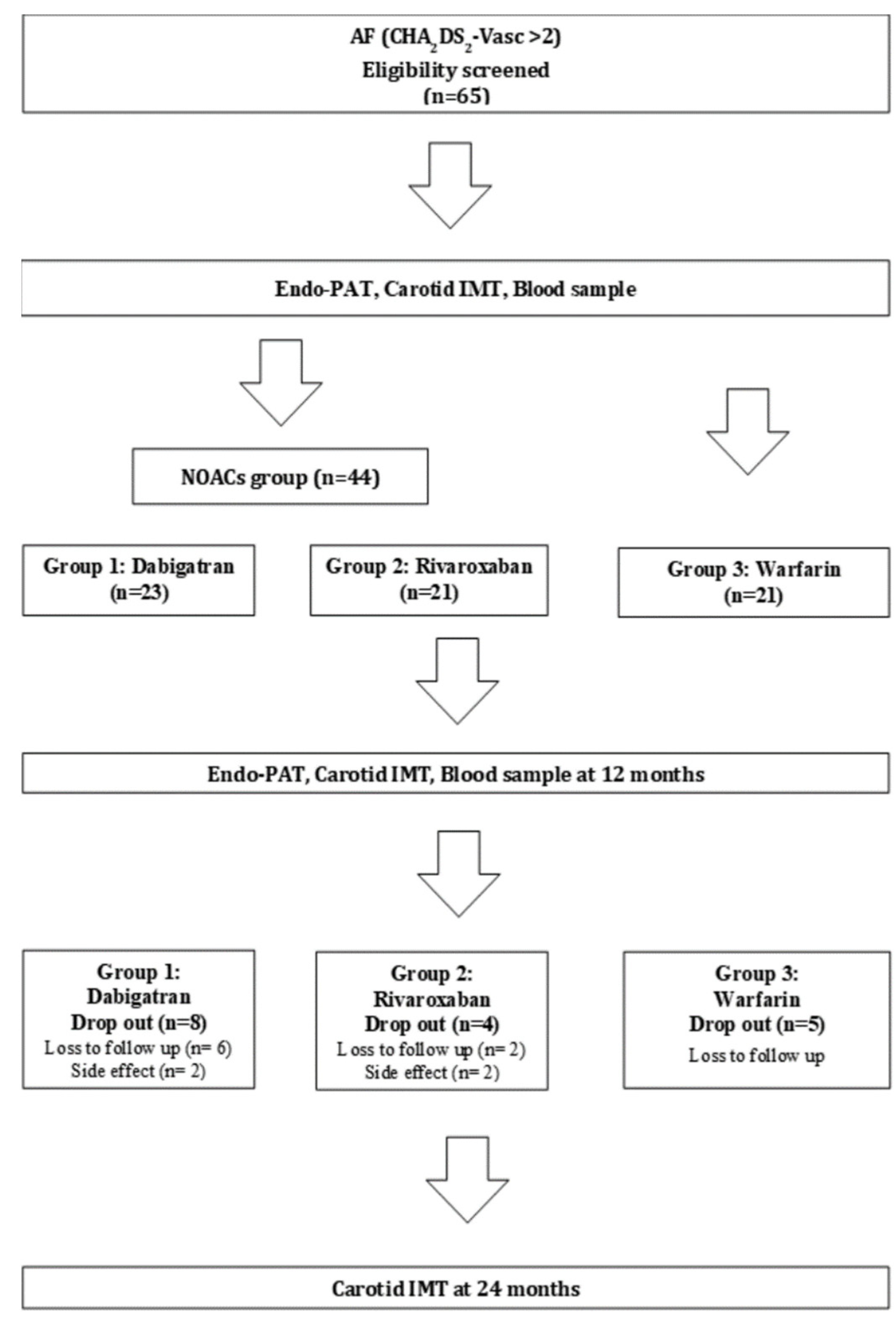
2.1. Study Subjects

We prospectively enrolled AF patients between 40 and 85 years of age and with CHA2DS2-VASc scores ≥ 2 (Table 1). The exclusion criteria were severe peripheral arterial disease (greater than Fontaine category IIb), grade 4 or higher cerebral infarction on the modified Rankin Scale, and proven coronary artery disease based on coronary angiogram. We also excluded patients with a range of concomitant comorbidities, including severe hepatic or renal dysfunction, uncontrolled congestive heart failure, hypertension, diabetes mellitus, hematological disorders, and allergy or hypersensitivity to the investigational drugs as well as pregnant or lactating women. Written informed consent was obtained from all patients, and the Ethics Review Board of Kyung Hee University Hospital approved this study. The two-year duration of the study period had a registration period from September 2015 to February 2016, with complete study duration from September 2017 to April 2018. For more information, please refer to our protocol article that has already been published [11].

2.2. Randomization

This study was a prospective, randomized, two-year follow-up study to further clarify the efficacy of NOAC in altering endothelial function and atherosclerosis progression in AF patients. The study design is shown in Figure 1. After enrollment, subjects were randomly assigned to the dabigatran group (110 or 150 mg twice/day; group 1), the rivaroxaban group (20 mg/day; group 2), or the warfarin group (controlled by international normalized ratio (INR) of 2–3; group 3). Clinical follow-up occurred at 1, 3, 12, and 24 months. Follow-up was conducted via telephone interviews or office visits. We assumed that NOACs would improve RHI values by an 8% difference, with no significant difference between the two NOACs. The expected difference in RHI was driven by clinical significance and previous medical treatments in other study populations [12,13,14]. To detect a statistically significant difference with a power of 80% with a two-sided α-level of 0.05, a sample of 165 patients (55 patients for each group) would be required. Assuming that the dropout rate would be 20%, the total sample size was set at 198 subjects [11]. However, due to the aforementioned reasons, it was difficult to enroll patients, and the registration speed slowed down due to the changes in the clinical guidelines and practice. Therefore, an interim analysis was conducted, and the results revealed no significant differences between the NOAC and warfarin groups, so fewer patients were enrolled.

2.3. Primary Outcome

The primary endpoint was defined as the change in the reactive hyperemia index (RHI) at 12 months. Secondary endpoints included changes in the right and left maximum IMT of the common carotid artery (CCA) and the internal carotid artery (ICA), mean IMT of the CCA and ICA at 12 and 24 months, 24-month cardiovascular events including cardiac death, stroke, acute myocardial infarction (AMI), overall cause of death, withdrawal of drug, or bleeding events.

2.4. RHI Measurement

Measurements were performed using a standard technique and device at baseline and at 12 months after randomization [15,16]. An RH-PAT 2000 device (Itamar Medical, Caesarea, Israel) was used for digital RH-PAT to evaluate endothelial function as previously described [5]. Impaired endothelial function was defined as logRHI <0.6, and favorable endothelial function was defined as logRHI ≥0.6.

2.5. Carotid IMT

CCA-IMT measurements were performed by experienced sonographers who were well trained in the use of 10-megahertz linear vascular probes (Vivid 7, GE Vingmed Ultrasound, Horten, Norway). Measurements were taken at baseline and 12 months after randomization. IMT was measured as the distance between two parallel echogenic lines corresponding to the blood–intima and media–adventitia interfaces on the posterior artery wall. Three IMT determinations were performed at the site of the thickest point with a maximum CCA-IMT and two adjacent points (1 cm upstream and 1 cm downstream from this site), and these three measurements were averaged (mean CCA-IMT). Carotid IMT was measured using dedicated software (Intimascope, Media Cross Co., Tokyo, Japan) by an examiner blinded to all clinical information.

2.6. Statistical Analyses

Demographic data were analyzed to identify pretreatment equivalencies and differences between the three study groups. Continuous variables are presented as the mean ± standard deviation and were compared using Student’s t-test or the Mann–Whitney test wherever appropriate. Non-normal distribution was identified from normality tests, and these data are presented as the median (interquartile range) and were compared using nonparametric methods (the Kruskal–Wallis or Wilcoxon signed-rank test). Categorical variables are presented as frequencies and percentages and were compared using the chi-squared or Fisher’s exact test wherever appropriate. Statistical significance was set at 0.05 (two-sided). All statistical analyses were performed using R software version 3.6.0. (R Foundation for Statistical Computing, Vienna, Austria).

2.7. Biomarkers of Atherosclerotic Plaque

Blood samples were obtained in serum separator tubes. After 20 min at room temperature, blood was centrifuged at 1000× g for 15 min. The serum was stored at −80 °C after dispensing 500 μL of serum into tubes. Concentrations of IL-6, TNF-α, p-selectin, and vWF were measured using the Quantikine and SimpleStep enzyme-linked immunosorbent assay (ELISA) kit according to the manufacturer’s protocol. A standard curve was created by reducing the data using computer software capable of generating a four-parameter logistic curve fit. The data were linearized by plotting the log of the concentration of the biomarkers versus the log of the optical density, and the best fit line could be determined by regression analysis.

3. Results

3.1. Patient Characteristics

From September 2015 to February 2016, 65 patients were randomly assigned. A total of 17 patients (26.2%) were excluded from the study due to loss at follow-up or side effects at 2-year follow-up (Figure 1). The patients’ characteristics are presented in Table 1. The average ages of participants in the dabigatran, rivaroxaban, and warfarin groups were 67.1 ± 9.4, 64.3 ± 7.7, and 67.7 ± 7.1 years, respectively. Males and non-smokers were more prevalent in all three groups, and more patients had hypertension than did not. There were no significant differences in patient characteristics between the three groups.

3.2. Reactive Hyperemia Index (RHI) and Carotid IMT

Table 2 presents the RHI and carotid IMT measurements at baseline, 12 months, and 24 months. At baseline, the dabigatran group’s RHI was 1.5 ± 0.4, the rivaroxaban RHI was 1.5 ± 0.4, and the warfarin RHI was 1.6 ± 0.5 (p = 0.487). The left and right carotid IMT was 0.7 mm in the NOAC groups and 0.8 mm (p = 0.697) in the warfarin group. At 12 months, the dabigatran group RHI was 1.6 ± 0.3, the rivaroxaban RHI was 1.6 ± 0.5, and the warfarin RHI was 1.6 ± 0.3 (p = 0.779, Figure 2). Carotid IMT values were not statistically different between the three groups. Notably, the three groups also did not differ statistically with respect to carotid IMT at 24 months.

3.3. Biomarkers of Atherosclerotic Plaque

Table 3 presents the endothelial and platelet activity biomarkers at baseline and 12 months. There was a trend of reduced p-selectin level in the NOAC groups compared to the warfarin group, but the results did not indicate statistically significant differences (Figure 3). The other biomarkers, namely IL-6, TNF-α, and vWF, showed no statistically significant differences between the NOAC and warfarin groups. There were no serious adverse events during this study.

4. Discussion

To the best of our knowledge, this is the first clinical study to investigate whether NOACs are indeed effective in altering endothelial function and atherosclerotic changes in patients with AF. Although AF may adversely affect endothelial function [15,17,18], it is not known whether vitamin K antagonists are helpful for such prevention. Previous studies showed that NOACs suppressed inflammatory cytokines and atherosclerotic cascades [3,4,19,20]. However, there was a lack of clinical data to corroborate the results. Interestingly, our trial showed that there was no difference in the ability to prevent endothelial dysfunction or atherosclerosis progression between the NOAC and warfarin groups. The results of this study provide new insights regarding our current understanding of NOAC mechanisms. The results should also help researchers to develop appropriate drug therapies for patients with AF and atherosclerosis. This will be much needed, as AF and related complications are becoming a significant health burden worldwide, with AF incidences rising at a staggering rate.
PARs are a family of G protein-coupled receptors which belong to four members (PAR-1 to -4). PAR-1 and PAR-2 are activated by factor Xa through a canonical G protein-dependent pathway in several cardiomyocytes and cardiac fibroblasts, which result in pathological cardiac remodeling in response to cardiac injury or stress [21]. In a mouse model, factor Xa inhibition was beneficial for prevention and regression of atherosclerosis, possibly mediated through reduced PAR activation [22]. Direct factor Xa inhibition was associated with slow progression of coronary atherosclerotic plaque compared with warfarin [23]. Experimental studies have suggested that a thrombin inhibitor improved endothelial function and decreased atherosclerosis in mice [24]. However, we found that there were no statistically significant differences between NOACs and warfarin with respect to vascular endothelial protection in high-risk patients with AF.
In a post hoc analysis of the X-VERT trial, rivaroxaban and warfarin showed similar influences on inflammatory biomarkers, including IL-6 [25]. Although the researchers found a similar reduction in inflammatory markers, we did not find a meaningful decrease in inflammation biomarkers in both NOAC and warfarin groups. It is possible that the results were different because of differences in the drugs that were used—the previous study only used rivaroxaban. Whereas, rivaroxaban and dabigatran were used in our study. However, in the RIVAL-AF trial, there were no significant differences in the changes in inflammatory cytokines such as IL-6 and TNF-α between the rivaroxaban and dabigatran groups. In a small observational study, a trend (p = 0.06) toward a reduction in P-selectin in the rivaroxaban treatment group compared to the control group was noted [26]. Our study also found a trend of reduced P-selectin level in NOAC-treated patients compared to warfarin-treated patients. The clinical significance of this finding is still unclear, thus further studies are needed. There are several other reasons why our results showed no significant differences. Firstly, previous animal studies administered higher doses of NOAC compared to the amount that was used in our study [22,24]. Secondly, our study was a small trial conducted over a short time period, and thus may not provide robust temporal statistical evidence.
Some potential limitations of this study should be considered. First, it included a relatively small number of patients due to early termination of the trial, with fewer patients enrolled than we had initially planned. Thus, our results should be interpreted with caution as the sample size may not be sufficient to detect significant differences between the warfarin and NOAC groups. Furthermore, the findings from this study cannot be generalized to all patients with AF. However, this issue is not unique to our study. Randomized controlled studies comparing the effects of NOACs and warfarin on human vascular endothelial cell function or biomarkers may suffer from the same ethical issues in the future. In this respect, this study provides invaluable data that will be difficult to obtain in future trials. Second, the primary outcome was not a hard endpoint for a clinical outcome but a surrogate marker that indirectly measures endothelial dysfunction. Therefore, future studies should directly explore clinical outcomes from vascular events that compare NOACs and warfarin.

5. Conclusions

There were no significant differences in the ability to prevent endothelial dysfunction and atherosclerosis progression between the NOAC and warfarin groups of AF patients.

Author Contributions

Conceptualization, J.M.L. and W.K.; methodology, G.-W.J., J.M.L. and W.K.; software, G.-W.J., J.M.L. and W.K.; validation, G.-W.J., J.M.L. and W.K.; formal analysis, G.-W.J., J.M.L. and W.K.; investigation, G.-W.J., J.M.L. and W.K.; resources, G.-W.J., S.W.C., J.K., Y.S.L., H.O.K., J.M.L., J.S.W., J.B.K., W.-S.K., H.C. and W.K.; data curation, G.-W.J., J.M.L. and W.K.; writing—original draft preparation, G.-W.J., J.M.L. and W.K.; writing—review and editing, G.-W.J., J.M.L. and W.K.; visualization, G.-W.J., J.M.L. and W.K.; supervision, J.M.L. and W.K.; project administration, W.K.; funding acquisition, W.K. All authors have read and agreed to the published version of the manuscript.

Funding

This research was supported by the National Research Foundation of the Ministry of Education, Science and Technology (NRF-2020R1F1A1076495).

Institutional Review Board Statement

All protocols were approved by the Ethics Review Board of Kyung Hee University Hospital (Approval Number 2015-10-202, Approval Date 21 December 2015).

Informed Consent Statement

Informed consent was obtained from all subjects involved in the study.

Data Availability Statement

All data are reported in the manuscript.

Conflicts of Interest

The authors declare no conflict of interest.

References

  1. Esmon, C.T. Targeting factor Xa and thrombin: Impact on coagulation and beyond. Thromb. Haemost. 2014, 111, 625–633. [Google Scholar] [CrossRef]
  2. Borissoff, J.I.; Spronk, H.M.H.; ten Cate, H. The Hemostatic System as a Modulator of atherosclerosis. N. Engl. J. Med. 2011, 364, 1746–1760. [Google Scholar] [CrossRef]
  3. Lee, I.O.; Kratz, M.T.; Schirmer, S.H.; Baumhakel, M.; Bohm, M. The effects of direct thrombin inhibition with dabigatran on plaque formation and endothelial function in apolipoprotein E-deficient mice. J. Pharmacol. Exp. Ther. 2012, 343, 253–257. [Google Scholar] [CrossRef] [Green Version]
  4. Borissoff, J.I.; Otten, J.J.; Heeneman, S.; Leenders, P.; van Oerle, R.; Soehnlein, O.; Loubele, S.T.; Hamulyak, K.; Hackeng, T.M.; Daemen, M.J.; et al. Genetic and pharmacological modifications of thrombin formation in apolipoprotein e-deficient mice determine atherosclerosis severity and atherothrombosis onset in a neutrophil-dependent manner. PLoS ONE 2013, 8, e55784. [Google Scholar] [CrossRef]
  5. Musz, P.; Podhajski, P.; Grzelakowska, K.; Umińska, J.M. Non-invasive assessment of endothelial function–a review of available methods. Med. Res. J. 2021, 6, 53–58. [Google Scholar] [CrossRef]
  6. Bonetti, P.O.; Pumper, G.M.; Higano, S.T.; Holmes, D.R., Jr.; Kuvin, J.T.; Lerman, A. Noninvasive identification of patients with early coronary atherosclerosis by assessment of digital reactive hyperemia. J. Am. Coll. Cardiol. 2004, 44, 2137–2141. [Google Scholar] [CrossRef] [Green Version]
  7. Rubinshtein, R.; Kuvin, J.T.; Soffler, M.; Lennon, R.J.; Lavi, S.; Nelson, R.E.; Pumper, G.M.; Lerman, L.O.; Lerman, A. Assessment of endothelial function by non-invasive peripheral arterial tonometry predicts late cardiovascular adverse events. Eur. Heart J. 2010, 31, 1142–1148. [Google Scholar] [CrossRef] [Green Version]
  8. Simon, A.; Gariepy, J.; Chironi, G.; Megnien, J.-L.; Levenson, J. Intima–media thickness: A new tool for diagnosis and treatment of cardiovascular risk. J. Hypertens. 2002, 20, 159–169. [Google Scholar] [CrossRef]
  9. Halcox, J.P.; Donald, A.E.; Ellins, E.; Witte, D.R.; Shipley, M.J.; Brunner, E.J.; Marmot, M.G.; Deanfield, J.E. Endothelial function predicts progression of carotid intima-media thickness. Circulation 2009, 119, 1005–1012. [Google Scholar] [CrossRef] [Green Version]
  10. Yoon, H.J.; Jeong, M.H.; Cho, S.H.; Kim, K.H.; Lee, M.G.; Park, K.H.; Sim, D.S.; Yoon, N.S.; Hong, Y.J.; Kim, J.H.; et al. Endothelial dysfunction and increased carotid intima-media thickness in the patients with slow coronary flow. J. Korean Med. Sci. 2012, 27, 614–618. [Google Scholar] [CrossRef]
  11. Kim, J.B.; Joung, H.J.; Lee, J.M.; Woo, J.S.; Kim, W.S.; Kim, K.S.; Lee, K.H.; Kim, W. Evaluation of the vascular protective effects of new oral anticoagulants in high-risk patients with atrial fibrillation (PREFER-AF): Study protocol for a randomized controlled trial. Trials 2016, 17, 422. [Google Scholar] [CrossRef] [Green Version]
  12. Clarkson, P.; Montgomery, H.E.; Mullen, M.J.; Donald, A.E.; Powe, A.J.; Bull, T.; Jubb, M.; World, M.; Deanfield, J.E. Exercise training enhances endothelial function in young men. J. Am. Coll. Cardiol. 1999, 33, 1379–1385. [Google Scholar] [CrossRef] [Green Version]
  13. Flammer, A.J.; Hermann, F.; Wiesli, P.; Schwegler, B.; Chenevard, R.; Hürlimann, D. Effect of losartan, compared with atenolol, on endothelial function and oxidative stress in patients with type 2 diabetes and hypertension. J. Hypertens 2007, 24, 785–791. [Google Scholar] [CrossRef]
  14. Reriani, M.K.; Dunlay, S.M.; Gupta, B.; West, C.P.; Rihal, C.S.; Lerman, L.O.; Lerman, A. Effects of statins on coronary and peripheral endothelial function in humans: A systematic review and meta-analysis of randomized controlled trials. Eur. J. Cardiovasc. Prev. Rehabil. 2011, 18, 704–716. [Google Scholar] [CrossRef] [PubMed]
  15. Wong, C.X.; Lim, H.S.; Schultz, C.D.; Sanders, P.; Worthley, M.I.; Willoughby, S.R. Assessment of endothelial function in atrial fibrillation: Utility of peripheral arterial tonometry. Clin. Exp. Pharmacol. Physiol. 2012, 39, 141–144. [Google Scholar] [CrossRef]
  16. Woo, J.S.; Jang, W.S.; Kim, H.S.; Lee, J.H.; Choi, E.Y.; Kim, J.B.; Kim, W.S.; Kim, K.S.; Kim, W. Comparison of peripheral arterial tonometry and flow-mediated vasodilation for assessment of the severity and complexity of coronary artery disease. Coron. Artery Dis. 2014, 25, 421–426. [Google Scholar] [CrossRef]
  17. Skalidis, E.I.; Zacharis, E.A.; Tsetis, D.K.; Pagonidis, K.; Chlouverakis, G.; Yarmenitis, S.; Hamilos, M.; Manios, E.G.; Vardas, P.E. Endothelial cell function during atrial fibrillation and after restoration of sinus rhythm. Am. J. Cardiol. 2007, 99, 1258–1262. [Google Scholar] [CrossRef]
  18. Yoshino, S.; Yoshikawa, A.; Hamasaki, S.; Ishida, S.; Oketani, N.; Saihara, K.; Okui, H.; Kuwahata, S.; Fujita, S.; Ichiki, H.; et al. Atrial fibrillation-induced endothelial dysfunction improves after restoration of sinus rhythm. Int. J. Cardiol. 2013, 168, 1280–1285. [Google Scholar] [CrossRef]
  19. Borissoff, J.I.; Heeneman, S.; Kilinc, E.; Kassak, P.; Van Oerle, R.; Winckers, K.; Govers-Riemslag, J.W.; Hamulyak, K.; Hackeng, T.M.; Daemen, M.J.; et al. Early atherosclerosis exhibits an enhanced procoagulant state. Circulation 2010, 122, 821–830. [Google Scholar] [CrossRef] [Green Version]
  20. Ragosta, M.; Gimple, L.W.; Gertz, S.; Dunwiddie, C.T.; Vlasuk, G.P.; Haber, H.; Powers, E.R.; Roberts, W.C.; Sarembock, I. Specific factor Xa inhibition reduces restenosis after balloon angioplasty of atherosclerotic femoral arteries in rabbits. Circulation 1994, 89, 1262–1271. [Google Scholar] [CrossRef] [Green Version]
  21. Antoniak, S.; Sparkenbaugh, E.; Pawlinski, R. Tissue factor, protease activated receptors and pathologic heart remodelling. Thromb. Haemost. 2014, 112, 893–900. [Google Scholar] [CrossRef] [PubMed] [Green Version]
  22. Posthuma, J.J.; Posma, J.J.N.; van Oerle, R.; Leenders, P.; van Gorp, R.H.; Jaminon, A.M.G.; Mackman, N.; Heitmeier, S.; Schurgers, L.J.; Ten Cate, H.; et al. Targeting Coagulation Factor Xa Promotes Regression of Advanced Atherosclerosis in Apolipoprotein-E Deficient Mice. Sci. Rep. 2019, 9, 3909. [Google Scholar] [CrossRef] [Green Version]
  23. Win, T.T.; Nakanishi, R.; Osawa, K.; Li, D.; Susaria, S.S.; Jayawardena, E.; Hamal, S.; Kim, M.; Broersen, A.; Kitslaar, P.H.; et al. Apixaban versus warfarin in evaluation of progression of atherosclerotic and calcified plaques (prospective randomized trial). Am. Heart J. 2019, 212, 129–133. [Google Scholar] [CrossRef] [PubMed]
  24. Pingel, S.; Tiyerili, V.; Mueller, J.; Werner, N.; Nickenig, G.; Mueller, C. Thrombin inhibition by dabigatran attenuates atherosclerosis in ApoE deficient mice. Arch. Med. Sci. 2014, 10, 154–160. [Google Scholar] [CrossRef] [Green Version]
  25. Kirchhof, P.; Ezekowitz, M.D.; Purmah, Y.; Schiffer, S.; Meng, I.L.; Camm, A.J.; Hohnloser, S.H.; Schulz, A.; Wosnitza, M.; Cappato, R. Effects of Rivaroxaban on Biomarkers of Coagulation and Inflammation: A Post Hoc Analysis of the X-VeRT Trial. TH Open 2020, 4, e20–e32. [Google Scholar] [CrossRef] [PubMed] [Green Version]
  26. Schultz, N.H.; Holme, P.A.; Bjornsen, S.; Henriksson, C.E.; Sandset, P.M.; Jacobsen, E.M. The impact of rivaroxaban on primary hemostasis in patients with venous thrombosis. Platelets 2020, 31, 43–47. [Google Scholar] [CrossRef]
Figure 1. Flow diagram of study participants.
Jcm 10 04332 g001
Figure 2. Reactive hyperemia index (RHI) at 12 months.
Jcm 10 04332 g002
Figure 3. Change in P-selectin at 12 months.
Jcm 10 04332 g003
Table 1. Baseline characteristics.
DabigatranRivaroxabanWarfarinp-Value
(n = 23)(n = 21)(n = 21)
Age (years)67.1 ± 9.464.3 ± 7.767.7 ± 7.10.360
Sex 0.083
  Female11 (47.8%)5 (23.8%)4 (19.0%)
  Male12 (52.2%)16 (76.2%)17 (81.0%)
BMI (kg/m2)25.3 ± 2.825.6 ± 3.125.3 ± 3.30.928
Smoking 0.586
  Current3 (13.0%)3 (14.3%)6 (28.6%)
  Former5 (21.7%)8 (38.1%)5 (23.8%)
  Never14 (60.9%)10 (47.6%)9 (42.9%)
Medical history
  Congestive heart failure6 (26.1%)9 (42.9%)6 (28.6%)0.447
  Diabetes mellitus8 (34.8%)2 (9.5%)6 (28.6%)0.133
  Hypertension17 (73.9%)15 (71.4%)11 (52.4%)0.265
  Dyslipidemia12 (52.2%)12 (57.1%)8 (38.1%)0.385
Current medication
  Aspirin2 (8.7%)4 (19.0%)2 (9.5%)0.519
  ACEi or ARB10 (43.5%)8 (38.1%)7 (33.3%)0.787
  Beta blocker15 (65.2%)12 (57.1%)9 (42.9%)0.323
  Calcium channel blocker8 (34.8%)8 (38.1%)9 (42.9%)0.859
  Statin11 (47.8%)11 (52.4%)7 (33.3%)0.430
  Atorvastatin4 (17.4%)4 (28.6%)4(14.3%)0.475
  Rosuvastatin4(17.4%)4((19.0%)2(9.5%)0.656
BMI = Body mass index; ACEi = Angiotensin-converting enzyme inhibitor; ARB = Angiotensin II type 1 receptor blocker. Data represent the number, frequency, or means ± SD.
Table 2. Reactive hyperemia index (RHI) and carotid IMT at baseline, 12 months, and 24 months.
DabigatranRivaroxabanWarfarinp-Value
(n = 23)(n = 21)(n = 21)
Baseline
  RHI 1.5 ± 0.41.5 ± 0.41.6 ± 0.50.487
  Lt carotid IMT (mm)0.7 ± 0.10.7 ± 0.10.8 ± 0.10.697
  Rt carotid IMT (mm)0.7 ± 0.10.7 ± 0.10.8 ± 0.20.495
  Maximal plaque of Lt carotid IMT (mm)1.9 ± 0.71.6 ± 0.52.0 ± 0.40.349
  Maximal plaque of Rt carotid IMT (mm)2.2 ± 0.71.9 ± 0.81.9 ± 0.60.452
12 months
  RHI1.6 ± 0.31.6 ± 0.51.6 ± 0.30.779
  Lt carotid IMT (mm)0.8 ± 0.10.7 ± 0.10.8 ± 0.20.629
  Rt carotid IMT (mm)0.7 ± 0.10.7 ± 0.10.8 ± 0.10.145
  Maximal plaque of Lt carotid IMT (mm) 1.8 ± 0.41.8 ± 0.51.6 ± 0.30.562
  Maximal plaque of Rt carotid IMT (mm)1.9 ± 0.71.6 ± 0.42.0 ± 0.40.218
24 months
  Lt carotid IMT (mm)0.7 ± 0.10.7 ± 0.10.8 ± 0.10.901
  Rt carotid IMT (mm)0.7 ± 0.10.7 ± 0.10.7 ± 0.10.850
  Maximal plaque of Lt carotid IMT (mm)1.7 ± 0.31.9 ± 0.91.9 ± 0.70.714
  Maximal plaque of Rt carotid IMT (mm)1.9 ± 0.61.6 ± 0.52.0 ± 0.40.113
IMT = intima–media thickness; Lt = left; Rt = right.
Table 3. Biomarkers of atherosclerotic plaque at baseline and 12 months.
NOACsWarfarinp-Value
Baseline
  IL-6 (pg/mL)32.2 ± 8.928.3 ± 5.80.312
  TNF-α (pg/mL)1.8 ± 5.21.3 ± 3.70.800
  P-selectin (ng/mL)161.2 ± 51.8166.0 ± 54.70.846
  vWF (μg/mL)6.7 ± 2.27.6 ± 2.60.416
12 months
  IL-6 (pg/mL)35.6 ± 10.428.2 ± 5.00.094
  TNF-α (pg/mL)3.7 ± 5.12.8 ± 4.70.710
  P-selectin (ng/mL)155.8 ± 71.6209.5 ± 94.30.161
  vWF (μg/mL)7.5 ± 2.96.8 ± 3.40.667
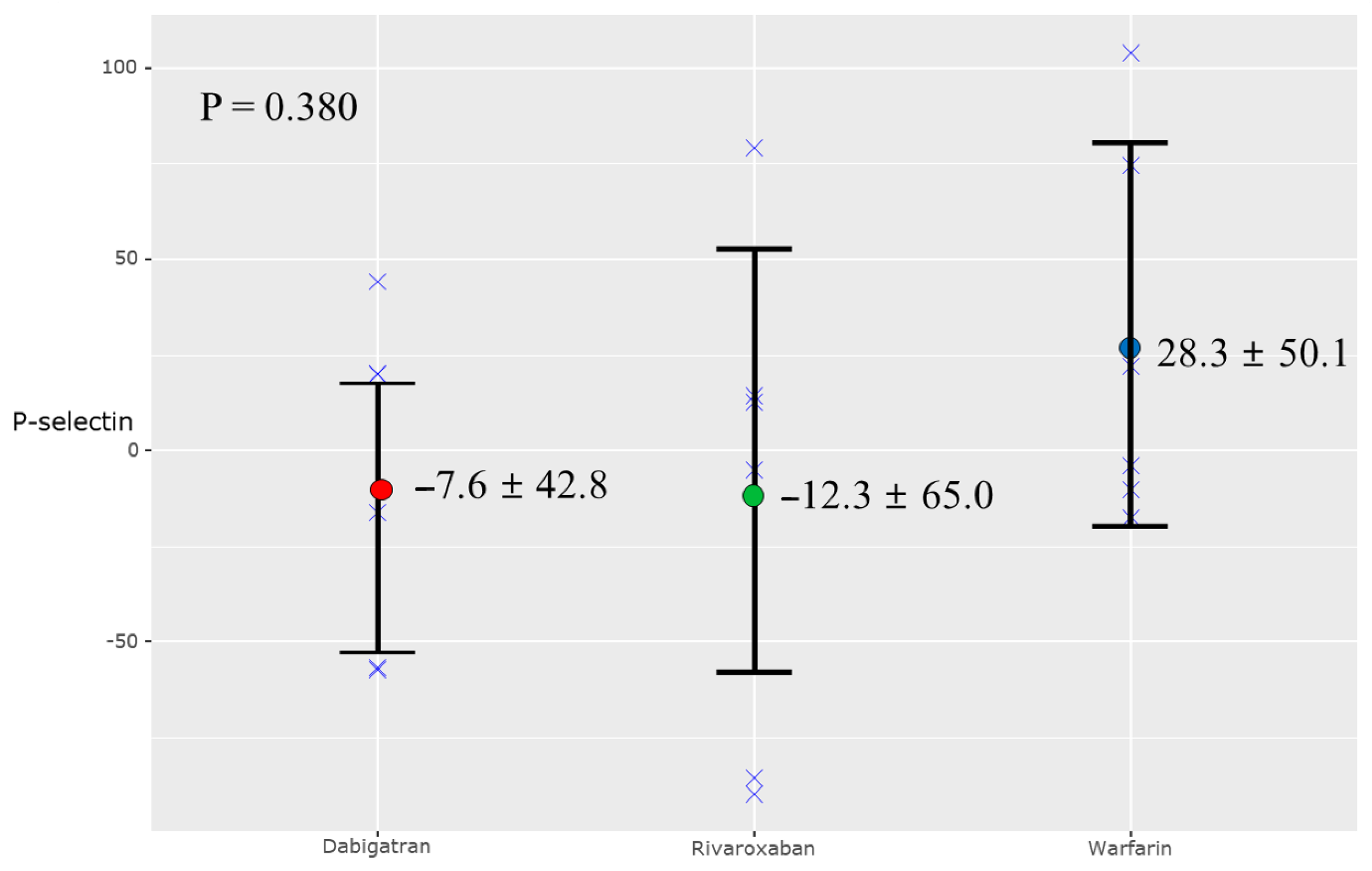
Change in biomarkers at 12 months
  IL-6 (pg/mL)3.5 ± 11.4−0.1 ± 1.60.279
  TNF-α (pg/mL)1.8 ± 5.61.5 ± 1.80.856
  P-selectin (ng/mL)−5.4 ± 54.743.5 ± 60.80.078
  vWF (μg/mL)0.7 ± 2.7−0.8 ± 2.40.225
IL-6 = Interleukin-6; TNF-α = Tumor necrosis factor alpha; vWF = Von Willebrand Factor.
Publisher’s Note: MDPI stays neutral with regard to jurisdictional claims in published maps and institutional affiliations.

