Article

Severe Myocardial Dysfunction after Non-Ischemic Cardiac Arrest: Effectiveness of Percutaneous Assist Devices

1
Department of Cardiology, Lariboisière Hospital, Assistance Publique-Hôpitaux de Paris (AP-HP), Denis Diderot University, INSERM UMRS 942, 75010 Paris, France
2
Abiomed Europe GmbH, Neuenhofer Weg 3, D-52074 Aachen, Germany
3
Department of Cardiovascular and Thoracic Surgery, Institute of Cardiology, Pitié-Salpêtrière Hospital, Assistance Publique-Hôpitaux de Paris (AP-HP), Sorbonne University, 47-83 Boulevard de l’Hôpital, 75013 Paris, France
4
XP-MED, 78100 Saint Germain en Laye, France
*
Author to whom correspondence should be addressed.
J. Clin. Med. 2021, 10(16), 3623; https://doi.org/10.3390/jcm10163623
Submission received: 30 June 2021 / Revised: 3 August 2021 / Accepted: 13 August 2021 / Published: 17 August 2021
(This article belongs to the Special Issue Cardiogenic Shock: Updates, Challenges and Opportunities)

Abstract

:
Introduction: Despite the improvements in standardized cardiopulmonary resuscitation, survival remains low, mainly due to initial myocardial dysfunction and hemodynamic instability. Our goal was to compare the efficacy of two left ventricular assist devices on resuscitation and hemodynamic supply in a porcine model of ventricular fibrillation (VF) cardiac arrest. Methods: Seventeen anaesthetized pigs had 12 min of untreated VF followed by 6 min of chest compression and boluses of epinephrine. Next, a first defibrillation was attempted and pigs were randomized to any of the three groups: control (n = 5), implantation of an percutaneous left ventricular assist device (Impella, n = 5) or extracorporeal membrane oxygenation (ECMO, n = 7). Hemodynamic and myocardial functions were evaluated invasively at baseline, at return of spontaneous circulation (ROSC), after 10–30–60–120–240 min post-resuscitation. The primary endpoint was the rate of ROSC. Results: Only one of 5 pigs in the control group, 5 of 5 pigs in the Impella group, and 5 of 7 pigs in the ECMO group had ROSC (p < 0.05). Left ventricular ejection fraction at 240 min post-resuscitation was 37.5 ± 6.2% in the ECMO group vs. 23 ± 3% in the Impella group (p = 0.06). No significant difference in hemodynamic parameters was observed between the two ventricular assist devices. Conclusion: Early mechanical circulatory support appeared to improve resuscitation rates in a shockable rhythm model of cardiac arrest. This approach appears promising and should be further evaluated.

1. Introduction

Cardiac arrest is a major cause of mortality and morbidity in Western countries. Each year, more than 350,000 people in the United States die suddenly [1]. Coronary artery disease (CAD) is a major cause of out-of-hospital cardiac arrest (OHCA) [2,3]. However, in 30% of patients, cardiac arrest is neither associated with CAD nor with an obvious extracardiac cause [4]. Despite recent advances in public education and resuscitation processes, only a few patients are transported alive to the hospital, and even less are discharged from the hospital without sequelae. Even among patients arriving at the hospital, survival rates remain low, ranging from 21–33%, and have not improved in recent years [5,6,7]. Successful treatment of cardiac arrest includes the rapid initiation of basic life support measures, including cardiopulmonary resuscitation (CPR). Immediate institution of such efforts can result in resuscitation rates as high as 30–40% [8,9].
In the cardiovascular post-resuscitation syndrome, cardiac filling pressures temporarily increase and the cardiac index dramatically decline [7,10]. Even after effective resuscitation and recovery of myocardial blood flow, ventricular functions are markedly depressed [10,11,12]. These findings underscore the need to restore spontaneous circulation via rapid and aggressive supportive therapy, with the expectation that part of myocardial stunning will recover. The need of an early-onset invasive strategy for OHCA was the central premise of the study evaluating the feasibility and safety of pre-hospital insertion of extra-corporeal membrane oxygenation (ECMO) [13]. Recently, the ARREST study reported improved survival in ECMO-facilitated resuscitation [14], as well as a Danish multicenter study [15].
In patients with acute heart failure, percutaneous mechanical circulatory support (MCS) devices are increasingly used [16]. Durable ventricular assist devices are complex, require surgical implantation, and are expensive. Thus, their use in the setting of cardiac arrest is not indicated given the uncertain prognosis and urgent nature of care. Percutaneous MCS devices are a viable option in cardiac arrest, owing to the rapid initiation of support and ease of use. The feasibility and superiority of ECMO compared to conventional CPR in adults with in-hospital cardiac arrest have been demonstrated [17,18]. Nevertheless, the use of ECMO involves large cannula, complex vascular access, and is associated with ischemic and infectious complications [19].
The Impella CP percutaneous assist device (Abiomed Inc., Danvers, MA, USA) is a valuable alternative to ECMO.
We previously reported that left-sided percutaneous support with Impella 2.5 was feasible in post-resuscitation shock with a high percentage of good neurological recovery [20]. These results were confirmed in a retrospective multicenter analysis [21] and a Danish multicenter study recently confirmed higher survival rate in cardiac arrested patient in adherence to national consensus selection criteria [15].
As ECMO has already been investigated as a rescue-device in refractory cardiac arrest with early implantation of the device in the field [13,22,23], the objective of this study was to compare the two percutaneous mechanical circulatory support devices that can be deployed early: The Impella CP versus ECMO. We compared ROSC obtention, hemodynamic parameters, myocardial recovery, and peripheral organ damage in a porcine model of ventricular fibrillation (VF) cardiac arrest, as the best model in cardiac arrest study [12,24,25].

2. Material and Methods

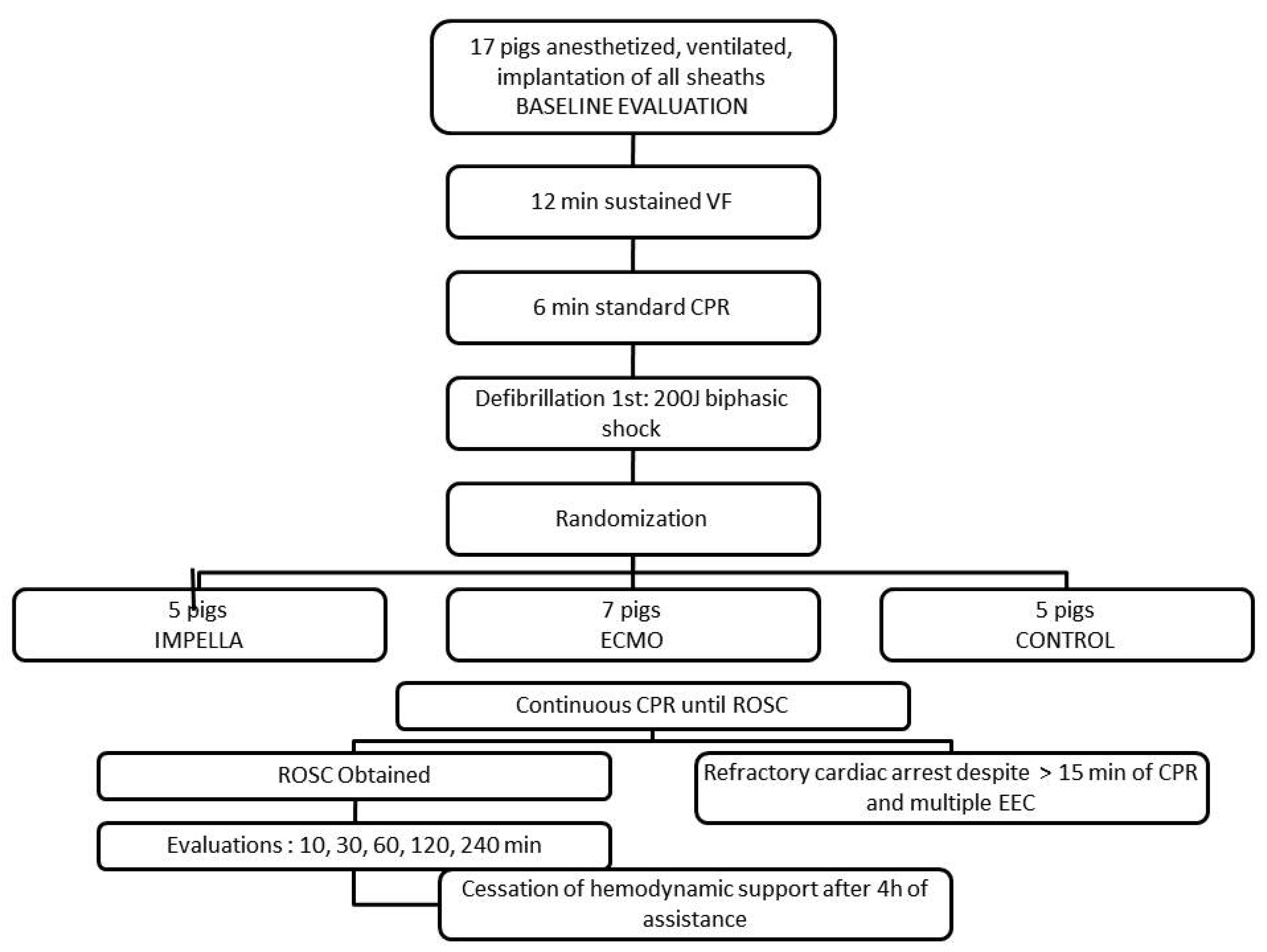
2.1. Animal Preparation

All procedures were conducted in accordance with the basic principles for the care and use of animals based on the Helsinki Declaration and were approved by the appropriate governmental institution. Seventeen male domestic pigs at 4 months of age (weighing 34.6 ± 4.6 kg) were studied in a model of untreated VF followed by mechanical support alone to mimic out-of-hospital cardiac arrest treatment.
Under anesthesia (Isoflurane) and mechanical endotracheal ventilation, a Swan-Ganz catheter was placed through the left external jugular vein and a 7F sheath was inserted in the right external jugular vein to place the electrodes for the required pacing [12]. A 6F sheath was introduced in the right carotid artery to place a pig tail catheter into the left ventricular cavity for the measurement of LV pressures and the angiography for LVEF estimation [12,26].
The left femoral artery was used as an access for continuous invasive pressure measurement.
The Impella was placed via the right femoral artery through a 13F sheath.
In the ECMO group, the arterial 15F cannula was placed in the right femoral artery and the venous cannula (21 F) was inserted through the left femoral vein.
ECG, pulse oximetry, and rectal temperature were continuously monitored using a standard patient monitor.
Peripheral tissue oxygen saturation (StO2) levels were measured using near infrared spectroscopy (NIRS) with an INVOS® Cerebral/Somatic Oximeter (Somanetics Corporation, Troy, MI, USA). The sensor was attached to the forehead.
Unfractioned heparin (5000 UI IV) was given as a bolus after sheath placement followed by additional boluses of 2500 UI IV to maintain an activated clotting time between 180 and 250 s.
Initial rapid IV infusion of 1000 mL of normal saline was given intravenously, followed by a continuous IV drip of 200 to 500 mL/h to reach and maintain a central venous pressure of 3–7 mmHg. Additional saline as rapid IV boluses of 100–250 mL was administered during the period of assistance.
Prior to the experimental protocol, a stabilization period of one hour was used to let the animal recover from instrumentation. Simple randomization was performed to define group allocation for each animal as experimentations were planned.

2.2. Experimental Protocol

VF was electrically induced by pacing the right ventricle at rates of 400–600 beats/min as confirmed by electrocardiography (ECG) and loss of the aortic pressure pulsations. Anesthesia and ventilation were discontinued during the arrest period. Cardiopulmonary resuscitation efforts were initiated after 12 min of untreated VF. The 12-min untreated VF period was chosen so that the myocardial insult would be severe enough to allow discrimination of outcomes among the three cardiac assistance groups.
Resuscitation [11,27,28,29] efforts included standard Basic Life Support, such as external chest compressions (by a pneumatically driven automatic piston device: Pneumatic Compression Controller, Ambu International, Glostrup, Denmark) and positive pressure ventilation with room air (FiO2 of 21%). The piston device provided a 4–6 cm compression, adjusted on the pig anatomy, at a rate of 100/min. The tidal volume was maintained at 10 mL/Kg. Epinephrine was administered as a 1-mg intravenous bolus at the initiation of CPR and 3 min after initiation. After 6 min of CPR, one external defibrillation (Zoll) was attempted with a 200 J biphasic shock, and additional defibrillation shocks, still biphasic 200 J, were delivered every 3 min preceded by an intravenous bolus of 1 mg epinephrine, if needed. After 6 min of CPR and the first external electric shock, cardiac assistance was started according to the group assignment (Figure 1). For logistic reasons, assignment to the groups was decided at the beginning of the experiment.
Resuscitation efforts were continued until ROSC was achieved or for a total of 15 min after delivery of the first external electric shock.
After ROSC was achieved, animals were mechanically ventilated, with supplemental oxygen if needed to obtain an arterial saturation greater than 90%. During the immediate post defibrillation stage, amiodarone, epinephrine or atropine was used, according to the advanced cardiac life support guidelines [7,28,29,30,31,32,33,34,35].
All groups received therapeutic hypothermia immediately after ROSC by infusion of 1.5 L of chilled saline with a target temperature between 34 and 36 °C. When the animals were stabilized, they were observed under general anesthesia and isoflurane was titrated to maintain adequate anesthesia and analgesia during the whole period of evaluation.
Animals remained instrumented for measurement of hemodynamic parameters. Fluid loading by saline was infused if required, likewise epinephrine boluses and, if needed infusion, depending on the hemodynamic parameters.
After the final data collection was completed, animals were euthanized by an intravenous solution of Doléthal® (Vétoquinol S.A., Lure, France) and necropsy studies were performed by a veterinarian. All the animals involved in the study were included in the analysis, no exclusion criteria were applied.

2.3. Data Analysis

In vivo evaluations were performed at baseline, before cardiac arrest, after successful resuscitation, and before initiation of hemodynamic support (if available), then 10–30–60–120–240 min after hemodynamic support.
The primary outcome of this study was the rate of ROSC.
Secondary outcomes were:
(1)
LV function (LVEF) after 4 h post-resuscitation measured by fluoroscopy (RAO 30° incidence) using an injection of 12 mL contrast medium (Visipaque®, GE Healthcare SAS, Boston, MA, USA) [26]
(2)
Myocardial function: Left ventricular end-diastolic pressure was measured by the pigtail, cardiac outflow, systolic ejection volume (mL/min), left ventricular systolic and diastolic volumes measured by fluoroscopy (RAO 30° incidence), and calculated using the standard software of the angiography suite;
(3)
Hemodynamic efficacy assessed based on systolic, mean and diastolic arterial pressures, central venous pressure, and pulmonary arterial pressure.

2.4. Statistical Analysis

Values are expressed as mean ± SD or number (%), as appropriate. Due to the small sample size and the likelihood of high heterogeneity, the experiments were planned as a hypothesis-generating study. The primary endpoint of successful ROSC was statistically evaluated using a Fisher’s exact test for a 2 × 3 contingency table. All secondary endpoints are merely descriptive. Except for the estimation of rate of ROSC, the control group was not included in the comparative analysis due to its poor outcome. In fact, within the control group, only one animal out of five was successfully resuscitated.
Non-normal distribution of the data was confirmed using the Shapiro test. Differences between groups were assessed using exact Wilcoxon-Mann-Whitney Test. All statistical analyses were performed using the R-statistical software (R Foundation for Statistical Computing, Vienna, Austria). A 2-sided p-value < 0.05 was considered statistically significant.

3. Results

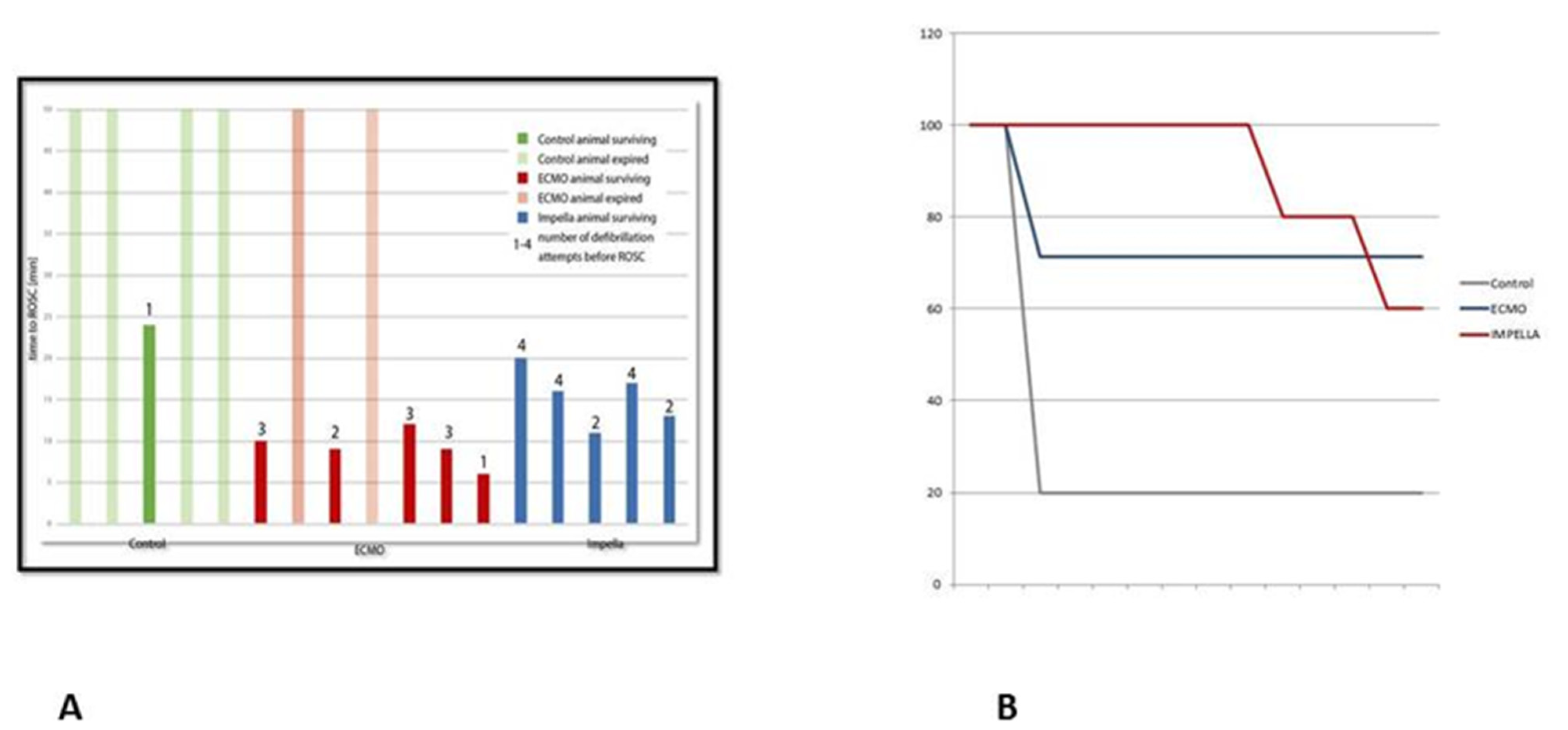
3.1. Resuscitation, ROSC and Survival

The characteristics of the resuscitation between the 3 groups are presented in Table 1. As shown in Figure 2A, pigs receiving hemodynamic support showed a higher rate of ROSC, with a 100% (5/5) success rate in the Impella group, 71.4% (5/7) in the ECMO group, and 20% (1/5) in the control group (p = 0.044). In pigs achieving ROSC, the duration to ROSC was shorter in the ECMO group. Nevertheless, two pigs treated with ECMO died during this period and post-mortem autopsy revealed hemorrhagic complications related to arterial (carotid or iliac) injuries. A third pig in the ECMO group also died despite initial ROSC within a few minutes due to iliac arterial bleeding. All pigs in the Impella group survived the initial phase of the experiment but 2 pigs died between 120–240 min. In one of the pigs, asystole occurred two hours after ROSC, CPR failed, and the post-mortem revealed a hemomediastinum, possibly a consequence of the mechanical chest compression. In the second pig, VF occurred after four hours of Impella support and multiple electrical cardioversion attempts failed. The post-mortem examination revealed severe cardiac hypertrophy as a pre-existing pathology and the presumable cause for the re-occurring and resistant arrhythmia.
As can be expected, the amount of epinephrine administered was lower in the mechanically assisted pigs. Despite a higher initial survival rate within the Impella group, no difference in survival was observed between the two assist devices at the end of the experiment (Figure 2B).

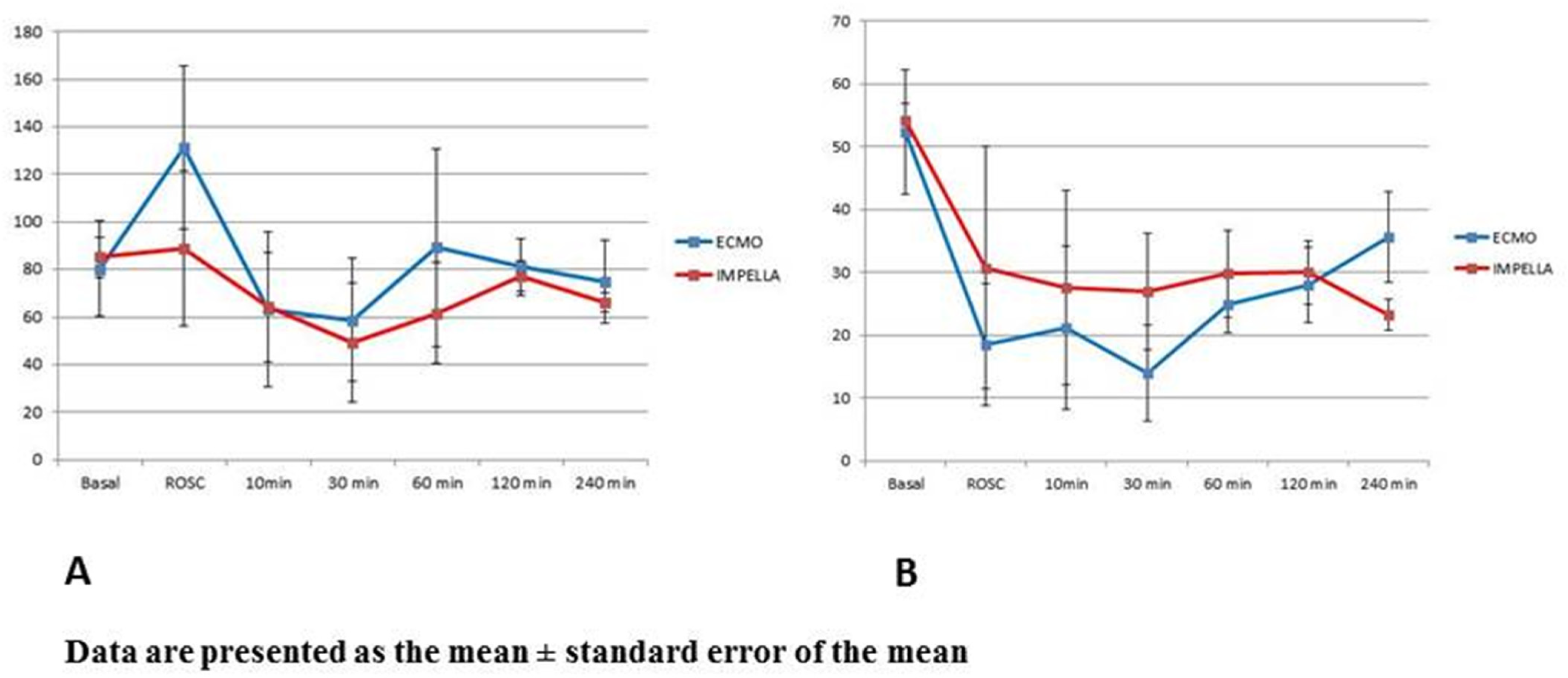
3.2. Mechanical Circulatory Support and Hemodynamic

The Impella system maintained sufficient mean aortic pressure (Figure 3A) during the entire experiment despite reduced flow rate than ECMO (Figure S1 in Supplementary Materials). Although the total flow was measured using a Swan-Ganz catheter (thermodilution method), these measurements are typically highly unreliable in the setting of mechanical circulatory support.
Even though pigs in the ECMO group showed a sustained reduction in LVEF in the early phase post-ROSC, there was no significant difference in LVEF between the two cardiac assist devices at 4 h post-resuscitation (Figure 3B).
Table 2 shows the different measurements of the myocardial function in the two groups receiving hemodynamic support.
No significant difference in hemodynamic parameters was observed between the support devices (Table 3). There was no difference in the hemodynamic variables between the devices at baseline. At the time of ROSC, the ECMO showed a trend towards better hemodynamics with higher MAP 131 vs. 89 mmHg (p = 0.15). Pulmonary arterial pressure was not different between the devices throughout the experiment.
As per the protocol, no correction of the acid-base balance was initiated during the 4 h of mechanical support. Blood gas analyses (Table 3) showed a numerically increased paO2 at the oxygenation membrane of the ECMO. The lactate levels did not differ between the two groups.

4. Discussion

This animal study demonstrated that the prompt initiation of percutaneous cardiac assistance during experimental cardiac arrest increased the rate of ROSC with a 100% success with the Impella device in the setting of prolonged VF. Compared to ECMO, no difference in hemodynamic parameters was observed with Impella.

4.1. Effectiveness of Prompt Cardiac Assistance

Cardiac assist devices are frequently used as a rescue therapy following failure of standard CPR. Data from clinical studies evaluating use of ECMO in the setting of OHCA are limited and they report low survival rates and disappointing neurologic outcomes [36,37]. However, ECMO is still useful in situations requiring high flow support and optimal oxygenation [15,38]. Schmidt et al. suggested 10 situations in which use of ECMO is often futile and included a case series of critically ill patients with long low-flow periods, lasting more than 30 min and more [39]. Recently, Lamhaut et al. demonstrated the feasibility and safety of initiation of pre-hospital ECMO for refractory CA in the field by an experienced team [13]. Their propensity analysis [22] also suggested that prehospital ECMO in a selected population may improve survival and neurological outcome, in particular if initiated earlier.
Our animal study supports this new paradigm of prompt initiation of cardiac support. The delay of 12 min was chosen to cause a significant myocardial dysfunction and could also reflects the clinical setting with a medium delay of emergency team arrival. The delay to start the assistance device would add approximately another ten minutes by experienced teams.
The use of Impella would involve a less invasive approach with lower vascular complication rates than ECMO, yet would require placement using radiography, thus currently limiting its use in the field.
In a similar animal experimental setup, Derwall et al. showed increased survival and improved outcome by CPR using Impella instead of chest compression [24]. Standard CPR with chest compression alone, even of high quality by a mechanical thumper demonstrated disappointing results [40]. The current growing evidence of epinephrine´s ambivalent results, showing higher rates of sustained ROSC but without any improvement in favorable neurological outcomes [41,42] supports the need for new approaches in resuscitation, in particular, a shift toward higher use of mechanical assist devices. The Danish multicenter study reported improved survival rate and good neurological outcome in a population of patient treated by mechanical circulatory support for OHCA [15]

4.2. Hemodynamic Support

Left ventricular assistance with Impella has been shown to have beneficial effects on coronary perfusion and the effective unloading of the LV reduced myocardial injury after reperfusion [43,44].
Based on these findings, active unloading with Impella in the acute phase may improve myocardial recovery after the initial stunning compared to the increase in afterload exerted by ECMO. Kern et al. showed complete recovery of myocardial stunning by 48 h after sustained VF [12]. However, in the animal model used in this study, measurements such as LVEF were taken only until 4 h after ROSC; nevertheless, the LVEF was also already improving by 4 h after ROSC.
Early after ROSC, LVEF in the Impella group seemed to be preserved during the first 2 h but started to decrease later. Within our experimental setting, the favorable effect of unloading by the Impella pump was counterbalanced by suction events that necessitated a reduction of flow. Automatic adaptation of flow, as available in the console for clinical use, was not effective in our setting. On the other hand, ECMO showed a reduction in LVEF during the initial phase, presumably due to an increase in afterload, and thus a delayed recovery. The young and healthy hearts might be able to compensate for the increase in the afterload over time with recovery within one hour post-ROSC. However, this increase in afterload might have severe consequences in the setting of heart condition with pre-existing comorbidities.
The expected reduction of LVEDP following LV unloading was not confirmed in our experiment. In fact, the pulmonary pressures did not decrease with unloading as shown previously in other experiments [24].

4.3. Limitations

We recognize several limitations in this hypothesis generating study without any sample size calculation. First, the pigs in the control group representing the clinical situation of cardiac arrest treatment without mechanical support had a very low rate of ROSC, thus limiting the comparisons. Nevertheless, the low rate of immediately successful resuscitation was comparable to the one observed in the literature [24]. The poor survival in the control group convinced us to not expose additional pigs to this condition.
Second, due to short-term evaluations, we did not perform clinical and neurological assessments. However, our measurement until four hours of support showed trends that can be interpreted. Invasive measurements of baseline hemodynamic parameters could have compromised survival and ROSC success rates.
Third, in order to mimic the constraints in the setting of OHCA, all biological measurements were analyzed after the experiment. As a result, ventilation was not adapted according to the blood gases for any of the groups, resulting in differences between the groups and hyperventilation in the ECMO group. Likewise, initial ventilation with room air (FiO2 of 21%) was chosen to be closer to the initial ventilation conditions “on the field”. A more intensive oxygenation would have been more efficient. We acknowledged that the resuscitation did not completely reflect current guideline recommendations.
Fourth, the complications observed in the ECMO group deserve consideration. The highly invasive procedure of cannulation in an emergency situation increases the risk of arterial misplacement and dissection, even in the relatively controlled setting of an animal experiment. This procedural aspect may partially explain the worse results observed in OHCA patients [13,36] compared to in-hospital cardiac arrest patients assisted with an ECMO [17]. The placement of the Impella requires vascular access and in patients with cardiac arrest, puncture of the femoral artery remains feasible and can be performed using ultrasound guidance [45].
Finally, all the pigs had normal myocardium without coronary artery disease and one can speculate that the myocardial stunning after prolonged VF could be even more marked in aged hearts with concomitant atherosclerotic disease and prompt LV support in this setting could improve outcomes significantly.

5. Conclusions

In this porcine model of VF cardiac arrest, early assistance with cardiac devices was associated with improved rates of return of spontaneous circulation. No significant difference was observed between Impella and ECMO percutaneous circulatory support, may be due to small sample size. The use of percutaneous circulatory support in the setting of cardiac arrest to increase the rate of ROSC is a promising strategy. Further clinical randomized studies are warranted to delineate the safety and effectiveness of these devices specifically.

Supplementary Materials

The following are available online at https://www.mdpi.com/article/10.3390/jcm10163623/s1, Figure S1: Flow delivered by the devices.

Author Contributions

S.M.-S. conceived of the study and its design, carried out the experiments, participated in the data acquisition, performed the statistical analysis, analyzed and interpreted the data and drafted the manuscript. C.N. and A.G. participated in the statistical analysis and helped to revise the manuscript. P.D., C.N., C.K. and M.B. helped in performing the experiments and revised the manuscript. P.L. and G.L. participated in the design of the study and revised the manuscript critically. A.C.-S., P.L. and G.L. participated in the design of the study and helped to interpret the data and revised the manuscript. All authors have read and agreed to the published version of the manuscript.

Funding

This research was founded by Abiomed Inc., Danvers, MA, USA, and MAQUET Cardiovascular provided all the ECMO equipment.

Institutional Review Board Statement

This study was approved (Avis 12/031) by the Ethics Committee applied to Animal Experimentation at the Jouy-en-Josas Center and AgroParisTech.

Data Availability Statement

All the data supporting reported results can be provided upon request to the corresponding author.

Acknowledgments

We thank Corinne Vrech and Francis Ballet for their assistance. The present submission has been decided in accordance with the study sponsor. We thank Renaud Tissier and Alain Berdeau for their experimental advices and Georges Moulin for his skillful technical assistance.

Conflicts of Interest

S.M.-S. is recipient of a grant provided by Abiomed Europe GmbH. C.N. and A.G. are employees of Abiomed Europe GmbH. The remaining authors declare that they have no competing interests.

Abbreviations

ANOVAAnalysis of variance
CACardiac arrest
CADCoronary artery disease
COCardiac output
CPRCardiopulmonary resuscitation
ECMOExtracorporeal membrane oxygenation
FiO2Inspired oxygen fraction
HRHeart rate
IVIntravenous
LVleft ventricle
LVEDPLeft ventricle end diastolic pressure
LVEDVLeft ventricle end diastolic volume
LVEFLeft ventricle ejection fraction
MAPMean arterial pressure
MPAPMean pulmonary artery pressure
OHCAOut hospital cardiac arrest
PaCO2Arterial carbon dioxide tension
PaO2Arterial oxygen tension
ROSCReturn of spontaneous circulation
VFVentricular fibrillation.

References

  1. Benjamin, E.J.; Virani, S.S.; Callaway, C.W.; Chamberlain, A.M.; Chang, A.R.; Cheng, S.; Chiuve, S.E.; Cushman, M.; Delling, F.N.; Deo, R.; et al. Heart Disease and Stroke Statistics-2018 Update: A Report from the American Heart Association. Circulation 2018, 137, e67–e492. [Google Scholar] [CrossRef] [PubMed]
  2. Eisenberg, M.S.; Mengert, T.J. Cardiac resuscitation. N. Engl. J. Med. 2001, 344, 1304–1313. [Google Scholar] [CrossRef]
  3. Bunch, T.J.; White, R.D.; Gersh, B.J.; Meverden, R.A.; Hodge, D.O.; Ballman, K.V.; Hammill, S.C.; Shen, W.K.; Packer, D.L. Long-term outcomes of out-of-hospital cardiac arrest after successful early defibrillation. N. Engl. J. Med. 2003, 348, 2626–2633. [Google Scholar] [CrossRef] [PubMed] [Green Version]
  4. Dumas, F.; Cariou, A.; Manzo-Silberman, S.; Grimaldi, D.; Vivien, B.; Rosencher, J.; Empana, J.-P.; Carli, P.; Mira, J.-P.; Jouven, X.; et al. Immediate Percutaneous Coronary Intervention Is Associated with Better Survival After Out-of-Hospital Cardiac Arrest: Insights from the PROCAT (Parisian Region Out of Hospital Cardiac Arrest) Registry. Circ. Cardiovasc. Interv. 2010, 3, 200–207. [Google Scholar] [CrossRef] [PubMed] [Green Version]
  5. Grubb, N.R.; Elton, R.A.; Fox, K.A. In-hospital mortality after out-of-hospital cardiac arrest. Lancet 1995, 346, 417–421. [Google Scholar] [CrossRef]
  6. Herlitz, J.; Engdahl, J.; Svensson, L.; Angquist, K.A.; Silfverstolpe, J.; Holmberg, S. Major differences in 1-month survival between hospitals in Sweden among initial survivors of out-of-hospital cardiac arrest. Resuscitation 2006, 70, 404–409. [Google Scholar] [CrossRef] [PubMed]
  7. Neumar, R.W.; Nolan, J.P.; Adrie, C.; Aibiki, M.; Berg, R.A.; Bottiger, B.W.; Callaway, C.; Clark, R.S.; Geocadin, R.G.; Jauch, E.C.; et al. Post-cardiac arrest syndrome: Epidemiology, pathophysiology, treatment, and prognostication. A consensus statement from the International Liaison Committee on Resuscitation (American Heart Association, Australian and New Zealand Council on Resuscitation, European Resuscitation Council, Heart and Stroke Foundation of Canada, InterAmerican Heart Foundation, Resuscitation Council of Asia, and the Resuscitation Council of Southern Africa); the American Heart Association Emergency Cardiovascular Care Committee; the Council on Cardiovascular Surgery and Anesthesia; the Council on Cardiopulmonary, Perioperative, and Critical Care; the Council on Clinical Cardiology; and the Stroke Council. Circulation 2008, 118, 2452–2483. [Google Scholar]
  8. Berdowski, J.; Berg, R.A.; Tijssen, J.G.; Koster, R.W. Global incidences of out-of-hospital cardiac arrest and survival rates: Systematic review of 67 prospective studies. Resuscitation 2010, 81, 1479–1487. [Google Scholar] [CrossRef]
  9. Wang, H.E.; Devlin, S.M.; Sears, G.K.; Vaillancourt, C.; Morrison, L.J.; Weisfeldt, M.; Callaway, C.W. Regional variations in early and late survival after out-of-hospital cardiac arrest. Resuscitation 2013, 83, 1343–1348. [Google Scholar] [CrossRef] [Green Version]
  10. Cerchiari, E.L.; Safar, P.; Klein, E.; Cantadore, R.; Pinsky, M. Cardiovascular function and neurologic outcome after cardiac arrest in dogs. The cardiovascular post-resuscitation syndrome. Resuscitation 1993, 25, 9–33. [Google Scholar] [CrossRef]
  11. Yannopoulos, D.; Matsuura, T.; McKnite, S.; Goodman, N.; Idris, A.; Tang, W.; Aufderheide, T.P.; Lurie, K.G. No assisted ventilation cardiopulmonary resuscitation and 24-hour neurological outcomes in a porcine model of cardiac arrest. Crit. Care Med. 2010, 38, 254–260. [Google Scholar] [CrossRef]
  12. Kern, K.B.; Hilwig, R.W.; Rhee, K.H.; Berg, R.A. Myocardial dysfunction after resuscitation from cardiac arrest: An example of global myocardial stunning. J. Am. Coll. Cardiol. 1996, 28, 232–240. [Google Scholar] [CrossRef] [Green Version]
  13. Lamhaut, L.; Jouffroy, R.; Soldan, M.; Phillipe, P.; Deluze, T.; Jaffry, M.; Dagron, C.; Vivien, B.; Spaulding, C.; An, K.; et al. Safety and feasibility of prehospital extra corporeal life support implementation by non-surgeons for out-of-hospital refractory cardiac arrest. Resuscitation 2013, 84, 1525–1529. [Google Scholar] [CrossRef] [PubMed]
  14. Yannopoulos, D.; Bartos, J.; Raveendran, G.; Walser, E.; Connett, J.; Murray, T.A.; Collins, G.; Zhang, L.; Kalra, R.; Kosmopoulos, M.; et al. Advanced reperfusion strategies for patients with out-of-hospital cardiac arrest and refractory ventricular fibrillation (ARREST): A phase 2, single centre, open-label, randomised controlled trial. Lancet 2020, 396, 1807–1816. [Google Scholar] [CrossRef]
  15. Mork, S.R.; Stengaard, C.; Linde, L.; Moller, J.E.; Jensen, L.O.; Schmidt, H.; Riber, L.P.; Andreasen, J.B.; Thomassen, S.A.; Laugesen, H.; et al. Mechanical circulatory support for refractory out-of-hospital cardiac arrest: A Danish nationwide multicenter study. Crit. Care 2021, 25, 174. [Google Scholar] [CrossRef]
  16. Rihal, C.S.; Naidu, S.S.; Givertz, M.M.; Szeto, W.Y.; Burke, J.A.; Kapur, N.K.; Kern, M.; Garratt, K.N.; Goldstein, J.A.; Dimas, V.; et al. 2015 SCAI/ACC/HFSA/STS Clinical Expert Consensus Statement on the Use of Percutaneous Mechanical Circulatory Support Devices in Cardiovascular Care: Endorsed by the American Heart Assocation, the Cardiological Society of India, and Sociedad Latino Americana de Cardiologia Intervencion; Affirmation of Value by the Canadian Association of Interventional Cardiology-Association Canadienne de Cardiologie d’intervention. J. Am. Coll. Cardiol. 2015, 65, e7–e26. [Google Scholar] [CrossRef] [Green Version]
  17. Chen, Y.S.; Lin, J.W.; Yu, H.Y.; Ko, W.J.; Jerng, J.S.; Chang, W.T.; Chen, W.J.; Huang, S.C.; Chi, N.H.; Wang, C.H.; et al. Cardiopulmonary resuscitation with assisted extracorporeal life-support versus conventional cardiopulmonary resuscitation in adults with in-hospital cardiac arrest: An observational study and propensity analysis. Lancet 2008, 372, 554–561. [Google Scholar] [CrossRef]
  18. Ouweneel, D.M.; Schotborgh, J.V.; Limpens, J.; Sjauw, K.D.; Engstrom, A.E.; Lagrand, W.K.; Cherpanath, T.G.V.; Driessen, A.H.G.; de Mol, B.; Henriques, J.P.S. Extracorporeal life support during cardiac arrest and cardiogenic shock: A systematic review and meta-analysis. Intensiv. Care Med. 2016, 42, 1922–1934. [Google Scholar] [CrossRef] [Green Version]
  19. Cheng, R.; Hachamovitch, R.; Kittleson, M.; Patel, J.; Arabia, F.; Moriguchi, J.; Esmailian, F.; Azarbal, B. Complications of extracorporeal membrane oxygenation for treatment of cardiogenic shock and cardiac arrest: A meta-analysis of 1866 adult patients. Ann. Thorac. Surg. 2014, 97, 610–616. [Google Scholar] [CrossRef]
  20. Manzo-Silberman, S.; Fichet, J.; Mathonnet, A.; Varenne, O.; Ricome, S.; Chaib, A.; Zuber, B.; Spaulding, C.; Cariou, A. Percutaneous left ventricular assistance in post cardiac arrest shock: Comparison of intra aortic blood pump and IMPELLA Recover LP2.5. Resuscitation 2013, 84, 609–615. [Google Scholar] [CrossRef]
  21. Panagides, V.; Vase, H.; Shah, S.P.; Basir, M.B.; Mancini, J.; Kamran, H.; Batra, S.; Laine, M.; Eiskjaer, H.; Christensen, S.; et al. Impella CP Implantation during Cardiopulmonary Resuscitation for Cardiac Arrest: A Multicenter Experience. J. Clin. Med. 2021, 10, 339. [Google Scholar] [CrossRef] [PubMed]
  22. Lamhaut, L.; Hutin, A.; Puymirat, E.; Jouan, J.; Raphalen, J.H.; Jouffroy, R.; Jaffry, M.; Dagron, C.; An, K.; Dumas, F.; et al. A Pre-Hospital Extracorporeal Cardio Pulmonary Resuscitation (ECPR) strategy for treatment of refractory out hospital cardiac arrest: An observational study and propensity analysis. Resuscitation 2017, 117, 109–117. [Google Scholar] [CrossRef] [Green Version]
  23. Lamhaut, L.; Jouffroy, R.; Kalpodjian, A.; Deluze, T.; Phillipe, P.; Vivien, B.; An, K.; Carli, P. Successful treatment of refractory cardiac arrest by emergency physicians using pre-hospital ECLS. Resuscitation 2012, 83, e177–e178. [Google Scholar] [CrossRef] [PubMed]
  24. Derwall, M.; Brucken, A.; Bleilevens, C.; Ebeling, A.; Fohr, P.; Rossaint, R.; Kern, K.B.; Nix, C.; Fries, M. Doubling survival and improving clinical outcomes using a left ventricular assist device instead of chest compressions for resuscitation after prolonged cardiac arrest: A large animal study. Crit. Care 2015, 19, 123. [Google Scholar] [CrossRef] [PubMed] [Green Version]
  25. Hutin, A.; Lamhaut, L.; Lidouren, F.; Kohlhauer, M.; Mongardon, N.; Carli, P.; Berdeaux, A.; Ghaleh, B.; Tissier, R. Early Coronary Reperfusion Facilitates Return of Spontaneous Circulation and Improves Cardiovascular Outcomes After Ischemic Cardiac Arrest and Extracorporeal Resuscitation in Pigs. J. Am. Heart Assoc. 2016, 5, e004588. [Google Scholar] [CrossRef]
  26. Greene, D.G.; Carlisle, R.; Grant, C.; Bunnell, I.L. Estimation of left ventricular volume by one-plane cineangiography. Circulation 1967, 35, 61–69. [Google Scholar] [CrossRef] [PubMed] [Green Version]
  27. Segal, N.; Matsuura, T.; Caldwell, E.; Sarraf, M.; McKnite, S.; Zviman, M.; Aufderheide, T.P.; Halperin, H.R.; Lurie, K.G.; Yannopoulos, D. Ischemic postconditioning at the initiation of cardiopulmonary resuscitation facilitates functional cardiac and cerebral recovery after prolonged untreated ventricular fibrillation. Resuscitation 2012, 83, 1397–1403. [Google Scholar] [CrossRef]
  28. Ong, M.E.H.; Perkins, G.D.; Cariou, A. Out-of-hospital cardiac arrest: Prehospital management. Lancet 2018, 391, 980–988. [Google Scholar] [CrossRef] [Green Version]
  29. Soar, J.; Donnino, M.W.; Maconochie, I.; Aickin, R.; Atkins, D.L.; Andersen, L.W.; Berg, K.M.; Bingham, R.; Bottiger, B.W.; Callaway, C.W.; et al. 2018 International Consensus on Cardiopulmonary Resuscitation and Emergency Cardiovascular Care Science with Treatment Recommendations Summary. Resuscitation 2018, 133, 194–206. [Google Scholar] [CrossRef] [Green Version]
  30. Guidelines 2000 for Cardiopulmonary Resuscitation and Emergency Cardiovascular Care. Part 1: Introduction to the International Guidelines 2000 for CPR and ECC: A consensus on science. Circulation 2000, 102 (Suppl. 8), I1–I11.
  31. Guidelines 2000 for Cardiopulmonary Resuscitation and Emergency Cardiovascular Care. Part 3: Adult basic life support. The American Heart Association in collaboration with the International Liaison Committee on Resuscitation. Circulation 2000, 102 (Suppl. 8), I22–I59. [Google Scholar]
  32. Guidelines 2000 for Cardiopulmonary Resuscitation and Emergency Cardiovascular Care. Part 6: Advanced cardiovascular life support: Section 3: Adjuncts for oxygenation, ventilation and airway control. The American Heart Association in collaboration with the International Liaison Committee on Resuscitation. Circulation 2000, 102 (Suppl. 8), I95–I104.
  33. Guidelines 2000 for Cardiopulmonary Resuscitation and Emergency Cardiovascular Care. Part 6: Advanced cardiovascular life support: Section 5: Pharmacology I: Agents for arrhythmias. The American Heart Association in collaboration with the International Liaison Committee on Resuscitation. Circulation 2000, 102 (Suppl. 8), I112–I128.
  34. Guidelines 2000 for Cardiopulmonary Resuscitation and Emergency Cardiovascular Care. Part 6: Advanced cardiovascular life support: Section 6: Pharmacology II: Agents to optimize cardiac output and blood pressure. The American Heart Association in collaboration with the International Liaison Committee on Resuscitation. Circulation 2000, 102 (Suppl. 8), I129–I135.
  35. Guidelines 2000 for Cardiopulmonary Resuscitation and Emergency Cardiovascular Care. Part 6: Advanced cardiovascular life support: Section 8: Postresuscitation care. The American Heart Association in collaboration with the International Liaison Committee on Resuscitation. Circulation 2000, 102 (Suppl. 8), I166–I171.
  36. Le Guen, M.; Nicolas-Robin, A.; Carreira, S.; Raux, M.; Leprince, P.; Riou, B.; Langeron, O. Extracorporeal life support following out-of-hospital refractory cardiac arrest. Crit. Care 2011, 15, R29. [Google Scholar] [CrossRef] [PubMed] [Green Version]
  37. Maekawa, K.; Tanno, K.; Hase, M.; Mori, K.; Asai, Y. Extracorporeal cardiopulmonary resuscitation for patients with out-of-hospital cardiac arrest of cardiac origin: A propensity-matched study and predictor analysis. Crit. Care Med. 2013, 41, 1186–1196. [Google Scholar] [CrossRef]
  38. Chieffo, A.; Dudek, D.; Hassager, C.; Combes, A.; Gramegna, M.; Halvorsen, S.; Huber, K.; Kunadian, V.; Maly, J.; Moller, J.E.; et al. Joint EAPCI/ACVC expert consensus document on percutaneous ventricular assist devices. Eur. Heart J. Acute Cardiovasc. Care 2021, 10, 570–583. [Google Scholar] [CrossRef] [PubMed]
  39. Schmidt, M.; Brechot, N.; Combes, A. Ten situations in which ECMO is unlikely to be successful. Intensive Care Med. 2016, 42, 750–752. [Google Scholar] [CrossRef]
  40. Rubertsson, S.; Lindgren, E.; Smekal, D.; Ostlund, O.; Silfverstolpe, J.; Lichtveld, R.A.; Boomars, R.; Ahlstedt, B.; Skoog, G.; Kastberg, R.; et al. Mechanical chest compressions and simultaneous defibrillation vs conventional cardiopulmonary resuscitation in out-of-hospital cardiac arrest: The LINC randomized trial. JAMA 2014, 311, 53–61. [Google Scholar] [CrossRef] [PubMed]
  41. Fisk, C.A.; Olsufka, M.; Yin, L.; McCoy, A.M.; Latimer, A.J.; Maynard, C.; Nichol, G.; Larsen, J.; Cobb, L.A.; Sayre, M.R. Lower-dose epinephrine administration and out-of-hospital cardiac arrest outcomes. Resuscitation 2018, 124, 43–48. [Google Scholar] [CrossRef] [PubMed]
  42. Perkins, G.D.; Ji, C.; Deakin, C.D.; Quinn, T.; Nolan, J.P.; Scomparin, C.; Regan, S.; Long, J.; Slowther, A.; Pocock, H.; et al. A Randomized Trial of Epinephrine in Out-of-Hospital Cardiac Arrest. N. Engl. J. Med. 2018, 379, 711–721. [Google Scholar] [CrossRef] [PubMed]
  43. Kapur, N.K.; Paruchuri, V.; Urbano-Morales, J.A.; Mackey, E.E.; Daly, G.H.; Qiao, X.; Pandian, N.; Perides, G.; Karas, R.H. Mechanically unloading the left ventricle before coronary reperfusion reduces left ventricular wall stress and myocardial infarct size. Circulation 2013, 128, 328–336. [Google Scholar] [CrossRef] [Green Version]
  44. Remmelink, M.; Sjauw, K.D.; Henriques, J.P.; de Winter, R.J.; Koch, K.T.; van der Schaaf, R.J.; Vis, M.M.; Tijssen, J.G.; Piek, J.J.; Baan, J., Jr. Effects of left ventricular unloading by Impella recover LP2.5 on coronary hemodynamics. Catheter. Cardiovasc. Interv. 2007, 70, 532–537. [Google Scholar] [CrossRef]
  45. Manning, J.E. Feasibility of blind aortic catheter placement in the prehospital environment to guide resuscitation in cardiac arrest. J. Trauma Acute Care Surg. 2013, 75 (Suppl. 2), S173–S177. [Google Scholar] [CrossRef] [PubMed]
Figure 1. Study design.
Jcm 10 03623 g001
Figure 2. ROSC delay and achievement (A) and Survival (B).
Jcm 10 03623 g002
Figure 3. Mean aortic pressure (A) and left ventricle ejection fraction (B).
Jcm 10 03623 g003
Table 1. Comparison of resuscitation parameters.
CONTROL
n = 5
ECMO
n = 7
IMPELLA
n = 5
p Value
ECMO vs. IMPELLA
Epinephrine total dose mg6.5 ± 1.93.6 ± 0.83.2 ± 0.80.623
External shock (n)4 ± 2.42.4 ± 0.83.2 ± 1.10.2
Total VF Duration (min)19 ± 1.720.7 ± 2.925 ± 4.80.13
Total duration chest compressions25 ± 11.410.4 ± 5.813.4 ± 5.40.28
ROSC before initiation of assistance n (%)1 (20)1 (14.3)1 (20)1
Success n (%)1(20)5 (71.4)5 (100)0.6
Mean ± Standard deviation.
Table 2. Myocardial function data.
Basal
ECMO = 7
Impella = 5
ROSC
ECMO = 5
Impella = 5
10 min
ECMO = 5
Impella = 5
30 min
ECMO = 4
Impella = 5
60 min
ECMO = 4
Impella = 5
120 min
ECMO = 4
Impella = 4
240 minECMO = 4
Impella = 3
LVEDV
ECMO =45.2 ± 11.245.9 ± 13.549.6 ± 12.156.3 ± 2.954.8 ± 8.949.5 ± 6.441.9 ± 15.8
Impella =53.8 ± 18.635.4 ± 12.831.9 ± 17.742.7 ± 15.544.7 ± 13.151.9 ± 15.862.6 ± 13.5
LVESV
ECMO =21.0 ± 7.838.6 ± 15.040.2 ± 15.948.6 ± 6.541.1 ± 8.836.0 ± 7.226.9 ± 7.2
Impella =24.6 ± 8.725.7 ± 15.928.48 ± 9.531.6 ± 14.031.7 ± 11.340.3 ± 15.448.1 ± 15.4
LVEDP (mmHg)
ECMO = 5.86 ± 7.911.6 ± 16.513.0 ± 10.114.5 ± 5.33.75 ± 1.54.5 ± 4.04.5 ± 5.8
Impella =8.6 ± 6.320.2 ± 17.916.4 ± 12.413.25 ± 9.411.6 ± 7.88.75 ± 7.712.67 ± 10.8
Cardiac Output (L/min)
ECMO = 6.20 ± 2.366.15 ± 3.535.78 ± 3.363.70 4.0 ± 2.125.56 ± 0.837.73 ± 2.75
Impella =5.98 ± 1.284.03 ± 3.214.47 ± 1.422.10 4.3 ± 2.497.68 ± 2.095.73 ± 1.09
Mean ± Standard deviation; : missing data, SD not available.
Table 3. Comparison of hemodynamics and blood gas data.
Basal
ECMO = 7
Impella = 5
ROSC
ECMO = 5
Impella = 5
10 min
ECMO = 5
Impella = 5
30 min
ECMO = 4
Impella = 5
60 min
ECMO = 4
Impella =5
120 min
ECMO = 4
Impella = 4
240 min ECMO = 4
Impella = 3
HR (bpm)
ECMO141 ± 35131 ± 26128 ± 16127 ± 16147 ± 21150 ± 12147 ± 35
Impella118 ± 21131 ± 15157 ± 29120 ± 34126 ± 43155 ± 18163 ± 6
MAP (mmHg)
ECMO = 84.14 ± 18.2137.8 ± 33.868.75 ± 34.867.33 ± 23.896.25 ± 44.583.25 ± 12.579.75 ± 15.4
Impella =85.20 ± 8.688.8 ± 32.664.20 ± 23.149.25 ± 24.861.6 ± 21.277.25 ± 6.266.33 ± 4.0
SPAP (mmHg)
ECMO = 29.9 ± 5.927.8 ± 14.824.4 ± 20.721.0 ± 11.742.0 ± 26.235.0 ± 14.840.5 ± 17.6
Impella =25.8 ± 1.840.6 ± 12.629.0 ± 7.835.8 ± 12.036.6 ± 10.742.8 ± 5.736.7 ± 9.3
CVP (mmHg)
ECMO = 6.0 ± 4.33.6 ± 3.43.2 ± 4.34.0 ± 2.93.75 ± 2.24.0 ± 4.25.0 ± 3.4
Impella =5.8 ± 3.612.2 ± 4.410.6 ± 4.98.75 ± 2.97.4 ± 3.65.5 ± 2.64.3 ± 2.1
PaO2 (mmHg)
ECMO = 485.6 ± 83.4558.8 ± 28.3435.6 ± 215.7349.7 ± 164.2501.7 ± 104.4456.9 ± 69.9373.0 ± 86.9
Impella =437.9 ± 61.4238.7 ± 138.2227.9 ± 142.3247.9 ± 163.2339.3 ± 144.8346.2 ± 171.6301.9 ± 230.9
PCo2 (mmHg)
ECMO = 37.6 ± 7.929.9 ± 9.930.1 ± 5.924.7 ± 6.427.4 ± 1.825.6 ± 7.528.1 ± 10.4
Impella =38.6 ± 9.849.0 ± 16.249.9 ± 18.651.0 ± 23.950.3 ± 10.9 *42.9 ± 13.361.1 ± 16.9
Lactate (mmol/L)
ECMO = 1.3 ± 0.67.2 ± 1.77.2 ± 1.17.9 ± 2.39.1 ± 1.98.4 ± 1.76.9 ± 2.4
Impella =2.4 ± 0.69.9 ± 2.39.9 ± 1.810.7 ± 2.510.9 ± 1.09.9 ± 1.39.6 ± 0.3
pH
ECMO = 7.45 ± 0.097.29 ± 0.077.23 ± 0.147.26 ± 0.157.29 ± 0.047.34 ± 0.147.34 ± 0.21
Impella =7.48 ± 0.097.13 ± 0.167.09 ± 0.097.07 ± 0.127.08 ± 0.087.15 ± 0.087.05 ± 0.10
Mean ± Standard deviation; * p = 0.03, for all other comparison p > 0.05.
Publisher’s Note: MDPI stays neutral with regard to jurisdictional claims in published maps and institutional affiliations.

