Article

A Novel Method for Adjusting the Taper and Adaption of Automatic Tooth Preparations with a High-Power Femtosecond Laser

by
Fusong Yuan
1,2,3,4,5,6,†,
Shanshan Liang
7,† and
Peijun Lyu
1,2,3,4,5,6,*
1
Center of Digital Dentistry, Peking University School and Hospital of Stomatology, Beijing 100081, China
2
Department of Prosthodontics, Peking University School and Hospital of Stomatology, Beijing 100081, China
3
National Engineering Laboratory for Digital and Material Technology of Stomatology, Beijing 100081, China
4
NHC Key Laboratory of Digital Technology of Stomatology, Beijing 100081, China
5
Beijing Key Laboratory of Digital Stomatology, Beijing 100081, China
6
National Clinical Research Center for Oral Diseases, Beijing 100871, China
7
Second Clinical Division, Peking University Hospital of Stomatology, Beijing 100081, China
*
Author to whom correspondence should be addressed.
These authors contributed equally to this work.
J. Clin. Med. 2021, 10(15), 3389; https://doi.org/10.3390/jcm10153389
Submission received: 24 May 2021 / Revised: 20 July 2021 / Accepted: 28 July 2021 / Published: 30 July 2021
(This article belongs to the Special Issue Advancements and New Technologies in Clinical Dentistry)

Abstract

:
This study explored the effect of the light-off delay setting in a robotically controlled femtosecond laser on the taper and adaption of resin tooth preparations. Thirty resin teeth (divided into six equal groups) were studied under different light-off delay conditions. Tapers from six vertical sections of the teeth were measured and compared among the light-off delay groups. The mean taper decreased from 39.268° ± 4.530° to 25.393° ± 5.496° as the light-off delay increased (p < 0.05). The average distance between the occlusal surfaces of the scanned data and the predesigned preparation data decreased from 0.089 ± 0.005 to 0.013 ± 0.030 μm as the light-off delay increased (p < 0.05). The light-off delay of the femtosecond laser is correlated with the taper and adaption of automatic tooth preparations; this setting needs to be considered during automatic tooth preparation.

1. Introduction

Clinical tooth preparation currently relies on the use of an air turbine handpiece or commercial dental lasers. However, these two methods have limitations, and their effectiveness relies greatly on the experience of the dentist. Misuse of these tools can result in excessive or inadequate preparation and potential injury to the gingiva, tongue, and buccal and labial mucosa. Additionally, high-speed dental turbines generate vibrations and sharp noises, which can make patients uncomfortable [1,2,3,4,5]. Furthermore, the currently used laser and dental turbines may stimulate the dental pulp, which can lead patients to endure varying degrees of pain [6,7,8,9]. Taper is one of the check points for achieving high-quality tooth preparation. It refers to the angle of aggregation of the tooth from the level of occlusion to the level of the gingiva after tooth preparation. The clinical standard for taper is 2–6°. However, due to differences in dentist proficiency and the difficulty of working in the narrow oral space with a limited visual field, it is often difficult to meet this requirement. An experiment by Aminian et al. showed that when preparing teeth on models, dentists often performed insufficient labial reduction and excessive incisional reduction [10]. Leempoel et al. found that the minimum average taper of a tooth preparation ranged from 7.75° to 15.15° [11]. Chen et al. showed that when the taper increased from 6° to 10°, cast retention decreased markedly, as did the success rate [12].
To overcome the drawbacks of manual tooth preparation techniques and to achieve an automatic ablation process, Yuan et al. utilized a three-axis, numerically controlled picosecond laser to prepare teeth successfully. Their findings validated a laser-based automatic tooth preparation technique (Figure 1). The robot contained (1) an intraoral scanner to obtain three-dimensional (3D) data of the target tooth and tooth fixture; (2) computer-aided design (CAD)/computer-aided manufacturing (CAM) software for generating the ablation path of the laser; (3) an effective low-heat ultrashort pulse-laser generator; (4) a six-degrees-of-freedom light-guiding arm; (5) a robotic system comprising two high-speed galvanometers and one focusing lens for tooth preparation; and (6) a tooth fixture that connected the robotic device to the target tooth [13].
Lasers have been used in the field of stomatology for a long time, with applications in oral mucosal disease, periodontal disease, dental caries, and peri-implantitis. In particular, it has a remarkable role in oral soft-tissue diseases. In recent years, due to the high precision of ablation and few thermal side effects [14,15,16,17,18,19], ultrashort pulse lasers have been widely used. According to previous studies, the hard tissue of teeth can be ablated at a low energy density by a femtosecond laser; the thresholds of the enamel and dentin were 0.6–2.2 and 0.3–1.4 J/cm2, respectively [20,21,22,23]. Sun et al. studied the efficacy of a titanium–sapphire femtosecond laser system (CPA-2001, Clark-MXR, Dexter, MI, USA) with respect to ablation efficiency and surface morphology of the dental hard tissues. The results showed that the surface of the hard tissue was smooth, and the boundary was clear after femtosecond laser ablation [24]. In previous studies, a femtosecond laser has been used to ablate a polylactic acid tubular product, producing a 20-μm-wide slit with a smooth inner wall and no residue or edge trimming [25,26]. Moreover, Wang et al. used femtosecond lasers to process 50-μm-thick poly(lactic-co-glycolic acid) to obtain microvascular stents [27]. Thus, the femtosecond laser is expected to become a tool for high-precision tooth preparation.
However, current research on the use of lasers in dentistry has mainly focused on the microscopic morphology, thermal effect, and ablation efficiency of lasers on the dental hard tissues [14,28,29,30,31]. There have been few studies on the accuracy of robotically controlled femtosecond laser ablation in tooth preparation. In a preliminary experiment, we have shown that when the light-off delay changed, the taper of the tooth preparation changed correspondingly. The light-off delay is a parameter of tooth preparation robotic control software. It refers to the time the laser beam stays on before the laser light is turned off. We hypothesized that if the laser light cannot be turned off immediately at the end of the laser-marking process, the marking will be too light; however, if the laser light-off delay is too long, the marking will be too heavy, which could then affect the taper.
The objective of the present study was to test our hypothesis that the light-off delay affects the results of tooth preparation.

2. Materials and Methods

Automatic tooth preparation was performed using a robot that controlled a femtosecond laser at 1030 nm, with a pulse width of <300 fs, a repetition rate of 200 kHz, an average output power of 6 W, a laser spot diameter of 80 μm, and a scan rate of 2580 mm/s. The TANGERINE femtosecond laser system (Amplitude Systemes, Bordeaux, France) was used.
Thirty standard composite resin teeth (molars) (A5SAN-500; Nissin Dental Products Inc., Kyoto, Japan) were randomly divided into six equal groups and were examined under different light-off delay conditions. In Groups 1–6, the period of light-off delay varied from 180 to 480 ms, with a 40-ms interval between groups, while the other parameters remained constant.

2.1. Data Acquisition

Resin teeth were fixed onto a dental model, and 3D data of the model with (Data I) and without (Data II) tooth fixture were obtained using an oral scanner. Then, Data I and Data II were registered into one coordinate by a common area registration method and the target tooth data were extracted using Geomagic Studio (Geomagic Studio 2013; Raindrop Geomagic, Research Triangle, NC, USA). The design of the tooth preparation was completed according to the predesigned preparation data of the full metal crown designed by self-developed CAD/CAM software. A 28-μm preparation depth and 400 total preparation layers were adopted to complete the discrete layered sections of the 3D CAD model of the prepared teeth. Scanning line filling and scanning path planning were performed according to each section’s outline information.

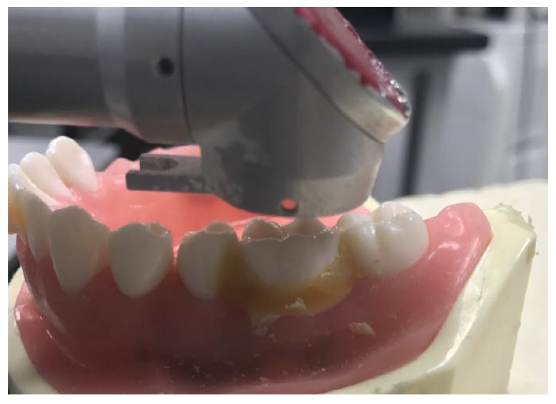
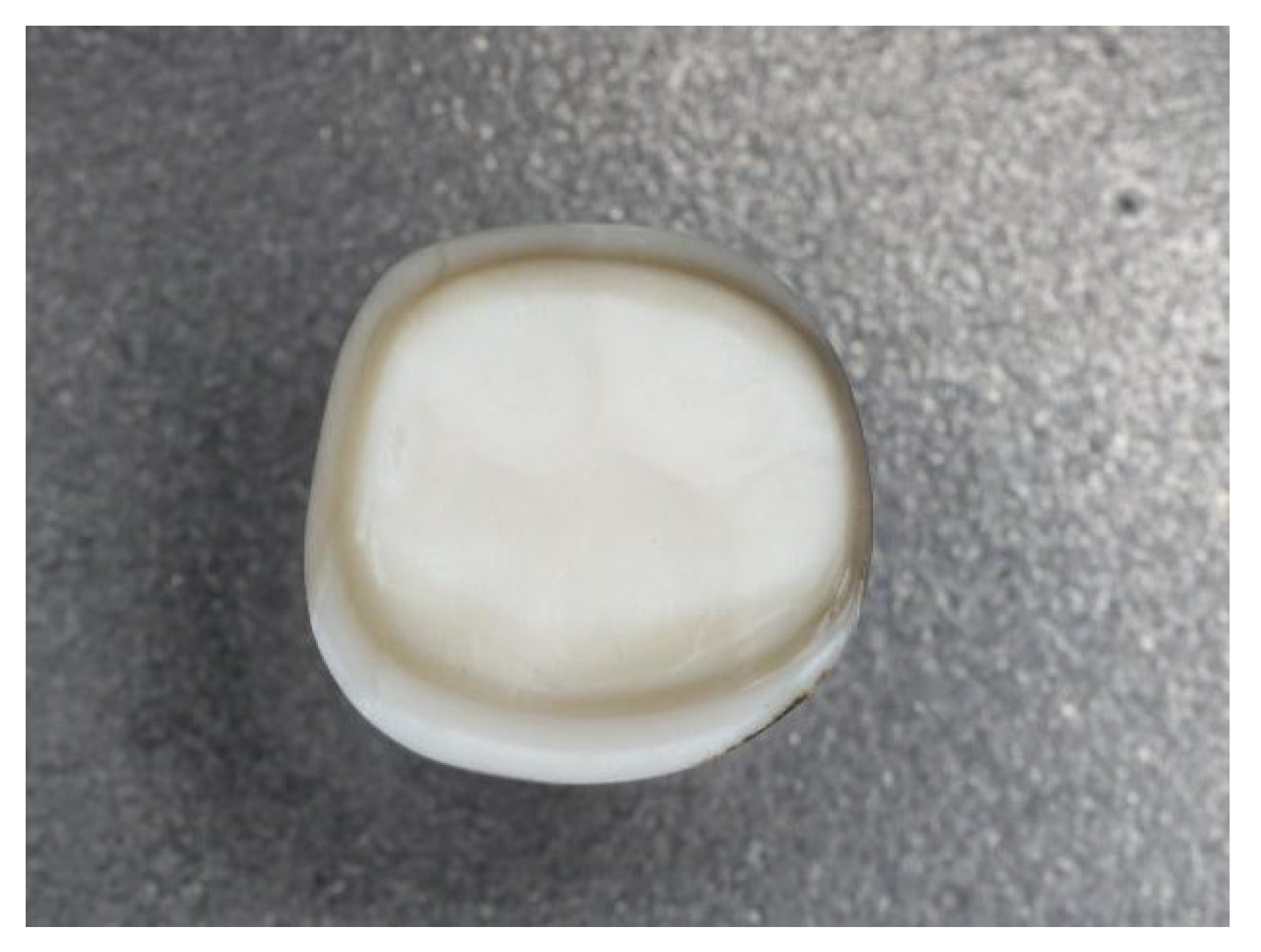
2.2. Tooth Preparation

Tooth preparation was automated with a femtosecond laser controlled by a dental robot (Figure 2) with a single depth of ablation at 28 μm. Preparation of the tooth surface was performed by the robot controlling the femtosecond laser to obtain a 2D-plane image of a specific layer (Figure 3). This process was conducted using high-speed 2D galvanometers. Five layers of pulse scanning and a 28-μm single-layer depth were prepared on the tooth surface; thus, the focusing lens moved forward by 28 μm. Each step was followed by another round of sectioning and scanning preparation. This process continued until a 3D virtual model of the tooth preparation was generated.

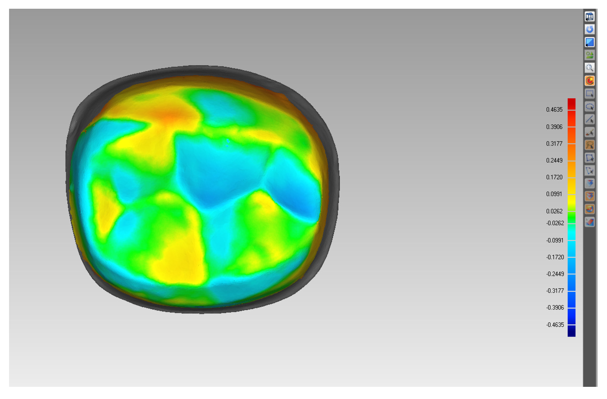
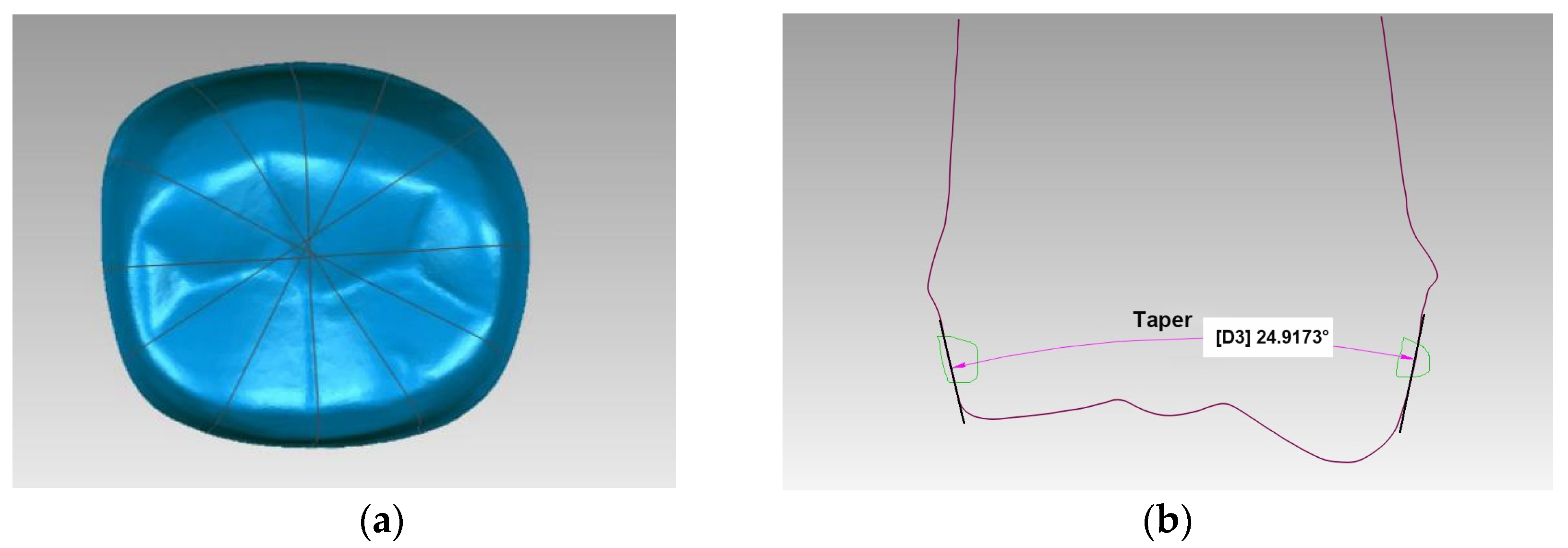
2.3. Software Measurement and Statistical Analysis
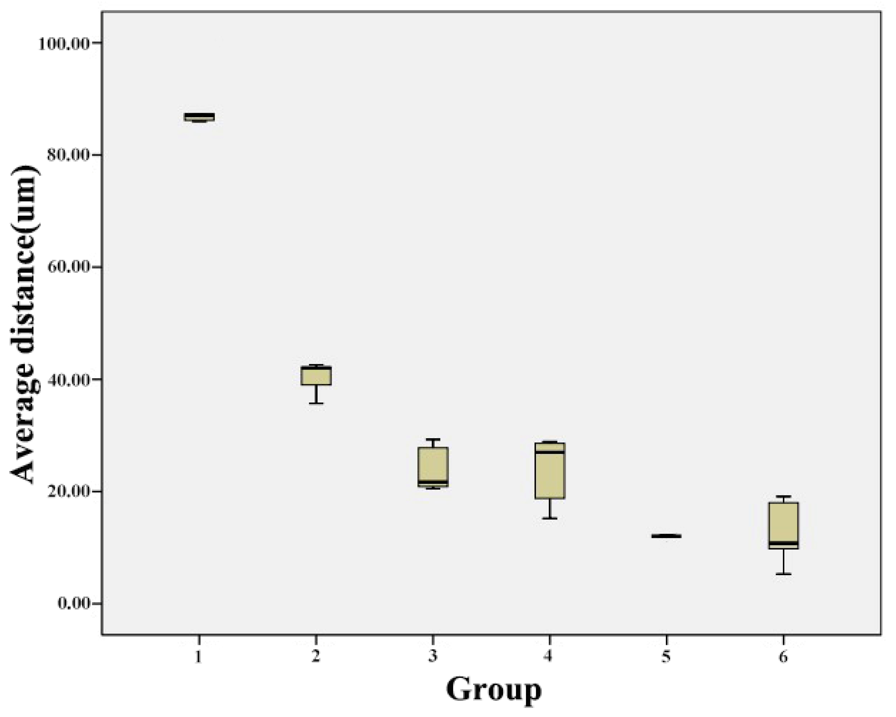
The 30 tooth preparations were digitally scanned with a 3D laser scanner (IScan D104i, Imetric 3D SA, Courgenay, Switzerland). Scanned data were converted to a stereolithography file. The 3D CAD software (Geomagic Studio 2013) was used to generate 3D reconstructions of these data (Figure 4). The tapers of six virtually vertical planes of each tooth preparation were measured using Geomagic Qualify software (Geomagic Qualify 2013) (Figure 5). Taper was determined by analyzing the taper between two opposing surfaces of all six planes. The mean values for each group were calculated, and a total of 30 tapers were measured for each light-off delay group. The predesigned preparation data and scanned data were input into the Geomagic Studio software. Eight different landmarks were selected from the predesigned data; these same landmarks were identified on scanned data and were used for manual registration. Then, the common regional register module was used to register the two sets of data. A bias module was subsequently used to measure the average distance (Figure 6). Five average distances were measured for each group. The overall average error between the predesigned data and scanned data was analyzed using the rank sum test in SPSS 19.0 (IBM SPSS Inc., Chicago, IL, USA) to compare the statistical significance of differences between the mean of the tapers and the average distance among groups.

3. Results

Automatic preparation of the resin teeth using a tooth preparation robot is shown in Figure 7.
The mean taper measurements for Groups 1–6 are shown in Table 1 and Figure 8; the taper decreased as the light-off delay increased. The mean taper decreased from 39.268° ± 4.530° to 25.393° ± 5.496° as the light-off delay increased. In this regard, there were significant differences between Groups 1 and 3, Groups 1 and 4, Groups 1 and 5, Groups 1 and 6, Groups 2 and 3, Groups 2 and 4, Groups 2 and 5, Groups 2 and 6, Groups 3 and 6, Groups 4 and 6, and Groups 5 and 6 (p-value < 0.05). In contrast, there were no significant differences between Groups 1 and 2, Groups 3 and 4, and Groups 4 and 5 (p-value > 0.05) (Table 2).
Table 3 and Figure 9 showed the comparison of the average distance between the scanned data and predesigned preparation data among Groups 1–6; these values decreased as the light-off delay increased; the average distance decreased from 0.089 ± 0.005 to 0.013 ± 0.030 μm as the light-off delay increased. In this regard, there were significant differences between Group 1 and the other groups, Group 2 and the other groups, and between Groups 3 and 5, Groups 3 and 6, Groups 4 and 5, and Groups 4 and 6 (p-value < 0.05); in contrast, no significant differences were observed between Groups 3 and 4 and between Groups 5 and 6 (p-value > 0.05) (Table 4).

4. Discussion

The convergence degree and the amount of prepared tooth are two important indicators of tooth preparation. This study explored a new method to improve the precise control of the convergence degree and the amount of prepared tooth and further enhance the accuracy of automated tooth preparation using a femtosecond laser. The automated tooth preparation system can help dentists improve the accuracy of tooth preparation, reduce dentists’ fatigue strength, and improve the patient experience.
In this experiment, we performed automatic tooth preparation using robotic control of a femtosecond laser for automatic 3D ablation of the target crown. Use of different light-off delay settings for the laser affected both the taper and distance between the scanned data and predesigned preparation data, with a decrease in these parameters as the light-off delay increased. However, a delay of 480 ms led to carbonization in the lower parts of the tooth preparation.
According to previous research, ideal 3D ablation should ensure that the thickness of a single slice is equal to the actual ablation depth of each laser layer. Therefore, obtaining a suitable single-depth ablation is a prerequisite for ensuring high-precision, high-efficiency, and high-quality 3D laser ablation. Yuan et al. conducted a preliminary study on the method of controlling the dentin depth error of a computerized, numerically controlled picosecond laser ablation. They found that the normal step size is positively correlated with the error of the ablation depth and the total depth-of-ablation error [32]. In the present study, we performed a single ablation of three layers using a single-layer ablation depth of 28 μm to reduce the error caused by multilayer ablation. This allowed us to explore the precision of this resin tooth preparation method and evaluate its effects.
In view of the anisotropy of the natural tooth structure, the irregular shape of the crown, and the different abilities of the laser to ablate enamel and dentin, resin teeth with the same shape as a natural tooth were used. Such a homogeneous sample can better reflect the effects of different laser parameters on the taper and adaption of the tooth preparation. The power and parameters used in this experiment could also be extended to natural teeth.
Laser energy exhibits a non-uniform Gaussian distribution. After the resin surface leaves the focal plane, the energy density at the periphery of the spot decreases, and the central energy density may still be higher than the target material’s ablation threshold. Therefore, the single-spot ablation surface may appear crater-like, and the taper of the laser ablation cannot meet the standard of the predesigned data. In the actual ablation process, the sidewall of each ablation layer contacting the resin material showed a reduced ablation phenomenon. Since ablation is accomplished through a layer-by-layer accumulative process, the accumulation of reduced ablation causes the tilt angle of the sidewall to increase. Consequently, the taper of the preparation body also increases, and this is not conducive to controlling the taper of the tooth preparation. In this study, we showed that when the light-off delay increased, taper decreased. In other words, the light-off delay could effectively reduce the taper of the preparation. However, the light-off delay cannot be increased indefinitely because a greater laser dwell time leads to heat accumulation and eventually carbonization of the target material.
The powerful peak power of the femtosecond laser enables the target tissue to form plasma directly [33,34], and the laser energy is used exclusively for material ablation, with the ablation rate increasing linearly with the energy density [35]. However, the actual ablation process has complex effects [36]. The arrival of the first laser pulse turns the surface tissue into plasma. If the formed plasma cannot be dissipated in time, it will form a barrier that absorbs or reflects the energy of the next laser pulse, preventing this laser energy from reaching the surface of the material. Excessive laser frequency, energy density, or a low scan rate can cause plasma buildup and a plasma-shielding effect [19]. The appearance of white powder on the resin tooth surface suggests that the ablated material had not entirely been converted to plasma. Thus, the increase in taper may be related to the incomplete formation of plasma, and these correlation parameters should be explored.
The larger tapers of the resin tooth preparation may also be a result of the poor position repeatability of the laser beam. The position repeatability of the laser beam used in this study was ±0.04 mm according to a preliminary experiment. This error may be one of the reasons for the increase in taper. Therefore, improved results may be obtained by using a robot with better position repeatability in future.
The average distance between the scanned data and predesigned data is an important measurement for the adaption of full-crown restoration. A large average distance indicates low adaption. During the registration process, the Geomagic Studio software calculated 3D suitability according to each point cloud; i.e., the software automatically calculated the paired root mean square for quantitative analysis. The magnitude of the difference can be specifically reflected in the positive and negative values on the deviation chromatogram for qualitative analysis. In the deviation chromatogram, the surface of the predesigned data was used as the origin. A positive value represents the distance away from the surface of the predesigned data, and a negative value reflects the distance from the surface of the predesigned data in the negative direction. This method calculates all paired point clouds for quantitative analysis, thereby obtaining more suitable information and avoiding the problem of data loss due to human handling of experimental objects [37]. In the ablation process, the current ablation plane and focal plane of the laser were in the same position (in the normal direction), so that the entire surface of the resin was irradiated by each spot, which reached the same or minimally different depths of ablation in a single ablation layer. This reduces the ablation error, resulting in more accurate depth control and a preferable adaption of the occlusal surface. However, the average distance that measures the deviation between the scanned data and predesigned data includes errors in scanning, data registration, software analysis, and laser ablation. This includes deviation of the occlusal and axial surfaces, in which the average distance of the occlusal surface is small and steady for the aforementioned reasons. When the light-off delay increased, the average distance of the axial surface decreased, similar to taper. Consequently, adaption also decreased.
This study has some limitations. In the actual application process, the effect of light-off delay on the taper would change according to the different materials used. One of the limitations of this study was that there is a certain difference between the tapers of tooth preparation and the tapering of clinical requirements. This might be solved by adjusting the taper of the model and exploring the relationship between other parameters and taper. Another limitation of this experiment was the use of resin teeth to explore the effect of the light-off delay on tooth preparation. During clinical tooth preparation, the dental hard tissues that need to be removed often contain both enamel and dentin. Using the same femtosecond laser parameters for materials with different ablation capabilities, the effect of the light-off delay on the taper and average distance of the teeth needs to be explored in the future. Nevertheless, our experiment revealed a clear trend.

5. Conclusions

We showed that the light-off delay setting of the femtosecond laser influences both taper and adaption in automatic tooth preparations. Thus, this setting needs to be considered during automatic tooth preparation.

Author Contributions

Conceptualization, F.Y. and P.L.; methodology, S.L.; software, F.Y.; validation, F.Y., S.L. and P.L.; formal analysis, F.Y.; investigation, P.L.; resources, F.Y.; data curation, S.L.; writing—original draft preparation, F.Y.; writing—review and editing, P.L.; visualization, S.L.; supervision, F.Y.; project administration, F.Y.; funding acquisition, F.Y. All authors have read and agreed to the published version of the manuscript.

Funding

This research was funded by the National Key Research and Development Program of China, grant number 2020YFB1312801; Natural National Science Foundation of China (NSFC), grant number 81571023; and Clinical Medicine Plus X—Young Scholars Project of Peking University, grant number PKU2019LCXQ024.

Institutional Review Board Statement

Not applicable.

Informed Consent Statement

Not applicable.

Data Availability Statement

The data presented in this study are available on request from the corresponding author.

Conflicts of Interest

The authors declare no conflict of interest.

References

  1. Theodoroff, S.M.; Folmer, R.L. Hearing loss associated with long-term exposure to high-speed dental handpieces. Gen. Dent. 2015, 63, 71–76. [Google Scholar] [PubMed]
  2. Yamada, T.; Kuwanom, S.; Ebisu, S.; Hayashi, M. Statistical analysis for subjective and objective evaluations of dental drill sounds. PLoS ONE 2016, 11, e0159926. [Google Scholar] [CrossRef] [PubMed]
  3. Willershausen, B.; Callaway, A.; Wolf, T.G.; Ehlers, V.; Scholz, L.; Wolf, D.; Letzel, S. Hearing assessment in dental practitioners and other academic professionals from an urban setting. Head Face Med. 2014, 10, 1. [Google Scholar] [CrossRef] [PubMed] [Green Version]
  4. Sun, J.; Xin, X.; Zhang, F. Noise level in prosthetic practice caused by dental handpieces. Chin. J. Prosthodont. 2010, 11, 2–4. [Google Scholar]
  5. Banga, H.K.; Goel, P.; Kumar, R.; Kumar, V.; Kalra, P.; Singh, S.; Singh, S.; Prakash, C.; Pruncu, C. Vibration exposure and transmissibility on dentist’s anatomy: A study of micro motors and air-turbines. Int. J. Environ. Res. Public Health 2021, 18, 4084. [Google Scholar] [CrossRef]
  6. Raucci-Neto, W.; Raquel Dos Santos, C.; Augusto de Lima, F.; Pecora, J.D.; Bachmann, L.; Palma-Dibb, R.G. Thermal effects and morphological aspects of varying Er:YAG laser energy on demineralized dentin removal: An in vitro study. Lasers Med. Sci. 2015, 30, 1231–1236. [Google Scholar] [CrossRef]
  7. Melo, M.A.; Lima, J.P.; Passos, V.F.; Rodrigues, L.K. The influence of dentin demineralization on morphological features of cavities using Er:YAG laser. Photomed. Laser. Surg. 2015, 33, 22–28. [Google Scholar] [CrossRef]
  8. Helene, C.; Joanne, J.E.C.; Rishi, S.R.; Ritu, G.; John, N.W. The cooling efficiency of different dental high-speed handpiece coolant port designs. Heliyon 2019, 5, e02185. [Google Scholar] [CrossRef] [Green Version]
  9. Albatayneh, O.B.; Seow, W.K.; Walsh, L.J. Assessment of Er:YAG laser for cavity preparation in primary and permanent teeth: A scanning electron microscopy and thermographic study. Pediatr. Dent. 2014, 36, 90. [Google Scholar]
  10. Aminian, A.; Brunton, P.A. A comparison of the depths produced using three different tooth preparation techniques. J. Prosthet. Dent. 2003, 89, 19–22. [Google Scholar] [CrossRef]
  11. Leempoel, P.J.; Lemmens, P.L.; Snoek, P.A.; van’t Hof, M.A. The convergence angle of tooth preparations for complete crowns. J. Prosthet. Dent. 1987, 58, 414–416. [Google Scholar] [CrossRef]
  12. Chen, L.; Hua, N.; Zhang, D. Influence of preparation height and taper on the retention of complete crown. Chin. J. Dent. Mater. Dev. 2005, 14, 179–181. [Google Scholar]
  13. Yuan, F.; Wang, Y.; Zhang, Y.; Sun, Y.; Wang, D.; Lyu, P. An automatic tooth preparation technique: A preliminary study. Sci. Rep. 2016, 6, 25281. [Google Scholar] [CrossRef] [PubMed] [Green Version]
  14. Braun, A.; Krillke, R.F.; Frentzen, M.; Bourauel, C.; Stark, H.; Schelle, F. Heat generation caused by ablation of dental hard tissues with an ultrashort pulse laser (USPL) system. Lasers Med. Sci. 2015, 30, 475–481. [Google Scholar] [CrossRef]
  15. Dausinger, F.; Lubatschowski, H.; Lichtner, F. Femtosecond Technology for Technical and Medical Applications; Springer: Berlin/Heidelberg, Germany, 2004. [Google Scholar]
  16. Serafetinides, A.A.; Khabbaz, M.G.; Makropoulou, M.I.; Kar, A.K. Picosecond laser ablation of dentine in endodontics. Lasers Med. Sci. 1999, 14, 168–174. [Google Scholar] [CrossRef]
  17. Mauersberger, S.; Schille, J.; Kujawa, K.; Schneider, L.; Million, C.; Hartung, K.; Oehlert, K.; Loeschner, U. High-precision surface profiling using multi-hundred watts ultrashort pulse lasers and ultrafast polygon-mirror based scanner. J. Laser Micro Nanoeng. 2020, 15, 1–9. [Google Scholar] [CrossRef]
  18. Rode, A.V.; Gamaly, E.G.; Luther-Davies, B.; Taylor, B.T.; Graessel, M.; Dawes, J.M.; Chan, A.; Lowe, R.M.; Hannaford, P. Precision ablation of dental enamel using a subpicosecond pulsed laser. Aust. Dent. J. 2003, 48, 233–239. [Google Scholar] [CrossRef]
  19. Sun, Y.; Vorobyev, A.; Liu, J.; Guo, C.; Lyu, P. Femtosecond pulsed laser ablation of dental hard tissues with numerical control: A roughness and morphology study. Chin. J. Stomatol. 2012, 47, 486–489. [Google Scholar] [CrossRef]
  20. Ji, L.; Li, L.; Devlin, H.; Liu, Z.; Jiao, J.; Whitehead, D. Ti:sapphire femtosecond laser ablation of dental enamel, dentine, and cementum. Lasers. Med. Sci. 2012, 27, 197–204. [Google Scholar] [CrossRef]
  21. Lizarelli, R.F.Z.; Costa, M.M.; Carvalho, E.; Nunes, F.D.; Bagnato, V.S. Selective ablation of dental enamel and dentin using femtosecond laser pulses. Laser Phys. Lett. 2008, 5, 63–69. [Google Scholar] [CrossRef] [Green Version]
  22. Daskalova, A.; Bashir, S.; Husinsky, W. Morphology of ablation craters generated by ultra-short laser pulses in dentin surfaces: AFM and ESEM evaluation. Appl. Surf. Sci. 2010, 257, 1119–1124. [Google Scholar] [CrossRef]
  23. Krüger, J.; Kautek, W.; Newesely, H. Femtosecond-pulse laser ablation of dental hydroxyapatite and single-crystalline fluoroapatite. Appl. Phys. A 1999, 69, S403–S407. [Google Scholar] [CrossRef]
  24. Sun, Y.; Vorobyev, A.; Chen, H.; Guo, C.; Lyu, P. Study on efficiency of femtosecond laser numerical control ablation of enamel and dentin. Chin. J. Stomatol. 2013, 48, 58–59. [Google Scholar]
  25. Wei, D.; Cheng, P.; Chen, X.; Wu, B.; Gao, F. Study on femtosecond laser processing of nonmetal vascular stent. Laser Optoelectron. Progr. 2013, 50, 117–122. [Google Scholar]
  26. Cheng, P.; Wei, D.; Wu, B.; Gao, F.; Chen, X. Femtosecond laser precision machine of biodegradable heart stent. Opt. Eng. 2014, 22, 63–68. [Google Scholar]
  27. Wang, H.W.; Cheng, C.W.; Li, C.W.; Chang, H.W.; Wu, P.H.; Wang, G.J. Fabrication of pillared PLGA microvessel scaffold using femtosecond laser ablation. Int. J. Nanomed. 2012, 7, 1865–1873. [Google Scholar] [CrossRef] [PubMed] [Green Version]
  28. Luengo, M.C.; Portillo, M.; Sanchez, J.M.; Peix, M.; Moreno, P.; Garcia, A.; Montero, J.; Albaladejo, A. Evaluation of micromorphological changes in tooth enamel after mechanical and ultrafast laser preparation of surface cavities. Lasers Med. Sci. 2013, 28, 267–273. [Google Scholar] [CrossRef] [PubMed]
  29. Sarathkumar, L.; Soundarapandian, S.; Ravi, B.; Muthukumaraswamy, A. Surface Processing: An Elegant Way to Enhance the Femtosecond Laser Ablation Rate and Ablation Efficiency on Human Teeth. Lasers Surg. Med. 2019, 51, 797–807. [Google Scholar] [CrossRef]
  30. Reinhard, E.F.; Maria, Q.; Nate, J.; Peter, K.; Michael, A.; Felix, K.K.; Jozef, Z.; Martin, G.; Hartmut, S. Ablation Precision and Thermal Effects of a Picosecond Infrared Laser (PIRL) on Roots of Human Teeth: A Pilot Study Ex Vivo. In Vivo 2020, 34, 2325–2336. [Google Scholar] [CrossRef]
  31. Straßl, M.; Kopecek, H.; Weinrotter, M.; Bäcker, A.; Al-Janabi, A.H.; Wieger, V.; Wintner, E. Novel applications of short and ultra-short pulses. Appl. Surf. Sci. 2005, 247, 561–570. [Google Scholar] [CrossRef]
  32. Yuan, F.; Lv, P.; Wang, D.; Wang, L.; Sun, Y.; Wang, Y. Controlling dental enamel-cavity ablation depth with optimized stepping parameters along the focal plane normal using a three axis, numerically controlled picosecond laser. Photomed. Laser Surg. 2015, 33, 92–97. [Google Scholar] [CrossRef] [PubMed] [Green Version]
  33. Stern, D.; Schoenlein, R.W.; Puliafito, C.A.; Dobi, E.T.; Birngruber, R.; Fujimoto, J.G. Corneal ablation by nanosecond, picosecond, and femtosecond lasers at 532 and 625 nm. Arch. Ophthalmol. 1989, 107, 587–592. [Google Scholar] [CrossRef]
  34. Elizabeth, J.K.; Jeremy, Y.; Bruce, E.B.; Mark, C.P.; Sivanandan, S.H. The role of ambient gas confinement, plasma chemistry, and focusing conditions on emission features of femtosecond laser-produced plasmas. J. Anal. At. Spectrom. 2020, 35, 1574–1586. [Google Scholar] [CrossRef]
  35. Lenzner, M.; Krüger, J.; Sartania, S.; Cheng, Z.; Spielmann, C.H.; Mourou, G.; Kautek, W.; Krausz, F. Femtosecond Optical Breakdown in Dielectrics. Phys. Rev. Lett. 1998, 80, 4076–4079. [Google Scholar] [CrossRef] [Green Version]
  36. Sallé, B.; Gobert, O.; Meynadier, P.; Perdrix, M.; Petite, G.; Semerok, A. Femtosecond and picosecond laser microablation: Ablation efficiency and laser microplasma expansion. Appl. Phys. A 1999, 69, S381–S383. [Google Scholar] [CrossRef]
  37. Xia, X.; Tan, F.; Wang, L.; Wu, S. Effects of different tooth preparations on three-dimensional adaption of crowns based on the reverse engineering. West China J. Stomatol. 2015, 33, 470–473. [Google Scholar]
Figure 1. Automatic tooth preparation robot.
Jcm 10 03389 g001
Figure 2. Resin tooth ablated automatically by the dental robot.
Jcm 10 03389 g002
Figure 3. Design of the path for laser scanning for single-layered preparation.
Jcm 10 03389 g003
Figure 4. Stereolithography file of the tooth preparation.
Jcm 10 03389 g004
Figure 5. Six vertical planes of the tooth preparation (a); measurement of the taper of a resin tooth preparation in Geomagic Qualify (b).
Jcm 10 03389 g005
Figure 6. Measurement of the average distance between the scanned data and predesigned preparation data.
Jcm 10 03389 g006
Figure 7. Resin tooth preparation.
Jcm 10 03389 g007
Figure 8. Influence of light-off delay on taper in the different groups.
Jcm 10 03389 g008
Figure 9. Influence of light-off delay on the average distance between the scanned data and predesigned preparation data.
Jcm 10 03389 g009
Table 1. Descriptive statistics of the taper (in °) of the tooth preparations made using different light-off delay settings.
GroupLight-Off Delay (ms)Mean (°)SD (°)
118039.2684.530
224038.8062.888
330033.6522.257
436032.4761.553
542031.1592.078
648025.3935.496
SD, standard deviation.
Table 2. Statistical analysis (p-value) of the taper (in °) of the tooth preparations made using different light-off delay settings.
Group23456
10.7490.0000.0000.0000.000
2-0.0010.0000.0000.000
3--0.4160.0870.000
4---0.3630.000
5----0.000
Table 3. Descriptive statistics of the average distance (in μm) between the scanned data and predesigned data of the tooth preparation with different light-off delays.
GroupLight-off Delay (ms)Mean (μm)SD (μm)
11800.0890.005
22400.0400.003
33000.024 0.042
43600.0240.006
54200.0130.025
64800.0130.030
SD, standard deviation.
Table 4. Statistical analysis (p-value) of the average distance (in μm) between the scanned data and predesigned data of the tooth preparation with different light-off delays.
Group23456
10.0000.0000.0000.0000.000
2-0.0000.0000.0000.000
3--0.9110.0010.001
4---0.0010.001
5----0.932
Publisher’s Note: MDPI stays neutral with regard to jurisdictional claims in published maps and institutional affiliations.

