Review

Pulmonary Complications in Hematopoietic Stem Cell Transplant Recipients—A Clinician Primer

1
Division of Pulmonary, Critical Care, Sleep Medicine, and Physiology, University of California San Diego, La Jolla, CA 92121, USA
2
Division of Hematology & Oncology, Scripps Clinic, La Jolla, CA 92037, USA
3
Sanford Stem Cell Clinical Center, Moores Cancer Center, Department of Medicine, Division of Regenerative Medicine, University of California San Diego, La Jolla, CA 92093, USA
4
Division of Cardiothoracic Radiology, University of California San Diego, La Jolla, CA 92121, USA
*
Author to whom correspondence should be addressed.
J. Clin. Med. 2021, 10(15), 3227; https://doi.org/10.3390/jcm10153227
Submission received: 22 June 2021 / Revised: 11 July 2021 / Accepted: 16 July 2021 / Published: 22 July 2021
(This article belongs to the Section Oncology)

Abstract

:
Hematopoietic stem cell transplants (HSCT) are becoming more widespread as a result of optimization of conditioning regimens and prevention of short-term complications with prophylactic antibiotics and antifungals. However, pulmonary complications post-HSCT remain a leading cause of morbidity and mortality and are a challenge to clinicians in both diagnosis and treatment. This comprehensive review provides a primer for non-pulmonary healthcare providers, synthesizing the current evidence behind common infectious and non-infectious post-transplant pulmonary complications based on time (peri-engraftment, early post-transplantation, and late post-transplantation). Utilizing the combination of timing of presentation, clinical symptoms, histopathology, and radiographic findings should increase rates of early diagnosis, treatment, and prognostication of these severe illness states.

1. Introduction

Hematopoietic stem cell transplant (HSCT) involves replacing a patient’s bone marrow with hematopoietic stem or progenitor cells from peripheral blood, bone marrow, or umbilical cord from another person or the same individual to restore immune–hematopoietic function after the underlying disease is eliminated [1]. Pulmonary complications post-HSCT affect between 45% and 60% of recipients [2,3] with a mortality rate exceeding 60% in mechanically ventilated patients after autologous HSCT [4]. This review is intended to form a framework for diagnosing and treating non-infectious and infectious pulmonary complications post-HSCT for the general clinician.

2. Hematopoietic Stem Cell Transplant Overview

Hematopoietic stem cell transplant (HSCT) is a multi-step process with a high risk for complications during marrow ablation, during engraftment, or afterwards [5,6,7]. Successful transplantation depends on the selection of the hematopoietic stem cell source, host preparation (conditioning regimen), and modulation of immune cell engraftment to minimize graft-versus-host disease (GVHD) [7].

2.1. Autologous versus Allogeneic Hematopoietic Stem Cell Transplantation (HSCT)

Hematopoietic stem cells can be obtained from self (autologous or auto-HSCT) or from a donor (allogeneic or allo-HSCT). Deciding whether to use autologous versus allogeneic transplant is dependent on multiple patient-specific factors and donor availability [8]. Autologous HSCTs have many advantages: no need to identify human leukocyte antigen (HLA)-matched donor, no risk of GVHD, no need for immunosuppressive therapy, more rapid hematopoietic recovery post-HSCT, and a lower risk of opportunistic infections. However, the relapse rate of underlying malignancy after autologous HSCT is often higher than in allogeneic HSCT, as autologous HSCT relies on the tumor response from cytoreductive therapy alone and does not induce a graft versus tumor response.
Advantages of allogeneic transplant include the ability to correct congenital or acquired defects and the immunologic, anti-tumor effects that can occur in response to persistent disease referred to as the graft versus tumor (GVT) effect [9]. Allogeneic transplant requires that the products be HLA-matched, as poorly HLA-matched products can lead to immune dysregulation and increased risk of GVHD. Furthermore, immune recovery is slower, and opportunistic infections are more common.

2.2. Engraftment

After selecting the source of stem cells, the host is prepared via high dose myeloablation therapy, reduced intensity myeloablation, or non-myeloablative conditioning [10]. Once a patient undergoes HSCT, engraftment occurs within a conditioning and host-dependent timeframe.
While there are several definitions of successful engraftment, patients typically develop a neutrophil count greater than 500/mm3, a platelet count greater than 20,000/microliter without any transfusions for one week, and a hematocrit greater than 25% for at least 20 days without any transfusions [11]. For autologous HSCT, white blood cell recovery typically occurs within two weeks while red blood cell and platelet recovery varies from patient to patient [6]. For allogeneic HSCT, peripheral blood granulocyte counts usually show signs of recovery within three weeks while platelet recovery is often delayed, taking on average 5–7 weeks [6]. Neutrophil engraftment is also dependent on the immunosuppressive regimen used, and recovery can average 10–30 days [12].

3. Timeline of Complications Following HSCT

The diagnosis and management of post-HSCT pulmonary manifestations requires a multidisciplinary approach. Diagnosis can be challenging as post-HSCT syndromes often have nonspecific clinical presentations. Utilizing common timelines of illness presentation post-HSCT (Figure 1) in combination with exposures to infectious agents, prior anti-microbial use, and unique radiographic findings aid in the diagnosis. Use of non-invasive tests including upper respiratory cultures, respiratory pathogen PCR, and serologies are useful adjuncts and can take the place of invasive testing to identify infectious pathogens [13]. Timely coordination and open communication between primary service, hematology–oncology, pulmonary, radiology, pathology, and infectious disease specialists is vital to the management of these patients.
Infectious and non-infectious pulmonary complications are classified by etiology and temporality in reference to HSCT, as it reflects the immunologic state of the patient. The often-cited time course for non-infectious entities consists of the pre/peri-engraftment phase (first 30 days), early post-transplantation phase (30–100 days), and the late post-transplantation phase (after 100 days). Infectious complications will be discussed separately.

4. Peri-Engraftment Period (0–30 Days Post-Transplant)

Conditions in the peri-engraftment period occur secondary to neutropenia, transfusion-related events, and toxicity of conditioning regimens. Non-infectious and infectious etiologies are equally likely to develop and often co-occur. An attempt should be made to identify and treat infection prior to initiation of immunosuppressive treatment for non-infectious complications; however, in critically ill patients, infectious and non-infectious complications may require concurrent treatment.
Cardiogenic causes of acute lung injury include volume overload resulting from large fluid volumes administered during chemotherapy, cardiac toxicity due to prior chemotherapy or radiation therapy, and transfusion-associated circulatory overload (TACO) after multiple blood products are transfused. Non-cardiogenic causes include chemotherapy pneumonitis and transfusion-related acute lung injury (TRALI). Chemotherapy-associated pneumonitis can occur due to a number of different chemotherapeutic agents including melphalan, busulfan, and bis(chloroethyl)nitrosourea (BCNU) [14]. Drug associated pneumonitis is often treated conservatively with withdrawal of offending agents and/or high-dose corticosteroids. TRALI should also be considered in patients requiring frequent blood product transfusions while awaiting engraftment. TRALI is typically seen within 6 h post-transfusion with acute respiratory failure, often requiring mechanical ventilation. Risk of TRALI is mitigated by conservative transfusion thresholds and protocols aimed at using low alloantibody products [15].

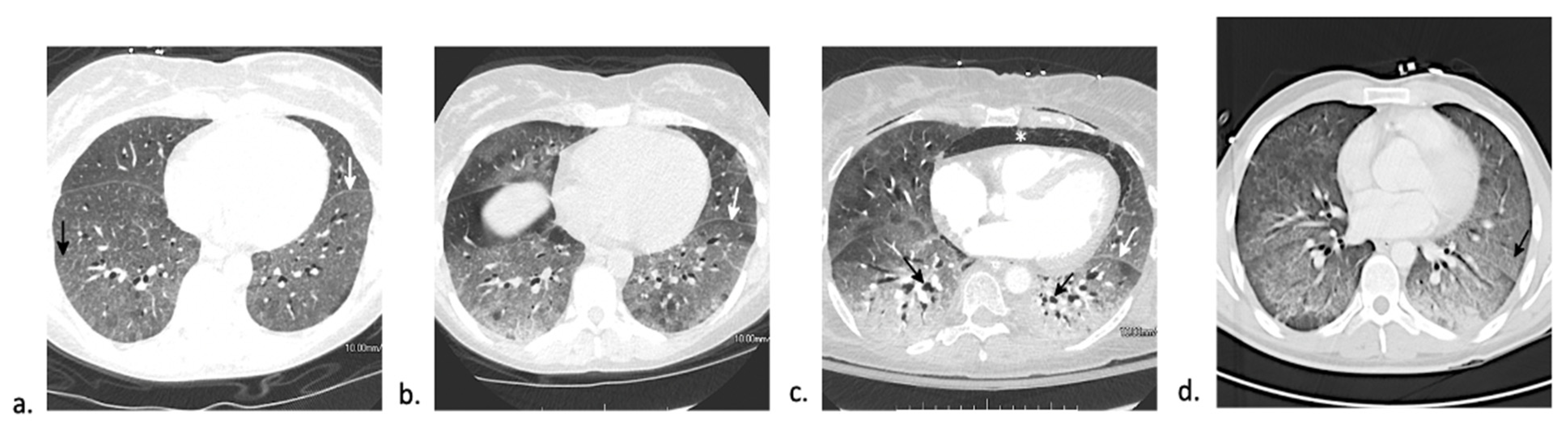
4.1. Peri-Engraftment Respiratory Distress Syndrome (PERDS)

Peri-engraftment respiratory distress syndrome (PERDS) has a wide range of reported incidence with a propensity to occur more frequently in allogeneic HSCT [9]. It is one of the earliest non-infectious pulmonary complication, occurring on average 7 days post-transplant with an average time to onset between 5 and 21 days [9]. The etiology is related to release of pro-inflammatory cytokines and early neutrophil engraftment, manifesting within 96 h of neutrophil recovery. PERDS has also been reported as an early sign of graft rejection, especially in cyclophosphamide-based non-myeloablative HSCTs with PERDS occurring independently from graft-versus-host disease (GVHD) [9]. PERDS is the pulmonary manifestation of engraftment syndrome and may be associated with multi-organ dysfunction. The Spitzer diagnostic criteria for engraftment syndrome include major criteria of non-infectious fever, erythrodermatous rash, and non-cardiogenic pulmonary edema, with minor criteria of weight gain, hepatic/renal dysfunction or transient encephalopathy [9]. Imaging during the early phase of PERDS demonstrates diffuse ground-glass opacities often with septal thickening and small pleural effusions (Figure 2a).
While most patients with PERDS respond favorably to treatment, some demonstrate progressive acute lung injury with lung consolidation and increasing alveolar collapse requiring intubation (Figure 2b,c). Up to one-third of patients also develop diffuse alveolar hemorrhage (DAH) [16]. Patients with respiratory failure requiring mechanical ventilation have a worse prognosis. Treatment consists of a short course of corticosteroids, with reports of rapid improvement within 24–48 h of corticosteroid initiation [9,16].

4.2. Diffuse Alveolar Hemorrhage (DAH)

Diffuse alveolar hemorrhage (DAH) has similar rates in both allogeneic (3–7%) and autologous (1–5%) HSCT recipients [17,18,19,20,21,22,23,24,25,26,27,28]. While classically considered an early pulmonary complication occurring within a month of transplant, up to 30% of cases of DAH occur >100 days post-transplantation [20]. The pathogenesis of DAH remains unclear and may be secondary to disruption of the alveolar–capillary basement membrane by conditioning, immune-mediated events, and return of neutrophils during marrow recovery. Older age, cord blood as transplant source, allogeneic HSCT, myeloablative conditioning, and acute GVHD are all considered risk factors for DAH [20,21].
DAH presents with fever, dyspnea, non-productive cough, and diffuse pulmonary infiltrates. Hemoptysis is relatively rare, making it a non-reliable sign for diagnosis. Computed tomography (CT) of the chest usually demonstrates patchy multifocal or diffuse ground-glass opacity with superimposed interlobular and intralobular septal thickening, creating a “crazy paving” pattern (Figure 2d). Bronchoscopy is diagnostic of DAH and is characterized by bloodier returns during bronchoalveolar lavage (BAL) with each instilled aliquot from at least three different samples from separate segmental bronchi or ≥20% hemosiderin-laden macrophages in the absence of infectious cause [18]. The majority of these patients require intensive care unit (ICU) admission and mechanical ventilation [18]. Treatment consists of high-dose corticosteroids. Despite treatment, patients with DAH have a poor prognosis with up to 80% case fatality [19,21]. Prognosis is better in those who present early after HSCT and in those receiving auto-HSCT (<30% mortality) compared to those observed later or in an allo-HSCT setting (>75% mortality) [18]. Additionally, underlying coagulopathy, need for mechanical ventilation, and identification of an organism on BAL are also associated with higher mortality and overall poor outcome [19,21].

5. Early Post-Transplantation Period (31–100 Days Post-Transplant)

This period is characterized by impaired cellular and humoral immunity and delayed lung injury from conditioning/preparative regimens. During this phase, the neutrophil count is generally normal, and the frequency of infectious complications typically decreases. Delayed complications of radiation and chemotherapy can produce manifestations similar to other HSCT-related complications during this period.

5.1. Idiopathic Pneumonia Syndrome (IPS)

Idiopathic pneumonia syndrome (IPS) represents a spectrum of diseases occurring between 20 and 42 days post-transplant, with the range extending up to 100+ days post-transplant. It is more common in allogeneic HSCT recipients [22]. Myeloablative conditioning with high-dose total body irradiation and acute GVHD are major risk factors for IPS development [24,25,26,27]. Patients often present with fever, non-productive cough, tachypnea, and hypoxemia. The diagnosis is based on three major requirements: widespread alveolar injury with symptoms and signs of pneumonia; absence of active lower respiratory tract infection; and absence of cardiac dysfunction, acute renal failure, or iatrogenic fluid overload as an etiology [26]. Imaging is non-specific, presenting as ground-glass opacity or consolidation in severe courses [27], which is often indistinguishable from other causes of acute lung injury (Figure 3a).
While both PERDS and DAH technically meet the diagnostic criteria of IPS, the three disease entities behave differently. High-dose corticosteroids continue to be the treatment of choice with variable response. Up to 85% of IPS cases are non-responsive to corticosteroids and progress to respiratory failure with up to 90% requiring mechanical ventilation [22,28]. Overall, IPS has a very poor prognosis with 60–80% case fatality [22,28,29,30]. The addition of etanercept has demonstrated promising early results with an overall improvement of 2-year survival although follow-up studies have suggested less durable benefit [30,31,32].

5.2. Pulmonary Cytolytic Thrombi

Pulmonary cytolytic thrombi are intravascular thromboemboli accompanied by the infiltration of monocytes into small and medium pulmonary vessels surrounded by areas of hemorrhage or infarct [33]. This phenomenon is most often seen post-allogeneic HSCT, primarily in children. It occurs a median 80 days, but can occur up to a year after transplant [34]. It is associated with acute and chronic GVHD. Symptoms are non-specific including fever, dyspnea, and chest pain. CT findings show multiple bilateral pulmonary nodules and peripheral opacities. Diagnosis of pulmonary cytolytic thrombi is made via open lung biopsy and cannot be treated empirically [34]. Treatment involves increasing immunosuppression, and the condition usually resolves within weeks to months [33].

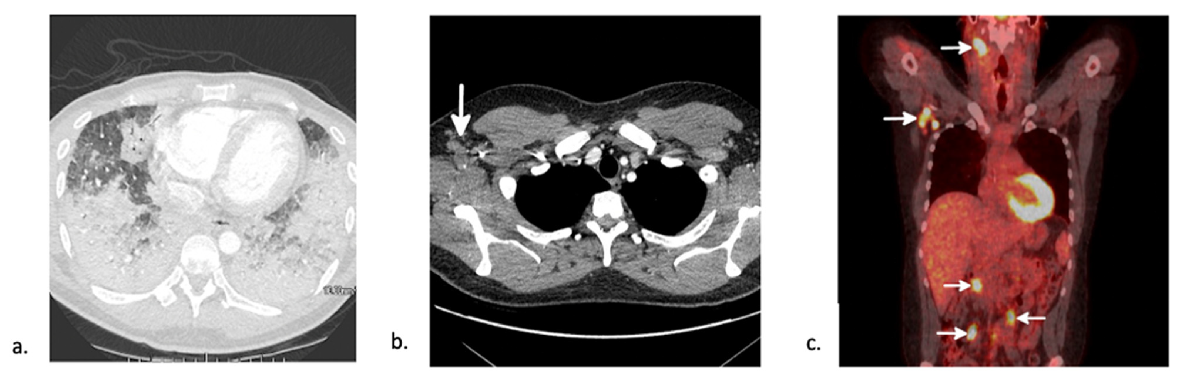
5.3. Post-Transplant Lymphoproliferative Disease (PTLD)

Post-transplant lymphoproliferative disease (PTLD) is a heterogenous group of lymphoid disorders that can complicate solid organ transplants as well as HSCTs. While in solid organ transplants, most PTLD is host-derived, the majority of PTLD (55–65%) in HSCT is due to reactivation and proliferation of Epstein–Barr virus (EBV)–infected donor derived B-lymphocytes due to weakened cytotoxic T-cell immunity [35]. However, EBV-negative cases of PTLD have been reported irrespective of donor or recipient’s EBV-status [36]. These can be differentiated into multiple subgroups: non-destructive PTLDs (including plasmocytic hyperplasia, infectious mononucleosis, and florid follicular hyperplasia), polymorphic PTLD, monomorphic PTLDs, and classical Hodgkin lymphoma PTLD [37]. It presents within 100 days in 75% of patients [38]. Risk factors for EBV-positive PTLD are directly related to the degree of T-cell depletion or impairment and thus include unrelated or human leukocyte antigen (HLA)-mismatched donors [39], EBV seropositivity status mismatch [39], cytomegalovirus (CMV) reactivation as a variable of EBV reactivation [40], splenectomy, and duration of immunosuppression [41]. Imaging often shows multifocal lymphadenopathy (Figure 3b,c). The leading treatment strategy is reduction of immunosuppression, with benefit seen with rituximab and chemotherapy in non-responders [35]. Survival is poor with an 80% mortality rate at 3 years [41].

6. Late Post-Transplantation Period (>100 Days Post-Transplant)

The term late-onset non-infectious pulmonary complications (LONIPCS) has been used to describe complications that occur 3 months after transplant [42]. These are caused by delayed immune recovery, immunosuppression, and immune-mediated reaction damage to the host tissue by the donor cells. Guidelines by the American Society of Blood and Bone Marrow Transplantation recommend pulmonary function testing (PFTs) at 6 months and then yearly post-HSCT for early diagnosis and treatment of LONIPCS including most notably bronchiolitis obliterans syndrome (BOS) [43].

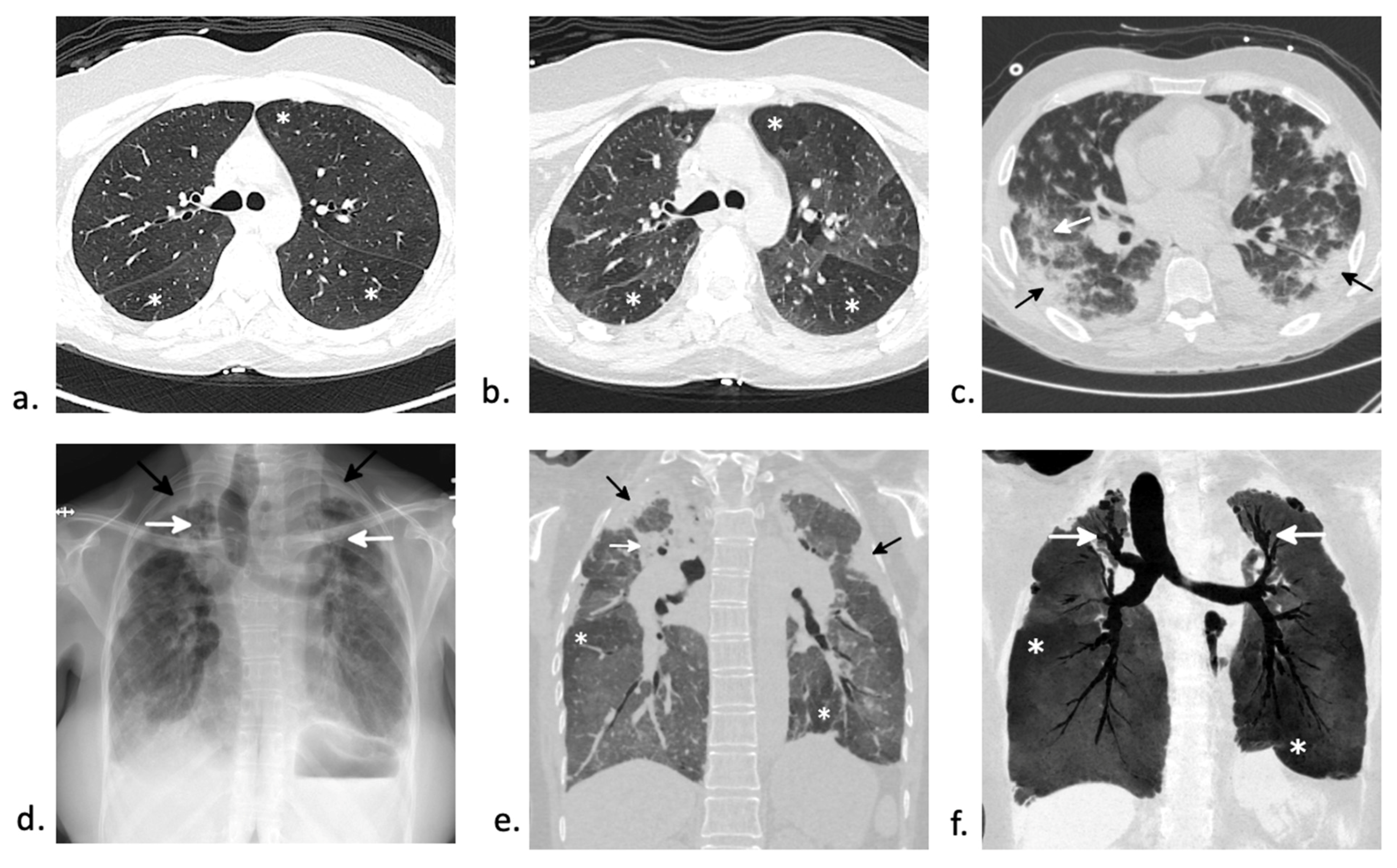
6.1. Bronchiolitis Obliterans Syndrome (BOS)

Chronic graft-versus-host disease (cGVHD) is currently the leading cause of long-term morbidity and mortality post-allogeneic HSCT [44]. Clinical diagnosis of bronchiolitis obliterans syndrome (BOS), in addition to one other distinctive manifestation of cGVHD, is sufficient to establish the diagnosis of cGVHD [45].
BOS is a clinical rather than a histopathological diagnosis [46]. BOS is characterized by (1) fixed airflow obstruction with forced expiratory volume in 1 s (FEV1)/vital capacity (VC) ratio <0.7 or the 5th percentile of predicted, (2) FEV1 < 75% of predicted with ≥10% decline in less than 2 years, (3) in the absence of respiratory tract infection (4) with signs of air trapping on PFTs or signs of air trapping or small airway thickening or bronchiectasis on CT chest [32]. BOS occurs between 6–24 months after transplant and occurs almost exclusively in allogeneic HSCT recipients [47]. The most common symptoms are progressive dyspnea, non-productive cough, and wheezing [46], although onset is generally insidious with up to 20% of patients remaining asymptomatic at diagnosis [47]. Imaging demonstrates a mosaic pattern on the inspiratory scan (Figure 4a,b). On expiratory imaging, the abnormal areas with air-trapping will not change in attenuation while normal lung will increase in attenuation (Figure 4c). The role of bronchoscopy is limited, often resulting in non-diagnostic findings of neutrophilic and/or lymphocytic inflammation [47]. As transbronchial biopsy has low sensitivity, the gold standard is open lung biopsy, although it is rarely performed. When performed, BO is characterized by submucosal bronchiolar fibrosis and luminal narrowing affecting the small airways.
BOS is irreversible with variable progression and mortality rates of up to 60% [46]. Management remains a challenge. Budesonide/formoterol, when compared to placebo, improved FEV1 by an average of 260 mL over one month although patients did not experience significant improvement in their respiratory symptoms [48]. Fluticasone, azithromycin, and montelukast (FAM regimen) with a brief steroid pulse suggested stabilization of pulmonary function tests [49] and is currently recommended as initial treatment of BOS [50]. The utility of azithromycin is unclear with a study showing no improvement in respiratory function with its use [51]. Furthermore, early azithromycin administration in patients undergoing allogeneic HSCT is associated with worse airflow decline–free survival [48]. A recent case series found that ruxolitinib in patients with steroid refractory cGVHD had an overall response rate of 70.7%; however, the response is less favorable in patients with lung involvement [52]. A randomized clinical trial of ruxolitinib in treatment of cGVHD is currently underway (NCT03112603). Extracorporeal photodynamic therapy may be associated with better overall survival [53]. Lung transplantation may also be an option for eligible candidates if medical therapy is unsuccessful [47].

6.2. Organizing Pneumonia

Organizing pneumonia (OP) has an incidence of approximately 10% after HSCT [54]. It occurs in both autologous and allogeneic HSCT, but it is more common in allogeneic recipients and can occur as a manifestation of cGVHD [55,56]. Symptoms include fever, shortness of breath, and non-productive cough with a median onset of 108 days [56]. Histopathologic confirmation is the gold standard for diagnosis with pathology demonstrating plugs of granulation tissue and fibrosis in the distal airways and alveoli with accompanying interstitial and alveolar inflammation [57]. There are numerous patterns of OP on CT, but common appearances include subpleural and peribronchovascular areas of ground-glass opacity, consolidation and linear opacities [58]. The “reverse halo” or “atoll” sign, due to areas of curvilinear consolidation with adjacent ground-glass opacity may also be present (Figure 4c) [58]. OP is very responsive to corticosteroid therapy [57], but recurrence is not uncommon [56]. Its prognosis is more favorable than BOS [57].

6.3. Interstitial Lung Disease

More recently, studies have focused on the prevalence of interstitial lung diseases (ILD) after HSCT [59]. While the incidence of non-infectious ILD after allo-HSCT is as low as 2–3% [60,61], ILD contributes to approximately 20% of LONIPCS [61]. Patients present with non-specific symptoms of shortness of breath and cough [61]. The main pulmonary function test (PFT) abnormality is a restrictive ventilatory defect [61]. Radiographic and pathologic findings vary, depending on the underlying entity. A histopathologic study including specimens from 18 different sites showed a variety of patterns of their interstitial lesions including non-specific interstitial pneumonia (NSIP), lymphoid interstitial pneumonia (LIP), diffuse alveolar damage (DAD), and pleuroparenchymal fibroelastosis (PPFE) [62].
One example of post-HSCT ILD is pleuroparenchymal fibroelastosis (PPFE), a rare form of interstitial pneumonia. PPFE can be idiopathic or secondary, the latter of which has been associated with HSCT. It occurs in <0.5% of patients post-HSCT and typically presents many years post-transplantation [63,64,65]. The incidence is higher after allogeneic HSCT [66]. This may occur secondary to diffuse alveolar damage with a failure of parenchymal lung injury to adequately resolve, promoting aberrant tissue repair [63]. CT imaging shows prominent superior pleural thickening often extending inferiorly to involve the major fissures. There is associated upper lobe fibrosis with superior displacement of the hila and traction bronchiectasis (Figure 4d–f). A histopathologic diagnosis of PPFE requires demonstration of interalveolar fibrosis and elastosis. In a review of 16 studies, Higo et al. also noted a high coexistence of pathologic findings of BO in addition to PPFE after allo-HSCT [66]. Pulmonary function tests typically show a restrictive ventilatory defect [65]. Several pharmacologic treatments including corticosteroids, everolimus, pirfenidone, and imatinib have been trialed with minimal success [67]. The most definitive treatment remains lung transplantation [67]. Overall, the prognosis is poor [65].

6.4. Pulmonary Veno-Occlusive Disease (PVOD)

Pulmonary veno-occlusive disease (PVOD) is a rare variant of pulmonary arterial hypertension (PAH) in which intimal proliferation occurs in the small pulmonary venules. A recent autopsy series suggests that it may be under-recognized with pathologic evidence of PVOD in 12 of the 35 autopsies in allo-HSCT recipients [68]. The etiology of PVOD has not been fully elucidated, but endothelial injury from cytotoxic chemotherapy or irradiation may be a risk factor [69]. It occurs in both autologous and allogeneic transplant recipients with time to onset ranging from several weeks to months [70]. PVOD is characterized by PAH with normal pulmonary capillary wedge pressure and pulmonary edema in the absence of left sided heart failure [69,70]. Histopathologic diagnosis may be required in patients who have an acceptable risk for biopsy [70]. Pathology is characterized by intimal proliferation and fibrosis of pulmonary venules and small veins, leading to progressive vascular obstruction and increased pulmonary capillary and arterial pressures [70]. Imaging demonstrates interstitial lung edema, diffuse ground-glass opacities with a centrilobular distribution, adenopathy, and prominent septal lines [71]. PFTs often show a severely low diffusing capacity for carbon monoxide (DLCO). Treatment remains controversial, given the risk for worsening pulmonary edema with the use of advanced pulmonary hypertension therapy. The only durable treatment is lung transplantation [70].

7. Infectious Complications

The peri-engraftment phase is hallmarked by mucositis due to conditioning therapy and prolonged neutropenia due to non-functional transplanted marrow [72]. The risk of infection is highest when absolute neutrophil count (ANC) is less than 500 and increases with increasing duration of neutropenia before engraftment [73]. During the early post-transplantation phase, infections are related to the defects in cellular immunity caused by immunosuppressive and conditioning regimens [72]. In the late post-transplantation phase, infection rates decline as immunosuppressant medications are tapered unless needed for cGVHD. Because of differences in immune recovery, autologous HSCT recipients are at higher risk of infectious complications during the peri-engraftment and early post-transplantation phase while allogeneic HSCT recipients are at higher risk of infection throughout the late post-transplantation phase due to cGVHD and prolonged immunosuppressive therapy [8,72].

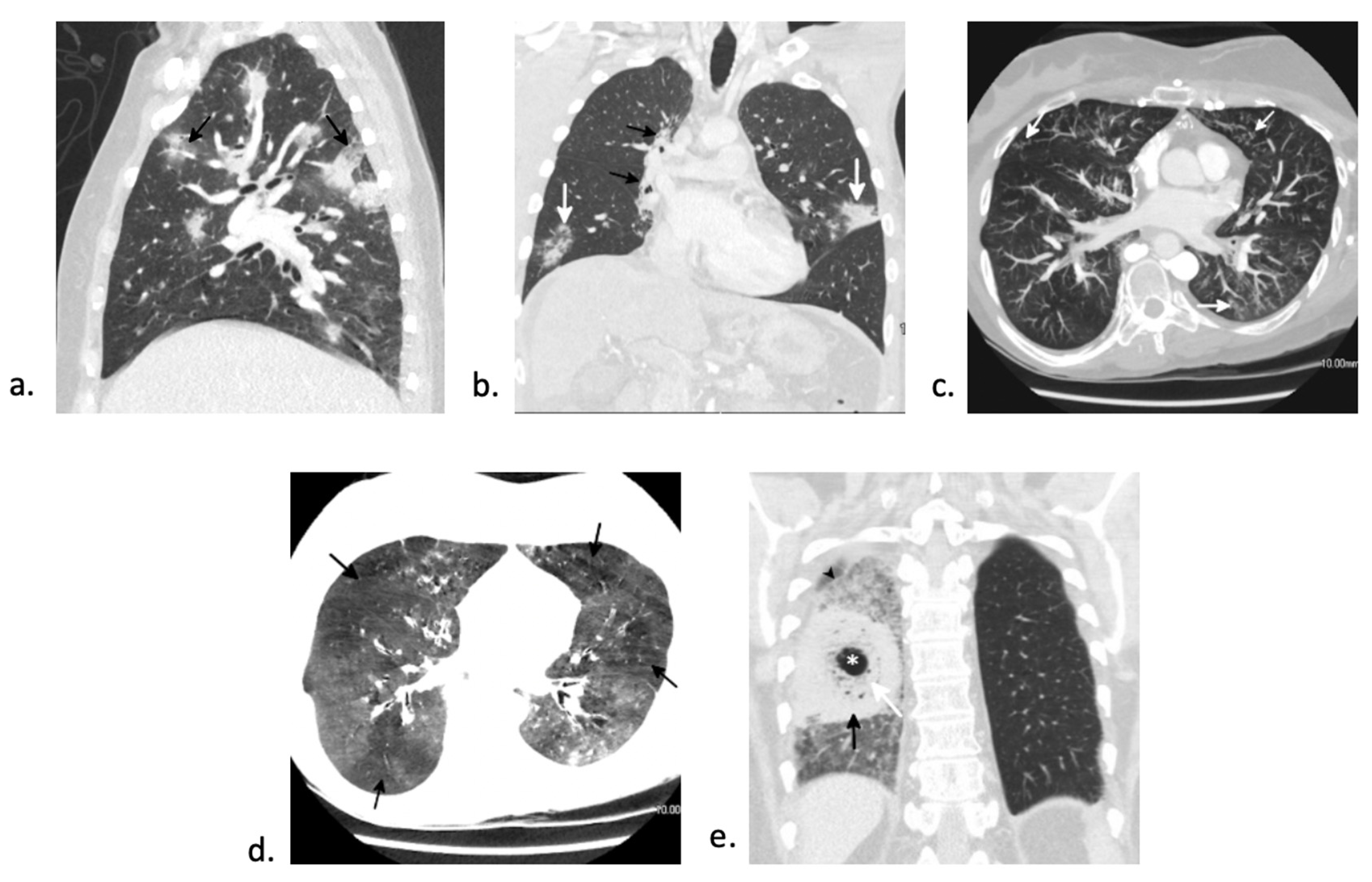
7.1. Bacterial Pneumonia

Bacterial infections account for approximately 90% of infections during the peri-engraftment period after allo-HSCT [74] and include bloodstream, respiratory, and gastrointestinal infections [75]. The sources of most bacterial infections in allo-HSCT recipients during peri-engraftment includes the gastrointestinal tract and indwelling vascular catheters [76]. Most studies report a high incidence of bacterial pneumonias [75,77,78] with up to 15% in in allogeneic and autologous HSCT recipients [79]. A nationwide study of allo-HSCT recipients revealed that 44% of pneumonias were due to bacterial etiology, compared to 29% of fungal and 19% of viral pneumonias [80]. Lung infections that occur in late post-transplantation phase are seen in patients with cGVHD and are frequently due to encapsulated organisms, particularly Streptococcus pneumoniae and Haemophilus influenzae (Figure 5b). Rarely, tubercular and non-tubercular mycobacterial infections can be seen in the post-transplant period especially in patients with poor immune reconstitution and cGVHD (Figure 5c,d) [81].

7.2. Fungal Pneumonia

Invasive fungal infections (IFI) can affect patients early in their post-transplant course. Invasive aspergillosis is the most common invasive fungal infection HSCT recipients [82,83]. The incidence is higher in allogeneic than in autologous HSCT recipients [83]. There are three separate “at risk” periods, with first peak around peri-engraftment associated with neutropenia, second peak between 40 and 70 days associated with acute GVHD, and late peak late after transplant with cGVHD [84]. Patients may present with fever, cough, and hemoptysis [85]. The classic radiographic finding on non-contrast CT of the chest are nodules surrounded by ground-glass opacity (“halo sign”), representing angioinvasion and hemorrhage in the surrounding tissue (Figure 5a) [83]. The addition of contrast to the CT can be useful for detection of the angioinvasive growth pattern of Aspergillus and even Mucor and generally includes a vessel occlusion sign, which was noted to have a sensitivity of 0.78 and specificity of 0.91 for angioinvasive disease in one study [86]. The diagnosis can be difficult and rely on clinical suspicion, radiographic presentation, laboratory studies (including serum or BAL galactomannan) and possible histopathologic findings [83]. Given its high incidence, many institutions preemptively utilize fungal prophylaxis in patients on immunosuppression therapy for cGVHD [87]. Posaconazole is the prophylactic medication of choice, as it is well tolerated and covers Aspergillus strains resistant to more commonly used azoles [82]. Voriconazole is the preferred primary therapy for invasive aspergillosis while liposomal Amphotericin B, isavuconazole, and posaconazole can be used as alternative therapies.
Zygomycosis is the second most common mold infection in HSCT with an increasing incidence over the past few decades [85]. Rare infections with fusarium and scedosporium can occur as well [88]. Invasive fungal infections with mucormycosis generally occurs > 3 months post-transplant and can make up to 8% of fungal infections in this patient population [89]. Although there are various imaging appearances of mucormycosis, one nearly pathognomonic imaging manifestation is the “bird’s nest” sign (Figure 5e) [90]. While mucormycosis infections are rare, they are frequently fatal and require early, aggressive management. Treatment involves combination of medical (with amphotericin B for zygomycosis) and surgical therapy [88].
Candidal infections occur less frequently as a result of universal prophylaxis with fluconazole. In high-risk patients, posaconazole may be the preferred prophylactic therapy due to the emergence of resistant non-albicans candida species including Candida glabrata and Candida krusei, especially in those with cGVHD [91]. Similarly, Pneumocystis jiroveci pneumonia (PJP) has a low incidence due to use of prophylaxis. Without adequate prophylaxis, it has an incidence of 5–16% [92] and often presents in late post-transplantation period if prophylaxis has been discontinued [93]. CT findings can vary but include diffuse infiltrates, consolidation in the upper lobes, and ground glass. Prophylaxis includes trimethoprim/sulfamethoxazole (TMP-SMX), pentamidine or atovaquone [92]. TMP-SMX is also the preferred treatment for PJP pneumonia [88].

7.3. Viral Pneumonia

Viral pneumonia from reactivation is common during the peri-engraftment phase and should be closely monitored while patients are immunosuppressed. Viral infection from herpes simplex virus (HSV)-1 and -2 occur in peri-engraftment and early post-transplantation phase, those from cytomegalovirus (CMV) and human herpes virus-6 (HHV-6) are often present in early post-transplantation phase, and infection from varicella zoster virus (VZV) is more common in the late post-transplantation phase [94]. The incidence of reactivation of viruses such as HSV, CMV, and VZV has drastically decreased since prophylaxis became routine.
CMV pneumonia has an incidence as high as 20% in allo-HSCT recipients [95]. In contrast, in a review of 795 auto-HSCT patients, only 16 (2%) were diagnosed with CMV pneumonia [96]. There is a higher frequency in CMV seropositive auto-HSCT recipients, compared to seronegative [96,97]. The diagnosis of CMV pneumonia is based on the combination of clinical presentation, radiologic evidence, and virus in BAL. CMV pneumonia presents with non-productive cough, dyspnea, and hypoxemia; fever is not always present [96]. Diagnosis of CMV pneumonia is difficult. CMV viremia in the blood may be helpful but is not diagnostic. Bronchoscopy is the most common method of confirming CMV in the lungs [98,99]. Although its presence is rare, inclusion bodies in BAL have a high sensitivity but low specificity for CMV pneumonia. Immunostaining for anti-CMV antibodies in BAL fluid has both high sensitivity and specificity and may be a helpful modality for diagnosis of CMV disease [100]. The absence of both findings can exclude CMV pneumonia with up to 99% specificity [100]. Viral shedding into the respiratory tract has been demonstrated without presence of invasive disease and should be interpreted cautiously [78]. CT findings of CMV pneumonia is nonspecific and may present with a mixture of patterns, most commonly ground-glass opacities, pulmonary nodules, and areas of consolidation [101]. The standard of therapy for CMV pneumonia is ganciclovir. The rate of mortality can be high with reports of 31–62% CMV pneumonia-related mortality [92,102] although the outcome has improved over the past few decades [103]. Lymphopenia prior to diagnosis and mechanical ventilation are associated with an increased overall mortality [72]. Given the high mortality rate, all CMV seropositive HSCT recipients and all CMV seronegative recipients with CMV seropositive donors should be placed on a CMV prevention program [89]. The prevention program is generally either universal prophylaxis with either ganciclovir, high-dose acyclovir, or valacyclovir for the first 100 days, or pre-emptive strategy of weekly monitoring and early therapy [103].
HSV can have pulmonary manifestations including tracheobronchitis and pneumonia [104]. The breadth of clinical presentation from an HSV infection is quite broad—from a simple cough to respiratory failure from diffuse alveolar damage or necrotizing pneumonia [105,106]. Hemoptysis can also occur, typically due to airway inflammation in tracheitis [106]. HSV pneumonia occurs in peri-engraftment and early post-transplantation phase. The mode of transmission is from continuous oropharyngeal spread [107]. Acyclovir prophylaxis should be offered to all HSV-seropositive allogeneic recipients to prevent HSV re-activation. This is initiated at the start of conditioning and continued until engraftment occurs or until mucositis resolves. Acyclovir prophylaxis for VZV seropositive allogeneic and autologous recipients is also routinely recommended to prevent recurrent VZV infection in the first year after HSCT [84].
Patients continue to be at high risk for seasonal community-acquired respiratory viruses (CARV). Respiratory syncytial virus (RSV), influenza A and B, parainfluenza, and rhinovirus cause the majority of non-CMV viral respiratory infections [108,109], especially in allo-HSCT recipients [90,92]. Routine immunization with inactivated influenza vaccine after HST can be helpful in preventing and minimizing infection severity [110,111]. A single-center study of 195 allo-HSCT reported a 30% incidence of severe viral pneumonia causing respiratory failure and requiring mechanical ventilation [109]. The overall mortality rate is high at 15–30%, depending on the offending viral agent [108,112].
RSV is one of the more common CARV that is associated with high mortality rate. In a single center retrospective study of 280 allo-HSCT recipients with RSV infection, 29% had progression to lower respiratory tract infection [113]. Older age, smoking history, conditioning with high-dose total body irradiation, and lymphopenia were associated with an increased risk of progression from upper respiratory to lower respiratory tract infection [113,114]. RSV is associated with a wide mortality rate, ranging from 5% to 43%, in HSCT patients [112,113,115,116,117]. Older age, male gender, bone marrow or cord blood as transplant source, corticosteroid use, lower respiratory tract infection, and oxygen requirement are all associated with increased overall mortality [113,116,117,118].
In 2019, the emergence of severe acute respiratory syndrome coronavirus 2 (SARS-CoV-2) led to a coronavirus disease 2019 (COVID-19) global pandemic. The majority of HSCT patients who contract COVID-19 have mild or moderate disease [119]. Studies have demonstrated that 14–25% of HSCT patients diagnosed with COVID-19 develop severe disease with respiratory failure requiring mechanical ventilation [119,120,121]. Patients typically present with fever, cough and shortness of breath [120]. Abnormal laboratory studies including anemia and lymphopenia may be present [120]. The most common radiographic abnormality in these patients is bilateral infiltrates [120]. A mortality rate of 16–21% has been reported in HSCT patients with COVID-19 [120,121,122,123]. Older age, male gender, use of immunosuppressive agents, and COVID-19 diagnosis within 1 year of HSCT are all associated with a higher risk of mortality in HSCT patients [119,120,122].
Updated guidelines have been published by European Society for Blood and Marrow Transplantation (EBMT) and American Society for Transplantation and Cellular Therapy (ASTCT) for the management of HSCT patients in regard to COVID-19 [124,125]. Patients should follow the same practices recommended for the general public including hand hygiene, masking, and social distancing [125]. Patients with upper or lower respiratory symptoms should undergo PCR testing for SARS-CoV-2 in addition to testing for other CARV [124]. Additional chest imaging may be required in post-HSCT recipients with lower respiratory symptoms with a negative SARS-CoV-2, given the discrepancy between upper and lower respiratory tract positivity [124]. Prolonged viral shedding may be seen in post-HSCT patients, as is seen with other viral infections in this patient population and may not equate to active infection [124]. Furthermore, HSCT patients may not mount an effective humoral response, so providers need to be cautious when interpreting serologic IgG and IgM assays [124]. Management of active COVID-19 infection for HSCT patients is similar to that of the general population [124]. There are no specific therapies recommended for HSCT-recipients with COVID-19. Prophylactic therapies should be continued in patients with active infection [124]. It is recommended that immunosuppression be carefully titrated. While no cases of graft rejection have been reported as a result of COVID-19, immunosuppression is recommended for anyone that demonstrates histologic evidence of GVHD regardless of COVID-19 infection [124,125].

8. Diagnostic Tools

Open communication between these primary services and these subspecialists is necessary for the initial evaluation of these patients. There are multiple diagnostic tools available if non-invasive methods do not yield a diagnosis. The most frequently utilized and widely considered safest diagnostic tool is flexible bronchoscopy. Bronchoscopic findings result in identification of infectious pathogens in 31–71% cases and change in therapy in 24–52% of patients [126,127]. Although yield is higher if performed early on and prior to initiation of anti-microbials, bronchoscopy may yield a treatment-altering diagnosis in up to half of cases, [128,129] even while patients are receiving empiric antimicrobial therapy [6]. Bronchoscopy with BAL can also identify organisms not evident on non-invasive testing and proves to be an important complementary strategy in the work-up of pulmonary complications in this population [130]. Furthermore, BAL cellularity can also help with the diagnosis of non-infectious entities including DAH. However, bronchoscopy becomes higher risk in those with hypoxemia, neutropenia and/or thrombocytopenia [131]. Most complications are reported as mild and self-limiting, but more severe adverse events like respiratory failure, arrhythmias, shock, and severe bleeding occurred at a rate of ≤5% [132].
While prior studies have shown that transbronchial biopsies (TBBx) can increase the diagnostic yield compared to bronchoscopy alone [133], more recent studies note that the addition of TBBx is not associated with a change in antibiotic therapy. However, positive results did increase the odds of a change in corticosteroids, suggesting a utility in non-infectious etiologies [134]. Not surprisingly, transbronchial biopsy confers an increased rate of complications and procedural mortality compared to bronchoscopy alone [134]. Endobronchial ultrasound guided transbronchial needle aspiration (EBUS-TBNA) has been demonstrated to be a safe and effective diagnostic method in sampling peripheral ground-glass opacities with high suspicion for primary malignancy [135,136], although the study of this technique as a diagnostic tool for infectious or non-infectious complications has been limited in immunocompromised patients [137]. However, limited data does demonstrate increased yield utilizing EBUS-TBNA compared to BAL alone in this pediatric population [137]. More research is required to elucidate the benefits and risks of EBUS-TBNA in the adult HSCT population. Trials comparing yield and safety of newer bronchoscopic interventions such as cryobiopsy to traditional TBBx in HSCT patients are lacking.
The use of other more invasive modalities is controversial [138]. Percutaneous CT-guided transthoracic lung biopsy has been performed in the past [139]. Surgical lung biopsy, either by video-assisted thoracoscopic surgery (VATS) or open thoracotomy [140,141], is now rarely performed due to risk of increased post-operative procedural complications and procedure-related mortality. The utility of these more invasive techniques has decreased over the past decade as other non-invasive diagnostic tests have improved [138]. Overall, the choice of invasive modality is individualized based on risks and benefits of the procedure for the patient [142]. The decision for and mode of bronchoscopic evaluation is institution- and practitioner-specific [142].

9. Summary

Collectively, the risk of both infectious and non-infectious pulmonary complications post-HSCT are high, but careful consideration of the timeline post-HSCT, an assessment of individual and local risk factors, and early involvement of a multidisciplinary team in the care of these individuals can help to identify and treat these complications in a timely manner.

Author Contributions

Conceptualization, J.J.; writing—original draft preparation, A.A., J.R., E.L., B.N. and J.J.; writing—review and editing, A.A., J.R., E.L., B.N., C.J., S.K., J.M., A.M. and J.J.; radiological images provided by S.K. All authors have read and agreed to the published version of the manuscript.

Funding

This research received no external funding.

Institutional Review Board Statement

Not applicable.

Informed Consent Statement

Not applicable.

Data Availability Statement

No new data were created or analyzed in this study. Data sharing is not applicable to this article.

Conflicts of Interest

The authors declare no conflict of interest.

References

  1. Barriga, F.; Ramirez, P.; Wietstruck, A.; Rojas, N. Hematopoietic stem cell transplantation: Clinical use and perspectives. Biol. Res. 2012, 45, 307–316. [Google Scholar] [CrossRef] [Green Version]
  2. Cordonnier, C.; Bernaudin, J.F.; Bierling, P.; Huet, Y.; Vernant, J.P. Pulmonary complications occurring after allogeneic bone marrow transplantation. A study of 130 consecutive transplanted patients. Cancer 1986, 58, 1047–1054. [Google Scholar] [CrossRef]
  3. Jules-Elysee, K.; Stover, D.E.; Yahalom, J.; White, D.A.; Gulati, S.C. Pulmonary complications in lymphoma patients treated with high-dose therapy autologous bone marrow transplantation. Am. Rev. Respir Dis. 1992, 146, 485–491. [Google Scholar] [CrossRef]
  4. Shorr, A.F.; Moores, L.K.; Edenfield, W.J.; Christie, R.J.; Fitzpatrick, T.M. Mechanical ventilation in hematopoietic stem cell transplantation: Can We effectively predict outcomes? Chest 1999, 116, 1012–1018. [Google Scholar] [CrossRef] [PubMed]
  5. Forman, S.J.; Negrin, R.S.; Antin, J.H.; Appelbaum, F.R. Thomas’ Hematopoietic Cell Transplantation; Wiley Blackwell: Hoboken, NJ, USA, 2016. [Google Scholar]
  6. Copelan, E.A. Hematopoietic stem-cell transplantation. N. Engl. J. Med. 2006, 354, 1813–1826. [Google Scholar] [CrossRef]
  7. Majhail, N.S.; Farnia, S.H.; Carpenter, P.A.; Champlin, R.E.; Crawford, S.; Marks, D.I.; Omel, J.L.; Orchard, P.J.; Palmer, J.; Saber, W.; et al. Indications for Autologous and Allogeneic Hematopoietic Cell Transplantation: Guidelines from the American Society for Blood and Marrow Transplantation. Biol. Blood Marrow Transplant. 2015, 21, 1863–1869. [Google Scholar] [CrossRef] [Green Version]
  8. Hatzimichael, E.; Tuthill, M. Hematopoietic stem cell transplantation. Stem Cells Cloning 2010, 3, 105–117. [Google Scholar] [CrossRef] [PubMed] [Green Version]
  9. Spitzer, T.R. Engraftment syndrome: Double-edged sword of hematopoietic cell transplants. Bone Marrow Transplant. 2015, 50, 469–475. [Google Scholar] [CrossRef] [PubMed] [Green Version]
  10. Bazinet, A.; Popradi, G. A general practitioner’s guide to hematopoietic stem-cell transplantation. Curr. Oncol. 2019, 26, 187–191. [Google Scholar] [CrossRef] [Green Version]
  11. Quesenberry, P.J.; Stewart, F.M.; Becker, P.; D’Hondt, L.; Frimberger, A.; Lambert, J.F.; Colvin, G.A.; Miller, C.; Heyes, C.; Abedi, M.; et al. Stem cell engraftment strategies. Ann. N. Y. Acad Sci. 2001, 938, 54–61. [Google Scholar] [CrossRef] [PubMed]
  12. Spitzer, T.R. Engraftment syndrome following hematopoietic stem cell transplantation. Bone Marrow Transplant. 2001, 27, 893–898. [Google Scholar] [CrossRef] [Green Version]
  13. Azar, M.M.; Gaston, D.C.; Kotton, C.N.; Malinis, M.F. Emerging Microbiology Diagnostics for Transplant Infections: On the Cusp of a Paradigm Shift. Transplantation 2020, 104, 1358–1384. [Google Scholar] [CrossRef] [PubMed]
  14. Cooke, K.R.; Yanik, G.A. Lung Injury Following Hematopoietic Cell Transplantation. Thomas’ Hematop. Cell Transplant. 2009, 95, 1456–1472. [Google Scholar]
  15. Ahya, V.N. Noninfectious Acute Lung Injury Syndromes Early After Hematopoietic Stem Cell Transplantation. Clin. Chest Med. 2017, 38, 595–606. [Google Scholar] [CrossRef]
  16. Capizzi, S.A.; Kumar, S.; Huneke, N.E.; Gertz, M.A.; Inwards, D.J.; Litzow, M.R.; Lacy, M.Q.; Gastineau, D.A.; Prakash, U.B.S.; Tefferi, A. Peri-engraftment respiratory distress syndrome during autologous hematopoietic stem cell transplantation. Bone Marrow Transplant. 2001, 27, 1299–1303. [Google Scholar] [CrossRef] [PubMed] [Green Version]
  17. Afessa, B.; Tefferi, A.; Litzow, M.R.; Krowka, M.J.; Wylam, M.E.; Peters, S.G. Diffuse Alveolar Hemorrhage in Hematopoietic Stem Cell Transplant Recipients. Am. J. Respir. Crit. Care Med. 2002, 166, 641–645. [Google Scholar] [CrossRef]
  18. Zhang, Z.; Wang, C.; Peters, S.G.; Hogan, W.J.; Hashmi, S.K.; Litzow, M.R.; Patnaik, M.S.; Niven, A.S.; Yadav, H. Epidemiology, Risk Factors, and Outcomes of Diffuse Alveolar Hemorrhage After Hematopoietic Stem Cell Transplantation. Chest 2021, 159, 2325–2333. [Google Scholar] [CrossRef]
  19. Keklik, F.; Alrawi, E.B.; Cao, Q.; Bejanyan, N.; Rashidi, A.; Lazaryan, A.; Arndt, P.; Dincer, E.H.; Bachanova, V.; Warlick, E.D.; et al. Diffuse alveolar hemorrhage is most often fatal and is affected by graft source, conditioning regimen toxicity, and engraftment kinetics. Haematologica 2018, 103, 2109–2115. [Google Scholar] [CrossRef]
  20. Majhail, N.S.; Parks, K.; Defor, T.E.; Weisdorf, D.J. Diffuse Alveolar Hemorrhage and Infection-Associated Alveolar Hemorrhage following Hematopoietic Stem Cell Transplantation: Related and High-Risk Clinical Syndromes. Biol. Blood Marrow Transplant. 2006, 12, 1038–1046. [Google Scholar] [CrossRef] [PubMed] [Green Version]
  21. Gupta, S.; Jain, A.; Warneke, C.L.; Gupta, A.; Shannon, V.R.; Morice, R.C.; Onn, A.; Jimenez, C.A.; Bashoura, L.; Giralt, S.A.; et al. Outcome of alveolar hemorrhage in hematopoietic stem cell transplant recipients. Bone Marrow Transplant. 2007, 40, 71–78. [Google Scholar] [CrossRef] [Green Version]
  22. Wenger, D.S.; Triplette, M.; Crothers, K.; Cheng, G.-S.; Hill, J.A.; Milano, F.; Shahrir, S.; Schoch, G.; Vusse, L.K.V. Incidence, Risk Factors, and Outcomes of Idiopathic Pneumonia Syndrome after Allogeneic Hematopoietic Cell Transplantation. Biol. Blood Marrow Transplant. 2020, 26, 413–420. [Google Scholar] [CrossRef] [Green Version]
  23. Gao, R.W.; Weisdorf, D.J.; DeFor, T.E.; Ehler, E.; Dusenbery, K.E. Influence of Total Body Irradiation Dose Rate on Idiopathic Pneumonia Syndrome in Acute Leukemia Patients Undergoing Allogeneic Hematopoietic Cell Transplantation. Int. J. Radiat. Oncol. Biol. Phys. 2019, 103, 180–189. [Google Scholar] [CrossRef]
  24. Zhu, K.E.; Hu, J.Y.; Zhang, T.; Chen, J.; Zhong, J.; Lu, Y.H. Incidence, risks, and outcome of idiopathic pneumonia syndrome early after allogeneic hematopoietic stem cell transplantation. Eur. J. Haematol. 2008, 81, 461–4666. [Google Scholar] [CrossRef]
  25. Fukuda, T.; Hackman, R.C.; Guthrie, K.A.; Sandmaier, B.M.; Boeckh, M.; Maris, M.B.; Maloney, D.G.; Deeg, H.J.; Martin, P.J.; Storb, R.F.; et al. Risks and outcomes of idiopathic pneumonia syndrome after nonmyeloablative and conventional conditioning regimens for allogeneic hematopoietic stem cell transplantation. Blood 2003, 102, 2777–2785. [Google Scholar] [CrossRef] [Green Version]
  26. Panoskaltsis-Mortari, A.; Griese, M.; Madtes, D.K.; Belperio, J.A.; Haddad, I.Y.; Folz, R.J.; Cooke, K.R. An official American Thoracic Society research statement: Noninfectious lung injury after hematopoietic stem cell transplantation: Idiopathic pneumonia syndrome. Am. J. Respir. Crit. Care Med. 2011, 183, 1262–1279. [Google Scholar] [CrossRef] [Green Version]
  27. Spira, D.; Faul, C.; Schaup, V.; Wirths, S.; Schulze, M.; Sauter, A.; Horger, M. HRCT findings in idiopathic pneumonia syndrome with documentation of the disease course. Eur. J. Radiol. 2012, 81, e147–e152. [Google Scholar] [CrossRef]
  28. Kantrow, S.P.; Hackman, R.C.; Boeckh, M.; Myerson, D.; Crawford, S.W. Idiopathic pneumonia syndrome: Changing spectrum of lung injury after marrow transplantation. Transplantation 1997, 63, 1079–1086. [Google Scholar] [CrossRef] [PubMed]
  29. Bilgrami, S.F.; Metersky, M.L.; McNally, D.; Naqvi, B.H.; Kapur, D.; Raible, D.; Bona, R.D.; Edwards, R.L.; Feingold, J.M.; Clive, J.M.; et al. Idiopathic pneumonia syndrome following myeloablative chemotherapy and autologous transplantation. Ann. Pharmacother. 2001, 35, 196–201. [Google Scholar] [CrossRef] [PubMed]
  30. Yanik, G.A.; Horowitz, M.M.; Weisdorf, D.J.; Logan, B.R.; Ho, V.T.; Soiffer, R.J.; Carter, S.L.; Wu, J.; Wingard, J.R.; DiFronzo, N.L.; et al. Randomized, double-blind, placebo-controlled trial of soluble tumor necrosis factor receptor: Enbrel (etanercept) for the treatment of idiopathic pneumonia syndrome after allogeneic stem cell transplantation: Blood and marrow transplant clinical trials network Protocol. Biol. Blood Marrow Transplant. J. Am. Soc. Blood Marrow Transplant. 2014, 20, 858–864. [Google Scholar] [CrossRef] [Green Version]
  31. Thompson, J.; Yin, Z.; D’Souza, A.; Fenske, T.; Hamadani, M.; Hari, P.; Rizzo, J.D.; Pasquini, M.; Saber, W.; Shah, N.; et al. Etanercept and Corticosteroid Therapy for the Treatment of Late-Onset Idiopathic Pneumonia Syndrome. Biol. Blood Marrow Transplant. J. Am. Soc. Blood Marrow Transplant. 2017, 23, 1955–1960. [Google Scholar] [CrossRef] [PubMed]
  32. Tizon, R.; Frey, N.; Heitjan, D.F.; Tan, K.S.; Goldstein, S.C.; O Hexner, E.; Loren, A.W.; Luger, S.M.; Reshef, R.; Tsai, D.; et al. High-dose corticosteroids with or without etanercept for the treatment of idiopathic pneumonia syndrome after allo-SCT. Bone Marrow Transplant. 2012, 47, 1332–1337. [Google Scholar] [CrossRef]
  33. Woodard, J.P.; Gulbahce, E.; Shreve, M.; Steiner, M.; Peters, C.; Hite, S.; Ralmsay, N.; DeFor, T.; Baker, K. Pulmonary cytolytic thrombi: A newly recognized complication of stem cell transplantation. Bone Marrow Transplant. 2000, 25, 293–300. [Google Scholar] [CrossRef] [Green Version]
  34. Gulbahce, H.E.; Manivel, J.C.; Jessurun, J. Pulmonary Cytolytic Thrombi: A Previously Unrecognized Complication of Bone Marrow Transplantation. Am. J. Surg. Pathol. 2000, 24, 1147–1152. [Google Scholar] [CrossRef] [PubMed]
  35. Al-Mansour, Z.; Nelson, B.P.; Evens, A.M. Post-Transplant Lymphoproliferative Disease (PTLD): Risk Factors, Diagnosis, and Current Treatment Strategies. Curr. Hematol. Malig. Rep. 2013, 8, 173–183. [Google Scholar] [CrossRef] [Green Version]
  36. Naik, S.; Riches, M.; Hari, P.; Kim, S.; Chen, M.; Bachier, C.; Shaughnessy, P.; Hill, J.; Ljungman, P.; Battiwalla, M.; et al. Survival outcomes of allogeneic hematopoietic cell transplants with EBV-positive or EBV-negative post-transplant lymphoproliferative disorder, A CIBMTR study. Transpl. Infect. Dis. 2019, 21, e13145. [Google Scholar] [CrossRef] [PubMed]
  37. Swerdlow, S.H.; Campo, E.; Pileri, S.A.; Harris, N.L.; Stein, H.; Siebert, R.; Advani, R.; Ghielmini, M.; Salles, G.A.; Zelenetz, A.D.; et al. The 2016 revision of the World Health Organization classification of lymphoid neoplasms. Blood 2016, 127, 2375–2390. [Google Scholar] [CrossRef] [Green Version]
  38. Uhlin, M.; Wikell, H.; Sundin, M.; Blennow, O.; Maeurer, M.; Ringden, O.; Winiarski, J.; Ljungman, P.; Remberger, M.; Mattsson, J.; et al. Risk factors for Epstein-Barr virus-related post-transplant lymphoproliferative disease after allogeneic hematopoietic stem cell transplantation. Haematologica 2014, 99, 346. [Google Scholar] [CrossRef] [PubMed]
  39. Sundin, M.; Le Blanc, K.; Ringdén, O.; Barkholt, L.; Omazic, B.; Lergin, C.; Levitsky, V.; Remberger, M. The role of HLA mismatch, splenectomy and recipient Epstein-Barr virus seronegativity as risk factors in post-transplant lymphoproliferative disorder following allogeneic hematopoietic stem cell transplantation. Haematologica 2006, 91, 1059–1067. [Google Scholar] [PubMed]
  40. Zallio, F.; Primon, V.; Tamiazzo, S.; Pini, M.; Baraldi, A.; Corsetti, M.T.; Gotta, F.; Bertassello, C.; Salvi, F.; Rocchetti, A.; et al. Epstein-Barr virus reactivation in allogeneic stem cell transplantation is highly related to cytomegalovirus reactivation. Clin. Transplant. 2013, 27, E491–E497. [Google Scholar] [CrossRef]
  41. Styczynski, J.; van der Velden, W.; Fox, C.P.; Engelhard, D.; de la Camara, R.; Cordonnier, C.; Ljungman, P. Management of Epstein-Barr Virus infections and post-transplant lymphoproliferative disorders in patients after allogeneic hematopoietic stem cell transplantation: Sixth European Conference on Infections in Leukemia (ECIL-6) guidelines. Haematologica 2016, 101, 803–811. [Google Scholar] [CrossRef] [PubMed]
  42. Patriarca, F.; Poletti, V.; Costabel, U.; Battista, M.; Sperotto, A.; Medeot, M.; Toffoletti, E.; Falnin, R. Clinical presentation, outcome and risk factors of late-onset non-infectious pulmonary complications after allogeneic stem cell transplantation. Curr. Stem Cell Res. Ther. 2009, 4, 161–167. [Google Scholar] [CrossRef]
  43. Majhail, N.S.; Rizzo, J.D.; Lee, S.J.; Aljurf, M.; Atsuta, Y.; Bonfim, C.; Burns, L.J.; Chaudhri, N.; Davies, S.; Okamoto, S.; et al. Recommended screening and preventive practices for long-term survivors after hematopoietic cell transplantation. Biol. Blood Marrow Transplant. J. Am. Soc. Blood Marrow Transplant. 2012, 18, 348–371. [Google Scholar] [CrossRef] [Green Version]
  44. Wingard, J.R.; Majhail, N.S.; Brazauskas, R.; Wang, Z.; Sobocinski, K.A.; Jacobsohn, D.; Sorror, M.L.; Horowitz, M.M.; Bolwell, B.; Rizzo, J.D.; et al. Long-term survival and late deaths after allogeneic hematopoietic cell transplantation. J. Clin. Oncol. Off. J. Am. Soc. Clin. Oncol. 2011, 29, 2230–2239. [Google Scholar] [CrossRef]
  45. Jagasia, M.H.; Greinix, H.T.; Arora, M.; Williams, K.M.; Wolff, D.; Cowen, E.W.; Palmer, J.; Weisdorf, D.; Treister, N.S.; Cheng, G.-S.; et al. National Institutes of Health Consensus Development Project on Criteria for Clinical Trials in Chronic Graft-versus-Host Disease: I. The 2014 Diagnosis and Staging Working Group report. Biol. Blood Marrow Transplant. 2015, 21, 389–401.e1. [Google Scholar] [CrossRef] [Green Version]
  46. Yoshihara, S.; Yanik, G.; Cooke, K.R.; Mineishi, S. Bronchiolitis obliterans syndrome (BOS), bronchiolitis obliterans organizing pneumonia (BOOP), and other late-onset noninfectious pulmonary complications following allogeneic hematopoietic stem cell transplantation. Biol. Blood Marrow Transplant. J. Am. Soc. Blood Marrow Transplant. 2007, 13, 749–759. [Google Scholar] [CrossRef] [PubMed] [Green Version]
  47. Soubani, A.O.; Uberti, J.P. Bronchiolitis obliterans following haematopoietic stem cell transplantation. Eur. Respir. J. 2007, 29, 1007–1019. [Google Scholar] [CrossRef] [PubMed]
  48. Bergeron, A.; Chevret, S.; Chagnon, K.; Godet, C.; Bergot, E.; De Latour, R.P.; Dominique, S.; De Revel, T.; Juvin, K.; Maillard, N.; et al. Budesonide/Formoterol for bronchiolitis obliterans after hematopoietic stem cell transplantation. Am. J. Respir. Crit. Care Med. 2015, 191, 1242–1249. [Google Scholar] [CrossRef]
  49. Williams, K.M.; Cheng, G.-S.; Pusic, I.; Jagasia, M.; Burns, L.; Ho, V.T.; Pidala, J.; Palmer, J.; Johnston, L.; Mayer, S.; et al. Fluticasone, Azithromycin, and Montelukast Treatment for New-Onset Bronchiolitis Obliterans Syndrome after Hematopoietic Cell Transplantation. Biol. Blood Marrow Transplant. J. Am. Soc. Blood Marrow Transplant. 2016, 22, 710–716. [Google Scholar] [CrossRef] [PubMed] [Green Version]
  50. Penack, O.; Marchetti, M.; Ruutu, T.; Aljurf, M.; Bacigalupo, A.; Bonifazi, F.; Ciceri, F.; Cornelissen, J.; Malladi, R.; Duarte, R.F.; et al. Prophylaxis and management of graft versus host disease after stem-cell transplantation for haematological malignancies: Updated consensus recommendations of the European Society for Blood and Marrow Transplantation. Lancet Haematol. 2020, 7, e157–e167. [Google Scholar] [CrossRef]
  51. Lam, D.C.; Lam, B.; Wong, M.K.; Lu, C.; Au, W.Y.; Tse, E.; Leung, A.Y.H.; Kwong, Y.L.; Liang, R.H.S.; Lam, W.K.; et al. Effects of azithromycin in bronchiolitis obliterans syndrome after hematopoietic SCT—A randomized double-blinded placebo-controlled study. Bone Marrow Transplant. 2011, 46, 1551–1556. [Google Scholar] [CrossRef] [PubMed] [Green Version]
  52. Wu, H.; Shi, J.; Luo, Y.; Tan, Y.; Zhang, M.; Lai, X.; Yu, J.; Liu, L.; Fu, H.; Huang, H.; et al. Evaluation of Ruxolitinib for Steroid-Refractory Chronic Graft-vs-Host Disease After Allogeneic Hematopoietic Stem Cell Transplantation. JAMA Netw. Open 2021, 4, e2034750. [Google Scholar] [CrossRef]
  53. Malik, M.I.; Litzow, M.; Hogan, W.; Patnaik, M.; Murad, M.H.; Prokop, L.J.; Wintetrs, J.L.; Halshmi, S. Extracorporeal photopheresis for chronic graft-versus-host disease: A systematic review and meta-analysis. Blood Res. 2014, 49, 100–106. [Google Scholar] [CrossRef] [PubMed]
  54. Adachi, Y.; Ozeki, K.; Ukai, S.; Sagou, K.; Fukushima, N.; Kohno, A. Patterns of onset and outcome of cryptogenic organizing pneumonia after allogeneic hematopoietic stem cell transplantation. Int. J. Hematol. 2019, 109, 700–710. [Google Scholar] [CrossRef] [PubMed]
  55. Ueda, K.; Watadani, T.; Maeda, E.; Ota, S.; Kataoka, K.; Seo, S.; Kumano, K.; Hangaishi, A.; Takahashi, T.; Imai, Y.; et al. Outcome and treatment of late-onset noninfectious pulmonary complications after allogeneic haematopoietic SCT. Bone Marrow Transplant. 2010, 45, 1719–1727. [Google Scholar] [CrossRef] [PubMed] [Green Version]
  56. Freudenberger, T.D.; Madtes, D.K.; Curtis, J.R.; Cummings, P.; Storer, B.E.; Hackman, R.C. Association between acute and chronic graft-versus-host disease and bronchiolitis obliterans organizing pneumonia in recipients of hematopoietic stem cell transplants. Blood 2003, 102, 3822–3828. [Google Scholar] [CrossRef] [Green Version]
  57. Afessa, B.; Litzow, M.R.; Tefferi, A. Bronchiolitis obliterans and other late onset non-infectious pulmonary complications in hematopoietic stem cell transplantation. Bone Marrow Transplant. 2001, 28, 425–434. [Google Scholar] [CrossRef] [Green Version]
  58. Pipavath, S.N.; Chung, J.H.; Chien, J.W.; Godwin, J.D. Organizing pneumonia in recipients of hematopoietic stem cell transplantation: CT features in 16 patients. J. Comput. Assist. Tomogr. 2012, 36, 431–436. [Google Scholar] [CrossRef] [PubMed]
  59. Schlemmer, F.; Chevret, S.; Lorillon, G.; De Bazelaire, C.; de Latour, R.P.; Meignin, V.; Michallet, M.; Hermet, E.; Wyplosz, B.; Houdouin, V.; et al. Late-onset noninfectious interstitial lung disease after allogeneic hematopoietic stem cell transplantation. Respir. Med. 2014, 108, 1525–1533. [Google Scholar] [CrossRef] [PubMed] [Green Version]
  60. Namkoong, H.; Ishii, M.; Mori, T.; Sugiura, H.; Tasaka, S.; Sakurai, M.; Koda, Y.; Kato, J.; Hasegawa, N.; Okamoto, S.; et al. Clinical and radiological characteristics of patients with late-onset severe restrictive lung defect after hematopoietic stem cell transplantation. BMC Pulm. Med. 2017, 17, 123. [Google Scholar] [CrossRef] [PubMed] [Green Version]
  61. Bergeron, A.; Chevret, S.; Peffault de Latour, R.P.; Chagnon, K.; De Margerie-Mellon, C.; Rivière, F.; Robin, M.; Mani, J.; Lorillon, G.; Socié, G.; et al. Noninfectious lung complications after allogeneic haematopoietic stem cell transplantation. Eur. Respir. J. 2018, 51, 1702617. [Google Scholar] [CrossRef] [Green Version]
  62. Meignin, V.; Thivolet-Bejui, F.; Kambouchner, M.; Hussenet, C.; Bondeelle, L.; Mitchell, A.; Chagnon, K.; Begueret, H.; Segers, V.; Cottin, V.; et al. Lung histopathology of non-infectious pulmonary complications after allogeneic haematopoietic stem cell transplantation. Histopathology. 2018, 73, 832–842. [Google Scholar] [CrossRef]
  63. Chua, F.; Desai, S.R.; Nicholson, A.G.; Detvaraj, A.; Renzoni, E.; Rice, A.; Wells, A.U. Pleuroparenchymal Fibroelastosis. A Review of Clinical, Radiological, and Pathological Characteristics. Ann. Am. Thorac. Soc. 2019, 16, 1351–1359. [Google Scholar] [CrossRef]
  64. Mariani, F.; Gatti, B.; Rocca, A.; Bonifazi, F.; Cavazza, A.; Fanti, S.; Tomassetti, S.; Piciucchi, S.; Poletti, V.; Zompaltori, M. Pleuroparenchymal fibroelastosis: The prevalence of secondary forms in hematopoietic stem cell and lung transplantation recipients. Diagn. Interv. Radiol. 2016, 22, 400–406. [Google Scholar] [CrossRef] [PubMed]
  65. Bondeelle, L.; Gras, J.; Michonneau, D.; Houdouin, V.; Hermet, E.; Blin, N.; Nicolini, F.; Michallet, M.; Dominique, S.; Huynh, A.; et al. Pleuroparenchymal fibroelastosis after allogeneic hematopoietic stem cell transplantation. Bone Marrow Transplant. 2020, 55, 982–986. [Google Scholar] [CrossRef] [PubMed]
  66. Higo, H.; Miyahara, N.; Taniguchi, A.; Maeda, Y.; Kiura, K. Cause of pleuroparenchymal fibroelastosis following allogeneic hematopoietic stem cell transplantation. Respir. Investig. 2019, 57, 321–324. [Google Scholar] [CrossRef] [PubMed]
  67. Takeuchi, Y.; Miyagawa-Hayashino, A.; Chen, F.; Kubo, T.; Handa, T.; Datet, H.; Halga, H. Pleuroparenchymal fibroelastosis and non-specific interstitial pneumonia: Frequent pulmonary sequelae of haematopoietic stem cell transplantation. Histopathology 2015, 66, 536–544. [Google Scholar] [CrossRef] [PubMed]
  68. Gazourian, L.; Spring, L.; Meserve, E.; Hwang, D.; Diaz, A.A.; Ash, S.Y.; Ho, V.T.; Sholl, L.M.; Washko, G.R. Pulmonary Clinicopathological Correlation after Allogeneic Hematopoietic Stem Cell Transplantation: An Autopsy Series. Biol Blood Marrow Transplant. 2017, 23, 1767–1772. [Google Scholar] [CrossRef] [Green Version]
  69. Bunte, M.C.; Patnaik, M.M.; Pritzker, M.R.; Burns, L.J. Pulmonary veno-occlusive disease following hematopoietic stem cell transplantation: A rare model of endothelial dysfunction. Bone Marrow Transplant. 2008, 41, 677–686. [Google Scholar] [CrossRef] [Green Version]
  70. Mandel, J.; Mark, E.J.; Hales, C.A. Pulmonary Veno-occlusive Disease. Am. J. Respir. Crit. Care Med. 2000, 162, 1964–1973. [Google Scholar] [CrossRef]
  71. Resten, A.; Maitre, S.; Humbert, M.; Rabiller, A.; Sitbon, O.; Capron, F.; Simonneau, G.; Musset, D. Pulmonary Hypertension: CT of the Chest in Pulmonary Venoocclusive Disease. Am. J. Roentgenol. 2004, 183, 65–70. [Google Scholar] [CrossRef]
  72. Dykewicz, C.A. Summary of the Guidelines for Preventing Opportunistic Infections among Hematopoietic Stem Cell Transplant Recipients. Clin. Infect. Dis. 2001, 33, 139–144. [Google Scholar] [CrossRef] [Green Version]
  73. Omrani, A.S.; Almaghrabi, R.S. Complications of hematopoietic stem cell transplantation: Bacterial infections. Hematol. Oncol. Stem Cell Ther. 2017, 10, 228–232. [Google Scholar] [CrossRef]
  74. Einsele, H.; Bertz, H.; Beyer, J.; Kiehl, M.G.; Runde, V.; Kolb, H.-J.; Holler, E.; Beck, R.; Schwerdfeger, R.; Schumacher, U.; et al. Infectious complications after allogeneic stem cell transplantation: Epidemiology and interventional therapy strategies—guidelines of the Infectious Diseases Working Party (AGIHO) of the German Society of Hematology and Oncology (DGHO). Ann. Hematol. 2003, 82 (Suppl. 2), S175–S185. [Google Scholar] [CrossRef]
  75. Balletto, E.; Mikulska, M. Bacterial Infections in Hematopoietic Stem Cell Transplant Recipients. Mediterr. J. Hematol. Infect. Dis. 2015, 7, e2015045. [Google Scholar] [CrossRef]
  76. Sahin, U.; Toprak, S.K.; Atilla, P.A.; Atilla, E.; Demirer, T. An overview of infectious complications after allogeneic hematopoietic stem cell transplantation. J. Infect. Chemother. 2016, 22, 505–514. [Google Scholar] [CrossRef] [Green Version]
  77. Diab, M.; ZazaDitYafawi, J.; Soubani, A.O. Major Pulmonary Complications After Hematopoietic Stem Cell Transplant. Exp. Clin. Transplant. 2016, 14, 259–270. [Google Scholar] [CrossRef]
  78. Kotloff, R.M.; Ahya, V.N.; Crawford, S.W. Pulmonary Complications of Solid Organ and Hematopoietic Stem Cell Transplantation. Am. J. Respir. Crit. Care Med. 2004, 170, 22–48. [Google Scholar] [CrossRef] [PubMed] [Green Version]
  79. Lossos, I.S.; Breuer, R.; Or, R.; Strauss, N.; Elishoov, H.; Naparstek, E.; Aker, M.; Nagler, A.; Moses, A.E.; Shapiro, M.; et al. Bacterial pneumonia in recipients of bone marrow transplantation. A five-year prospective study. Transplantation 1995, 60, 672–678. [Google Scholar] [CrossRef]
  80. Aguilar-Guisado, M.; Jiménez-Jambrina, M.; Espigado, I.; Rovira, M.; Martino, R.; Oriol, A.; Borrell, N.; Ruiz, I.; Martín-Dávila, P.; de la Camara, R.; et al. Pneumonia in allogeneic stem cell transplantation recipients: A multicenter prospective study. Clin. Transplant. 2011, 25, E629–E638. [Google Scholar] [CrossRef]
  81. Cordonnier, C.; Martino, R.; Trabasso, P.; Held, T.K.; Akan, H.; Ward, M.S.; Fabian, K.; Ullmann, A.J.; Wulffraat, N.; Ljungman, P.; et al. Mycobacterial Infection: A Difficult and Late Diagnosis in Stem Cell Transplant Recipients. Clin. Infect. Dis. 2004, 38, 1229–1236. [Google Scholar] [CrossRef] [Green Version]
  82. Gea-Banacloche, J. Pulmonary infectious complications after hematopoietic stem cell transplantation: A practical guide to clinicians. Curr. Opin. Organ. Transplant. 2018, 23, 375–380. [Google Scholar] [CrossRef]
  83. De La Cruz, O.; Silveira, F.P. Respiratory Fungal Infections in Solid Organ and Hematopoietic Stem Cell Transplantation. Clin. Chest Med. 2017, 38, 727–739. [Google Scholar] [CrossRef]
  84. Gea-Banacloche, J. Risks and Epidemiology of Infections After Hematopoietic Stem Cell Transplantation. In Transplant Infections, 4th ed.; Ljungman, P., Snydman, D., Boeckh, M., Eds.; Springer International Publishing: Cham, Switzerland, 2016; pp. 81–99. [Google Scholar]
  85. Chuzi, S.; Tavora, F.; Cruz, M.; Costa, R.; Chaet, Y.K.; A Carneiro, B.; Giles, F.J. Clinical features, diagnostic challenges, and management strategies in checkpoint inhibitor-related pneumonitis. Cancer Manag. Res. 2017, 9, 207–213. [Google Scholar] [CrossRef] [PubMed] [Green Version]
  86. Henzler, C.; Henzler, T.; Buchheidt, D.; Nance, J.W.; Weis, C.A.; Vogelmann, R.; Benck, U.; Viergutz, T.; Becher, T.; Boch, T.; et al. Diagnostic Performance of Contrast Enhanced Pulmonary Computed Tomography Angiography for the Detection of Angioinvasive Pulmonary Aspergillosis in Immunocompromised Patients. Sci. Rep. 2017, 7, 4483. [Google Scholar] [CrossRef] [PubMed] [Green Version]
  87. Young, J.-A.H. Infectious complications of acute and chronic GVHD. Best Pract. Res. Clin. Haematol. 2008, 21, 343–356. [Google Scholar] [CrossRef] [PubMed]
  88. Limper, A.H.; Knox, K.S.; Sarosi, G.A.; Ampel, N.M.; Bennett, J.E.; Catanzaro, A.; Davies, S.F.; Dismukes, W.E.; Hage, C.A.; Marr, K.A.; et al. An official American Thoracic Society statement: Treatment of fungal infections in adult pulmonary and critical care patients. Am. J. Respir. Crit. Care Med. 2011, 183, 96–128. [Google Scholar] [CrossRef] [PubMed] [Green Version]
  89. Lanternier, F.; Sun, H.-Y.; Ribaud, P.; Singh, N.; Kontoyiannis, D.P.; Lortholary, O. Mucormycosis in Organ and Stem Cell Transplant Recipients. Clin. Infect. Dis. 2012, 54, 1–8. [Google Scholar] [CrossRef] [Green Version]
  90. Jung, J.; Kim, M.Y.; Lee, H.J.; Park, Y.; Lee, S.-O.; Choi, S.-H.; Kim, Y.; Woo, J.; Kim, S.-H. Comparison of computed tomographic findings in pulmonary mucormycosis and invasive pulmonary aspergillosis. Clin. Microbiol. Infect. 2015, 21, 684.e11–684.e18. [Google Scholar] [CrossRef] [Green Version]
  91. Tacke, D.; Buchheidt, D.; Karthaus, M.; Krause, S.; Maschmeyer, G.; Neumann, S.; Ostermann, H.; Penack, O.; Rieger, C.; Ruhnke, M.; et al. Primary prophylaxis of invasive fungal infections in patients with haematologic malignancies. 2014 update of the recommendations of the Infectious Diseases Working Party of the German Society for Haematology and Oncology. Ann. Hematol. 2014, 93, 1449–1456. [Google Scholar] [CrossRef]
  92. Stern, A.; Green, H.; Paul, M.; Vidal, L.; Leibovici, L. Prophylaxis for Pneumocystis pneumonia (PCP) in non-HIV immunocompromised patients. Cochrane Database Syst. Rev. 2014, Cd005590. [Google Scholar] [CrossRef]
  93. Bondeelle, L.; Bergeron, A. Managing pulmonary complications in allogeneic hematopoietic stem cell transplantation. Expert Rev. Respir. Med. 2019, 13, 105–119. [Google Scholar] [CrossRef]
  94. Tomblyn, M.; Chiller, T.; Einsele, H.; Gress, R.; Sepkowitz, K.; Storek, J.; Wingalrd, J.R.; Young, J.-A.; Boeckh, M.A. Guidelines for preventing infectious complications among hematopoietic cell transplantation recipients: A global perspective. Biol. Blood Marrow Transplant. 2009, 15, 1143–1238. [Google Scholar] [CrossRef] [PubMed] [Green Version]
  95. Machado, C.M.; Dulley, F.L.; Boas, L.S.; Castetlli, J.B.; A Macedo, M.C.; Silva, R.L.; Pallota, R.; Saboya, R.S.; Pannuti, C.S. CMV pneumonia in allogeneic BMT recipients undergoing early treatment of pre-emptive ganciclovir therapy. Bone Marrow Transplant. 2000, 26, 413–417. [Google Scholar] [CrossRef] [PubMed] [Green Version]
  96. Konoplev, S.; Champlin, R.E.; Giralt, S.; Ueno, N.T.; Khouri, I.; Raad, I.; Rolston, K.; Jacobson, K.; Tarrand, J.; Luna, M.; et al. Cytomegalovirus pneumonia in adult autologous blood and marrow transplant recipients. Bone Marrow Transplant. 2001, 27, 877–881. [Google Scholar] [CrossRef] [PubMed] [Green Version]
  97. Wieruszewski, P.M.; Herasevich, S.; Gajic, O.; Yadav, H. Respiratory failure in the hematopoietic stem cell transplant recipient. World J. Crit. Care Med. 2018, 7, 62–72. [Google Scholar] [CrossRef] [PubMed]
  98. Travi, G.; Pergam, S.A. Cytomegalovirus pneumonia in hematopoietic stem cell recipients. J. Intensive Care Med. 2014, 29, 200–212. [Google Scholar] [CrossRef] [Green Version]
  99. Ison, M.G.; Fishman, J.A. Cytomegalovirus pneumonia in transplant recipients. Clin. Chest Med. 2005, 26, 691–705. [Google Scholar] [CrossRef]
  100. Tamm, M.; Traenkle, P.; Grilli, B.; Solèr, M.; Bolliger, C.T.; Grilli, B.; et Dalquen, P.; Cathomas, G. Pulmonary cytomegalovirus infection in immunocompromised patients. Chest 2001, 119, 838–843. [Google Scholar] [CrossRef] [Green Version]
  101. Franquet, T.; Lee, K.S.; Müller, N.L. Thin-section CT findings in 32 immunocompromised patients with cytomegalovirus pneumonia who do not have AIDS. AJR Am. J. Roentgenol. 2003, 181, 1059–1063. [Google Scholar] [CrossRef]
  102. Meng, X.Y.; Fu, H.X.; Zhu, X.L.; Wang, J.-Z.; Liu, X.; Yan, C.-H.; Zhang, Y.-Y.; Mo, X.-D.; Wang, Y.; Han, W.; et al. Comparison of different cytomegalovirus diseases following haploidentical hematopoietic stem cell transplantation. Ann. Hematol. 2020, 99, 2659–2670. [Google Scholar] [CrossRef]
  103. Erard, V.; Guthrie, K.A.; Seo, S.; Smith, J.; Huang, M.; Chietn, J.; Flowers, M.E.D.; Corey, L.; Boeckh, M. Reduced Mortality of Cytomegalovirus Pneumonia After Hematopoietic Cell Transplantation Due to Antiviral Therapy and Changes in Transplantation Practices. Clin. Infect. Dis. 2015, 61, 31–39. [Google Scholar] [CrossRef]
  104. Ljungman, P.; Ellis, M.N.; Hackman, R.C.; Shepp, D.H.; Meyers, J.D. Acyclovir—resistant herpes simplex virus caresuepneumonia after marrow transplantation. J. Infect. Dis. 1990, 162, 244–248. [Google Scholar] [CrossRef]
  105. Jellinge, M.E.; Hansen, F.; Coia, J.E.; Song, Z. Herpes Simplex Virus Type 1 Pneumonia-A Review. J. Intensive Care Med. 2021, 885066620965941. [Google Scholar] [CrossRef]
  106. Taplitz, R.A.; Jordan, M.C. Pneumonia caused by herpesviruses in recipients of hematopoietic cell transplants. Semin. Respir. Infect. 2002, 17, 121–129. [Google Scholar] [CrossRef]
  107. Dadwal, S.S. Herpes Virus Infections Other than Cytomegalovirus in the Recipients of Hematopoietic Stem Cell Transplantation. Infect. Dis. Clin. N. Am. 2019, 33, 467–484. [Google Scholar] [CrossRef]
  108. Chemaly, R.F.; Ghosh, S.; Bodey, G.P.; Rohatgi, N.; Safdar, A.; Keating, M.J.; Champlin, R.E.; Aguilera, E.A.; Tarrand, J.J.; Raad, I.I. Respiratory viral infections in adults with hematologic malignancies and human stem cell transplantation recipients: A retrospective study at a major cancer center. Medicine 2006, 85, 278–287. [Google Scholar] [CrossRef]
  109. Wang, L.; Allen, J.; Diong, C.; Goh, Y.-T.; Gopalakrishnan, S.; Ho, A.; Hwang, W.; Lim, F.; Oon, L.; Tan, T.-T.; et al. Respiratory virus infection after allogeneic hematopoietic stem cell transplant in a tropical center: Predictive value of the immunodeficiency scoring index. Transpl. Infect. Dis. 2017, 19, e12693. [Google Scholar] [CrossRef]
  110. Schiffer, J.T.; Kirby, K.; Sandmaier, B.; Storb, R.; Corey, L.; Boeckh, M. Timing and severity of community acquired respiratory virus infections after myeloablative versus non-myeloablative hematopoietic stem cell transplantation. Haematologica 2009, 94, 1101–1108. [Google Scholar] [CrossRef] [PubMed]
  111. Rubin, L.G.; Levin, M.J.; Ljungman, P.; Davies, E.G.; Avery, R.; Tomblyn, M.; Bousvaros, A.; Dhanireddy, S.; Sung, L.; Keyserling, H.; et al. 2013 IDSA Clinical Practice Guideline for Vaccination of the Immunocompromised Host. Clin. Infect. Dis. 2014, 58, E44–E100. [Google Scholar] [CrossRef] [Green Version]
  112. Ljungman, P.; Ward, K.N.; Crooks, B.N.; Parker, A.; Martino, R.; Shaw, P.; Brinch, L.; Brune, M.; De La Camara, R.; Dekker, A.W.; et al. Respiratory virus infections after stem cell transplantation: A prospective study from the Infectious Diseases Working Party of the European Group for Blood and Marrow Transplantation. Bone Marrow Transplant. 2001, 28, 479–484. [Google Scholar] [CrossRef] [PubMed] [Green Version]
  113. Shah, D.P.; Ghantoji, S.S.; Shah, J.N.; El Taoum, K.K.; Jiang, Y.; Popat, U.; Hosing, C.; Rondon, G.; Tarrand, J.J.; Champlin, R.E.; et al. Impact of aerosolized ribavirin on mortality in 280 allogeneic haematopoietic stem cell transplant recipients with respiratory syncytial virus infections. J. Antimicrob. Chemother. 2013, 68, 1872–1880. [Google Scholar] [CrossRef] [Green Version]
  114. Kim, Y.J.; Guthrie, K.A.; Waghmare, A.; Walsh, E.E.; Falsey, A.R.; Kuypers, J.; Cent, A.; Etnglund, J.A.; Boeckh, M. Respiratory syncytial virus in hematopoietic cell transplant recipients: Factors determining progression to lower respiratory tract disease. J. Infect. Dis. 2014, 209, 1195–1204. [Google Scholar] [CrossRef] [Green Version]
  115. Pilie, P.; Werbel, W.A.; Riddell Jt Shu, X.; Schaubel, D.; Gregg, K.S. Adult patients with respiratory syncytial virus infection: Impact of solid organ and hematopoietic stem cell transplantation on outcomes. Transpl. Infect. Dis. 2015, 17, 551–557. [Google Scholar] [CrossRef]
  116. Renaud, C.; Xie, H.; Seo, S.; Kuypers, J.; Cent, A.; Corey, L.; Leisenring, W.; Boeckh, M.; Etnglund, J.A. Mortality rates of human metapneumovirus and respiratory syncytial virus lower respiratory tract infections in hematopoietic cell transplantation recipients. Biol. Blood Marrow Transplant. 2013, 19, 1220–1226. [Google Scholar] [CrossRef] [Green Version]
  117. McCarthy, A.J.; Kingman, H.M.; Kelly, C.; Taylor, G.S.; O Caul, E.; Grier, D.; Moppett, J.; Foot, A.B.M.; Cornish, J.M.; Oakhill, A.; et al. The outcome of 26 patients with respiratory syncytial virus infection following allogeneic stem cell transplantation. Bone Marrow Transplant. 1999, 24, 1315–1322. [Google Scholar] [CrossRef] [PubMed] [Green Version]
  118. Seo, S.; Campbell, A.P.; Xie, H.; Chien, J.W.; Leisenring, W.; Etnglund, J.A.; Boeckh, M. Outcome of respiratory syncytial virus lower respiratory tract disease in hematopoietic cell transplant recipients receiving aerosolized ribavirin: Significance of stem cell source and oxygen requirement. Biol. Blood Marrow Transplant. 2013, 19, 589–596. [Google Scholar] [CrossRef] [PubMed] [Green Version]
  119. Sharma, A.; Bhatt, N.S.; St Martin, A.; Abid, M.B.; Bloomquist, J.; Chemaly, R.F.; Dandoy, C.; Gauthier, J.; Gowda, L.; Perales, M.-A.; et al. Clinical characteristics and outcomes of COVID-19 in haematopoietic stem-cell transplantation recipients: An observational cohort study. Lancet Haematol. 2021, 8, e185–e193. [Google Scholar] [CrossRef]
  120. Varma, A.; Kosuri, S.; Ustun, C.; lbrahim, U.; Moreira, J.; Bishop, M.R.; Nathan, S.; Mehta, J.; Moncayo, D.; Heng, J.; et al. COVID-19 infection in hematopoietic cell transplantation: Age, time from transplant and steroids matter. Leukemia 2020, 34, 2809–2812. [Google Scholar] [CrossRef]
  121. Shah, G.L.; DeWolf, S.; Lee, Y.J.; Tamari, R.; Dahi, P.B.; Lavery, J.A.; Ruiz, J.D.; Devlin, S.M.; Cho, C.; Peled, J.U.; et al. Favorable outcomes of COVID-19 in recipients of hematopoietic cell transplantation. J. Clin. Investig. 2020, 130, 6656–6667. [Google Scholar] [CrossRef]
  122. Altuntas, F.; Ata, N.; Yigenoglu, T.N.; Bascı, S.; Dal, M.S.; Korkmaz, S.; Namdaroglu, S.; Basturk, A.; Hacıbekiroglu, T.; Dogu, M.H.; et al. COVID-19 in hematopoietic cell transplant recipients. Bone Marrow Transplant. 2021, 56, 952–955. [Google Scholar] [CrossRef]
  123. Piñana, J.L.; Martino, R.; García-García, I.; Parody, R.; Morales, M.D.; Benzo, G.; Gómez-Catalan, I.; Coll, R.; De La Fuente, I.; Luna, A.; et al. Risk factors and outcome of COVID-19 in patients with hematological malignancies. Exp. Hematol. Oncol. 2020, 9, 21. [Google Scholar] [CrossRef] [PubMed]
  124. Waghmare, A.; Abidi, M.Z.; Boeckh, M.; Chemaly, R.F.; Dadwal, S.; EBoghdadly, Z.; Kamboj, M.; Papanicolaou, G.A.; Pergam, S.A.; Shahid, Z.; et al. Guidelines for COVID-19 Management in Hematopoietic Cell Transplantation and Cellular Therapy Recipients. Biol. Blood Marrow Transplant. 2020, 26, 1983–1994. [Google Scholar] [CrossRef] [PubMed]
  125. Ljungman, P.; Mikulska, M.; de la Camara, R.; Basak, G.W.; Chabannon, C.; Corbacioglu, S.; Duarte, R.; Dolstra, H.; Lankester, A.C.; Mohty, M.; et al. The challenge of COVID-19 and hematopoietic cell transplantation; EBMT recommendations for management of hematopoietic cell transplant recipients, their donors, and patients undergoing CAR T-cell therapy. Bone Marrow Transplant. 2020, 55, 2071–2076. [Google Scholar] [CrossRef]
  126. Sakata, K.K.; Klassen, C.L.; Bollin, K.B.; Grys, T.E.; Slack, J.L.; Wesselius, L.J.; Vikra, H.R. Microbiologic yield of bronchoalveolar lavage specimens from stem cell transplant recipients. Transpl. Infect. Dis. 2017, 19, e12684. [Google Scholar] [CrossRef] [PubMed]
  127. Wahla, A.S.; Chatterjee, A.; Khan, I.I.; Conforti, J.F.; Haponik, E. Survey of academic pulmonologists, oncologists, and infectious disease physicians on the role of bronchoscopy in managing hematopoietic stem cell transplantation patients with pulmonary infiltrates. J. Bronchol. Interv. Pulmonol. 2014, 21, 32–39. [Google Scholar] [CrossRef] [PubMed]
  128. Shannon, V.R.; Andersson, B.S.; Lei, X.; Champlin, R.E.; Kontoyiannis, D.P. Utility of early versus late fiberoptic bronchoscopy in the evaluation of new pulmonary infiltrates following hematopoietic stem cell transplantation. Bone Marrow Transplant. 2010, 45, 647–655. [Google Scholar] [CrossRef] [Green Version]
  129. Jorge, L.; Torres, D.; Languasco, A.; Rodriguez, P.; Bonvehí, P.; Temporiti, E.; Relloso, S.; Videlal, C.; Herrera, F. Clinical Usefulness of Bronchoalveolar Lavage in the Management of Pulmonary Infiltrates in Adults with Hematological Malignancies and Stem Cell Transplantation. Mediterr. J. Hematol. Infect. Dis. 2020, 12, e2020025. [Google Scholar] [CrossRef]
  130. Feinstein, M.B.; Habtes, I.; Giralt, S.; Stover, D.E. Utility of Bronchoscopy with Bronchoalveolar Lavage among Hematologic Transplant Recipients in the Era of Noninvasive Testing. Respiration 2021, 100, 339–346. [Google Scholar] [CrossRef]
  131. Harris, B.; Lowy, F.D.; Stover, D.E.; Arcasoy, S.M. Diagnostic bronchoscopy in solid-organ and hematopoietic stem cell transplantation. Ann. Am. Thorac. Soc. 2013, 10, 39–49. [Google Scholar] [CrossRef]
  132. Gilbert, C.R.; Lerner, A.; Baram, M.; Awsare, B.K. Utility of flexible bronchoscopy in the evaluation of pulmonary infiltrates in the hematopoietic stem cell transplant population—A single center fourteen year experience. Arch. Bronconeumol. 2013, 49, 189–195. [Google Scholar] [CrossRef]
  133. Mulabecirovic, A.; Gaulhofer, P.; Auner, H.W.; Popper, H.; Krause, R.; Hesse, C.; Sill, H. Pulmonary infiltrates in patients with haematologic malignancies: Transbronchial lung biopsy increases the diagnostic yield with respect to neoplastic infiltrates and toxic pneumonitis. Ann. Hematol. 2004, 83, 420–422. [Google Scholar] [CrossRef]
  134. O’Dwyer, D.N.; Duvall, A.S.; Xia, M.; Hoffman, T.C.; Bloye, K.S.; Bulte, C.A.; Zhou, X.; Murray, S.; Moore, B.; Yanik, G.A. Transbronchial biopsy in the management of pulmonary complications of hematopoietic stem cell transplantation. Bone Marrow Transplant. 2018, 53, 193–198. [Google Scholar] [CrossRef] [PubMed]
  135. Ikezawa, Y.; Shinagawa, N.; Sukoh, N.; Morimoto, M.; Kikuchi, H.; Watanabe, M.; Nakano, K.; Oizumi, S.; Nishimura, M. Usefulness of Endobronchial Ultrasonography With a Guide Sheath and Virtual Bronchoscopic Navigation for Ground-Glass Opacity Lesions. Ann. Thorac. Surg. 2017, 103, 470–475. [Google Scholar] [CrossRef] [PubMed] [Green Version]
  136. Hayama, M.; Okamoto, N.; Suzuki, H.; Tamiya, M.; Shiroyama, T.; Tanaka, A.; Nishida, T.; Nishihara, T.; Uehara, N.; Morishita, N.; et al. Radial endobronchial ultrasound with a guide sheath for diagnosis of peripheral cavitary lung lesions: A retrospective study. BMC Pulm. Med. 2016, 16, 76. [Google Scholar] [CrossRef] [PubMed] [Green Version]
  137. Bouso, J.M.; Yendur, O.; Hysinger, E.; Planet, P.J.; Haas, A.; Goldfarb, S.; Piccione, J. Endobronchial Ultrasound–guided Biopsy Is Feasible, Safe, and Improves Diagnostic Yields in Immunocompromised Children. Am. J. Respir. Crit. Care Med. 2019, 201, 384–386. [Google Scholar] [CrossRef] [PubMed]
  138. Harris, B.; Geyer, A.I. Diagnostic Evaluation of Pulmonary Abnormalities in Patients with Hematologic Malignancies and Hematopoietic Cell Transplantation. Clin. Chest Med. 2017, 38, 317–331. [Google Scholar] [CrossRef] [PubMed]
  139. Carrafiello, G.; Laganà, D.; Nosari, A.M.; Guffanti, C.; Morra, E.; Recaldini, C.; D’Alba, M.J.; Sonvico, U.; Vanzulli, A.; Fugazzola, C. Utility of computed tomography (CT) and of fine needle aspiration biopsy (FNAB) in early diagnosis of fungal pulmonary infections. Study of infections from filamentous fungi in haematologically immunodeficient patients. Radiol. Med. 2006, 111, 33–41. [Google Scholar] [CrossRef] [PubMed]
  140. Snyder, C.L.; Ramsay, N.K.; McGlave, P.B.; Ferrell, K.L.; Leonard, A.S. Diagnostic open-lung biopsy after bone marrow transplantation. J. Pediatr. Surg. 1990, 25, 871–876. [Google Scholar] [CrossRef]
  141. Chellapandian, D.; Lehrnbecher, T.; Phillips, B.; Fisher, B.T.; Zaoutis, T.E.; Steinbach, W.J.; Beyene, J.; Sung, L. Bronchoalveolar lavage and lung biopsy in patients with cancer and hematopoietic stem-cell transplantation recipients: A systematic review and meta-analysis. J. Clin. Oncol. 2015, 33, 501–509. [Google Scholar] [CrossRef]
  142. Wingard, J.R.; Hiemenz, J.W.; Jantz, M.A. How I manage pulmonary nodular lesions and nodular infiltrates in patients with hematologic malignancies or undergoing hematopoietic cell transplantation. Blood 2012, 120, 1791–1800. [Google Scholar] [CrossRef] [Green Version]
Figure 1. A timetable organized by peri-engraftment, early post-transplantation and late post-transplantation periods encompassing both infectious and non-infectious pulmonary complications. Important to note that all of the above vary in time of presentation and can often overlap. For example, the increase in non-myeloablative conditioning can make this timeline less reliable. Infectious and non-infectious complications tend to occur more commonly in allogeneic transplants are typically secondary to chronic graft-versus-host disease and prolonged immunosuppressive medications. Abbreviations: PERDS = peri-engraftment respiratory distress syndrome; DAH = diffuse alveolar hemorrhage; PVOD = pulmonary veno-occlusive disease; BO = bronchiolitis obliterans; HSV = herpes simplex virus; VZV = varicella zoster virus; RSV = Respiratory syncytial virus; CMV = cytomegalovirus; PJP = Pneumocystis jirovecii.
Jcm 10 03227 g001
Figure 2. CT findings of peri-engraftment non-infectious pulmonary complications. a–c: Peri-engraftment respiratory distress syndrome (PERDS) in a 28-year-old woman with a fever and rash who is 7 days post-autologous HSCT for acute myelogenous leukemia (AML). (a) Axial image from a chest CT shows diffuse ground-glass opacity with interlobular and intralobular septal thickening creating a “crazy paving” pattern (black arrow). This is a nonspecific finding that can be seen in PERDS, DAH, viral pneumonia, and pulmonary edema. At the time of this scan, there was no lower lobe volume loss (white arrow). Despite steroid therapy, the patient’s symptoms worsened. (b) Axial image from a chest CT four days later shows increased lower lobe ground-glass opacity and septal thickening. The left major fissure is posteriorly displaced (white arrow) due to increasing lower lobe volume loss. (c) Axial image from a chest CT 11 days later shows findings of acute respiratory distress syndrome (ARDS) with extensive lower lobe predominant consolidation and ground-glass opacity with bronchial dilation (black arrow) and increasing lower lobe volume loss (white arrow). The patient developed pneumomediastinum (asterisk) due to barotrauma from intubation. (d) Diffuse alveolar hemorrhage (DAH) in a 32-year-old man 14 days status post-autologous HSCT. Axial CT image shows lower lobe predominant ground-glass opacity with a small focus of consolidation at the left base. Diffuse interlobular and intralobular septal thickening creating a “crazy paving” pattern (black arrow) is present. While this pattern has a broad differential diagnosis, DAH was confirmed on bronchoscopy.
Jcm 10 03227 g002
Figure 3. CT findings in early-post transplant non-infectious complications. (a) Axial CT in a 25-year-old man 89-days post-autologous HSCT for aplastic anemia shows lower lobe predominant consolidation. After an extensive work-up, the patient was diagnosed with idiopathic pneumonia syndrome (IPS). He died 24 days later. (b) Axial chest CT shows numerous enlarged right axillary lymph nodes (white arrow). (c) Coronal image from a subsequent positron emission tomography (PET)-CT shows increased FDG uptake in the right axillary nodes as well as uptake in right cervical and mesenteric lymph nodes (white arrows) consistent with a diagnosis of PTLD.
Jcm 10 03227 g003
Figure 4. CT findings in late non-infectious complications. (a,b) Bronchiolitis obliterans syndrome (BOS) in a 42-year old woman two years after autologous HSCT. (a) Axial imaging during inspiration demonstrates a subtle mosaic attenuation with multiple areas of increased lucency (asterisks) adjacent to more normal lung. (b) On expiratory scan, the normal lung increases in attenuation. However, the areas with BO do not change in attenuation (asterisks) on expiration due to air trapping. (c) Chronic graft-versus-host disease (cGVHD) manifesting as organizing pneumonia (OP) in a 53-year-old man 5 months after autologous HSCT. Axial CT shows lower lobe and subpleural predominant consolidation and ground-glass opacity (black arrows) with a few scattered nodules. An “atoll” sign is present in the right lower lobe (white arrow). Extensive infectious work-up was negative and open lung biopsy showed organizing pneumonia. (df) Pleuroparenchymal fibroelastosis (PPFE) and bronchiolitis obliterans syndrome (BOS) in a 36-year-old woman 21 years after autologous HSCT for myelodysplastic syndrome. (d) Posteroanterior (PA) radiograph shows pronounced pleural thickening superiorly (black arrows) with superior displacement of the hila and upper lobe bronchiectasis (white arrows). (e) Corresponding coronal image from a chest CT better shows the exuberant superior pleural thickening extending inferior along the periphery of the upper lobes (black arrows). There is extensive upper lobe volume loss with superior displacement of the hila and areas of conglomerate perihilar and upper lobe fibrosis (white arrow). A mosaic attenuation is present in the lower lobes with areas of relative increased attenuation (asterisks). (f) A 10 mm thick minimum intensity projection image (MinIP) better shows the upper lobe predominant bronchiectasis (white arrows) and the areas of relative hypoattenuation in the lower lobes (asterisks). The patient underwent lung transplant which confirmed both PPFE and BO.
Jcm 10 03227 g004
Figure 5. CT findings in early and late infectious complications. (a)Angioinvasive aspergillus in a 37-year-old woman status post-autologous HSCT for acute myeloid leukemia (AML). Sagittal chest CT shows numerous areas of nodular consolidation with surrounding ground-glass halos (black arrows) consistent with an angioinvasive infection. Aspergillus was confirmed on bronchoscopy. (b) Streptococcus pneumoniae infection in a 34-year-old man status post-autologous HSCT for Hodgkin lymphoma. Coronal chest CT shows nonspecific areas of consolidation in the left upper lobe and right lower lobe (white arrows). Streptococcus pneumoniae infection was confirmed on bronchoscopy. Paramediastinal fibrosis in the right lung (black arrows) due to radiation therapy for HL can be seen. (c,d) Nontuberculous mycobacterial infection (NTM) and bronchiolitis obliterans syndrome (BOS) in a 51-year-old woman three years after autologous stem cell transplant for acute lymphoblastic leukemia. (c) A 5 mm thick maximum intensity projection image (MIP) shows bronchial wall thickening and multifocal tree-in-bud nodularity (white arrows) due to NTM infection. (d) Corresponding 5 mm thick minimum intensity projection image (MinIP) at the same level nicely shows the pronounced mosaic attenuation (black arrows) due to BOS. (e) Mucor infection in a 49-year-old man nine months after autologous HSCT. Coronal image from a chest CT shows a mass-like lesion with dense circular consolidation (black arrow) with central necrosis manifesting as “bubbly”-appearing areas of ground-glass opacity (white arrow) and cavitation (asterisk). This “bird’s nest” sign is highly suggesting of mucormycosis. Surrounding ground-glass opacity with a “crazy paving” pattern (black arrowhead) is due to hemorrhage.
Jcm 10 03227 g005
Publisher’s Note: MDPI stays neutral with regard to jurisdictional claims in published maps and institutional affiliations.

