Article

External Validation of the IOTA Classification in Women with Ovarian Masses Suspected to Be Endometrioma

by
Lee Cohen Ben-Meir
1,
Roy Mashiach
2 and
Vered H. Eisenberg
1,*
1
Endometriosis Center of Excellence, Department of Obstetrics and Gynecology, Sheba Medical Center, Ramat-Gan 5262000, Israel
2
Department of Gynecology, Sheba Medical Center, Ramat-Gan 5262000, Israel
*
Author to whom correspondence should be addressed.
J. Clin. Med. 2021, 10(13), 2971; https://doi.org/10.3390/jcm10132971
Submission received: 30 May 2021 / Revised: 24 June 2021 / Accepted: 27 June 2021 / Published: 1 July 2021

Abstract

:
The study aimed to perform external validation of the International Ovarian Tumor Analysis (IOTA) classification of adnexal masses as benign or malignant in women with suspected endometrioma. A retrospective study including women referred to an endometriosis tertiary referral center for dedicated transvaginal ultrasound (TVUS). Adnexal masses were evaluated using the IOTA classification simple descriptors, simple rules and expert opinion. The reference standard was definitive histology after mass removal at laparoscopy. In total, 621 women were evaluated and divided into four groups: endometrioma on TVUS and confirmed on surgery (Group 1 = 181), endometrioma on TVUS but other benign cysts on surgery (Group 2 = 9), other cysts on TVUS but endometrioma on surgery (Group 3 = 2), masses classified as other findings or suspicious for malignancy on TVUS and confirmed on surgery (Group 4 = 5 potentially malignant, 11 benign). This gave a sensitivity 98.9%, specificity 64%, positive 95.3% and negative 88.9% predictive values, positive 2.74 and negative 0.02 likelihood ratios and 94.7% overall accuracy. The surgical diagnosis for the five masses suspected to be malignant was: borderline serous tumor (2), borderline mucinous tumor (2), and endometrioid lesion with complex hyperplasia without atypia (1). The conclusions were that the IOTA classification simple descriptors, simple rules and expert opinion performs well for classifying adnexal masses suspected to be endometrioma. The most common potentially malignant masses in these women were borderline ovarian tumors.

1. Introduction

Transvaginal ultrasound (TVUS) is the first-line imaging technique for evaluation of adnexal masses. The importance of characterizing adnexal masses as benign or malignant is crucial for decision making and subsequent invasive or non-invasive management [1,2,3]. However, standardization of classification has always been a challenge in this field [4,5]. To overcome subjectivity and professional experience of TVUS operators, the International Ovarian Tumor Analysis (IOTA) Group developed a system of standardization in characterization of adnexal masses. A set of “simple descriptors” and “simple rules” characteristics were described and validated in order to introduce simplicity, reproducibility and ease of use. Using the combination of IOTA’s two steps has been shown to yield high conclusive results when determining whether a mass is benign or malignant prior to surgery [6,7,8,9,10].
TVUS plays a significant role in non-invasive investigation in endometriosis patients who do not undergo surgery [11,12]. Endometriosis is a common gynecological disease, with a prevalence of 30–50% in symptomatic women [13] and 1–2% in low-risk populations [14]. Over fifty percent of women with deep infiltrating endometriosis (DIE) present with an endometrioma (ovarian endometriotic cyst) and are referred for high level ultrasound evaluation. Endometrioma is usually a benign adnexal mass, however previous studies have described endometrioma as a risk factor for ovarian cancer [15,16], With the most common histological cancer types being clear cell and endometrioid types [17,18,19,20,21].
The aim of this study was to perform an external validation of the IOTA classification in the evaluation of endometrioma as benign or malignant findings, using TVUS performed during tertiary evaluation of endometriosis patients.

2. Materials and Methods

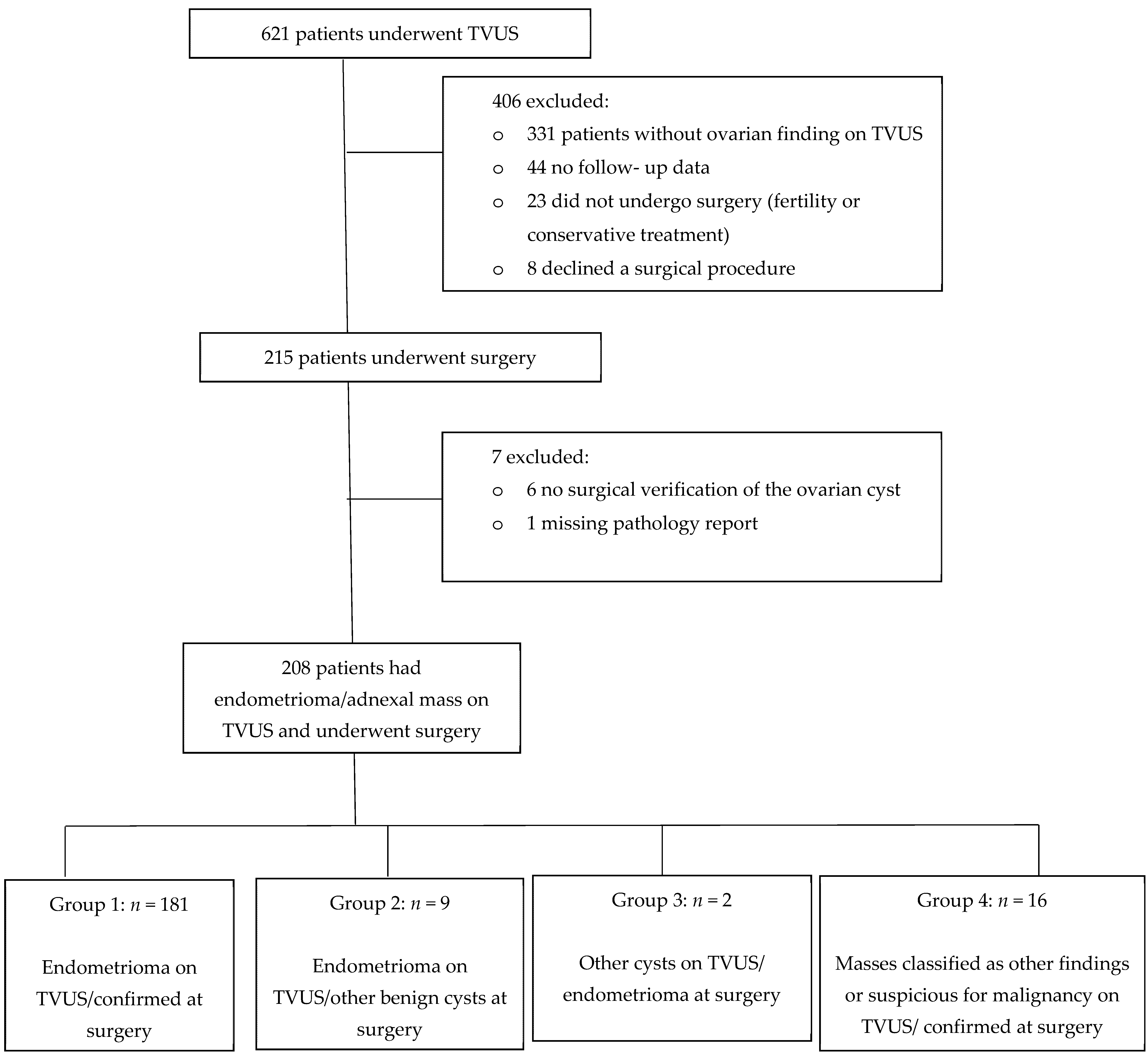
2.1. Study Design and Participants

This was a retrospective cohort study in an endometriosis tertiary referral center at the Sheba Medical Center in Tel Hashomer, a center of excellence open for referrals from the entire country, between May 2011–August 2017. Women were referred due to symptoms suggestive of endometriosis (such as severe dysmenorrhea, chronic pelvic pain, dyspareunia, infertility, gastrointestinal and urinary complaints) and suspicious TVUS findings (such as adnexal masses, adenomyosis, deep infiltrative lesions, etc.). The study included women above 18 years of age, who could undergo TVUS, for whom we had complete clinical, sonographic, surgical reports and pathological results. Electronic hospital records and outpatient referral files were used to extract patient information, which included demographic data (age, body mass index, marital status, parity, smoking habits), clinical history (previous cesarean sections, surgical history for endometriosis, fertility treatment history), clinical symptoms (dysmenorrhea, dyspareunia, pelvic pain, infertility, urinary and gastrointestinal symptoms), TVUS findings, and surgical and pathology reports. Patients were followed until September 2020.

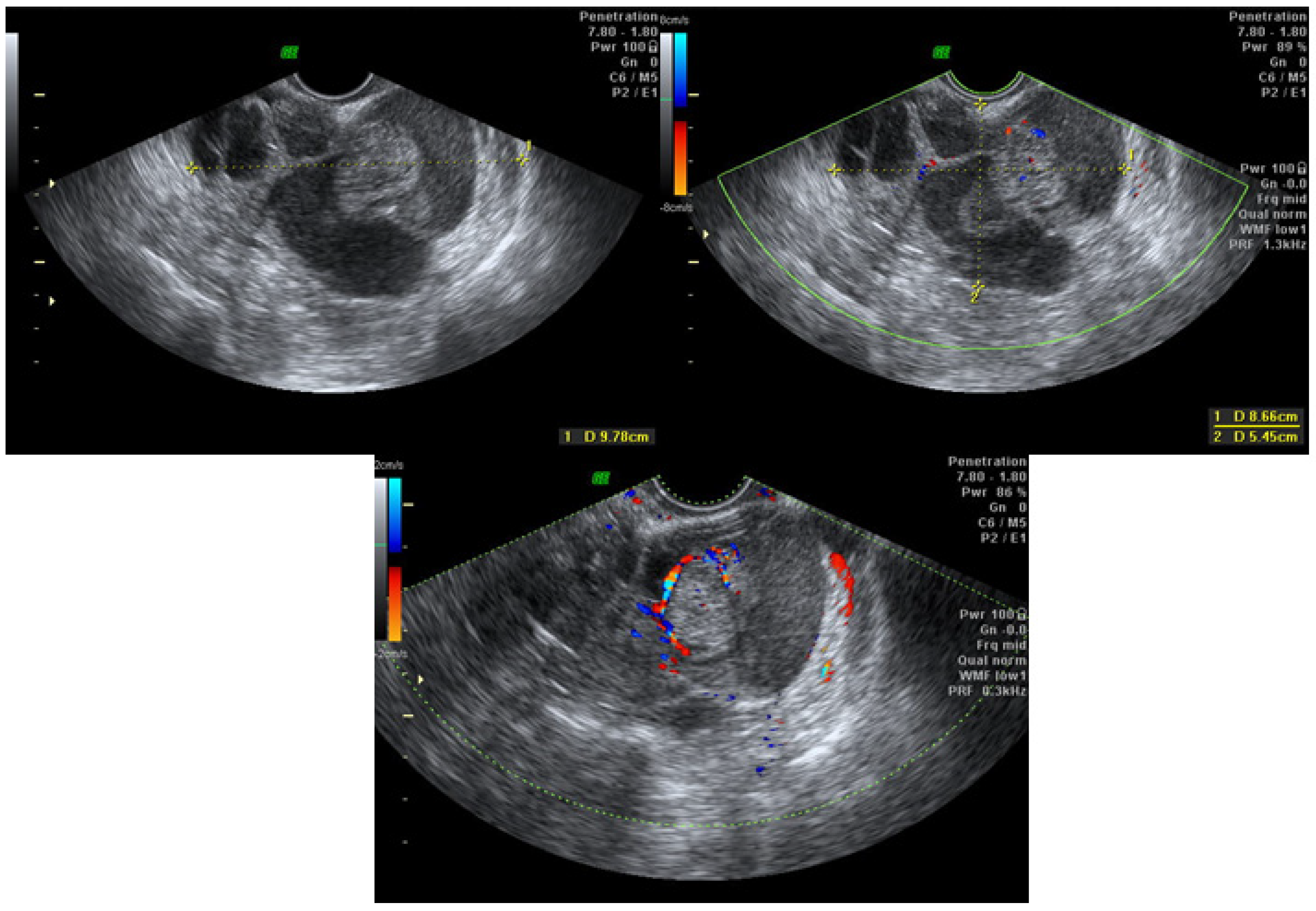
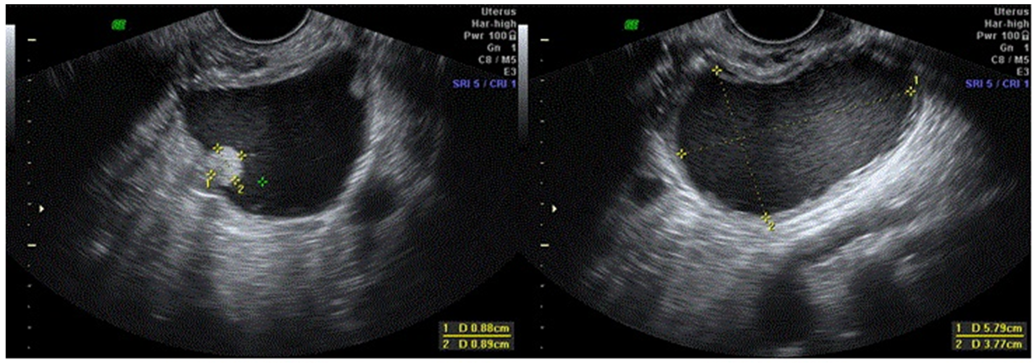
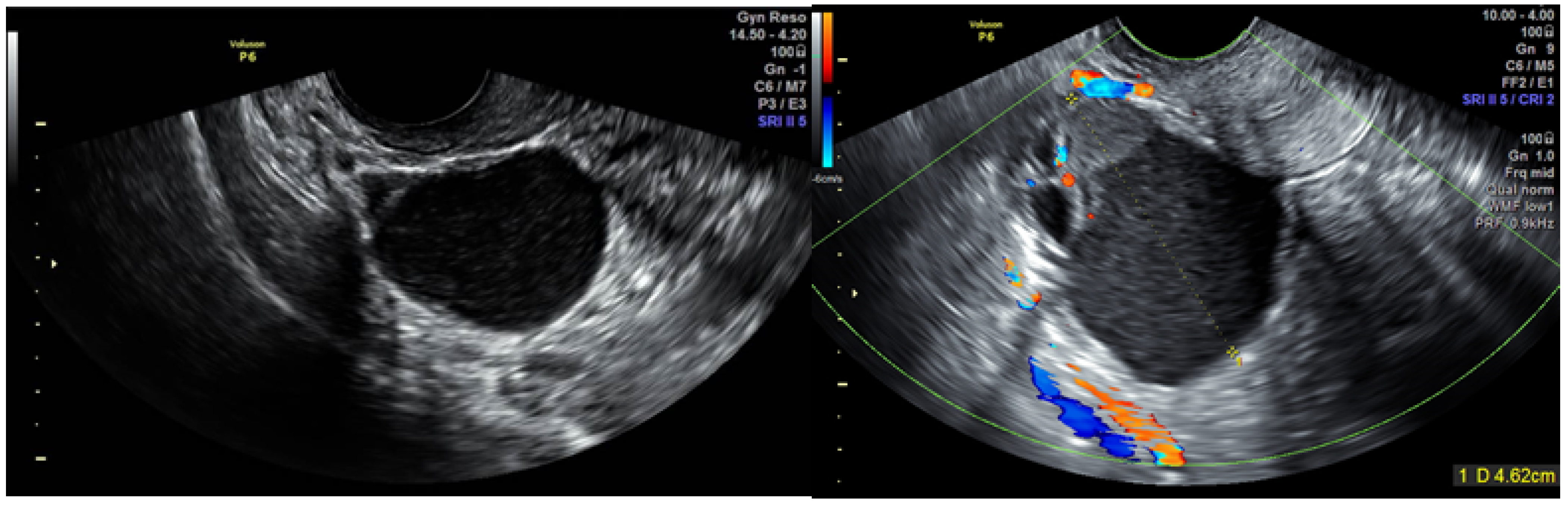
2.2. TVUS Evaluation of Adnexal Masses

All TVUS scans were carried out using a 7.5 MHz transvaginal probe (Voluson GE Medical Systems, Villach, Austria). The examinations were performed by expert physician sonographers, fully trained gynecologists with more than ten years’ experience in this field (Level-III examiners). Grey scale and color Doppler ultrasound were used to obtain morphological and blood flow variables to characterize each adnexal mass. The examination included a thorough evaluation of all pelvic viscera without bowel preparation. IDEA’s (International Deep Endometriosis Analysis group) systemic approach of sonographic evaluation of endometriosis was taken into consideration in the TVUS examinations following their publication [22]; Endometriosis was diagnosed based on the presence of deeply infiltrative endometriotic (DIE) nodules (for instance uterosacral ligament, bowel and bladder nodules), signs of pelvic adhesions (kissing ovaries or absent sliding of viscera, intestinal adhesions), presence of ovarian endometrioma or overt tubal disease as previously described [23,24,25,26,27,28,29,30,31].
Adnexal masses were evaluated using the etc. IOTA classification [4,6]. An endometrioma was diagnosed using simple descriptors as a unilocular tumor with ground glass echogenicity. When the mass did not classify as an endometrioma, the IOTA simple rules [8] were used in order to predict malignancy: (1) irregular solid tumor; (2) presence of ascites; (3) at least four papillary structures; (4) irregular multilocular–solid tumor with a largest diameter of at least 100 mm; and (5) very high color content on color Doppler examination. When at least one of the malignancy features were present and without benign sonographic features: ((1) unilocular cyst; (2) largest diameter of the largest solid component <7 mm; (3) acoustic shadows; (4) smooth multilocular tumor with largest diameter <100 mm and (5) absence of color flow on Doppler examination) we classified the mass as malignant. Benignity of a mass was classified if at least one of the benign features was present, and in the absence of malignancy features [9]. When the IOTA simple rules did not allow to reach a diagnosis (e.g., both malignant and benignity features were present or absence), expert subjective assessment was taken into consideration. In cases of bilateral adnexal mass, the largest or most complex cyst was used for analysis.

2.3. Laparoscopic Surgeries

All surgeries were carried out by highly qualified surgeons who specialized in laparoscopic surgery for endometriosis. The indications for surgery were: severe unmanageable symptoms, medical treatment failure, infertility and repeated IVF failure, ovarian cysts larger than 4 cm that was detected on TVUS and DIE involvement of other organs such as the ureter, bladder, rectum, or colon. Patients who wished to conceive and suffered from severe symptoms, or had findings on pelvic examination and/or US evaluation, and/or infertility, were counseled to have surgery before attempting to achieve pregnancy. Other aspects in the decision-making process were age, previous surgery, and ovarian reserve. Yet, the final decision whether to undergo surgery or continue medical or conservative treatment depended on patient preference.
Comparison between the TVUS examination and the surgical reports of each patient was performed, with the reference standard being definitive histology after mass removal. The patients were allocated into four groups based on diagnosis of endometrioma on TVUS and confirmation or contradiction on surgery and pathological report. Many of the patients presented with a suspected endometrioma, since this is a center that specializes in endometriosis diagnosis and treatment.

2.4. Ethical Aspects

Ethical approval was obtained from the local research ethics committee (IRB). Written informed consent was not required since the ultrasound assessment was offered as part of standard clinical care at the center. No procedure was performed specifically for the purpose of the study and no identifying information is included in the data presented here.

2.5. Statistical Analysis

Categorical variables were described as numbers and percentages. Continuous variables were evaluated for normal distribution using histograms and Q-Q plots. Normally distributed continuous variables (such as age) were reported as means ± SD’s and non-normally distributed variables were reported as medians and IQR. The diagnostic performance was estimated by calculating the sensitivity, specificity, positive and negative predictive values, positive and negative likelihood ratios and overall accuracy, with their corresponding 95% CI’s. All statistical analyses were 2-tailed and statistical significance in all tests was set at p < 0.05. Statistical analysis was performed using SPSS software (IBM SPSS statistics version 25, IBM Corporation, Armonk, NY, USA, 2017).

3. Results

3.1. Participants and Clinical Data

Six hundred and twenty-one women were evaluated for endometriosis and underwent TVUS examination, of whom 331 patients had no adnexal finding and were excluded from the statistical analysis. We further excluded 44 patients for whom we did not have complete follow-up data and 31 women who did not undergo surgery at our center. Two hundred and fifteen women underwent surgery, seven of whom were excluded (six patients had no surgical verification of the ovarian cyst and one with a missing pathology report), leaving 208 women who had an adnexal mass on TVUS and underwent surgery at our center (see Figure 1). Demographic, clinical data and prevalence of TVUS findings are presented in Table 1. Women presented with symptoms of endometriosis including dysmenorrhea in 177 women (85.1%), dyspareunia in 103 (49.5%), gastrointestinal complaints in 96 (46.2%) and urinary complaints in 47 (22.65%). Out of 134 women (64.4%) who tried to conceive, 88 (65.6%) suffered from subfertility. All women were premenopausal. Overall follow-up was on average 32.75 (±25.94) months.

3.2. TVUS Evaluation of Adnexal Mass and Surgery Results

Patients were allocated into 4 groups based on a comparison between TVUS findings and surgery results: (1) endometrioma on TVUS which was confirmed on surgery (Group 1, n = 181); (2) endometrioma on TVUS but other benign cysts at surgery (Group 2, n = 9); (3) other cysts on TVUS and endometrioma at surgery (Group 3, n = 2); (4) masses classified as other findings or suspicious for malignancy on TVUS and confirmed at surgery (Group 4, n= 16).
In group 2 the pathological results were: 5 follicular luteinizing cysts, 2 hemorrhagic corpus luteal cysts, 1 serous cystadenofibroma. One of the patients had an inconclusive pathology result, the differential diagnosis was: epidermal inclusion cyst, benign cystic Brenner tumor and/or benign mucinous cyst with extensive squamous metaplasia.
In group 3, two women presented with clear cysts on TVUS which were found to be endometrioma on pathology. Both of the patients had extensive endometriosis with adenomyosis and DIE. One of the patients had bilateral clear cysts on TVUS with a maximal diameter of 27 mm on the left ovary, while on surgery and pathology an endometrioma was confirmed. For the second patient, both TVUS and surgery misdiagnosed a clear cyst (23 mm clear cyst without flow was evaluated on TVUS), while pathology confirmed an endometrioma.
Clinical data, TVUS and surgical findings and pathology results of group 4 are presented in Table 2. Out of six masses suspected to be malignant, five were potentially malignant on pathology (two borderline serous tumor, two borderline mucinous tumor and one endometrioid lesion with complex hyperplasia without atypia) (Figure 2, Figure 3, Figure 4 and Figure 5, Videos S1–S3 in Supplementary Materials). The benign mass was a hydro salpinx.
The results of the comparison between TVUS evaluation and pathology results of the cysts were as follows: sensitivity 98.9% (95% CI, 96.11–99.87%), specificity 64% (95% CI, 45.52–82.03%), positive 95.3% (95% CI, 92.26–97.14%) and negative 88.9% (95% CI, 66.15–97.04%) predictive values, positive 2.74 (95% CI, 1.63–4.63) and negative 0.02 (95% CI, 0.00–0.07) likelihood ratios, and 94.7% (95% CI, 90.73–97.33%) overall accuracy.

4. Discussion

In this study we performed an external validation of the IOTA classification of adnexal masses in the evaluation of masses suspected to be endometriomas in women referred for dedicated endometriosis TVUS. We found a very high sensitivity and positive predicted values, confirming that the IOTA criteria are an excellent tool for non-invasive diagnoses of adnexal masses in endometriosis patients.
In the original IOTA study, as in our study, ultrasound examinations were performed by expert sonographers and obtained a sensitivity and a specificity of 92% [6]. Eighty-one percent of 1938 masses were diagnosed successfully using the simple descriptors and simple rules approach. In subsequent studies, external validations have been reported. A study in the U.K. by Sayaneh et al. [32] assessed 301 women with adnexal masses who underwent surgery and reported that 89% of the masses could be classified using IOTA’s simple descriptors and simple rules together. Similar to our study, the reported sensitivity was high, however the specificity was higher than ours, both 95%. Adding the examiners’ subjective assessment, in case the mass could not be fully assessed, reduced the sensitivity and specificity to 93% and 92%, respectively. It is of note that the sonographers in their study were not experts in the field (i.e., Level II examiners), which may explain the higher measured sensitivity in our study (98.9%) compared to that of the original study.
Testa et al. [33] conducted a multicentric study of 2403 women in 18 centers and six European countries, externally validating the IOTA strategy. Using simple descriptors and simple rules allowed correct classification of 81% of the masses, with a sensitivity and specificity of 95.7% and 73.6%, respectively. Adding a subjective examiner assessment gave a comparable sensitivity of 92.5% and raised the specificity to 87.6%. Similar to our study the most common benign diagnoses were endometrioma (14.3%) and serous cystadenoma (10.8%). Additionally, all ultrasound examinations were performed by level-III examiners. Alcazar et al. [34] reported external validation using the three-step strategy of IOTA with a high sensitivity (94.3%) and specificity (94.9%). In their study, non-expert examiners took expert subjective opinion into account when the mass classification was inconclusive. The study was conducted in two tertiary-care hospitals and included 362 women.
A small percentage of the masses (4.3%) was misdiagnosed as endometrioma, while other benign cysts were found at surgery (group 2). Our results are similar to the original IOTA study that suggested sonographic characterization for diagnosis of endometrioma [35]. In their study, 66 of 652 (10.1%) masses were misdiagnosed as endometrioma by an expert subjective assessment. A meta-analysis describing the diagnostic accuracy of TVUS for endometrioma [36], showed that most of the cysts, falsely thought to be endometrioma, were in fact hemorrhagic cysts, dermoid cysts, simple cysts, or cystadenomas. The results are not surprising considering the fact that most acute hemorrhagic cysts (as hemorrhagic corpus luteal cysts) have the same ultrasound morphology as an endometrioma, not to mention, ground glass appearance of cyst fluid can be present in different benign masses. Even the most expert of examiners sometimes can be misled by the clinical presentation. In group 4, the IOTA simple descriptors and simple rules for malignancy performed as expected. We confirmed the vast majority of the cysts thought to be malignant. In previous studies, [32,33,34] the false-negative cases were borderline tumors, while in our study, all of those tumors were accurately detected. We had one false-positive case of hydrosalpinx. The description of a multilocular solid cyst with pappillations misled the examiner to suspect a malignancy.
The main strengths of our study are the validation by experienced gynecologic examiners, familiar with the IOTA classification strategy of adnexal masses. Our study used the best reference standard (i.e., histological results), and the ultrasound examinations, surgeries, and histological analysis were conducted in one center, which was important for uniformity of diagnostic methods, laparoscopic procedures, and accuracy of patient follow-up.
Our study has several limitations which must be addressed: The fact that we are a tertiary referral center for endometriosis evaluation and treatment means that we see a high-risk population, most of whom do have endometriosis. The relatively low specificity that was found can be attributed to observer bias. However, to minimize this bias, we applied IOTA’s strict protocol for the evaluation of all adnexal masses. Additionally, using IOTA’s simple rules method by non-expert examiners does in fact yield a higher specificity compared to that reached by expert examiners [37].
Another limitation is that the simple descriptors, simple rules, and expert’s subjective assessment were assessed simultaneously by experienced sonographers. As a result, a comparison between the different steps of the IOTA strategies was difficult. Moreover, comparing the impact of expert and non-expert examiners during endometriosis evaluation using the IOTA strategy may be beneficial to reducing the time delay to diagnosis for these patients.

5. Conclusions

The IOTA classification simple descriptors, simple rules and expert opinion performs well for classifying adnexal masses suspected to be endometrioma, in women evaluated for endometriosis in a tertiary referral center. The most common potentially malignant masses in this population were borderline ovarian tumors. Operators who perform endometriosis diagnosis can use the IOTA classification when evaluating masses that they encounter during the examination. Additionally, the clinicians referring the patient can use the IOTA classification for diagnosis and need not wait for the tertiary exam for evaluation of the mass. It seems that teaching clinical operators the IOTA classification along with the IDEA consensus will benefit patients being referred for endometriosis evaluation.

Supplementary Materials

The following are available online at https://www.mdpi.com/article/10.3390/jcm10132971/s1, Video S1: case number 7—serous cystadenoma, Video S2: case number 8—mucinous cystadenoma, Video S3: case number 10—mucinous cystadenoma.

Author Contributions

Conceptualization, L.C.B.-M. and V.H.E.; methodology, L.C.B.-M. and V.H.E.; writing—original draft preparation, L.C.B.-M. and V.H.E.; writing—review and editing, L.C.B.-M., R.M. and V.H.E. All authors have read and agreed to the published version of the manuscript.

Funding

This research received no external funding.

Institutional Review Board Statement

The study was conducted according to the guidelines of the Declaration of Helsinki, and approved by the Institutional Review Board of Sheba Medical Center (protocol code 4706-17-SMC, 26 November 2017).

Informed Consent Statement

Informed consent was obtained from all subjects involved in the study.

Data Availability Statement

Not applicable.

Acknowledgments

Tomer Ziv-Baran for statistical analysis.

Conflicts of Interest

The authors declare no conflict of interest.

References

  1. Guerriero, S.; Ajossa, S.; Garau, N.; Piras, B.; Paoletti, A.M.; Melis, G.B. Ultrasonography and color Doppler-based triage for adnexal masses to provide the most appropriate surgical approach. Am. J. Obstet. Gynecol. 2005, 192, 401–406. [Google Scholar] [CrossRef] [PubMed]
  2. Alcázar, J.L.; Royo, P.; Jurado, M.; Minguez, J.A.; Laparte, C.; Galván, R.; García-Manero, M.; López-García, G. Triage for surgical management of ovarian tumors in asymptomatic women: Assessment of an ultrasound-based scoring system. Ultrasound Obstet. Gynecol. 2008, 32, 220–225. [Google Scholar] [CrossRef] [PubMed]
  3. American College of Obstetricians and Gynecoclogists. Practice bulletin No. 174: Evaluation and Management of Adnexal Masses. Obstet. Gynecol. 2016, 128, e210. [Google Scholar] [CrossRef] [PubMed]
  4. Timmerman, D.; Valentin, L.; Bourne, T.; Collins, W.P.; Verrelst, H.; Vergote, I. Terms, definitions and measurements to describe the sonographic features of adnexal tumors: A consensus opinion from the International Ovarian Tumor Analysis (IOTA) group. Ultrasound Obstet. Gynecol. 2000, 16, 500–505. [Google Scholar] [CrossRef]
  5. Van Holsbeke, C.; Daemen, A.; Yazbek, J.; Holland, T.K.; Bourne, T.; Mesens, T.; Lannoo, L.; Boes, A.S.; Joos, A.; Van De Vijver, A.; et al. Ultrasound experience substan-tially impacts on diagnostic performance and confidence when adnexal masses are classified using pattern recognition. Gynecol. Obstet. Investig. 2010, 69, 160–168. [Google Scholar] [CrossRef]
  6. Ameye, L.; Timmerman, D.; Valentin, L.; Paladini, D.; Zhang, J.; Van Holsbeke, C.; Lissoni, A.A.; Savelli, L.; Veldman, J.; Testa, A.C.; et al. Clinically oriented three-step strategy for assessment of adnexal pathology. Ultrasound Obstet. Gynecol. 2012, 40, 582–591. [Google Scholar] [CrossRef]
  7. Ueland, F.R.; DePriest, P.D.; Pavlik, E.J.; Kryscio, R.J.; Van Nagell, J.R. Preoperative differentiation of malignant from benign ovar-ian tumors: The efficacy of morphology indexing and Doppler flow sonography. Gynecol. Oncol. 2003, 91, 46–50. [Google Scholar] [CrossRef]
  8. Timmerman, D.; Testa, A.C.; Bourne, T.; Ameye, L.; Jurkovic, D.; Van Holsbeke, C.; Paladini, D.; Van Calster, B.; Vergote, I.; Van Huffel, S.; et al. Simple ultrasound-based rules for the diagnosis of ovarian cancer. Ultrasound Obstet. Gynecol. 2008, 31, 681–690. [Google Scholar] [CrossRef]
  9. Timmerman, D.; Ameye, L.; Fischerova, D.; Epstein, E.; Melis, G.B.; Guerriero, S.; Van Holsbeke, C.; Savelli, L.; Fruscio, R.; Lissoni, A.A.; et al. Simple ultrasound rules to distinguish between be-nign and malignant adnexal masses before surgery: Prospective validation by IOTA group. Bmj 2011, 342, 94. [Google Scholar]
  10. Froyman, W.; Wynants, L.; Landolfo, C.; Bourne, T.; Valentin, L.; Testa, A.; Sladkevicius, P.; Franchi, D.; Fischerova, D.; Savelli, L.; et al. Validation of the performance of International Ovarian Tumor Analysis (IOTA) methods in the diagnosis of early stage ovarian cancer in a non-screening population. Diagnostics 2017, 7, 32. [Google Scholar] [CrossRef]
  11. Kennedy, S.; Bergqvist, A.; Chapron, C.; D’Hooghe, T.; Dunselman, G.; Greb, R.; Hummelshoj, L.; Prentice, A. ESHRE guideline for the diagnosis and treatment of endometriosis. Hum. Reprod. 2005, 20, 2698–2704. [Google Scholar] [CrossRef]
  12. Kontoravdis, A.; Chryssikopoulos, A.; Hassiakos, D.; Liapis, A.; Zourlas, P.A. The diagnostic value of laparoscopy in 2365 patients with acute and chronic pelvic pain. Int. J. Obstet. Gynaecol. 1996, 52, 243–248. [Google Scholar] [CrossRef]
  13. Rogers, P.A.; Adamson, G.D.; Al-Jefout, M.; Becker, C.M.; D’Hooghe, T.M.; Dunselman, G.A.; Fazleabas, A.; Giudice, L.C.; Hoenr, A.W.; Hull, M.L.; et al. Research priorities for endometriosis: Recommendations from a global consortium of investigators in en-dometriosis and for the WES/WERF consortium for research priorities in endometriosis. Reprod. Sci. 2017, 24, 202–226. [Google Scholar] [CrossRef] [Green Version]
  14. Eisenberg, V.H.; Weil, C.; Chodick, G.; Shalev, V. Epidemiology of endometriosis: A large population-based database study from a healthcare provider with 2 million members. BJOG Int. J. Obstet. Gynaecol. 2018, 125, 55–62. [Google Scholar] [CrossRef] [Green Version]
  15. Kobayashi, H.; Sumimoto, K.; Kitanaka, T.; Yamada, Y.; Sado, T.; Sakata, M.; Yoshida, S.; Kawaguchi, R.; Kanayama, S.; Shigetomi, H.; et al. Ovarian endometrioma—Risks factors of ovarian cancer development. Eur. J. Obstet. Gynecol. Reprod. Biol. 2008, 138, 187–193. [Google Scholar] [CrossRef] [PubMed]
  16. Brinton, L.A.; Gridley, G.; Persson, I.; Baron, J.; Bergqvist, A. Cancer risk after a hospital discharge diagnosis of endometriosis. Am. J. Obstet. Gynecol. 1997, 176, 572–579. [Google Scholar] [CrossRef]
  17. Jimbo, H.; Yoshikawa, H.; Onda, T.; Yasugi, T.; Sakamoto, A.; Taketani, Y. Prevalence of ovarian endometriosis in epithelial ovar-ian cancer. Int. J. Gynecol. Obstet. 1997, 59, 245–250. [Google Scholar] [CrossRef]
  18. Crozier, M.A.; Copeland, L.J.; Silva, E.G.; Gershenson, D.M.; Stringer, C. Clear cell carcinoma of the ovary: A study of 59 cases. Gynecol. Oncol. 1989, 35, 199–203. [Google Scholar] [CrossRef]
  19. DePriest, P.D.; Banks, E.R.; Powell, D.E.; van Nagell, J.R.; Gallion, H.H.; Puls, L.E.; Hunter, J.E.; Kryscio, R.J.; Royalty, M.B. Endometrioid carcinoma of the ovary and endometriosis: The association in postmenopausal women. Gynecol. Oncol. 1992, 47, 71–75. [Google Scholar] [CrossRef]
  20. Thomsen, L.H.; Schnack, T.H.; Buchardi, K.; Hummelshoj, L.; Missmer, S.A.; Forman, A.; Blaakaer, J. Risk factors of epithelial ovarian carcinomas among women with endometriosis: A systematic review. Acta Obstet. Gynecol. Scand. 2016, 96, 761–778. [Google Scholar] [CrossRef]
  21. Testa, A.C.; Van Holsbeke, C.; Zannoni, G.F.; Fransis, S.; Moerman, P.; Vellone, V.G.; Mascilini, F.; Licameli, A.; Ludovisi, M.; Di Legge, A.; et al. Ovarian cancer arising in endometrioid cysts: Ultrasound findings. Ultrasound Obstet. Gynecol. 2011, 38, 99–106. [Google Scholar] [CrossRef] [PubMed]
  22. Guerriero, S.; Condous, G.; Bosch, T.V.D.; Valentin, L.; Leone, F.P.G.; Van Schoubroeck, D.; Exacoustos, C.; Installé, A.J.F.; Martins, W.P.; Abrao, M.S.; et al. Systematic approach to sonographic evaluation of the pelvis in women with suspected endometriosis, including terms, definitions and measurements: A consensus opinion from the International Deep Endometriosis Analysis (IDEA) group. Ultrasound Obstet. Gynecol. 2016, 48, 318–332. [Google Scholar] [CrossRef] [PubMed]
  23. Bazot, M.; Thomassin, I.; Hourani, R.; Cortez, A.; Darai, E. Diagnostic accuracy of transvaginal sonography for deep pelvic en-dometriosis. Ultrasound Obstet. Gynecol. 2004, 24, 180–185. [Google Scholar] [CrossRef] [PubMed]
  24. Hudelist, G.; English, J.; Thomas, A.; Tinelli, A.; Singer, C.F.; Keckstein, J. Diagnostic accuracy of transvaginal ultrasound for non-invasive diagnosis of bowel endometriosis: Systematic review and meta-analysis. Ultrasound Obstet. Gynecol. 2011, 37, 257–263. [Google Scholar] [CrossRef]
  25. Piketty, M.; Chopin, N.; Dousset, B.; Millischer-Bellaische, A.-E.; Roseau, G.; Leconte, M.; Borghese, B.; Chapron, C. Preoperative work-up for patients with deeply infiltrating endometriosis: Transvaginal ultrasonography must definitely be the first-line imaging examination. Hum. Reprod. 2008, 24, 602–607. [Google Scholar] [CrossRef] [Green Version]
  26. Menakaya, U.; Reid, S.; Infante, F.; Condous, G. Systematic evaluation of women with suspected endometriosis using a 5-domain sonographically based approach. J. Ultrasound Med. 2015, 34, 937–947. [Google Scholar] [CrossRef]
  27. Abrao, M.S.; Goncalves, M.O.D.C.; Dias, J.A., Jr.; Podgaec, S.; Chamie, L.P.B.R. Comparison between clinical examination, transvagi-nal sonography and magnetic resonance imaging for the diagnosis of deep endometriosis. Hum. Reprod. 2007, 22, 3092–3097. [Google Scholar] [CrossRef] [Green Version]
  28. Guerriero, S.; Ajossa, S.; Gerada, M.; Virgilio, B.; Angioni, S.; Melis, G.B. Diagnostic value of transvaginal ’tenderness-guided’ ultrasonography for the prediction of location of deep endometriosis. Hum. Reprod. 2008, 23, 2452–2457. [Google Scholar] [CrossRef] [Green Version]
  29. Martin, D.C.; Batt, R.E. Retrocervical, retrovaginal pouch, and rectovaginal septum endometriosis. J. Am. Assoc. Gynecol. Laparosc. 2001, 8, 12–17. [Google Scholar] [CrossRef]
  30. Leon, M.; Alcazar, J.L. High sliding sign: A new soft marker of uterine fundus compromise in deep infiltrating endometriosis. Ultrasound Obstet. Gynecol. 2015, 45, 624. [Google Scholar] [CrossRef]
  31. Guerriero, S.; Alcazar, J.L.; Ajossa, S.; Pilloni, M.; Melis, G.B. Three-dimensional sonographic characteristics of deep endometrio-sis. J. Ultrasound Med. 2009, 28, 1061–1066. [Google Scholar] [CrossRef]
  32. Sayasneh, A.; Kaijser, J.; Preisler, J.; Johnson, S.; Stalder, C.; Husicka, R.; Guha, S.; Naji, O.; Abdallah, Y.; Raslan, F.; et al. A multicenter prospective external vali-dation of the diagnostic performance of IOTA simple descriptors and rules to characterize ovarian masses benign simple descriptor malignant simple descriptor. Gynecol. Oncol. 2013, 130, 140–146. [Google Scholar] [CrossRef]
  33. Testa, A.; Kaijser, J.; Wynants, L.; Fischerová, D.; Van Holsbeke, C.; Franchi, D.; Savelli, L.; Epstein, E.; Czekierdowski, A.; Guerriero, S.; et al. Strategies to diagnose ovarian cancer: New evidence from phase 3 of the multicentre international IOTA study. Br. J. Cancer 2014, 111, 680–688. [Google Scholar] [CrossRef] [Green Version]
  34. Alcázar, J.L.; Pascual, M.A.; Graupera, B.; Aubá, M.; Errasti, T.; Olartecoechea, B.; Ruiz-Zambrana, A.; Hereter, L.; Ajossa, S.; Guerriero, S. External validation of IOTA simple descriptors and simple rules for classifying adnexal masses. Ultrasound Obstet. Gynecol. 2016, 48, 397–402. [Google Scholar] [CrossRef]
  35. Van Holsbeke, C.; Van Calster, B.; Guerriero, S.; Savelli, L.; Paladini, D.; Lissoni, A.A.; Czekierdowski, A.; Fischerova, D.; Zhang, J.; Mestdagh, G.; et al. Endometriomas: Their ultrasound characteristics. Ultrasound Obstet. Gynecol. 2010, 35, 730–740. [Google Scholar] [CrossRef]
  36. Moore, J.; Copley, S.; Morris, J.; Lindsell, D.; Golding, S.; Kennedy, S. A systematic review of the accuracy of ultrasound in the diagnosis of endometriosis. Ultrasound Obstet. Gynecol. 2002, 20, 630–634. [Google Scholar] [CrossRef]
  37. Alcazar, J.L.; Pascual, M.A.; Olartecoechea, B.; Graupera, B.; Auba, M.; Ajossa, S.; Hereter, L.; Julve, R.; Gaston, B.; Peddes, C.S.F. IOTA simple rules for discriminating between benign and malignant adnexal masses: Prospective external validation. Ultrasound Obstet. Gynecol. 2013, 4, 467–471. [Google Scholar] [CrossRef]
Figure 1. Study flowchart. TVUS (transvaginal ultrasound).
Jcm 10 02971 g001
Figure 2. Case number 1; 46 years old woman, presented with dysmenorrhea, infertility (11 cycles of IVF), 1 parity. CA125 = 30. On TVUS multilocular cyst with high suspicious of malignancy, hystological result was borderline mucinous tumor.
Jcm 10 02971 g002
Figure 3. Case number 3; 37 years old woman, presented with dysmenorrhea, infertility (13 cycles of IVF), 0 parity. On TVUS unilocular solid cyst, hystological result was a borderline serous tumor.
Jcm 10 02971 g003
Figure 4. Case number 7; 36 years old woman, presented without symptoms. On TVUS multilocular cyst, hystological result was a serous cystadenoma.
Jcm 10 02971 g004
Figure 5. Case number 8; 30 years old woman, presented without symptoms. On TVUS multilocular cyst, hystological result was a mucinous cystadenoma.
Jcm 10 02971 g005
Table 1. Demographic, clinical data and prevalence of TVUS findings of patients attending an endometriosis tertiary referral center and included in the study (n = 208).
ParameterGroup 1
(n = 181)
Group 2
(n = 9)
Group 3
(n = 2)
Group 4
(n = 16)
Total
(n = 208)
Age, mean ± SD, years33.6 ± 6.130.4 ± 10.443 ± 1.434.1 ± 7.233.6 ± 6.5
BMI, median (IQR)22.5
(19.9–25.6)
22.3
(20.7–22.8)
2323.4
(20.6–26.6)
22.5
(20.1–25.5)
Smoking (%)56 (30.9%)4 (44.4%)1 (50%)6 (37.5%)67 (32.2%)
Nulliparous (%)115 (63.5%)5 (55.6%)010 (62.5%)130 (62.5%)
Previous Cesarean section (%)17 (9.4%)1 (11.1%)02 (12.5%)20 (9.6%)
Previous surgery (%)74 (40.9%)4 (44.4%)1 (50%)10 (62.5%)89 (42.8%)
Additional TVUS findings (%)
Maximal size of cyst, median (IQR), mm50
(41–66)
44.5
(39.5–53)
4046
(38.5–64.7)
49
(40.5–65.5)
Kissing ovaries53 (29.8%)2 (22.2%)03 (18.8%)58 (28.3%)
Uterosacral ligaments nodule85 (47.2%)5 (55.6%)02 (12.5%)92 (44.4%)
Retro cervical nodule46 (25.4%)00046 (22.1%)
Recto sigmoid nodule57 (31.5%)3 (33.3%)02 (12.5%)62 (29.8%)
Bladder nodule8 (4.4%)001 (6.3%)9 (4.3%)
Intestinal nodule26 (14.4%)1 (11.1%)01 (6.3%)28 (13.5%)
Pouch of Douglas obliteration85 (47%)4 (44.4%)03 (18.8%)92 (44.2%)
SD—standard deviation; BMI—body mass index; TVUS—transvaginal ultrasound; IQR—interquartile range.
Table 2. Clinical data, findings on TVUS and surgery and pathology results of women with malignant adnexal masses and different benign adnexal masses.
Case NumberAge (Years)ParitySide of FindingMaximal Size of Finding (mm)TVUS * DescriptionSuspected MalignancyAdditional FindingsPathology Result of Adnexal MassFollow-Up (Months)Comments
1461Left98Multilocular cysticYesUterus myomatosus, right endometrioma, rectosigmoid noduleBorderline mucinous tumor53Normal follow-up, no recurrence.
2322Right100Multilocular cysticYesNoneBorderline mucinous tumor5Normal follow-up, no recurrence.
3370Left58Unilocular solidYesNoneBorderline serous tumor92Recurrence after 4 years, underwent USO **, normal follow-up.
4280Right40Unilocular cystic low-level echogenicityYesNoneBorderline serous tumor54Normal follow-up, no recurrence. Spontaneous pregnancy.
5360Right47Unilocular cystic low-level echogenicityYesRectovaginal noduleEndometrioid lesion (complex hyperplasia without atypia)34Normal follow-up. Infertility-repeated IVF cycles, no pregnancy.
6220Left46Multilocular solid cyst with papillationsYesUterosacral ligament nodulesHydrosalpinx22Normal follow-up, no recurrence.
7363Left45Multilocular cysticNoUterosacral ligament nodulesSerous cystadenoma15Normal follow-up, no recurrence.
8300Right66Multilocular cysticNoNoneMucinous cystadenoma31Recurrence after 2.5 years, underwent cystectomy for the third time, normal follow-up.
9380Left33Unilocular cystic low-level echogenicityNoDeep lesion around right ureterMucinous cystadenoma45Normal follow-up, no recurrence. Conceived twice with fertility treatments.
10320Left98Unilocular cysticNoNoneMucinous cystadenoma19Normal follow-up, no recurrence. Spontaneous pregnancy.
11403Left61Unilocular solidNoNoneFibro-thecoma4Normal follow-up, no recurrence.
12240Right53Multilocular cysticNoBladder nodule, left endometrioma.Multi-cystic benign mesothelioma with decidual changes11Normal follow-up, no recurrence.
13392Right20Unilocular solidNoBladder nodule, uterosacral ligament noduleMature cystic teratoma25Normal follow-up, no recurrence.
14431Right74Multilocular cysticNoIntestinal adhesionsHemorrhagic corpus luteum5Normal follow-up, no recurrence.
15402Left35Unilocular cysticNoUterosacral ligament noduleHemorrhagic corpus luteum78Normal follow-up, no recurrence.
16230Left69Unilocular cystic low-level echogenicity with papillation (para ovarian)NoNonePara-ovarian cyst: Serous Cyst adenofibroma31Normal follow-up, no recurrence. Spontaneous pregnancy.
* TVUS—transvaginal ultrasound; ** USO—unilateral salpingo-oophorectomy.
Publisher’s Note: MDPI stays neutral with regard to jurisdictional claims in published maps and institutional affiliations.

