Article

Diagnostic Ability of Peguero-Lo Presti Electrocardiographic Left Ventricular Hypertrophy Criterion in Severe Aortic Stenosis

by
Aleksandra Gamrat
1,
Katarzyna Trojanowicz
1,
Michał A. Surdacki
1,
Aleksandra Budkiewicz
1,
Adrianna Wąsińska
1,
Ewa Wieczorek-Surdacka
2,
Andrzej Surdacki
3 and
Bernadeta Chyrchel
3,*
1
Students’ Scientific Group at the Second Department of Cardiology, Jagiellonian University Medical College, 2 Jakubowskiego Street, 30-688 Cracow, Poland
2
Chair and Department of Nephrology, Faculty of Medicine, Jagiellonian University Medical College, 2 Jakubowskiego Street, 30-688 Cracow, Poland
3
Second Department of Cardiology, Institute of Cardiology, Faculty of Medicine, Jagiellonian University Medical College, 2 Jakubowskiego Street, 30-688 Cracow, Poland
*
Author to whom correspondence should be addressed.
J. Clin. Med. 2021, 10(13), 2864; https://doi.org/10.3390/jcm10132864
Submission received: 31 May 2021 / Revised: 20 June 2021 / Accepted: 27 June 2021 / Published: 28 June 2021
(This article belongs to the Section Cardiology)

Abstract

:
Traditional electrocardiographic (ECG) criteria for left ventricular hypertrophy (LVH), introduced in the pre-echocardiographic era of diagnosis, have a relatively low sensitivity (usually not exceeding 25–40%) in detecting LVH. A novel Peguero-Lo Presti ECG-LVH criterion was recently shown to exhibit a higher sensitivity than the traditional ECG-LVH criteria in hypertension. Our aim was to test the diagnostic ability of the novel Peguero-Lo Presti ECG-LVH criterion in severe aortic stenosis. We retrospectively analyzed 12-lead ECG tracings and echocardiographic records from the index hospitalization of 50 patients with isolated severe aortic stenosis (mean age: 77 ± 10 years; 30 women and 20 men). Exclusion criteria included QRS > 120 ms, bundle branch blocks or left anterior fascicular block, a history of myocardial infarction, more than mild aortic or mitral regurgitation, and significant LV dysfunction by echocardiography. We compared the agreement of the novel Peguero-Lo Presti criterion and traditional ECG-LVH criteria with echocardiographic LVH (LV mass index > 95 g/m2 in women and >115 g/m2 in men). Echocardiographic LVH was found in 32 out of 50 study patients. The sensitivity of the Peguero-Lo Presti criterion in detecting LVH was improved (55% vs. 9–34%) at lower specificity (72% vs. 78–100%) in comparison to 8 single traditional ECG-LVH criteria. Additionally, the positive predictive value (77% vs. 72%), positive likelihood ratio (2.0 vs. 1.5), and odds ratio (3.2 vs. 2.4) were higher for the Peguero-Lo Presti criterion versus the presence of any of these 8 traditional ECG-LVH criteria. Cohen’s Kappa, a measure of concordance between ECG and echocardiography with regard to LVH, was 0.24 for the Peguero-Lo Presti criterion, −0.01–0.13 for single traditional criteria, and 0.20 for any traditional criterion. However, by the receiver operating characteristics (ROC) curve analysis, the overall ability to discriminate between patients with and without LVH was insignificantly lower for the Peguero-Lo Presti versus Cornell voltage as a continuous variable (area under the ROC curve: 0.65 (95% CI, 0.48–0.81) vs. 0.71 (0.55–0.86), p = 0.5). In conclusion, our preliminary results suggest a slightly better, albeit still low, agreement of the novel Peguero-Lo Presti ECG criterion compared to the traditional ECG-LVH criteria with echocardiographic LVH in severe aortic stenosis.

1. Introduction

Left ventricular hypertrophy (LVH), defined as increased left ventricular mass, has long been recognized as a predictor of adverse clinical events, including death from cardiovascular causes [1,2,3,4]. In the pre-echocardiographic era, traditional ECG criteria were the only practical technique in the diagnosis of LVH [5]. However, their drawback is a low sensitivity, generally not exceeding 30–40%, in detecting LVH diagnosed by magnetic resonance or echocardiography, the current standards in LVH diagnosis [5,6], Nevertheless, ECG is still used as a screening tool owing to its wide availability. Moreover, recent findings suggest that LVH on ECG and anatomic LVH have independent prognostic values for cardiovascular mortality [6,7,8,9,10].
A novel ECG criterion for LVH, proposed by Peguero et al. [11], was recently shown to be more sensitive than traditional ECG-LVH criteria in patients with arterial hypertension. To date, the ability of the novel criterion to discriminate patients with and without anatomic LVH have been estimated in various clinical settings, including cardiac patients and the general population, demonstrating generally rather modest (and, at best, moderate) superiority over the classical ECG-LVH criteria in some [12,13,14,15,16], but not all [17,18,19,20,21,22,23]. However, to the best of our knowledge, only one study was focused on patients with aortic stenosis [24], the most prevalent valvular heart disease. Nevertheless, although in that report the Peguero-Lo Presti criterion had higher sensitivity than Cornell and Sokolow–Lyon LVH voltage criteria to predict anatomic LVH, a complex analysis of the agreement between the novel criterion, traditional ECG-LVH criteria, and anatomic LVH in aortic stenosis has not been reported so far.
Our aim was to compare the concordance of the novel Peguero-Lo Presti LVH criterion and traditional ECG-LVH criteria with echocardiographic LVH in severe aortic stenosis.

2. Materials and Methods

Out of 83 pre-screened subjects previously hospitalized in our center with the final diagnosis of severe aortic stenosis by means of echocardiography [25], we selected 50 previously described subjects (30 women and 20 men; average age 77 ± 10 years) with isolated severe aortic stenosis without relevant coexistent diseases [26]. As reported [26], we excluded subjects with QRS duration >120 ms, His bundle branch or left anterior fascicular block, more than mild aortic regurgitation or disease of another valve, a history of myocardial infarction, and left ventricular (LV) ejection fraction below 40%.
We compared the agreement of the novel Peguero-Lo Presti criterion [11] and traditional ECG-LVH criteria on routine in-hospital 12-lead ECG tracing [5,26] with echocardiographic LVH, defined in accordance with current recommendations as a LV mass (by the Devereux equation) over 95 g/m2 in women and over 115 g/m2 in men [27].
Traditional ECG-LVH criteria include R wave amplitudes (R I > 1.5 mV, R aVL ≥ 1.1 mV, max. R V5/V6 > 2.6 mV), R V6 > R V5 (Holt–Spodick criterion), and the combined voltage criteria introduced by Sokolow and Lyon (S V1 + max. R V5/V6 > 3.5 mV), Romhilt (S V2 + max. R V5/V6 > 4.5 mV), Cornell (R aVL + S V3 > 2.0 in women or >2.8 mV in men) as well as Gubner and Ungerleider (R I + S III > 2.5 mV) [5]. We also calculated the novel Peguero-Lo Presti voltage criterion, defined as the sum of the amplitude of the deepest S wave in any lead and the S wave in lead V4 ≥ 2.8 mV in men and ≥2.3 mV in women [11].
The protocol was approved by the institutional ethics committee, including the fact that patients’ informed consent was not obtained due to a retrospective design (Approval No.: 1072.6120.260.2020 issued on 24 September 2020).
Beyond sensitivity, specificity, positive predictive value (PPV), and negative predictive value (NPV), we calculated positive likelihood ratio, odds ratio, and relative risk as estimates of the diagnostic ability of ECG-LVH criteria with regard to echocardiographic LVH. Positive likelihood ratio was defined as the probability of having LVH on ECG in patients with versus without echocardiographic LVH, i.e., sensitivity/(1 − specificity), odds ratio as the odds of echocardiographic LVH (i.e., the probability of having/not having echocardiographic LVH) in patients with versus without LVH on ECG, while relative risk as the probability of echocardiographic LVH in patients with versus without LVH on ECG.
Additionally, we computed Cohen’s kappa to estimate concordance of echocardiographic LVH and LVH on ECG, or mutual agreement between two different ECG-LVH criteria, corrected for the agreement which might be expected by random chance [28]. The presence of a systematic difference between echocardiographic LVH and the ECG-LVH criteria was assessed by McNemar’s test. A Cohen’s kappa of 0.01–0.20, 0.21–0.40, 0.41–0.60, 0.61–0.80, and >0.8 is equivalent to slight, fair, moderate, substantial, and almost perfect, respectively, degree of concordance between the results obtained by two different methods [28].
Additionally, since by means of the receiver operating characteristics (ROC) curve analysis we previously observed the highest ability of the Cornell voltage to predict echocardiographic LVH among 13 classical LVH-related QRS voltages in our study group [26], the diagnostic performance of the Peguero-Lo Presti voltage against the Cornell voltage was also compared as a continuous variable by the ROC curve analysis.
Finally, using multiple regression, we estimated potential confounding effects of transaortic pressure gradient, averaged in-hospital mean arterial blood pressure, age, gender, and body mass index on the association between LV mass index and the Peguero-Lo Presti voltage.
All statistical analyses were made with STATISTICA version 13.3.704.0 (TIBCO Software Inc., 2017; Palo Alto, CA, USA).

3. Results

Patients’ characteristics are presented in Table 1.
The sensitivity of the Peguero-Lo Presti criterion in predicting echocardiographic LVH (present in 32 of 50 study subjects, out of whom 90% presented concentric LVH [24]) was improved (55%) in comparison to 8 single traditional ECG-LVH criteria (9–34%) at lower specificity (72% vs. 78–100%) (Table 2). In addition, NPV (48% vs. 36–41%) and overall accuracy (61% vs. 38–50%) were slightly higher for the Peguero-Lo Presti criterion than the 8 classical ECG-LVH criteria analyzed separately (Table 2).
Additionally, PPV (77% vs. 72%), positive likelihood ratio (2.0 vs. 1.5) and odds ratio (3.2 vs. 2.4) were higher for the Peguero-Lo Presti compared to the presence of any of these 8 traditional criteria (Table 2 and Table 3).
Regarding the presence of echocardiographic LVH, Cohen’s kappa was 0.24 for the Peguero-Lo Presti criterion, −0.01–0.13 for single traditional ECG criteria analyzed separately, and 0.20 for the presence of any of the traditional criteria (Table 2).
However, mean area under ROC curve (AUC [95% confidence interval]) was insignificantly lower for the Peguero-Lo Presti compared to the Cornell voltage (0.65 (0.48–0.81) vs. 0.71 (0.55–0.86), p = 0.5) (Figure 1), whose AUC was highest among the traditional voltages as previously shown [26].
The degree of agreement between the Peguero-Lo Presti criterion and the traditional ECG-LVH criteria was highest for the Cornell voltage, followed by the Sokolow–Lyon voltage, Romhilt voltage, R aVL, Holt-Spodick criterion, Gubner–Ungerleider voltage, max. R V5/V6 and R I (Cohen’s kappa: 0.28, 0.18, 0.16, 0.12, 0.07, 0.0, −0.02, and −0.05, respectively).
By multiple regression (adjusted coefficient of multiple determination (R2), 0.30, p < 0.001), the Peguero-Lo Presti voltage was associated with gender (men vs. women: mean non-standardized regression coefficient, 0.70 ± 0.27, p = 0.01) and LV mass index (mean standardized regression coefficient (β) ± standard error of the mean, 0.30 ± 0.13, p = 0.03), being unrelated to peak aortic pressure gradient (β = 0.17 ± 0.13, p = 0.2), averaged in-hospital mean arterial pressure (β = 0.15 ± 0.13, p = 0.3), age (β = −0.23 ± 0.12, p = 0.06), and body mass index (β = −0.03 ± 0.13, p = 0.8).

4. Discussion

Our results suggest a slightly better—albeit still rather low—ability of the Peguero-Lo Presti criterion to predict echocardiographic LVH than the traditional ECG-LVH criteria in severe isolated aortic stenosis.
A higher sensitivity of the Peguero-Lo Presti criterion compared to the traditional criteria in predicting LVH (62% vs. 35%) was reported for the first time in 2017 among patients with arterial hypertension at a similar specificity (~90%) [11]. It was postulated that a greater accuracy of the novel criterion might result, inter alia, from the measurement of the deepest S waves in any ECG lead, not in fixed leads [11,23]. This flexibility is likely to attenuate the interference of such variables as body habitus, distance between the heart and the skin, and tissue electrical conductivity, all of which can non-uniformly influence the voltage recorded by ECG electrodes in different leads. Moreover, Peguero et al. [11] suggested that the second part of the QRS complex, corresponding to the S wave, may have a better association with LV mass, because it reflects the propagation of the depolarization wave front through intramyocardial and epicardial fibers of the LV free wall, which changes the mean depolarization vector already in patients with mild LVH in the horizontal plane, represented by precordial ECG leads. Additionally, in the same study group as the present study, we recently demonstrated a higher predictive value of S waves versus R waves with regard to echocardiographic LVH in both precordial and limb leads by means of the ROC curve analysis across all possible threshold voltage values as continuous predictors [26]. Accordingly, as R waves reflect earlier phases of ventricular depolarization than S waves, their net amplitude is more influenced by multiple competing vectors which can partially cancel each other, thereby decreasing R wave voltage and its association with LV mass.
In our hands, the Peguero-Lo Presti criterion exhibited the highest sensitivity, followed by the Cornell criterion (55% vs. 34% for the Peguero-Lo Presti and Cornell criterion, respectively), in agreement with the pivotal retrospective study by Peguero et al. [11], a report by Ramchand et al. [24] in aortic stenosis (48–49% vs. 24–26%) and prospective studies by Keskin et al. [23] in 310 hypertensive subjects (19% vs. 12%) and Guerreiro et al. [13] in 240 consecutive cardiac patients referred for cardiac magnetic resonance (47% vs. 29%). However, in the present study this benefit was achieved at the cost of reduced specificity, in contrast to preserved specificity in those studies (90% vs. 92% [11], 84–92% vs. 82–92% [24], 93% vs. 94% [23] and >94% [13]). This discrepancy is unlikely a consequence of different patients’ demographics because the specificity of the novel criterion is high in older patients [14,29], as our study subjects. An improved sensitivity of the Peguero-Lo Presti versus Cornell criterion (42–57% vs. 19–21%) at reduced specificity (67–83% vs. 96%) was also observed by Sun et al. [19] in a large Asian general population, out of whom about 50% had hypertension, which was a likely cause of a slightly lower discriminating ability of the Peguero-Lo Presti voltage by the ROC curve analysis in that report (0.665 vs. 0.699 in men and 0.689 vs. 0.721 in women) [19] and in the present study.
These inconsistencies could result from confounding effects of some interfering factors, such as the LV remodeling pattern, stenosis severity, or LV dysfunction which might attenuate the relationship between LVH on ECG and echocardiography. First, concentric LVH was linked to improved sensitivity of some ECG-LVH criteria [30,31]. Nevertheless, concentric LVH, predominant in our patients and in the study by Peguero et al. [11], accounted for only 61% of LVH in the report by Keskin et al. [23]. On the other hand, Ye et al. [32] observed more pronounced associations of the Peguero-Lo Presti than Cornell voltage with relative LV wall thickness beyond LV mass. This could preferentially impair the specificity of the Peguero-Lo Presti criterion via false positive LVH diagnosis in patients with concentric LV geometry, common in aortic stenosis.
Second, peak transvalvular velocity or pressure gradient were previously shown to influence ECG voltage criteria irrespective of LV mass [26,33,34]. Importantly, both Bula et al. [34] and our study group [26] have recently described positive associations of the Sokolow–Lyon and Romhilt voltage but not Cornell voltage with peak aortic jet velocity [34] or gradient [26] by multiple regression. Accordingly, the lack of such correlations for the Peguero-Lo Presti voltage in the same study group might have enhanced the diagnostic performance of the Peguero-Lo Presti voltage against the voltages other than the Cornell voltage. That we observed lower AUC for the Peguero-Lo Presti voltage compared to the Cornell voltage is consistent with the proposed hypothesis.
Third, a low final number of patients analyzed in the present study strongly limits the interpretation of our results. However, in order to limit potential confounding effects of coexistent diseases we had excluded subjects with a history of myocardial infarction, relevant heart valve disorders other than aortic stenosis or LV ejection fraction below 40%. Nevertheless, subclinical LV dysfunction, frequently accompanying aortic stenosis despite preserved global LV systolic function [35], was shown to increase the Sokolow–Lyon and Cornell voltages regardless of LV mass in hypertensive subjects [36]. Moreover, relations between the Peguero-Lo Presti voltage and the degree of subclinical LV dysfunction have not been described so far, to the best of our knowledge. However, owing to a retrospective study design based on routine medical records, we were not able to perform such an analysis because novel echocardiographic techniques were unavailable for the vast majority of the study patients.

5. Conclusions

Our preliminary results suggest a slightly better, albeit still low, agreement of the novel Peguero-Lo Presti ECG criterion than the traditional ECG-LVH criteria with echocardiographic LVH in severe aortic stenosis. Larger studies are warranted to validate the Peguero-Lo Presti LVH criterion and identify its potential confounders in patients with LVH of different etiologies.

Author Contributions

Conceptualization, B.C., A.G., K.T., M.A.S., A.B., A.S., E.W.-S.; methodology, B.C., A.G., K.T., M.A.S., A.B., A.S.; validation, B.C.; formal analysis, B.C., A.S.; investigation, A.G., K.T., M.A.S., A.B., A.W., B.C.; resources, B.C., A.S.; data curation, B.C., A.S.; writing—original draft preparation, A.G.; writing—review and editing, B.C., K.T., M.A.S., A.B., A.W., E.W.-S., A.S.; visualization, A.S.; supervision, B.C., A.S.; project administration, B.C., A.S.; funding acquisition, A.S. All authors have read and agreed to the published version of the manuscript.

Funding

The APC was covered by the Faculty of Medicine, Jagiellonian University Medical College (Cracow, Poland).

Institutional Review Board Statement

The study was conducted in accordance with the guidelines of the Declaration of Helsinki, and approved by the Bioethical Committee of Jagiellonian University (Cracow, Poland) (Approval No.: 1072.6120.260.2020 issued on 24 September 2020).

Informed Consent Statement

Patient consent was waived due to a retrospective study design.

Data Availability Statement

The data presented in this study are available on request from the corresponding author.

Acknowledgments

The results of this study were presented at the International Medical Congress of Silesia (SIMC) on 13 May 2021 (Katowice, Poland) and at the Peripheral Interventions in Krakow (PINC) annual conference on 17 May 2021 (Cracow, Poland).

Conflicts of Interest

The authors declare no conflict of interest. The funder of the APC had no role in the design of the study; in the collection, analyses, or interpretation of data; in the writing of the manuscript, or in the decision to publish the results.

References

  1. Andersson, C.; Johnson, A.D.; Benjamin, E.J.; Levy, D.; Vasan, R.S. 70-year legacy of the Framingham Heart Study. Nat. Rev. Cardiol. 2019, 16, 687–698. [Google Scholar] [CrossRef]
  2. Kannel, W.B.; Gordon, T.; Offutt, D. Left ventricular hypertrophy by electrocardiogram. Prevalence, incidence, and mortality in the Framingham study. Ann. Intern. Med. 1969, 71, 89–105. [Google Scholar] [CrossRef] [PubMed]
  3. Kannel, W.B.; Doyle, J.T.; McNamara, P.M.; Quickenton, P.; Gordon, T. Precursors of sudden coronary death. Factors related to the incidence of sudden death. Circulation 1975, 51, 606–613. [Google Scholar] [CrossRef] [PubMed] [Green Version]
  4. Porthan, K.; Kenttä, T.; Niiranen, T.J.; Nieminen, M.S.; Oikarinen, L.; Viitasalo, M.; Hernesniemi, J.; Jula, A.M.; Salomaa, V.; Huikuri, H.V.; et al. ECG left ventricular hypertrophy as a risk predictor of sudden cardiac death. Int. J. Cardiol. 2019, 276, 125–129. [Google Scholar] [CrossRef] [Green Version]
  5. Hancock, E.W.; Deal, B.J.; Mirvis, D.M.; Okin, P.; Kligfield, P.; Gettes, L.S.; Bailey, J.J.; Childers, R.; Gorgels, A.; Josephson, M.; et al. AHA/ACCF/HRS recommendations for the standardization and interpretation of the electrocardiogram: Part V: Electrocardiogram changes associated with cardiac chamber hypertrophy: A scientific statement from the American Heart Association Electrocardiography and Arrhythmias Committee, Council on Clinical Cardiology; the American College of Cardiology Foundation; and the Heart Rhythm Society: Endorsed by the International Society for Computerized Electrocardiology. Circulation 2009, 119, e251–e261. [Google Scholar] [PubMed] [Green Version]
  6. Aro, A.L.; Chugh, S.S. Clinical Diagnosis of Electrical Versus Anatomic Left Ventricular Hypertrophy: Prognostic and Therapeutic Implications. Circ. Arrhythm. Electrophysiol. 2016, 9, e003629. [Google Scholar] [CrossRef] [PubMed] [Green Version]
  7. Sundström, J.; Lind, L.; Arnlöv, J.; Zethelius, B.; Andrén, B.; Lithell, H.O. Echocardiographic and electrocardiographic diagnoses of left ventricular hypertrophy predict mortality independently of each other in a population of elderly men. Circulation 2001, 103, 2346–2351. [Google Scholar] [CrossRef] [Green Version]
  8. Narayanan, K.; Reinier, K.; Teodorescu, C.; Uy-Evanado, A.; Chugh, H.; Gunson, K.; Jui, J.; Chugh, S.S. Electrocardiographic versus echocardiographic left ventricular hypertrophy and sudden cardiac arrest in the community. Heart Rhythm 2014, 11, 1040–1046. [Google Scholar] [CrossRef] [Green Version]
  9. Bacharova, L.; Chen, H.; Estes, E.H.; Mateasik, A.; Bluemke, D.A.; Lima, J.A.; Burke, G.L.; Soliman, E.Z. Determinants of discrepancies in detection and comparison of the prognostic significance of left ventricular hypertrophy by electrocardiogram and cardiac magnetic resonance imaging. Am. J. Cardiol. 2015, 115, 515–522. [Google Scholar] [CrossRef] [Green Version]
  10. Greve, A.M.; Boman, K.; Gohlke-Baerwolf, C.; Kesäniemi, Y.A.; Nienaber, C.; Ray, S.; Egstrup, K.; Rossebø, A.B.; Devereux, R.B.; Køber, L.; et al. Clinical implications of electrocardiographic left ventricular strain and hypertrophy in asymptomatic patients with aortic stenosis: The Simvastatin and Ezetimibe in Aortic Stenosis study. Circulation 2012, 125, 346–353. [Google Scholar] [CrossRef] [Green Version]
  11. Peguero, J.G.; Lo Presti, S.; Perez, J.; Issa, O.; Brenes, J.C.; Tolentino, A. Electrocardiographic criteria for the diagnosis of left ventricular hypertrophy. J. Am. Coll. Cardiol. 2017, 69, 1694–1703. [Google Scholar] [CrossRef] [PubMed]
  12. Shao, Q.; Meng, L.; Tse, G.; Sawant, A.C.; Zhuo Yi Chan, C.; Bazoukis, G.; Baranchuk, A.; Li, G.; Liu, T. Newly proposed electrocardiographic criteria for the diagnosis of left ventricular hypertrophy in a Chinese population. Ann. Noninvasive Electrocardiol. 2019, 24, e12602. [Google Scholar] [CrossRef] [Green Version]
  13. Guerreiro, C.; Azevedo, P.; Ladeiras-Lopes, R.; Ferreira, N.; Barbosa, A.R.; Faria, R.; Almeida, J.; Primo, J.; Melica, B.; Braga, P. Peguero-Lo Presti criteria for diagnosis of left ventricular hypertrophy: A cardiac magnetic resonance validation study. J. Cardiovasc. Med. 2020, 21, 437–443. [Google Scholar] [CrossRef] [PubMed]
  14. Gürdal, A.; Keskin, K.; Sığırcı, S.; Balaban Koçaş, B.; Çetin, Ş.; Orta Kılıçkesmez, K. Assessment of electrocardiographic criteria for the diagnosis of left ventricular hypertrophy in the octogenarian population. Int. J. Clin. Pract. 2021, 75, e13643. [Google Scholar] [CrossRef] [PubMed]
  15. Yu, Z.; Song, J.; Cheng, L.; Li, S.; Lu, Q.; Zhang, Y.; Lin, X.; Liu, D. Peguero-Lo Presti criteria for the diagnosis of left ventricular hypertrophy: A systematic review and meta-analysis. PLoS ONE 2021, 16, e0246305. [Google Scholar]
  16. Noubiap, J.J.; Agbaedeng, T.A.; Nyaga, U.F.; Nkoke, C.; Jingi, A.M. A meta-analytic evaluation of the diagnostic accuracy of the electrocardiographic Peguero-Lo Presti criterion for left ventricular hypertrophy. J. Clin. Hypertens. 2020, 22, 1145–1153. [Google Scholar] [CrossRef]
  17. MacFarlane, P.W.; Clark, E.N.; Cleland, J.G.F. New criteria for LVH should be evaluated against age. J. Am. Coll. Cardiol. 2017, 70, 2206–2207. [Google Scholar] [CrossRef]
  18. Nyaga, U.F.; Boombhi, J.; Menanga, A.; Mokube, M.; Ndomo Mevoula, C.S.; Kingue, S.; Noubiap, J.J. Accuracy of the novel Peguero Lo-Presti criterion for electrocardiographic detection of left ventricular hypertrophy in a black African population. J. Clin. Hypertens. 2021, 23, 1186–1193. [Google Scholar] [CrossRef]
  19. Sun, G.Z.; Wang, H.Y.; Ye, N.; Sun, Y.X. Assessment of novel Peguero-Lo Presti electrocardiographic left ventricular hypertrophy criteria in a large Asian population: Newer may not be better. Can. J. Cardiol. 2018, 34, 1153–1157. [Google Scholar] [CrossRef]
  20. Ricciardi, D.; Vetta, G.; Nenna, A.; Picarelli, F.; Creta, A.; Segreti, A.; Cavallaro, C.; Carpenito, M.; Gioia, F.; Di Belardino, N.; et al. Current diagnostic ECG criteria for left ventricular hypertrophy: Is it time to change paradigm in the analysis of data? J. Cardiovasc. Med. 2020, 21, 128–133. [Google Scholar] [CrossRef]
  21. Sparapani, R.; Dabbouseh, N.M.; Gutterman, D.; Zhang, J.; Chen, H.; Bluemke, D.A.; Lima, J.A.C.; Burke, G.L.; Soliman, E.Z. Detection of left ventricular hypertrophy using Bayesian additive regression trees: The MESA. J. Am. Heart Assoc. 2019, 8, e009959. [Google Scholar] [CrossRef] [PubMed]
  22. Snelder, S.M.; van de Poll, S.W.E.; de Groot-de Laat, L.E.; Kardys, I.; Zijlstra, F.; van Dalen, B.M. Optimized electrocardiographic criteria for the detection of left ventricular hypertrophy in obesity patients. Clin. Cardiol. 2020, 43, 483–490. [Google Scholar] [CrossRef]
  23. Keskin, K.; Ser, O.S.; Dogan, G.M.; Cetinkal, G.; Yildiz, S.S.; Sigirci, S.; Kilickesmez, K. Assessment of a new electrocardiographic criterion for the diagnosis of left ventricle hypertrophy: A prospective validation study. North. Clin. Istanb. 2020, 7, 231–236. [Google Scholar] [CrossRef] [PubMed]
  24. Ramchand, J.; Sampaio Rodrigues, T.; Kearney, L.G.; Patel, S.K.; Srivastava, P.M.; Burrell, L.M. The Peguero-Lo Presti electrocardiographic criteria predict all-cause mortality in patients with aortic stenosis. J. Am. Coll. Cardiol. 2017, 70, 1831–1832. [Google Scholar] [CrossRef] [PubMed]
  25. Baumgartner, H.; Hung, J.; Bermejo, J.; Chambers, J.B.; Edvardsen, T.; Goldstein, S.; Lancellotti, P.; LeFevre, M.; Miller, F.; Otto, C.M. Recommendations on the Echocardiographic Assessment of Aortic Valve Stenosis: A Focused Update from the European Association of Cardiovascular Imaging and the American Society of Echocardiography. J. Am. Soc. Echocardiogr. 2017, 30, 372–392. [Google Scholar] [CrossRef]
  26. Budkiewicz, A.; Surdacki, M.A.; Gamrat, A.; Trojanowicz, K.; Surdacki, A.; Chyrchel, B. Electrocardiographic Versus Echocardiographic Left Ventricular Hypertrophy in Severe Aortic Stenosis. J. Clin. Med. 2021, 10, 2362. [Google Scholar] [CrossRef] [PubMed]
  27. Lang, R.M.; Badano, L.P.; Mor-Avi, V.; Afilalo, J.; Armstrong, A.; Ernande, L.; Flachskampf, F.A.; Foster, E.; Goldstein, S.A.; Kuznetsova, T.; et al. Recommendations for cardiac chamber quantification by echocardiography in adults: An update from the American Society of Echocardiography and the European Association of Cardiovascular Imaging. Eur. Heart J. Cardiovasc. Imaging 2015, 16, 233–270. [Google Scholar] [CrossRef]
  28. Watson, P.F.; Petrie, A. Method agreement analysis: A review of correct methodology. Theriogenology 2010, 73, 1167–1179. [Google Scholar] [CrossRef] [Green Version]
  29. Clark, E.; MacFarlane, P.W. Specificity of new diagnostic criteria for left ventricular hypertrophy. Comput. Cardiol. 2017, 44, 1–4. [Google Scholar]
  30. Oikonomou, E.; Theofilis, P.; Mpahara, A.; Lazaros, G.; Niarchou, P.; Vogiatzi, G.; Tsalamandris, S.; Fountoulakis, P.; Christoforatou, E.; Mystakidou, V.; et al. Diagnostic performance of electrocardiographic criteria in echocardiographic diagnosis of different patterns of left ventricular hypertrophy. Ann. Noninvasive Electrocardiol. 2020, 25, e12728. [Google Scholar] [CrossRef] [Green Version]
  31. Tomita, S.; Ueno, H.; Takata, M.; Yasumoto, K.; Tomoda, F.; Inoue, H. Relationship between electrocardiographic voltage and geometric patterns of left ventricular hypertrophy in patients with essential hypertension. Hypertens. Res. 1998, 21, 259–266. [Google Scholar] [CrossRef] [PubMed] [Green Version]
  32. Ye, N.; Sun, G.Z.; Zhou, Y.; Wu, S.J.; Sun, Y.X. Influence of relative wall thickness on electrocardiographic voltage measures in left ventricular hypertrophy: A novel factor contributing to poor diagnostic accuracy. Postgrad. Med. 2020, 132, 141–147. [Google Scholar] [CrossRef] [PubMed]
  33. Greve, A.M.; Gerdts, E.; Boman, K.; Gohlke-Baerwolf, C.; Rossebø, A.B.; Hammer-Hansen, S.; Køber, L.; Willenheimer, R.; Wachtell, K. Differences in cardiovascular risk profile between electrocardiographic hypertrophy versus strain in asymptomatic patients with aortic stenosis (from SEAS data). Am. J. Cardiol. 2011, 108, 541–547. [Google Scholar] [CrossRef]
  34. Bula, K.; Ćmiel, A.; Sejud, M.; Sobczyk, K.; Ryszkiewicz, S.; Szydło, K.; Wita, M.; Mizia-Stec, K. Electrocardiographic criteria for left ventricular hypertrophy in aortic valve stenosis: Correlation with echocardiographic parameters. Ann. Noninvasive Electrocardiol. 2019, 24, e12645. [Google Scholar] [CrossRef] [PubMed] [Green Version]
  35. Dahl, J.S.; Magne, J.; Pellikka, P.A.; Donal, E.; Marwick, T.H. Assessment of subclinical left ventricular dysfunction in aortic stenosis. JACC Cardiovasc. Imaging 2019, 12, 163–171. [Google Scholar] [CrossRef]
  36. Beladan, C.C.; Popescu, B.A.; Calin, A.; Rosca, M.; Matei, F.; Gurzun, M.M.; Popara, A.V.; Curea, F.; Ginghina, C. Correlation between global longitudinal strain and QRS voltage on electrocardiogram in patients with left ventricular hypertrophy. Echocardiography 2014, 31, 325–334. [Google Scholar] [CrossRef] [PubMed]
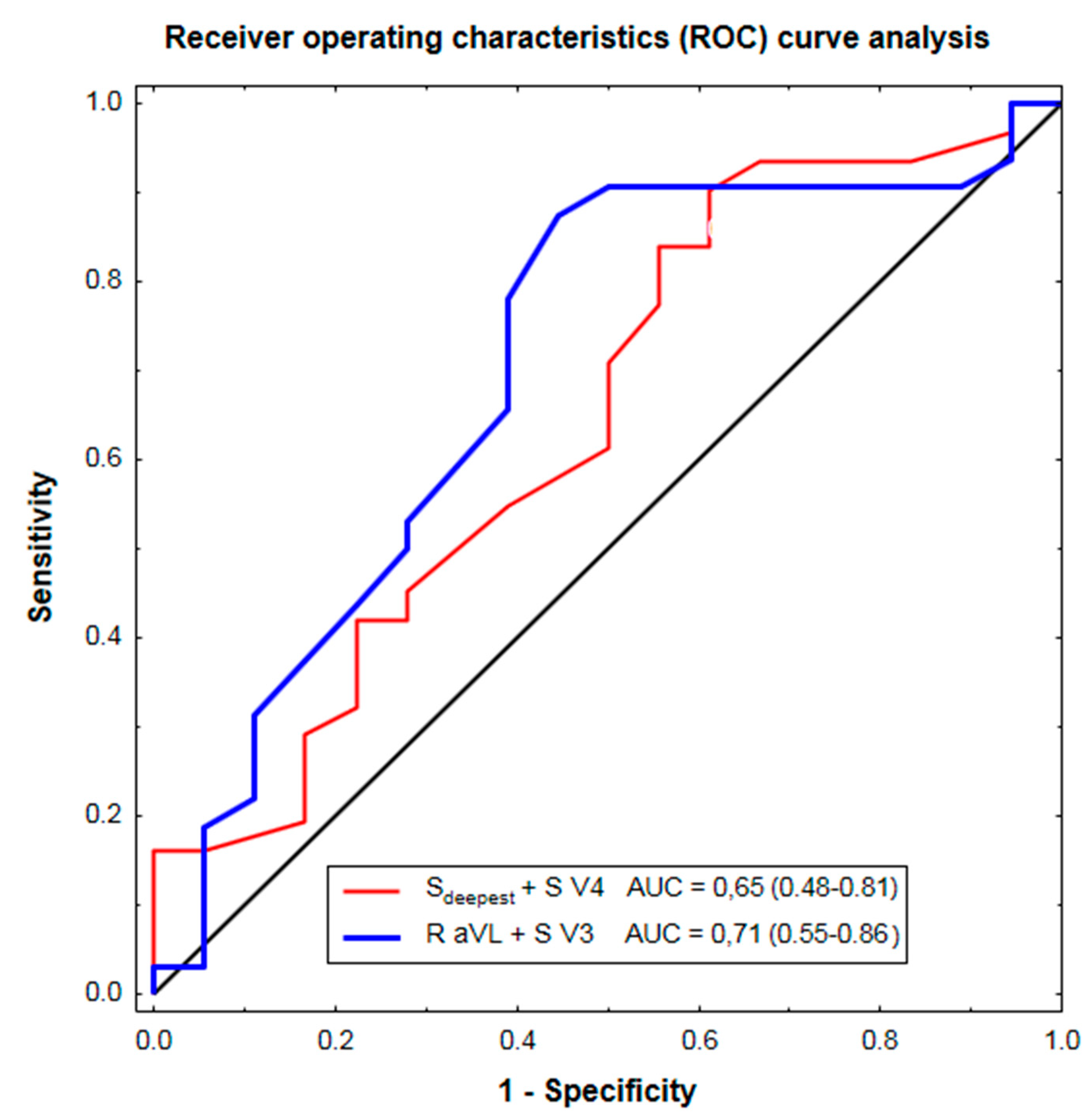
Figure 1. The ability of the Peguero-Lo Presti (Sdeepest + S V4) and Cornell (R aVL + S V3) voltage as a continuous variable to discriminate aortic stenosis patients with and without echocardiographic LVH, depicted as mean area under the ROC curve (AUC) and 95% confidence interval.
Jcm 10 02864 g001
Table 1. Patients’ characteristics.
CharacteristicMean ± SD or n (%)
Age, years77 ± 10
Women/men, n30/20
Hypertension, n (%)46 (92%)
Mean arterial pressure, mm Hg94 ± 11
Diabetes, n (%)26 (52%)
Body mass index, kg/m226.9 ± 4.2
eGFR, mL/min/1.73 m270 ± 16
LV mass index, g/m2121 ± 39
LV end-diastolic diameter, mm46 ± 7
Relative LV wall thickness0.54 ± 0.13
Aortic valve area, cm20.7 ± 0.2
Mean aortic gradient, mm Hg53 ± 19
Peak aortic gradient, mm Hg85 ± 27
Abbreviations: eGFR: estimated glomerular filtration rate by the CKD-EPI formula; LV: left ventricular; n: number; SD: standard deviation.
Table 2. Diagnostic performance of the Peguero-Lo Presti criterion and traditional ECG-LVH criteria to predict echocardiographic LVH.
ECG Criteria for LVHSensitivitySpecificityPPVNPVAccuracyCohen’s KappaMcNemar Test
Traditional ECG-LVH criteria
Single traditional criteria9–34%78–100%60–100%36–41%38–50%−0.01–0.13≤0.0014
≥1 traditional criterion66%56%72%48%62%0.200.6
Peguero-Lo Presti criterion
Sdeepest + S V4 ≥ 2.3 mV (W)
Sdeepest + S V4 ≥ 2.8 mV (M)55%72%77%48%61%0.240.07
LVH: left ventricular hypertrophy; PPV: positive predictive value; NPV: negative predictive value; W: women; M: men.
Table 3. Positive likelihood ratio of electrocardiographic LVH in patients with versus without echocardiographic LVH, and odds ratio and relative risk of echocardiographic LVH in patients with versus without any of the traditional ECG-LVH criteria.
LVH Criteria by ECGPositive Likelihood RatioOdds Ratio (95% CI)Relative Risk (95% CI)
≥1 traditional criterion1.52.4 (0.7–7.8)1.4 (0.6–3.5)
Peguero-Lo Presti criterion
Sdeepest + S V4 ≥ 2.3 mV (W)
Sdeepest + S V4 ≥ 2.8 mV (M)
2.03.2 (0.9–11.0)1.5 (0.6–3.7)
CI: confidence interval; other abbreviations as in Table 1.
Publisher’s Note: MDPI stays neutral with regard to jurisdictional claims in published maps and institutional affiliations.