
Review

P-Loop Channels: Experimental Structures, and Physics-Based and Neural Networks-Based Models
I.M. Sechenov Institute of Evolutionary Physiology and Biochemistry, Russian Academy of Sciences, 194223 St. Petersburg, Russia
*
Author to whom correspondence should be addressed.
Membranes 2022, 12(2), 229; https://doi.org/10.3390/membranes12020229

Submission received: 27 December 2021 / Revised: 9 February 2022 / Accepted: 9 February 2022 / Published: 16 February 2022
(This article belongs to the Special Issue Membrane Channel of Cells)

Abstract

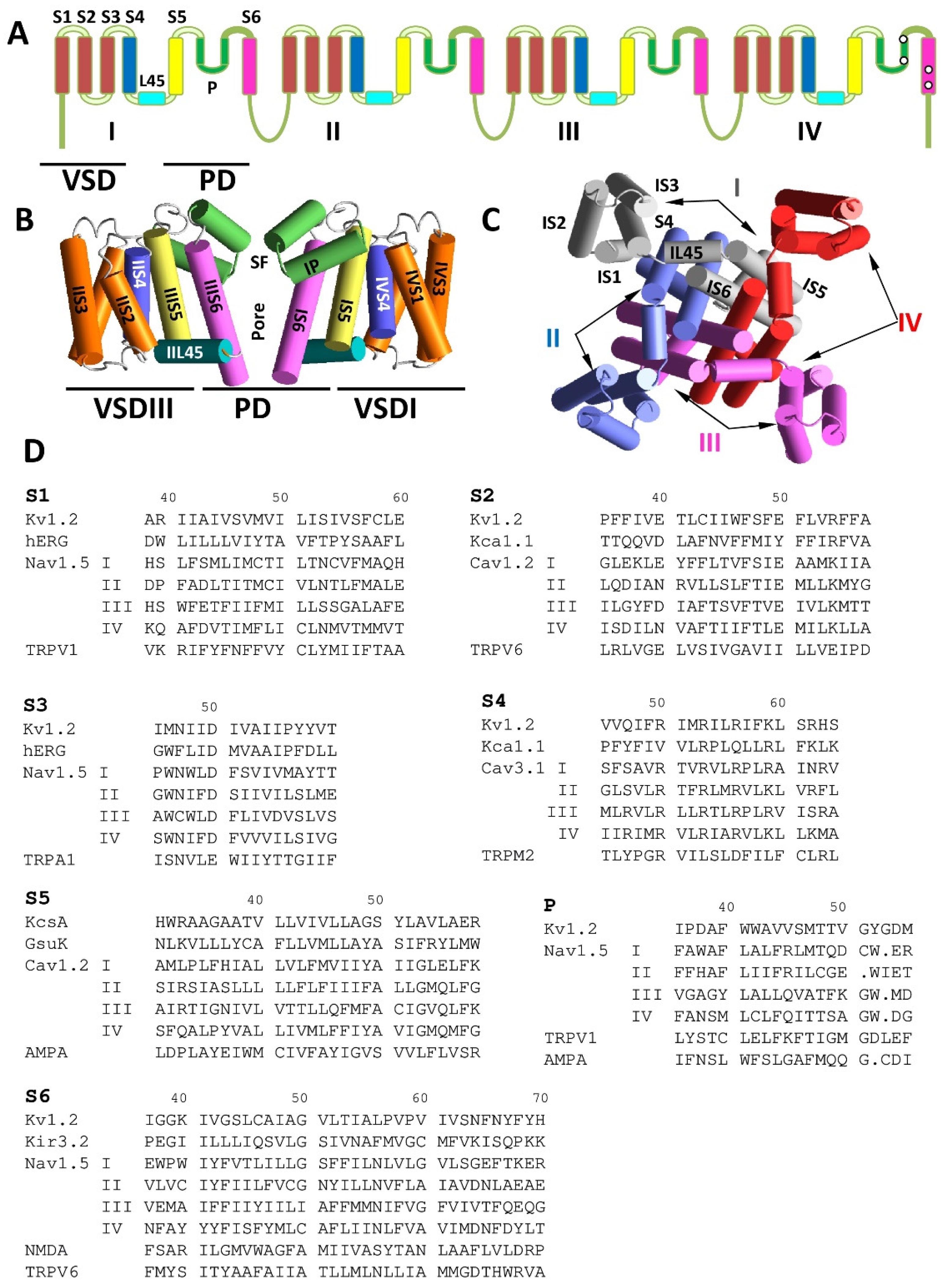
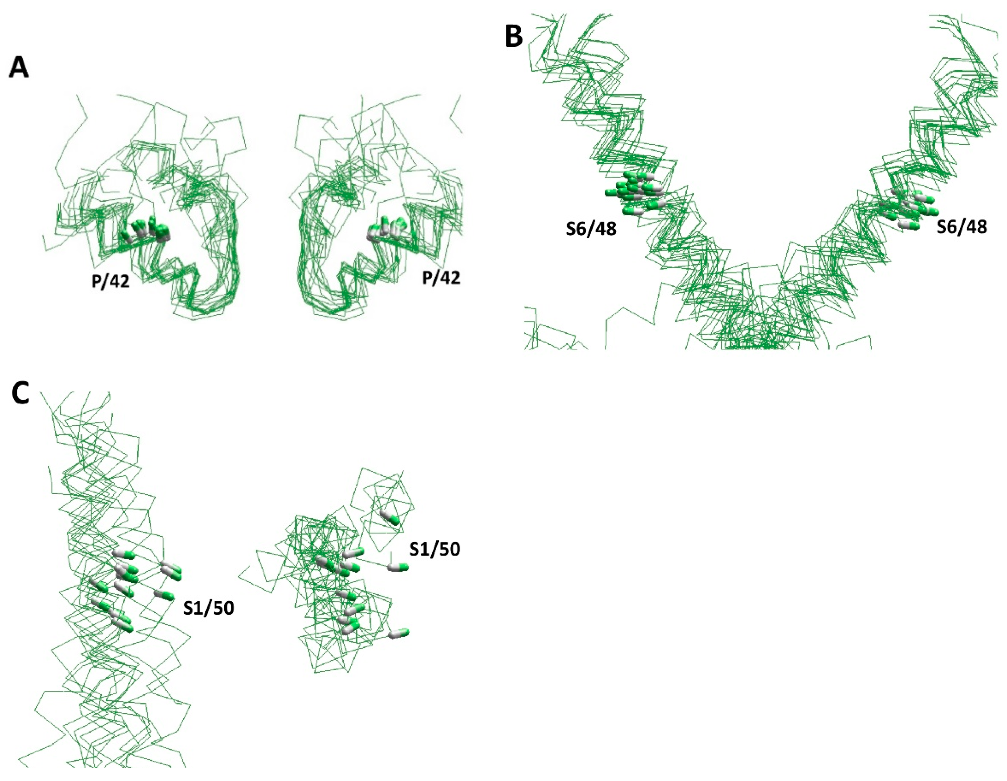
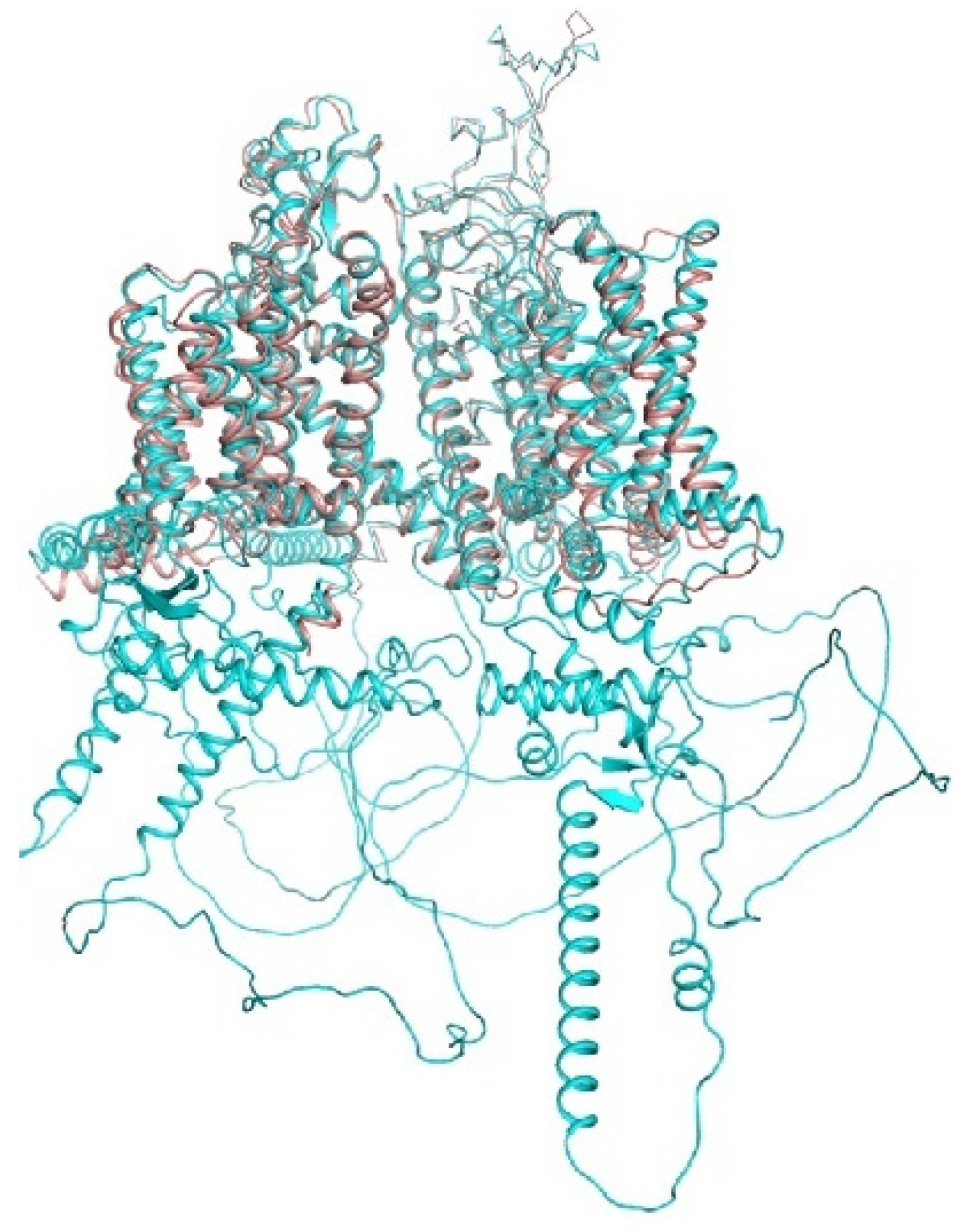
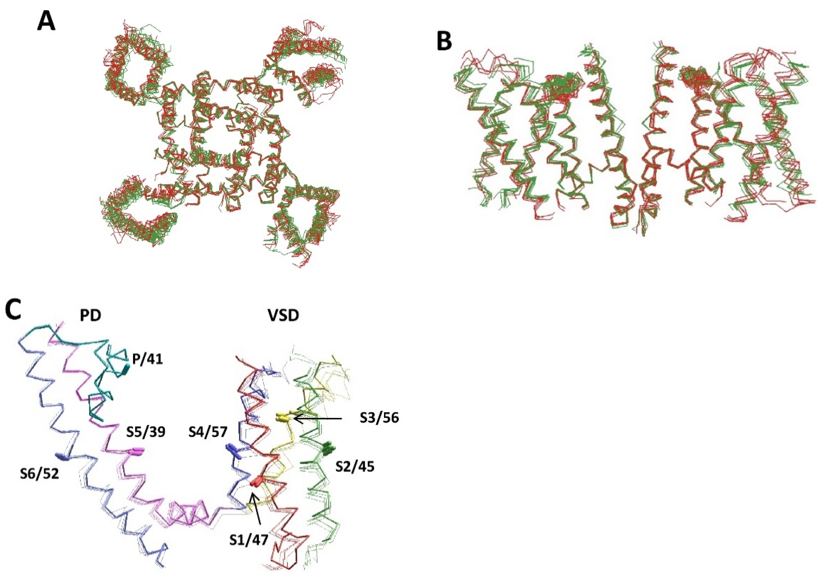
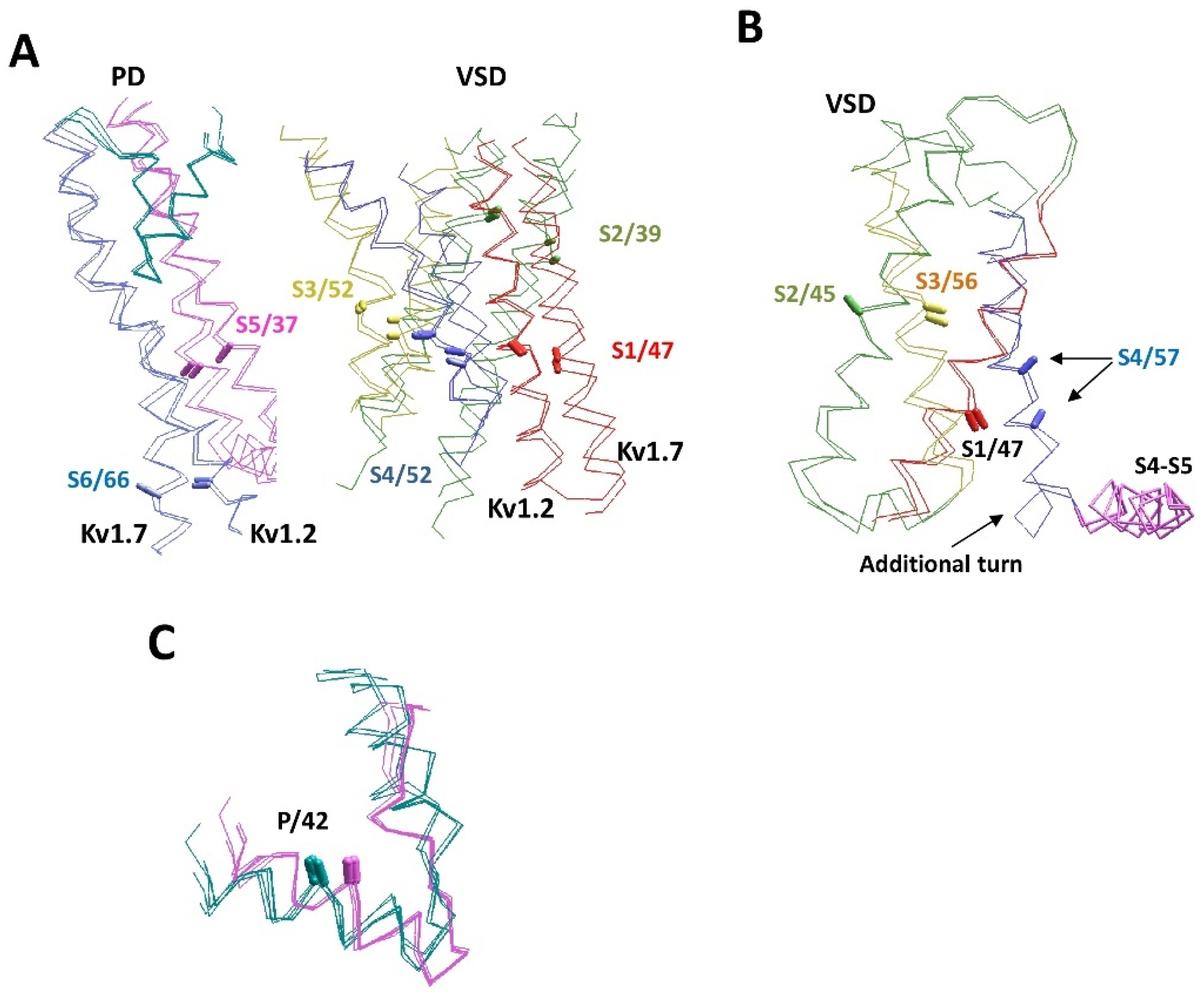
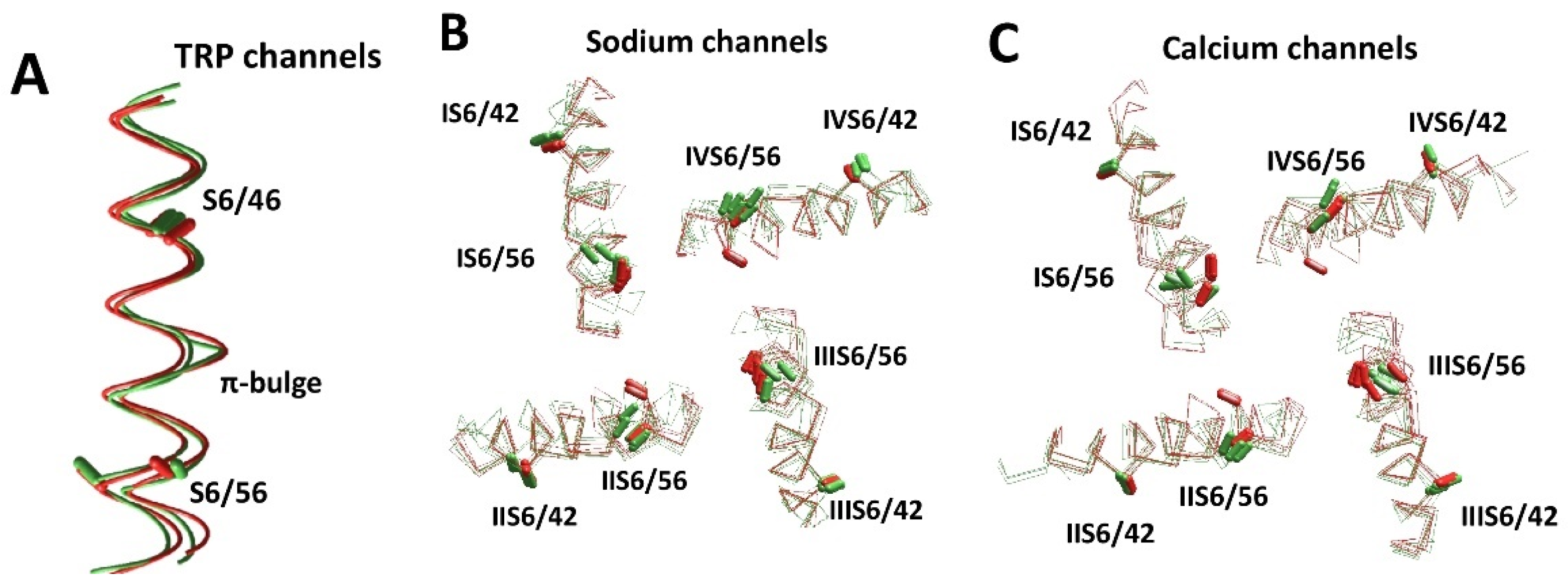
The superfamily of P-loop channels includes potassium, sodium, and calcium channels, as well as TRP channels and ionotropic glutamate receptors. A rapidly increasing number of crystal and cryo-EM structures have revealed conserved and variable elements of the channel structures. Intriguing differences are seen in transmembrane helices of channels, which may include π-helical bulges. The bulges reorient residues in the helices and thus strongly affect their intersegment contacts and patterns of ligand-sensing residues. Comparison of the experimental structures suggests that some π-bulges are dynamic: they may appear and disappear upon channel gating and ligand binding. The AlphaFold2 models represent a recent breakthrough in the computational prediction of protein structures. We compared some crystal and cryo-EM structures of P-loop channels with respective AlphaFold2 models. Folding of the regions, which are resolved experimentally, is generally similar to that predicted in the AlphaFold2 models. The models also reproduce some subtle but significant differences between various P-loop channels. However, patterns of π-bulges do not necessarily coincide in the experimental and AlphaFold2 structures. Given the importance of dynamic π-bulges, further studies involving experimental and theoretical approaches are necessary to understand the cause of the discrepancy.
Keywords: ligand–channel interactions; sequence alignment; π-bulges; crystal structures; cryo-EM structures; potassium channels; sodium channels; calcium channels; TRP channels; ionotropic glutamate receptors ligand–channel interactions; sequence alignment; π-bulges; crystal structures; cryo-EM structures; potassium channels; sodium channels; calcium channels; TRP channels; ionotropic glutamate receptors