Article

Oral Immunization of Larvae and Juvenile of Lumpfish (Cyclopterus lumpus) against Vibrio anguillarum Does Not Influence Systemic Immunity

1
Marine Microbial Pathogenesis and Vaccinology Laboratory, Department of Ocean Sciences, Memorial University of Newfoundland, St. John’s, NL A1C 5S7, Canada
2
Department of Ocean Sciences, Ocean Frontier Institute, Memorial University of Newfoundland, St. John’s, NL A1C 5S7, Canada
3
Aquatic Research Cluster, CREAIT Network, Department of Ocean Sciences, Memorial University of Newfoundland, St. John’s, NL A1C 5S7, Canada
4
Dr. Joe Brown Aquatic Research Building (JBARB), Memorial University of Newfoundland, St. John’s, NL A1C 5S7, Canada
5
Fisheries and Marine Institute, Memorial University of Newfoundland, St. John’s, NL A1C 5S7, Canada
*
Author to whom correspondence should be addressed.
Vaccines 2021, 9(8), 819; https://doi.org/10.3390/vaccines9080819
Submission received: 27 May 2021 / Revised: 13 July 2021 / Accepted: 16 July 2021 / Published: 23 July 2021
(This article belongs to the Special Issue Vaccines for Aquaculture)

Abstract

:
Vibrio anguillarum, a marine bacterial pathogen that causes vibriosis, is a recurrent pathogen of lumpfish (Cyclopterus lumpus). Lumpfish is utilized as a cleaner fish in the Atlantic salmon (Salmo salar) aquaculture in the North Atlantic region because of its ability to visualize and prey on the ectoparasite sea lice (Lepeophtheirus salmonis) on the skin of Atlantic salmon, and its performance in cold environments. Lumpfish immunity is critical for optimal performance and sea lice removal. Oral vaccine delivery at a young age is the desired method for fish immunization because is easy to use, reduces fish stress during immunization, and can be applied on a large scale while the fish are at a young age. However, the efficacy of orally delivered inactivated vaccines is controversial. In this study, we evaluated the effectiveness of a V. anguillarum bacterin orally delivered to cultured lumpfish and contrasted it to an intraperitoneal (i.p.) boost delivery. We bio-encapsulated V. anguillarum bacterin in Artemia salina live-feed and orally immunized lumpfish larvae. Vaccine intake and immune response were evaluated by microscopy and quantitative polymerase chain reaction (qPCR) analysis, respectively. qPCR analyses showed that the oral immunization of lumpfish larvae resulted in a subtle stimulation of canonical immune transcripts such as il8b, il10, igha, ighmc, ighb, ccl19, ccl20, cd8a, cd74, ifng, and lgp2. Nine months after oral immunization, one group was orally boosted, and a second group was both orally and i.p. boosted. Two months after boost immunization, lumpfish were challenged with V. anguillarum (7.8 × 10⁵ CFU dose−1). Orally boosted fish showed a relative percentage of survival (RPS) of 2%. In contrast, the oral and i.p. boosted group showed a RPS of 75.5% (p < 0.0001). V. anguillarum bacterin that had been orally delivered was not effective in lumpfish, which is in contrast to the i.p. delivered bacterin that protected the lumpfish against vibriosis. This suggests that orally administered V. anguillarum bacterin did not reach the deep lymphoid tissues, either in the larvae or juvenile fish, therefore oral immunization was not effective. Oral vaccines that are capable of crossing the epithelium and reach deep lymphoid tissues are required to confer an effective protection to lumpfish against V. anguillarum

1. Introduction

Ectoparasitic sea lice infestations are a serious health issue for wild and cultured Atlantic salmon (Salmo salar) [1]. The sea louse (e.g., Lepeophtheirus salmonis) feeds on salmonid mucus and tissues leading to physiological and immune failures and may lead to death if left untreated [1,2]. Over the last few decades, the Atlantic salmon industry has used chemical and physical methods to control sea lice infestations [3,4]. The use of chemotherapeutants generates the emergence of resistance in sea lice [3] and enhances negative public opinion on the impacts of aquaculture activity on the environment. Routine mechanical treatments (e.g., abrasion and thermal treatment) used in the salmon industry to physically remove the lice have adverse effects on salmon health and welfare [4]. Currently, multiple non-medicinal methods are used in different combinations (e.g., curtains and skirts, anti-lice diets, laser, water jets, and ultrasound) as part of an integrated pest management (IPM) strategy [5]. Another current strategy to control sea lice in the North Atlantic farms is the use of cleaner fish species as a biological control [4,6]. The use of cleaner fish is considered an eco-friendly alternative, as it can reduce the use of chemotherapeutants, and it is less stressful to farmed fish [7]. In Atlantic Canada, both lumpfish (Cyclopterus lumpus) and cunner (Tautogolabrus adspersus) are used as cleaner fish [8,9]. However, lumpfish is the preferred species for the biocontrol of sea lice due to its performance in cold environments and biomass availability [10,11]. In contrast to other cleaner fish species, lumpfish actively remove sea lice from farmed salmon in cold environments, and they have been industrialized in the North Atlantic region [12,13,14,15]. Use and demand for lumpfish in salmon farms in Ireland, the United Kingdom (UK), Norway, the Faroe Islands, Iceland, and Canada have increased in recent years [13,16]. For example, 11.8 and 0.8 million juveniles were used in Norway and in the UK in 2015, respectively [17]. In 2018, approximately 30 million juvenile lumpfish were used in Norway [18]; in the UK, approximately 6 million lumpfish were used; in Iceland, approximately 3.5 million were used [19]; and approximately 300 thousand lumpfish were used in Ireland [20]. In Canada, the use of lumpfish is a very recent practice, and approximately 1 million lumpfish were used in Atlantic Canada in 2019 [19].
Similar to other finfish species, lumpfish is susceptible to different types of bacterial pathogens such as Aeromonas salmonicida, Pasteurella spp., Tenacibaculum spp., Pseudomonas anguilliseptica, and Moritella viscosa [21,22]. Vibriosis, typically caused by Vibrio anguillarum, is a prevalent pathogen in lumpfish in Newfoundland [23], but others such as V. ordalii, V. salmonicida, and V. splendidus also infect lumpfish elsewhere [12,24]. V. anguillarum is a Gram-negative, motile, rod-shaped bacterium and a ubiquitous marine pathogen of invertebrates and vertebrates [25,26]. Formalin-killed V. anguillarum (bacterins) serotypes O1 and O2 mixed with adjuvants are often used to formulate vaccines for different fish species, including Atlantic salmon, Atlantic cod (Gadus morhua) [27], gilthead seabream (Sparus aurata), and lumpfish [28]. Intraperitoneal (i.p.) injection is the most common method of vaccine delivery [29], but it cannot be applied to small fish. An oral-immersion vaccine is ideal for fish immunization, as it is needle-free and causes minimum stress during application [30]. However, oral vaccine delivery to small fish or larvae has been evaluated with controversial results [31]. Vaccine bio-encapsulation strategies, such as the used of Artemia salina fed with bacterin, can increase the delivery of the vaccine to small lumpfish larvae by increasing intake and can directly deliver the antigen to the fish gut [32,33,34].
Here, we evaluated the efficacy of a V. anguillarum bacterin bio-encapsulated in A. salina delivered as live-feed to lumpfish larvae. We determined that the oral V. anguillarum vaccine delivered in A. salina live-feed reached the lumpfish gut. Real-time quantitative polymerase chain reaction (qPCR) analyses of immune-relevant genes revealed that oral immunization modestly immune stimulated the lumpfish larvae. Nine months later, a lumpfish group was orally boosted, and an independent second group was orally and i.p. boosted. Two months later, the immunized fish groups were i.p. challenged with ten times the lethal dose 50 (LD50) of V. anguillarum (7.8 × 10⁵ colony forming units (CFU) dose−1). Oral immunization of lumpfish delayed mortality but did not confer protective immunity against the V. anguillarum challenge, which contrasts with the i.p. vaccination, which protected the fish against the lethal infection.

2. Materials and Methods

2.1. Vibrio anguillarum J360 Culture Conditions

V. anguillarum J360 serotype O2 (NCBI IDs: Chromosome 1 CP034672; Chromosome 2 CP034673; and plasmid CP034674), a local lumpfish isolate, was used in this study [23]. V. anguillarum J360 was grown in 3 mL of tryptic soy broth (TSB, Difco) for 24 h at 28 °C with aeration (180 rpm, in an orbital shaker). Bacterial growth was monitored using spectrophotometry (Genesys 10 UV spectrophotometer, Thermo Fisher Scientific Inc., Waltham, MA, USA) and by plating to determine the CFU mL−1. When the optical density (OD600 nm) reached ~0.7 (1 × 10⁸ CFU mL−1), the cells were harvested by centrifugation at 4200× g for 10 min at room temperature. The cell suspension was washed twice with phosphate-buffered saline (PBS; 136 mM NaCl, 2.7 mM KCl, 10.1 mM Na2HPO4, 1.5 mM KH2PO4; pH 7.2) [35] at 4200× g and resuspended in 300 μL of PBS 1X (~1 × 10¹⁰ CFU mL−1). The final inoculum was serially diluted (1:10), and the concentration was determined by plate counting [36] in TSB supplemented with 1.5% bacto agar (TSA).

2.2. Bacterin Preparation

The bacterin preparation was conducted according to established protocols and quantified using flow cytometry enumeration [37] with modifications. First, V. anguillarum J360 was grown in 500 mL of TSB supplemented with a final concentration of 150 μM 2,2-dipyridyl (Sigma-Aldrich, St. Louis, MO, USA) at 28 °C with aeration (180 rpm, in an orbital shaker) to induce synthesis of the iron-regulated outer membrane proteins (IROMPs) [38]. Bacterial growth was monitored spectrophotometrically until it reached ~1 × 10⁸ CFU−1. V. anguillarum cells were harvested using centrifugation (4200× g at 4 °C for 10 min) and washed twice with PBS. V. anguillarum cells were fixed with buffered formalin 6% (Sigma) at room temperature for 3 d with gentle agitation in a rocker shaker. Cell viability was determined each day by plating onto TSA. Formalin was removed using centrifugation, and cells were then dialyzed (Spectrum™ Spectra/Por™ dialysis membrane 12–14,000 Dalton molecular weight cut-off, Thermo) in 2 L of PBS at 6 °C for 3 d with agitation. V. anguillarum bacterins were quantified using the BD FACS Aria II flow cytometer (BD Biosciences, San Jose, CA, USA) and BD FACS Diva v7.0 software as described previously [39]. Bacterin cells (1 × 10¹⁰ CFU mL−1) were stored at 4 °C until use.

2.3. V. anguillarum Bacterin Fluorescent Labeling

V. anguillarum bacterin was labeled with 5-([4,6-dichlorotriazinyl] amino) fluorescein hydrochloride (DTAF; Sigma) according to previously described protocols [40] with minor modifications. First, V. anguillarum bacterin (~1 × 10¹⁰ CFU mL−1) was centrifuged at 4200× g for 10 min and then resuspended in 950 µL of bicarbonate buffer (0.1 M, pH 9). Following that, the cells were mixed with 50 µL of DTAF solution (100 µg in dimethyl sulfoxide (DMSO); Sigma) and incubated overnight at 4 °C in dark conditions. After incubation, the bacterin cells were centrifuged (4200× g for 10 min) and washed three times with bicarbonate buffer and were finally resuspended in PBS and kept at 4 °C until use.
Next, we describe the optimization of V. anguillarum bacterin bio-encapsulation in A. salina. To optimize bacterin bio-encapsulation in A. salina, we used the method described by Campbell et al. [32] with modifications. Additionally, we developed a semi-quantitative method to estimate the levels of bacterin bio-encapsulation in A. salina. First, A. salina nauplii were hatched from cysts according to the supplier’s instructions (INVE, Salt Lake City, UT, USA) (Figure S1). After the A. salina nauplii hatched (~20 h at 20 °C), nauplii were washed with seawater for 30 min (Figure S1). A. salina cultures were nutritionally supplemented with Ori-One (Skretting, Fontaine les Vervins, France) and Ori-Green (Skretting, Fontaine les Vervins, France) commercial dry microalgae extract at a ratio of 1:1 (Ori-One:Ori-green) for 3 h at 20 °C. To determine the optimal time for bacterin bio-encapsulation in A. salnina, supplemented A. salina nauplii were inoculated into 6 well plates with a 3 mL total volume per well at a density of 1000 nauplii mL−1. Additionally, a separate plate was inoculated with non-supplemented A. salina nauplii to determine the possible effect of nutrient supplementation on bio-encapsulation. Both groups were inoculated with DTAF-labeled V. anguillarum bacterin (5 × 10⁷ cells mL−1). The control group was mock inoculated with seawater and used to evaluate autofluorescence. The nauplii were incubated at 20 °C for 48 h to determine the optimal time for bacterin bio-encapsulation (Figure S1A). A. salina samples (1 mL) were collected at 1, 3, 5, 24, 36, and 48 h, and fixed in 10% buffered formalin. The presence of V. anguillarum inactivated bacterin was examined and counted using confocal microscopy (Nikon Eclipse Ti, Melville, NY, USA) to determine the number of A. salina containing 0%, 25%, 50%, 75%, 100% bacterin (Figure S1A). After determining the optimal time for bacterin bio-encapsulation in A. salina nauplii, we evaluated the bio-encapsulation stability at 6 °C for 6 d (Figure S1B). A. salina nauplii were supplemented with Ori-One, Ori-Green, and DTAF-labeled V. anguillarum bacterin at 20 °C for 3 h and then placed at 6 °C for 6 d. The A. salina control group was mock inoculated with seawater. A. salina samples were collected each day and fixed with buffered 10% formalin. The levels of bio-encapsulation in A. salina were determined using confocal microscopy (Figure S1B). DTAF-labeled V. anguillarum inactivated bacterin bio-encapsulated in A. salina nauplii were used to feed the lumpfish larvae (Figure S2) and to determine the presence of V. anguillarum bacterin in the larvae gut compared to the non-orally immunized fish (Figure S3). Fifty lumpfish larvae were orally immunized and maintained at 6 °C for 24 h. The larvae gut was observed at 0, 0.5, 1, 2, 4, 6, and 24 h post-oral immunization using epi-fluorescence microscopy (Optika, Italy) (Figure S3).

2.4. V. anguillarum Bacterin Bio-Encapsulation in A. salina

Based on the vaccine bio-encapsulation optimization results, larger volumes of A. salina were prepared for lumpfish larvae immunization. A. salina nauplii were hatched and washed with seawater for 30 min and placed in 20 L buckets containing 15 L of seawater. A. salina nauplii were maintained at a density of ~2.5 million A. salina per liter. A. salina cultures were enriched with nutritional supplements derived from microalgae, Ori-One (Skretting, France) and Ori-Green (Skretting, France) according to the manufacturer’s instructions. A. salina cultures were inoculated with V. anguillarum bacterin (107 cells mL−1) and incubated at 20 °C for 3 h with aeration. Controls were mock inoculated with PBS. After 3 h of enrichment and bacterin bio-encapsulation, the A. salina cultures were maintained at 6 °C for 6 d under constant light. These cultures were used for the daily oral immunization of the lumpfish larvae (Figure S4).

2.5. Aquafeed Coating with V. anguillarum Bacterin

Commercial dry feed was coated with dry V. anguillarum bacterin to orally boost immunized fish. Ficoll, a non-toxic polymer, was used as a cryoprotectant for bacterial lyophilization [41]. Ficoll also serves as an antigen and adjuvant carrier [41,42,43,44,45,46]. To freeze-dry the bacterin, a formalin-killed V. anguillarum (2 × 10⁹ CFU mL−1) suspension was mixed with Ficoll solution (20% Ficoll400 (GE Healthcare, Sweden), 300 mM NaCl) at a 1:1 ratio to prevent cell lysis during lyophilization. The cells were lyophilized (Edwards super module E2-M5, Edwards, UK) for 3 days. The bacterin powder was mixed with 3–4 mm commercial dry pellet (Skretting-Europa 15: crude protein (55%), crude fat (15%), crude fiber (1.5%), calcium (3%), phosphorus (2%), sodium (1%), vitamin A (5000 IU kg−1), vitamin D (3000 IU kg−1), and vitamin E (200 IU kg−1)) at the ratio of 0.9 g bacterin per 100 g aquafeed. After mixing the feed with the bacterin powder, a layer of cod liver oil was added (3 mL 100 g feed−1), and the feed was then dried at room temperature to complete the coating process. The coated feed was stored at 4 °C until used.

2.6. Fish Culture Conditions

All animal protocols required for this research were approved by the Institutional Animal Care Committee and the Biosafety Committee at Memorial University of Newfoundland (MUN). Experiments were conducted under protocols #18-01-JS, #18-03-JS, and biohazard license L-01. Lumpfish egg masses were in vitro fertilized and maintained in 5 L buckets containing UV treated (300 mW cm2), filtered, flow-through seawater (33 ppt) at 10–10.5 °C, with 95–110% air saturation, and held under ambient photoperiod (spring–summer) at the Joe Brown Aquatic Research Building (JBARB), Department of Ocean Sciences, Memorial University of Newfoundland, Canada. After embryo development was complete, the larvae were hatched and maintained in the same seawater conditions and air saturation at 10 °C until full egg yolk sac absorption and the establishment of independent feeding was achieved (Figure S2).

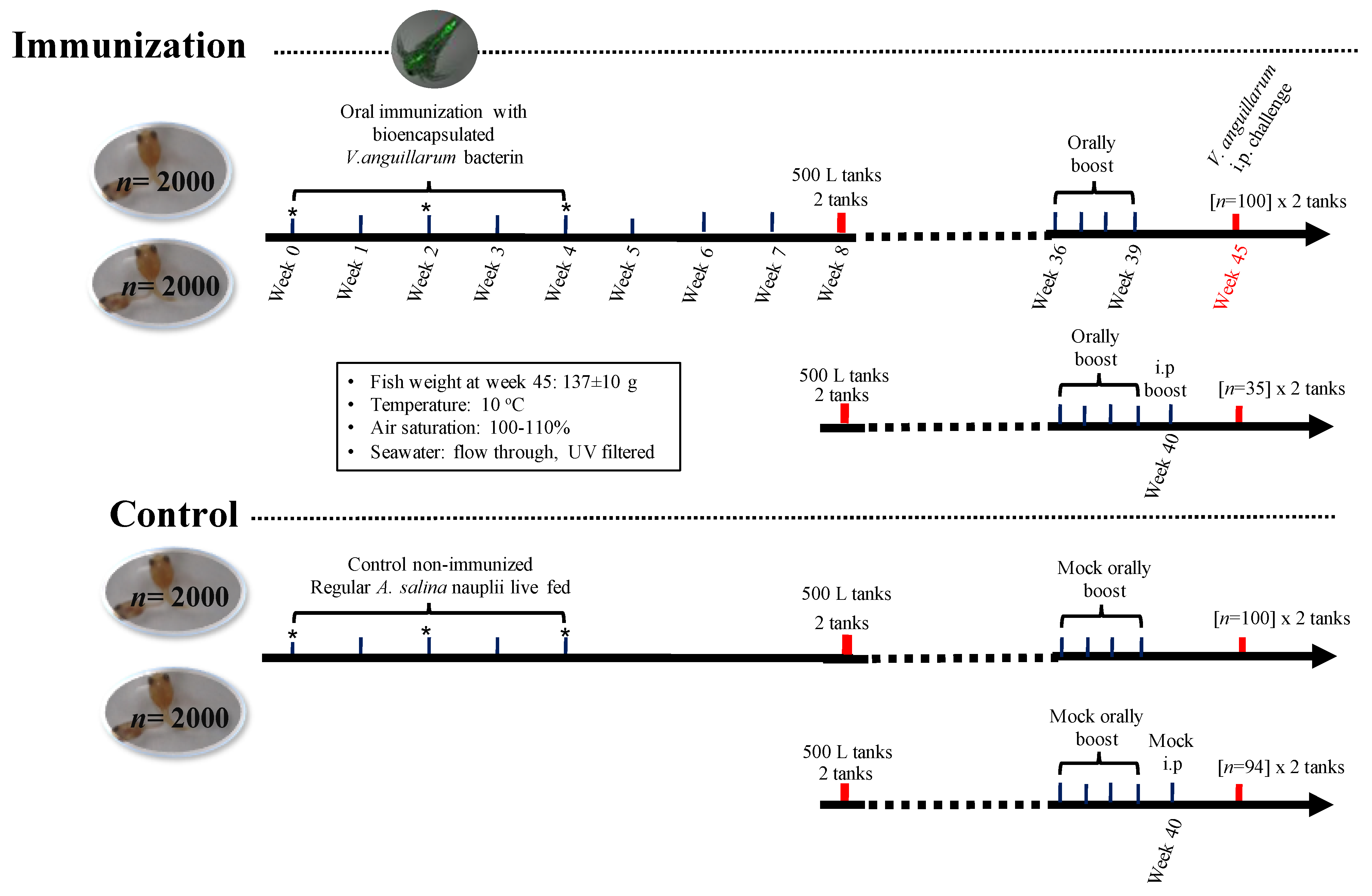
2.7. Lumpfish Immunization Assays

Lumpfish larvae were stocked in 4 tanks at a density of 2000 larvae per tank (20 L) with UV treated (300 mW cm2), filtered, flow-through seawater at 8–10 °C, and with 95–110% air saturation (Figure 1). Two tanks containing lumpfish larvae (1-week post-hatch (wph)) were orally immunized with the bio-encapsulated V. anguillarum vaccine daily for 4 weeks. Two tanks fed containing only A. salina served as controls (Figure 1). Lumpfish larvae were fed with A. salina nauplii or bio-encapsulated vaccine 3 times per day (350 mL of A. salina culture per 2000 larvae) (Figure S4). Thereafter, the fish were fed A. salina for additional 10 d and then fed with dry pelleted diet daily (0.75–2% body weight). Whole larvae pool samples were collected at 0, 2, and 4 weeks post-immunization (wpi) (Figure 1). At each time point, triplicate pools of 5–10 larvae were sampled from each tank and placed in a 1.5 mL RNase-free tube containing 300 μL of TRIzol Reagent (Invitrogen, Waltham, MA, USA), flash-frozen in liquid nitrogen, and stored at −80 °C until processing. After 8 weeks, the juvenile lumpfish were transferred into eight 500 L tanks (Figure 1). The lumpfish were fed an assorted size pelleted diet for the remainder of the experiment (15 months). Nine months post-hatch, two tanks with 100 fish each (~72.1 ± 30 g) were orally boost immunized using commercial pellets coated with the V. anguillarum bacterin. Lumpfish were starved for 24 h pre-oral vaccination. Lumpfish were fed with V. anguillarum bacterin coated dry pellets three times (every 2 weeks for 3 days) at 0.75% body weight (Figure 1). Two control tanks were mock-orally boosted with ficoll (vaccine vehicle) coated pellets (Figure 1). Two independent groups orally boosted were additionally i.p. boosted at 40 wph (~145 ± 31.8 g) with V. anguillarum bacterin (6.3 × 10⁸ cells dose−1). Control groups were mock-orally and i.p. boosted with the respective vaccine vehicle (Figure 1). Two months later, the animals (~132–244 g) were transferred to the AQ3 aquatic biocontainment facility at the Cold-Ocean Deep-Sea Research Facility (CDRF) for challenge assays.

2.8. V. anguillarum J360 Challenge Assays in Lumpfish

The challenge assays were conducted at the CDRF AQ3 biocontainment facility under the Institutional Animal Care Committee approved protocol (18-02-JS) and established protocols [28]. First, after transfer to the AQ3 biocontainment facility, lumpfish were acclimated for 1 week under optimal conditions prior to the commencement of the challenge. Lumpfish were challenged by an i.p. injection with 7 times the lethal dose 50 (7.8 × 10⁵ CFU dose−1) of V. anguillarum J360. Fish survival was monitored for 30 days post-challenge. The relative percent of survival (RPS) of the control and vaccinated fish was calculated using the formula: RPS = [1 − (% mortality in vaccinated fish/% mortality in control fish)] × 100 [47].

2.9. Total RNA Extraction

RNA was extracted from the larvae pools pre-immunized (n = 3 individual pools of 10 larvae each), 2 wpi (n = 3 pools of 10 larvae each), and 4 wpi (n = 3 pools of 5 larvae each). RNA was also extracted from the larvae post mock immunization(control) at 2 and 4 wpi. Lumpfish larvae pools, previously flash frozen in a 1.5 mL RNase-free tube containing 300 μL of TRIzol reagent (Invitrogen), were homogenized using a micro-tube homogenizer (ThermoFisher Scientific, Waltham, MA, USA). An additional 700 μL of TRIzol was added to the tube, and the extractions were then completed following the manufacturer’s instructions. The TRIzol extracted samples were then purified using the RNeasy® Mini Kit (QIAGEN, Mississauga, ON, Canada) following the manufacturer’s instructions. RNA samples were treated with 2 U mL−1 of TURBO DNase (TURBO DNA-free™ Kit, Invitrogen) following the manufacturer’s instructions for the complete digestion of genomic DNA and the removal of the remaining DNase and divalent cations, such as magnesium and calcium. Purified RNA samples were quantified and evaluated for purity using a Genova Nano spectrophotometer (Jenway, Staffordshire, UK) and evaluated for integrity using 1% agarose gel electrophoresis [35]. A PCR test was conducted using the 60S ribosomal protein L32 (rpl32) reference gene primers and the RNA as a template to rule out the presence of DNA.

2.10. cDNA Synthesis and qPCR Parameters

cDNA was synthesized in 20 μL reactions from 1 μg of RNA using SuperScript IV VILO Master Mix (Invitrogen) following the manufacturer’s instructions. PCR amplifications were performed in 13 μL reactions using 1X Power SYBR Green PCR Master Mix (Applied Biosystems, Waltham, MA, USA), 50 nM of both the forward and reverse primers, and the indicated cDNA quantity. Amplifications were performed using the QuantStudio 6 Flex Real-Time PCR system (384-well format) (Applied Biosystems, Waltham, MA, USA). The real-time analysis program consisted of 1 cycle of 50 °C for 2 min, 1 cycle of 95 °C for 10 min, 40 cycles of 95 °C for 15 s, and 60 °C for 1 min, with fluorescence detection at the end of each 60 °C step and followed by dissociation curve analysis.

2.11. qPCR Primer Quality Assurance Testing

All primer pairs for the transcripts of interest (TOIs) and the endogenous control transcripts were designed and quality control (QC) tested using the larvae RNA samples generated herein. cDNAs were synthesized from the individual pooled larvae RNA samples, including pre-immunized control, mock-immunized control (2 and 4 wpi), and immunized larvae (2 and 4 wpi). The control and immunized cDNA samples were independently pooled and used for primer quality evaluation. To calculate amplification efficiencies for each primer pair [48], standard curves were generated for both cDNA pools (control and immunized) using a 5-point 1:3 dilution series starting with cDNA representing 10 ng of input total RNA. The reported efficiencies represent an average of the two values (Supplementary Table S1). Each primer pair was also tested to ensure that a single product was amplified and that there was no primer dimer present in the no-template control. Finally, amplicons were electrophoretically separated on 2% agarose gels and compared using a 1 kb plus ladder (Invitrogen) to verify that the correct size fragment was amplified.
Eighteen TOIs were well expressed in larvae, and as such, amplification efficiencies could be calculated using larvae cDNA template (see fluorescence threshold cycle (CT) values for studies 1 to 3; Supplementary Table S2). However, fourteen of these transcripts were expressed at low levels in larvae (see CT values for studies 4 to 6; Supplementary Table S2). For the latter TOIs, technical replicates and spacing were acceptable over the first three points of the cDNA dilution series. As the experimental input cDNA amount (8 ng) for these TOIs lies within the first 2 dilutions, these assays were deemed acceptable for analysis in larvae. However, for these fourteen transcripts, the amplification efficiencies reported in Supplementary Table S1 and inputted into the QuantStudio Real-Time PCR Software (version 1.3) (Applied Biosystems) were those that had been previously generated for head kidney samples.

2.12. Endogenous Control (Normalizer) Selection

Expression levels of the TOIs were normalized to the transcript levels of the two endogenous controls. To select these endogenous controls, 5 transcripts (rpl32, elongation factor 1-alpha (ef1a), eukaryotic translation initiation factor 3 subunit D (etif3d), polyadenylate-binding protein 1a (pabpc1a), and polyadenylate-binding protein 1b (pabpc1b)) were analyzed. First, the CT values of all 27 samples in the study were measured (in duplicate) for each of these transcripts using cDNA representing 3.25 ng of input total RNA and then analyzed using geNorm [49]. Based on this analysis, rpl32 (geNorm M = 0.169) and etif3d (geNorm M = 0.177) were selected as the two endogenous controls.

2.13. Experimental qPCR Analyses

To study the effects of oral immunization with the V. anguillarum bacterin bio-encapsulated in A. salina on the immunome of lumpfish larvae, expression levels of 32 TOIs with immune-relevant functional annotations were assessed (Supplementary Table S1). Individual larvae pools (n = 27 pools) were subjected to qPCR analyses (Figure 1). In the qPCR analyses, cDNA representing 3.25 ng (study 1 to 3, see Supplementary Table S2) and 8 ng (study 3 to 6, see Supplementary Table S2) of input RNA was used as a template in the PCR reactions. The input RNA concentration was increased to 8 ng due to lower expression transcript levels in the larvae. In each qPCR study, expression levels of a given transcript were measured across two plates. On each plate, the TOIs and endogenous controls were tested in triplicate, and a no-template control was included for every sample. A plate linker sample was also included to ensure that there was no plate-to-plate variability. The relative quantity (RQ) of each transcript was determined using the QuantStudio Real-Time PCR Software (version 1.3), with normalization to both the rpl32 and etif3d transcript levels and with the amplification and efficiencies incorporated. For each TOIs, the sample with the lowest normalized expression (mRNA) level was set as the calibrator sample (i.e., assigned an RQ value = 1) (Supplementary Table S2). Additionally, the transcript expression levels were determined using the comparative 2−ΔΔCt method [50] with two reference genes [51] (Supplementary Table S3).

2.14. Statistical Analysis

All data are expressed as the mean ± standard error (SE). Assumptions of normality and homogeneity were tested for variances. A one-way ANOVA followed by Tukey’s multiple comparison post hoc test was used to determine significant differences between the survival of the control and infected groups. The Kaplan–Meier estimator was applied for the estimation of the survival fractions after the V. anguillarum challenges, and the log-rank test was used to identify differences between treatment groups (p < 0.0001). A two-way ANOVA was used to analyze the gene expression data followed by Sidak’s multiple comparisons post hoc test to identify significant differences between each treatment in the control and immunized groups at each time point (2 weeks and 4 weeks). All statistical tests were performed using Graphpad Prism version 8.0 (Graphpad Software, USA, www.GraphPad.com (accessed on 15 June 2021)), and p-values < 0.05 were considered statistically significant.

3. Results

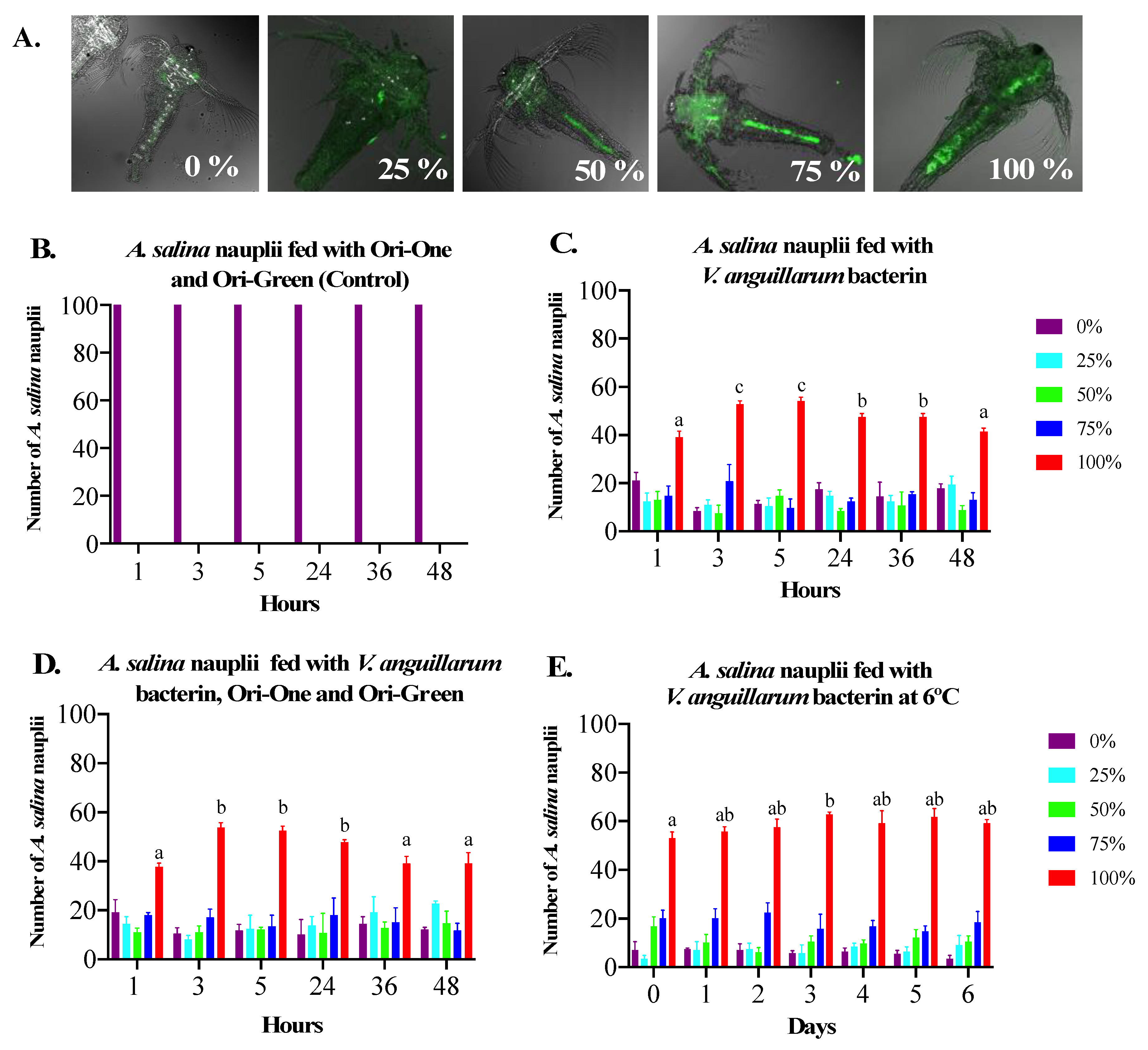
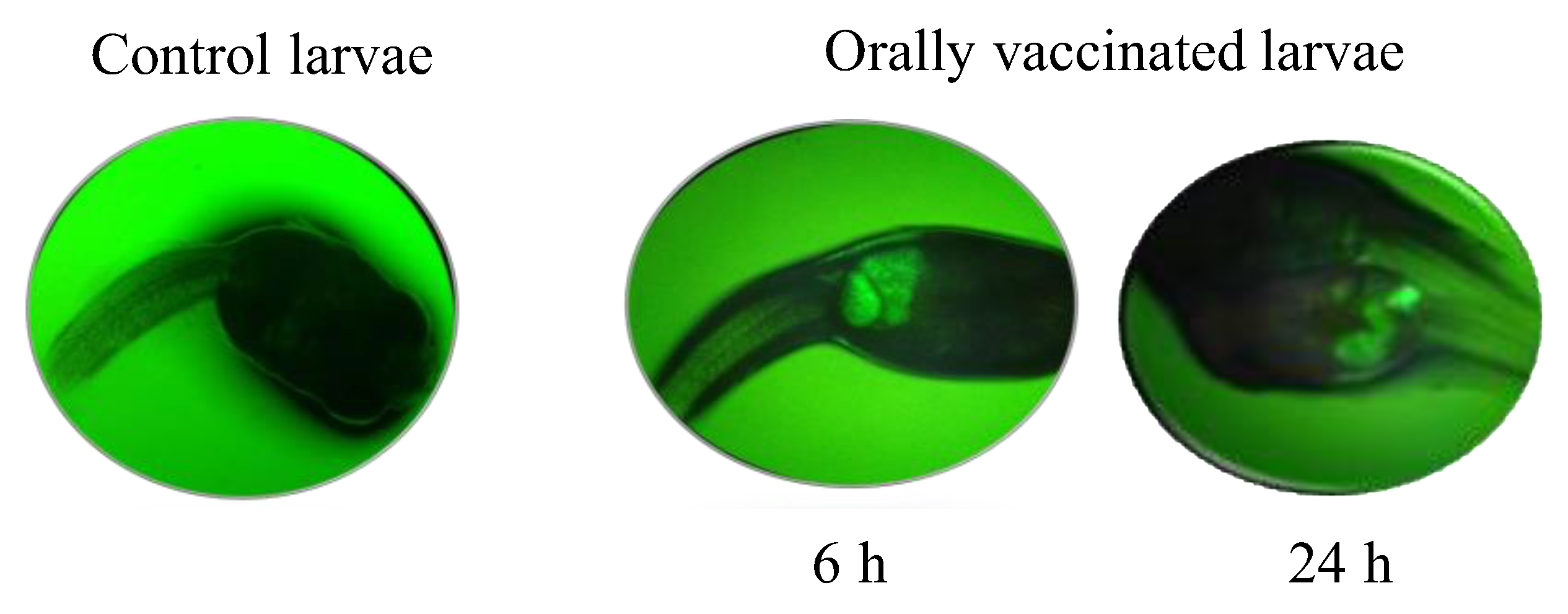
3.1. Bio-Encapsulation of V. anguillarum Bacterin in A. salina Nauplii

We established a semi-quantitative method to estimate the levels of V. anguillarum bacterin bio-encapsulation in A. salina (Figure 2A). Autofluorescence was ruled out using A. salina nauplii inoculated with nutritional supplements (Ori-One and Ori-Green) (Figure 2B). Using this method, we determined that approximately 100% of the A. salina nauplii reached the maximum capacity for V. anguillarum bacterin bio-encapsulation 3 h post-inoculation at 20 °C in both the absence (Figure 2C) or presence (Figure 2D) of nutritional supplements. In the absence of supplements, a significant decrease in the percentage of the A. salina nauplii with 100% bio-encapsulation levels was observed 24 h post-inoculation, which gradually declined thereafter (Figure 2C). In the presence of nutritional supplements, a significant decrease occurred 36 h post-inoculation (Figure 2D). We determined that the optimal V. anguillarum bacterin bio-encapsulation in A. salina method is the presence of nutritional supplementation with a bio-encapsulation time of 3 h at 20 °C. As we wanted to produce a bio-encapsulated vaccine batch that could be used for several days, we evaluated the stability of the V. anguillarum bacterin in the intestine of A. salina nauplii post bio-encapsulation at 6 °C (Figure S1B). This temperature was chosen as it is similar to the water temperature at which lumpfish are cultured. We determined that the bacterin concentration in A. salina nauplii remained stable for up to 6 d post-inoculation (Figure 2E). DTAF-labeled V. anguillarum bacterin bio-encapsulated in A. salina nauplii were then fed to lumpfish larvae and fluorescence microscopy was used to determine if the V. anguillarum bacterin reached the larvae gut (Figure S3). Fluorescence microscopy demonstrated the presence of the V. anguillarum bacterin in the gut of the lumpfish larvae after 6 and 24 h, whereas fluorescence was not detected in the gut of lumpfish larvae who had been fed A. salina nauplii only (Figure 3).

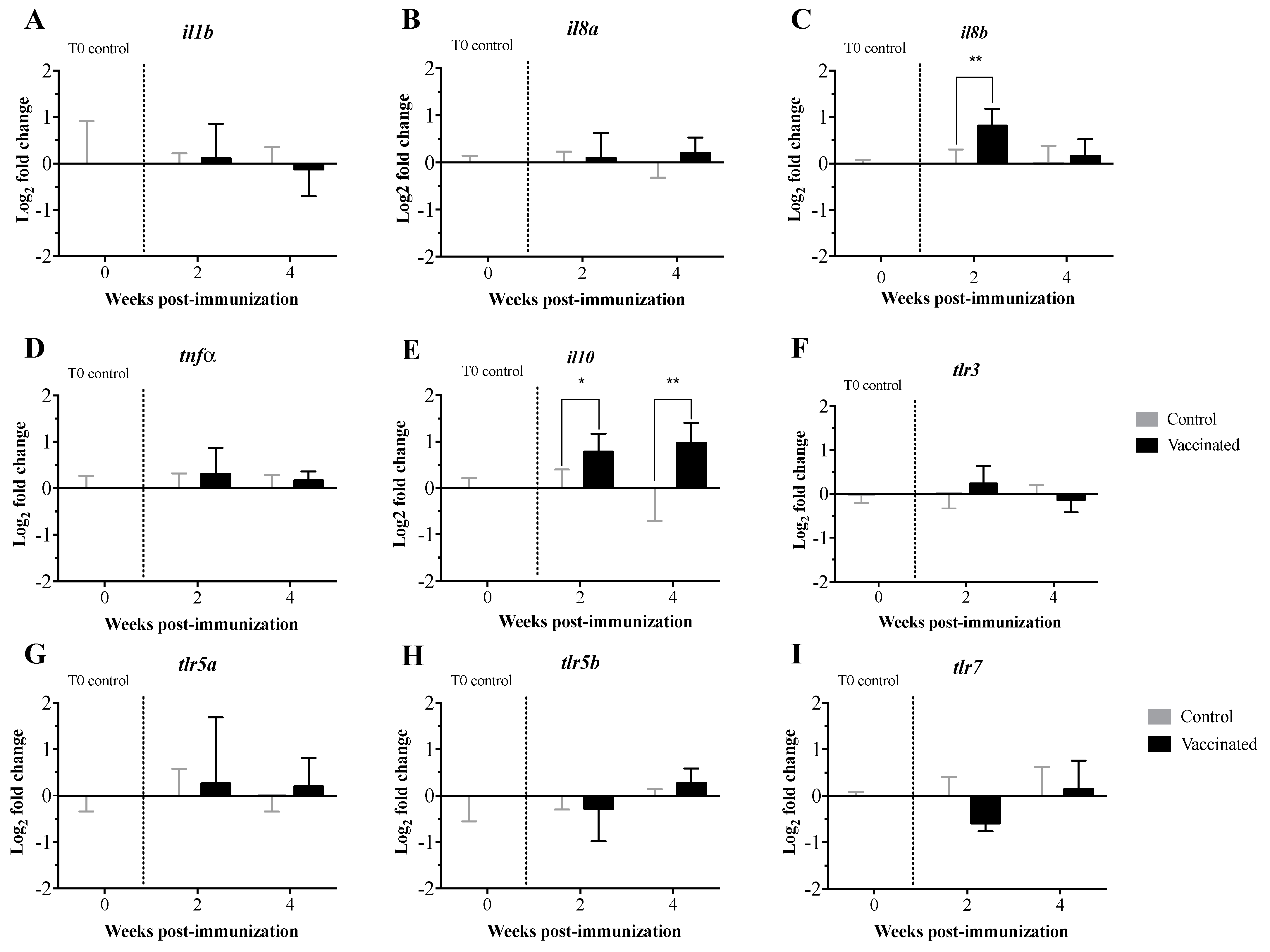
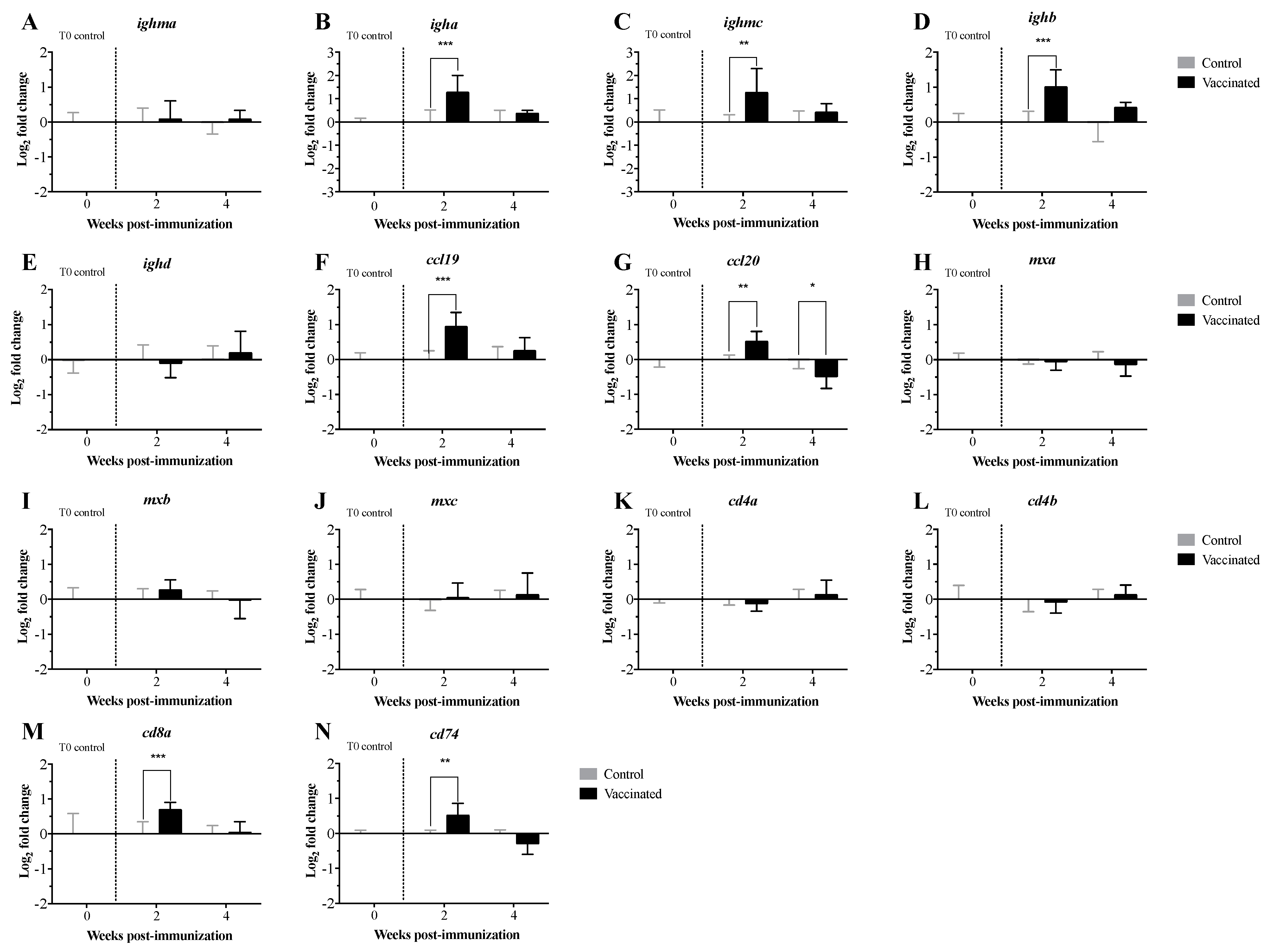
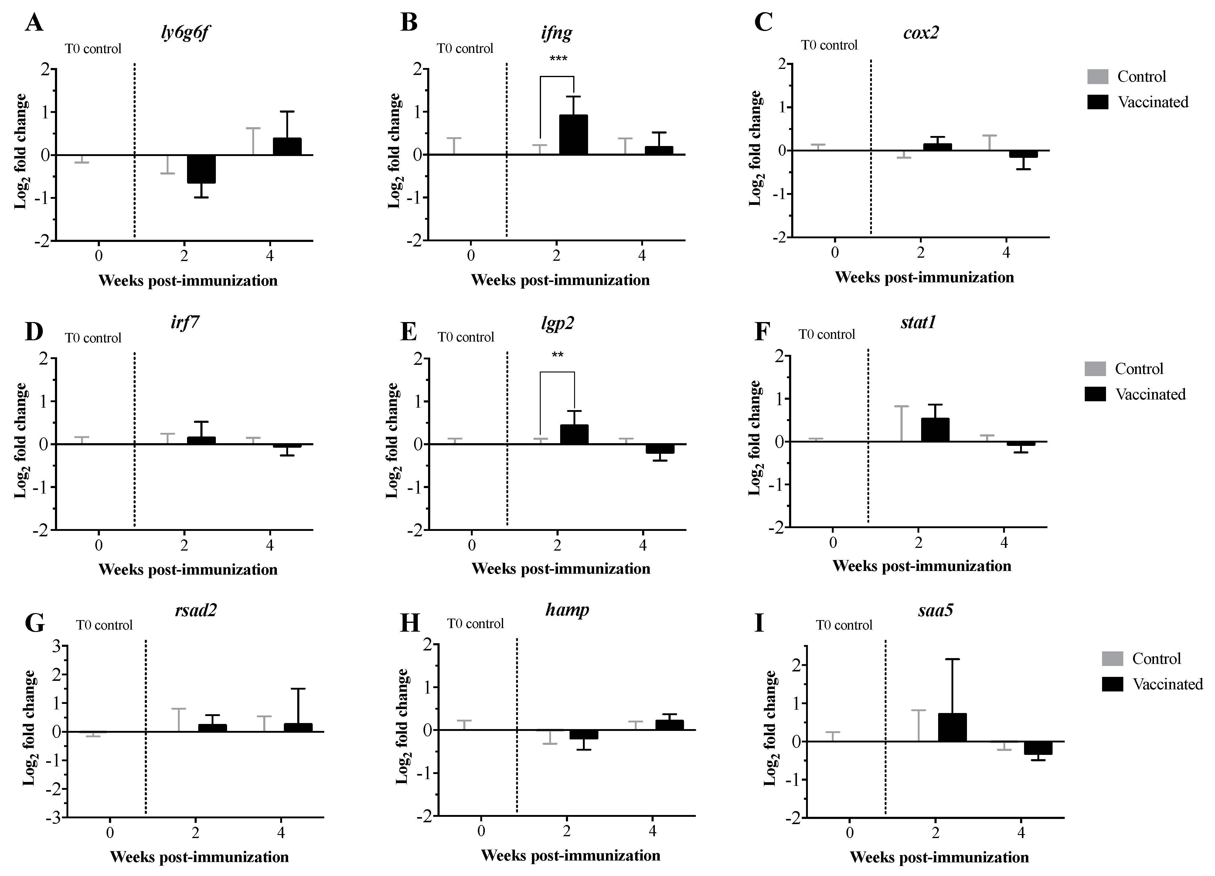
3.2. Transcript Expression Profile of the Immunome of Orally Immunized Lumpfish Larvae

Expression levels of the transcripts related to the innate and adaptive immune response were measured in pre-immunized larvae and at 2 and 4 wpi with the V. anguillarum bacterin bio-encapsulated in A. salina nauplii. The transcript expression levels were analyzed using both the 2−ΔΔCt (Figure 4 and Figure 5) and the RQ methods (Supplementary Figures S5 and S6). Both methods showed similar results, with the exception of the statistical significance demonstrated for toll-like receptor 7 (tlr7) and immunoglobulin heavy chain b (ighb) (Figure 4, Figure 5 and Figure 6; Supplementary Figures S5–S7). Transcript expression levels were compared statistically in orally immunized compared to control larvae at 2 and 4 wpi only. Comparisons over time could not be assessed due to developmental and considerable size differences of the larvae over time. Significant up-regulation of interleukin 8b (il8b) (Figure 4A and Figure S5A), immunoglobulin heavy chain a (igha), ighb and immunoglobulin mu heavy chain c (ighmc) (Figure 5A and Figure S6A), chemokines (ccl19 and ccl20) (Figure 5B and Figure S6B), cluster of differentiation 8 alpha (cd8a) and HLA class II histocompatibility antigen gamma chain (cd74) (Figure 5C and Figure S6C), interferon-gamma (ifng) (Figure 6 and Figure S7), and ATP-dependent RNA helicase lgp2 (lgp2) (Figure 6 and Figure S7) occurred at 2 wpi. Significant up-regulation of interleukin 10 (il10) occurred at 4 wpi (Figure 4A and Figure S5A). Significant downregulation of lymphocyte antigen 6 family member G6F (ly6g6f) (Figure 6 and Figure S7) and ccl20 (Figure 6B and Figure S6B) occurred at 2 and 4 wpi, respectively. There were no significant differences in the expression levels of the remaining transcripts at either time point.

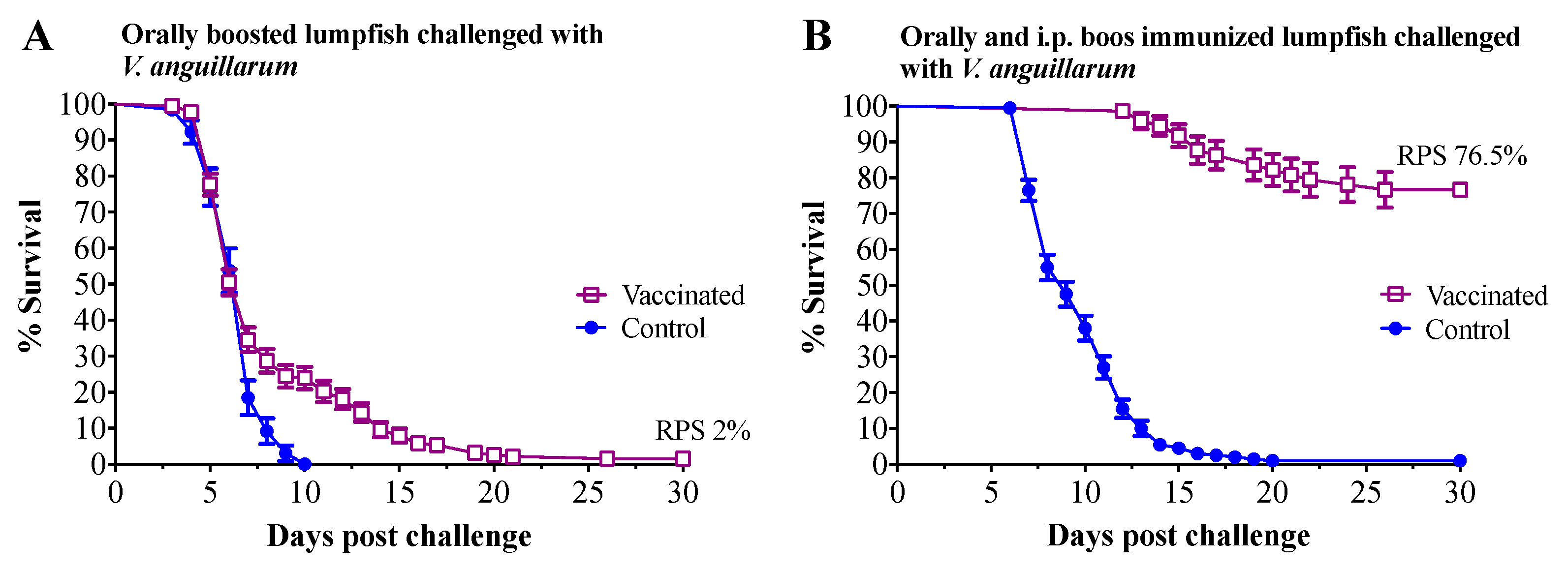
3.3. Vaccine Challenge

Immunized lumpfish were challenged at 45 wpi with 7 times the LD50 dose for V. anguillarum J360 (7.8 × 10⁵ CFU dose−1) (Figure 1). Mortality in lumpfish who had been orally mock immunized as larvae and then mock-orally boosted as juveniles started at 3 days post-challenge, with 100% mortality by day 10 post-challenge. Mortality in lumpfish that had been orally immunized as larvae and then orally boosted as juveniles was delayed and started at 3 days post-challenge, with a final RPS of 2% (Figure 7A). Mortality in lumpfish that had been mock orally immunized as larvae and then both mock orally and i.p. boosted as juveniles started at 7 days post-challenge, with 100% mortality by 20 days post-challenge (Figure 7B). Lumpfish that had been orally immunized as larvae and then both orally and i.p. boosted as juveniles survived the i.p. challenge with V. anguillarum, with a RPS of 76.5% (p < 0.0001) (Figure 7B).

4. Discussion

Vibriosis is one of the most common bacterial diseases affecting lumpfish aquaculture [39]. As mentioned previously, immunization of fish at an early age with a needle-free vaccine and with minimal stress during immunization is the ideal vaccine delivery method for finfish aquaculture [30]. However, the effectiveness of bath and oral inactivate vaccine delivery to small fish or larvae has been evaluated with varied results [52]. Commercial bath vaccines have been used in lumpfish with low effectiveness against local V. anguillarum isolates [28]. However, the efficacy of an orally administered vaccine in lumpfish during the early-life stages remains unknown. Here, we evaluated the efficacy of an orally delivery V. anguillarum bacterin bio-encapsulated in A. salina nauplii in lumpfish larvae. The molecular immune response to oral immunization was evaluated in larvae by qPCR. Approximately 9 months after the initial oral immunization, lumpfish were then either orally or both orally and i.p. boosted, and the effectiveness of the vaccines was evaluated by assessing the RPS after a lethal i.p. V. anguillarum challenge.
First, we evaluated the V. anguillarum bacterin uptake in A. salina nauplii and, thereafter this bio-encapsulated bacterin, in the gut of lumpfish larvae. Our observations indicated that the V. anguillarum bacterin was fully bio-encapsulated by the A. salina nauplii after 3 h and was maintained for at least 6 d at 6 °C (Figure 2). Similar results were observed by Campbell et al. [32], where the V. anguillarum bacterin showed maximum bio-encapsulation after 1 h or 2 h using 1.5 × 10⁷ CFU mL−1 or 1.5 × 10⁶ cells mL−1, respectively. Vaccine bio-encapsulation in A. salina nauplii protects the antigens from the intestinal tract of the fish and facilitates the recognition of the antigens using the macrophages in the mucosal layer of the hindgut [53]. The effectiveness of protecting the antigen from gastrointestinal digestion and its delivery to the hindgut of fish larvae has been demonstrated in previous studies [33,53]. Here, we confirmed the presence of the V. anguillarum bacterin in the A. salina nauplii and in the gut of fish larvae 6 h post-oral immunization (Figure 3). These results validated the internalization of the V. anguillarum bacterin in the lumpfish gut.
The expression profiles of 32 TOIs related to innate and adaptive immunity were evaluated in larvae pre-immunization and at 2 and 4 wpi. In pre-immunized larvae, we did not see any expression of the TOIs, which was expected. When considering orally immunized compared to larvae who had been orally mock immunized at 2 and 4 wpi, there were no significant differences in the levels of pro-inflammatory cytokines (tnfa, il1b, il8a; Figure 4A and Figure S6A), toll-like receptors (tlr3, tlr5a, tlr5b; Figure 4B and Figure S6B), immunoglobulin heavy chain transcripts (ighma, ighd; Figure 5A and Figure S7A), interferon-induced effectors (mxa, mxb, mxc; Figure 5B and Figure 7B), cluster of differentiation transcripts (cd4a, cd4b; Figure 5C and Figure S7C), or other immune-related transcripts (cox2, irf7, lgp2, stat1, rsad2, hamp, saa5; Figure 6 and Figure S8). In contrast, levels of il8b, igha, ighmc, ighb, ccl19, ccl20, cd8a, cd74, ifng, and lgp2 were significantly upregulated, and levels of ly6g6f and tlr7 were significantly downregulated at 2 wpi (Figure 4A, Figure 5 and Figure 6, Figures S6A, S7 and S8). Levels of il10 were significantly upregulated, and levels of ccl20 were significantly downregulated at 4 wpi (Figure 4A and Figure 5B, Figures S6A and S7B). These results indicate that 35 d old lumpfish larvae are not highly immune stimulated by oral immunization, suggesting that the interaction between the lymphoid tissues and the vaccine was not enough to trigger adaptive immune protection.
il8 and ifng play important roles in the recruitment of monocytes and neutrophils to sites of inflammation. Whereas il10 acts as anti-inflammatory cytokine and as such, plays a crucial role in the regulation of the inflammatory response [54]. Although there are studies on the expression of il8 and il10 in fish, the role of these interleukins in the early developmental stages of lumpfish is still unknown. In the current study, in orally immunized larvae compared to larvae who had been orally mock immunized, il8 and ifng were significantly upregulated at 2 wpi, il10 significantly upregulated at 4 wpi (Figure 4A, Figure 5 and Figure 6; Figures S6 and S8A). Similar expression profiles for ifng and il10 have been observed in Atlantic salmon that had been infected with the salmonid alphavirus subtype-3 (SAV-3) [55] or immunized with the A. salmonicida vaccine [56]. lgp2 is a member of the RLR family that participates in the recognition of viral RNA pathogen-associated molecular patterns (PAMPs) in the cytoplasm and induces the synthesis of ifng [57,58,59,60]. Similarly, our results showed that lgp2 and ifng were significantly upregulated at 2 wpi (Figure 6 and Figure S8). The results suggest that the oral immunization of lumpfish larvae triggers an innate immune response that is later regulated via the canonical anti-inflammatory cytokine il10.
TLRs play an important role in early innate and adaptive immunity by detecting PAMPs in bacteria and viruses [61,62,63]. TLRs activate the transcription factor NF-κB, resulting in the production of several pro-inflammatory cytokines such as il1b, tnfa, il8, il10, il6, il12, il17, ifng, and the tumour necrosis factor (tnf) [64,65]. Expression levels of tlr3, tlr5, and tlr7 were not significantly different in orally immunized compared to larvae who had been orally mock immunized at either time point (Figure 4B), however tlr7 was significantly downregulated at 2 wpi in the RQ statistical analysis (Figure S6B). These results suggest that the vaccine does not induce a full immune response in larvae.
ly6g6f is a member of the superfamily lymphocyte antigen-6 (Ly6)/urokinase-type plasminogen activator receptor (uPAR) [66]. Ly6/uPAR proteins have functions in cell proliferation, migration, cell–cell interaction, immune cell maturation, macrophage activation, T lymphocyte development, differentiation, and cytokine production [67,68,69]. The function of ly6g6f in fish is not yet defined, and in this study, the expression of ly6g6f was significantly downregulated at 2 wpi in lumpfish larvae (Figure 6 and Figure S8). These results agree with the low level of immune protection.
The igh (immunoglobulin heavy locus) encodes the IgM heavy chains, and these loci have been characterized in several fish species, including fugu, rainbow trout, zebrafish, and Atlantic salmon [70,71,72]. It has been established that igh plays a role during the adaptive immune response by recognizing foreign antigens for phagocytosis, and the complement system [73]. Here, we found that igha, ighd, and ighmc expression were significantly upregulated in orally immunized compared to larvae who had been orally mock immunized at 2 wpi (Figure 5A and Figure S7A). These results suggest the oral immunization of larvae triggers some level of an adaptive immune response, but it seems insufficient to trigger memory immune protection.
ccl19 is a chemokine known to orchestrate the migration of dendritic cells (DCs) and T cells into lymphoid tissue or vaccination sites and is also involved in immune tolerance and inflammatory responses [74,75]. ccl20 attracts lymphocytes and DCs towards epithelial cells to mucosal immune sites under inflammatory conditions [76]. In orally immunized compared to larvae who had been orally mock immunized, there was a significant increase in levels of ccl19 and ccl20 at 2 wpi, while there was a significant decrease in ccl20 levels at 4 wpi (Figure 5B and Figure S7B). These results aligned with the expression patterns of other transcripts evaluated here, supporting the idea that the lumpfish larvae did respond to the oral immunization.
CD4 (a classical marker of T helper cells) and CD8 (a marker of cytotoxic lymphocytes) are polypeptides that play an important role in signal transduction and the activation of T-helper cells and cytotoxic T cells, respectively [77]. We found that cd8 was significantly upregulated in orally immunized compared to larvae who had been orally mock immunized at 2 wpi (Figure 5C and Figure S7C). However, cd4 was not significantly dysregulated. These results suggest that CD8 cellular-mediated adaptive immunity, but not the CD4 response, was activated in lumpfish larvae aligning with the lack of immune protection triggered by the oral immunization (Figure 7A).
CD74 is the MHC class II-associated invariant chain, which plays a role in antigen presentation [78,79]. CD4 and CD74 lost their original functions in anglerfish (Lophius piscatorius) and Atlantic cod (Gadus morhua) [80,81,82,83]. Here, we found that these transcripts are present in lumpfish, and although cd74 was upregulated in orally immunized compared to larvae who had been orally mock immunized at 2 wpi, cd4a and cd4b were not (Figure 5C and Figure S7C). These results revealed that oral immunization in lumpfish larvae triggers a partial adaptive immune response.
The lumpfish larvae were vaccinated after the yolk sac was absorbed, after which the larvae exhibited an active feeding behavior. Although there is no literature about the immunity of lumpfish larvae, it is well known that lumpfish larvae are more mature and active than other marine fish [84]. It has also been shown that the main immune organs of lumpfish develop after hatching [85]. These reports, in addition to our current results, suggest that lumpfish larvae are immune competent, and antigens need to be delivered across the epithelia to trigger full immunity. The transcript expression levels (Figure 4, Figure 5 and Figure 6) also indicated that lumpfish larvae are immune stimulated by oral immunization, but not enough to trigger immune protection. For instance, the expression of il8b, il0, igha, ighmc, ighb, cd8, and c74 was upregulated in orally immunized larvae (Figure 4, Figure 5 and Figure 6). It seems that oral immunization with V. anguillarum bacterin in lumpfish larvae triggered Th1-like immune response and cellular immunity, which is related to il10 and cd8 upregulation. This is the first study on lumpfish larvae molecular immunity and provides novel knowledge and a baseline to study the ontogeny of the immune system in lumpfish.
The effectiveness of vaccination in fish depends on the delivery, vaccine design, and the fish species. For instance, mortality in lumpfish bath immunized and i.p. boosted with a commercial polyvalent formalin-inactivated V. anguillarum O1 and O2 vaccine was only delayed in an i.p. challenge using V. anguillarum [28]. Similar to our current results, a commercial bivalent whole-cell V. anguillarum O1 and O2 vaccine delivered by immersion and followed by an i.p. boost immunization in European sea bass (Dicentrarchus labrax) conferred approximately 99% survival against a V. anguillarum i.p. challenge [86]. In this study, we observed that lumpfish orally immunized as larvae and then orally boosted as juveniles did not survive the V. anguillarum i.p. challenge (Figure 7A). Nevertheless, we determined that oral vaccination delayed mortality in lumpfish challenged with V. anguillarum, suggesting that the oral vaccination did stimulate fish immunity, but not enough to confer protection. Similar results were found in salmonids orally immunized against Yersinia and V. anguillarum, where oral immunization conferred no or low immunity to juvenile immunized fish [87,88,89,90]. In contrast, lumpfish orally immunized as larvae and then both orally and i.p. boosted as juveniles showed a significant RPS (76.5%) to the V. anguillarum i.p. challenge (Figure 7B). This suggests that the orally administered vaccines were not reaching the deep lymphoid tissues, either in the larvae or juvenile fish, and as such, oral immunization was not effective in contrast to the i.p. delivered vaccine. Therefore, it is suggested that inactivated V. anguillarum vaccines for lumpfish should be administered using the i.p. route to confer acceptable levels of immune protection.

5. Conclusions

Oral immunization of lumpfish larvae using bio-encapsulated bacterin demonstrated that it reached the gut and immune, stimulating the fish larvae. However, oral immunization did not trigger an evident adaptive immune response, even after oral boost immunization. V. anguillarum bacterin that had been orally administered delayed mortality and did not confer protection against the i.p. V. anguillarum. In contrast, i.p. immunization conferred significant immune protection. These results suggest the need for oral vaccines that have the capability of crossing the epithelium and reaching the deep lymphoid tissues to trigger immune protection.

Supplementary Materials

The following are available online at https://www.mdpi.com/article/10.3390/vaccines9080819/s1, Figure S1: Experimental design for optimization of V. anguillarum bacterin bio-encapsulation in A. salina nauplii, Figure S2: Lumpfish culture conditions and embryonic development, Figure S3: Oral immunization of lumpfish larvae with the DTAF-labeled V. anguillarum bacterin bio-encapsulated in Artemia nauplii, Figure S4: V. anguillarum bacterin bio-encapsulation in A. salina nauplii for industrial application, Figure S5: Transcript expression levels of cytokines (A) and toll-like receptors (B) in lumpfish larvae orally immunized with the V. anguillarum bacterin bio-encapsulated in A. salina nauplii, Figure S6: Transcript expression levels of immunoglobulin heavy locus genes (A), cytokine CC genes and interferon-induced GTP-binding proteins (B), and cluster of differentiation genes (C) in lumpfish larvae orally immunized with the V. anguillarum bacterin bio-encapsulated in A. salina nauplii, Figure S7: Transcript expression levels of other immune-related genes in lumpfish larvae orally immunized with the V. anguillarum bacterin bio-encapsulated in A. salina nauplii, Table S1: Sequences of primer pairs used in qPCR analyses of transcript expression levels in lumpfish (Cyclopterus lumpus) larvae, Table S2: Ct, ∆Ct, and RQ values of transcript expression levels of immune-related transcripts in lumpfish (Cyclopterus lumpus) larvae orally immunized with the V. anguillarum bacterin bio-encapsulated in A. salina nauplii, Table S3: Conversion and normalization of Ct values using the 2(−∆∆Ct) method.

Author Contributions

Conceptualization, M.D., J.S., D.B. and J.W.; methodology, M.D., T.C., A.H., I.V., H.G., S.K., J.R.H., J.M., D.B. and J.S.; investigation, M.D., J.M., D.B., J.R.H., J.W. and J.S.; writing—original draft, M.D. and J.S.; resources: J.S., D.B. and J.W.; writing—review and editing, M.D., T.C., I.V., H.G., S.K., J.M., D.B., J.W. and J.S.; visualization, M.D. and J.S.; supervision, J.S. and J.W.; funding acquisition, J.S., J.W. and D.B. All authors have read and agreed to the published version of the manuscript.

Funding

Research funding was provided by the Ocean Frontier Institute through an award from the Canada First Research Excellence Fund (sub-module J3), the Vietnam International Education Development (VIED) fellowship, the Government of Newfoundland and Labrador, Department of Fisheries and Land Resources, NSERC-Discovery grant (RGPIN-2018-05942), and the Atlantic Fisheries Fund, Canada.

Institutional Review Board Statement

Animal protocols #18-01-JS, #18-03-JS (21 January 2018), and biohazard licence L-01 were reviewed and approved by the Institutional Animal Care Committee (https://www.mun.ca/research/about/acs/acc/) (accessed on 15 June 2021) following the Canadian Council of Animal Care guidelines (https://www.ccac.ca/) (accessed on 15 June 2021).

Informed Consent Statement

Not applicable.

Data Availability Statement

Not applicable.

Acknowledgments

We thank the staff at the Cold-Ocean Deep-Sea Research Facility (CDRF) and the Joe Brown Aquaculture Research Building (JBARB) for their expert technical assistance with the fish assays. Primers for qPCR analysis were designed and quality-tested, and qPCR were data generated at the Functional Genomics and Molecular Biology laboratory, Aquatic Research Cluster, CREAIT Network, Memorial University of Newfoundland.

Conflicts of Interest

The authors declare no conflict of interest.

References

  1. Costello, M.J. The global economic cost of sea lice to the salmonid farming industry. J. Fish Dis. 2009, 32, 115–118. [Google Scholar] [CrossRef] [PubMed]
  2. Fast, M.D. Fish immune responses to parasitic copepod (namely sea lice) infection. Dev. Comp. Immunol. 2014, 43, 300–312. [Google Scholar] [CrossRef]
  3. Aaen, S.M.; Helgesen, K.O.; Bakke, M.J.; Kaur, K.; Horsberg, T.E. Drug resistance in sea lice: A threat to salmonid aquaculture. Trends Parasitol. 2015, 31, 72–81. [Google Scholar] [CrossRef]
  4. Overton, K.; Barrett, L.T.; Oppedal, F.; Kristiansen, T.S.; Dempster, T. Sea lice removal by cleaner fish in salmon aquaculture: A review of the evidence base. Aquacult. Environ. Interact. 2020, 12, 31–44. [Google Scholar] [CrossRef] [Green Version]
  5. Brooks, K.M. Considerations in developing an integrated pest management programme for control of sea lice on farmed salmon in Pacific Canada. J. Fish Dis. 2009, 32, 59–73. [Google Scholar] [CrossRef] [PubMed]
  6. Brooker, A.J.; Papadopoulou, A.; Gutierrez, C.; Rey, S.; Davie, A.; Migaud, H. Sustainable production and use of cleaner fish for the biological control of sea lice: Recent advances and current challenges. Vet. Rec. 2018, 183, 383. [Google Scholar] [CrossRef] [Green Version]
  7. Treasurer, J.W. A review of potential pathogens of sea lice and the application of cleaner fish in biological control. Pest Manag. Sci. 2002, 58, 546–558. [Google Scholar] [CrossRef] [PubMed]
  8. Boyce, D.; Ang, K.P.; Prickett, R. Cunner and lumpfish as cleaner fish species in Canada. In Cleaner Fish Biology and Aquaculture Applications; Treasurer, J.W., Ed.; 5M Publishing Ltd.: Sheffield, UK, 2018. [Google Scholar]
  9. Umasuthan, N.; Valderrama, K.; Vasquez, I.; Segovia, C.; Hossain, A.; Cao, T.; Gnanagobal, H.; Monk, J.; Boyce, D.; Santander, J. A novel marine pathogen isolated from wild cunners (Tautogolabrus adspersus): Comparative genomics and transcriptome profiling of Pseudomonas sp. strain J380. Microorganisms 2021, 9, 812. [Google Scholar] [CrossRef]
  10. Monk, J.; Boyce, D.L.; Ang, K.P.; George, S.; Tucker, D.; Jeannot, K.; Fry, J.; Gianasi, B.; Hickey, S.; O’Brien, N. Cleaner fish research and production in Newfoundland. Bull. Aqaucult. Assoc. Can. 2016, 2, 39–45. [Google Scholar]
  11. Charmley, K.J. Habitat selection by two species of cleaner fishes that may be beneficial in removing sea lice from cultured salmon. SURG J. 2019, 11. [Google Scholar] [CrossRef]
  12. Marcos-Lopez, M.; Donald, K.; Stagg, H.; McCarthy, U. Clinical Vibrio anguillarum infection in lumpsucker Cyclopterus lumpus in Scotland. Vet. Rec. 2013, 173, 319. [Google Scholar] [CrossRef]
  13. Imsland, A.K.; Reynolds, P.; Eliassen, G.; Hangstad, T.A.; Foss, A.; Vikingstad, E.; Elvegård, T.A. The use of lumpfish (Cyclopterus lumpus L.) to control sea lice (Lepeophtheirus salmonis krøyer) infestations in intensively farmed atlantic salmon (Salmo salar L.). Aquaculture 2014, 424, 18–23. [Google Scholar] [CrossRef]
  14. Whittaker, B.A.; Consuegra, S.; Garcia de Leaniz, C. Genetic and phenotypic differentiation of lumpfish (Cyclopterus lumpus) across the North Atlantic: Implications for conservation and aquaculture. PeerJ 2018, 6, e5974. [Google Scholar] [CrossRef] [Green Version]
  15. Toffan, A.; De Salvador, M.; Scholz, F.; Pretto, T.; Buratin, A.; Rodger, H.D.; Toson, M.; Cuenca, A.; Vendramin, N. Lumpfish (Cyclopterus lumpus, Linnaeus) is susceptible to viral nervous necrosis: Result of an experimental infection with different genotypes of Betanodavirus. J. Fish Dis. 2019, 42, 1667–1676. [Google Scholar] [CrossRef] [PubMed]
  16. Rueness, E.; Berg, P.R.; Gulla, S.; Halvorsen, K.; Järnegren, J.; Malmstrøm, M.; Mo, T.A.; Rimstad, E.; de Boer, H.; Eldegard, K.; et al. Assessment of The Risk to Norwegian Biodiversity from Import of Wrasses and Other Cleaner Fish for Use in Aquaculture. Norwegian Scientific Committee for Food and Environment (VKM). 2019, pp. 2535–4019. Available online: https://vkm.no/download/18.22fe061816d90c026b4a890d/1570782153468/Assessment%20of%20the%20risk%20to%20Norwegian%20biodiversity%20from%20import%20of%20wrasses%20and%20other%20cleaner%20fish%20for%20use%20in%20aquaculture.pdf (accessed on 15 June 2021).
  17. Powell, A.; Treasurer, J.W.; Pooley, C.L.; Keay, A.J.; Lloyd, R.; Imsland, A.K.; Garcia de Leaniz, C. Use of lumpfish for sea-lice control in salmon farming:challenges and opportunities. Rev. Aquac. 2017, 10, 1–20. [Google Scholar] [CrossRef] [Green Version]
  18. NDF Fisheries. Norwegian Directorate for Fisheries. 2019. Available online: https://www.fiskeridir.no/English/Aquaculture/Statistics/Cleanerfish-Lumpfish-and-Wrasse (accessed on 15 June 2021).
  19. Foss, A.; Imsland, A.K.D.; Roth, B.; Nytrø, A.V. Catch me if you can: How to recapture lumpfish using light as an attractant. Aquac. Eng. 2020, 90, 102074. [Google Scholar] [CrossRef]
  20. Bolton-Warberg, M. An overview of cleaner fish use in Ireland. J. Fish Dis. 2018, 41, 935–939. [Google Scholar] [CrossRef] [PubMed] [Green Version]
  21. Ronneseth, A.; Haugland, G.T.; Colquhoun, D.J.; Brudal, E.; Wergeland, H.I. Protection and antibody reactivity following vaccination of lumpfish (Cyclopterus lumpus L.) against atypical Aeromonas salmonicida. Fish Shellfish Immunol. 2017, 64, 383–391. [Google Scholar] [CrossRef] [PubMed]
  22. Ellul, R.M.; Bulla, J.; Brudal, E.; Colquhoun, D.; Wergeland, H.; Ronneseth, A. Protection and antibody reactivity in lumpsucker (Cyclopterus lumpus L.) following vaccination against Pasteurella sp. Fish Shellfish Immunol. 2019, 95, 650–658. [Google Scholar] [CrossRef] [PubMed]
  23. Vasquez, I.; Cao, T.; Chakraborty, S.; Gnanagobal, H.; O’Brien, N.; Monk, J.; Boyce, D.; Westcott, J.D.; Santander, J. Comparative Genomics Analysis of Vibrio anguillarum Isolated from Lumpfish (Cyclopterus lumpus) in Newfoundland Reveal Novel Chromosomal Organizations. Microorganisms 2020, 8, 1666. [Google Scholar] [CrossRef] [PubMed]
  24. Erkinharju, T.; Dalmo, R.A.; Hansen, M.; Seternes, T. Cleaner fish in aquaculture: Review on diseases and vaccination. Rev. Aquac. 2021, 13, 189–237. [Google Scholar] [CrossRef]
  25. Frans, I.; Michiels, C.W.; Bossier, P.; Willems, K.A.; Lievens, B.; Rediers, H. Vibrio anguillarum as a fish pathogen: Virulence factors, diagnosis and prevention. J. Fish Dis. 2011, 34, 643–661. [Google Scholar] [CrossRef]
  26. Holm, K.O.; Nilsson, K.; Hjerde, E.; Willassen, N.P.; Milton, D.L. Complete genome sequence of Vibrio anguillarum strain NB10, a virulent isolate from the Gulf of Bothnia. Stand. Genomic Sci. 2015, 10, 60. Available online: https://www.ncbi.nlm.nih.gov/pubmed/26380645 (accessed on 15 June 2021). [CrossRef] [Green Version]
  27. Mikkelsen, H.; Lund, V.; Larsen, R.; Seppola, M. Vibriosis vaccines based on various sero-subgroups of Vibrio anguillarum O2 induce specific protection in Atlantic cod (Gadus morhua L.) juveniles. Fish Shellfish Immunol. 2011, 30, 330–339. [Google Scholar] [CrossRef]
  28. Chakraborty, S.; Cao, T.; Hossain, A.; Gnanagobal, H.; Vasquez, I.; Boyce, D.; Santander, J. Vibrogen-2 vaccine trial in lumpfish (Cyclopterus lumpus) against Vibrio anguillarum. J. Fish Dis. 2019, 42, 1057–1064. [Google Scholar] [CrossRef]
  29. Dadar, M.; Chakraborty, S.; Dhama, K.; Prasad, M.; Khandia, R.; Hassan, S.; Munjal, A.; Tiwari, R.; Karthik, K.; Kumar, D.; et al. Advances in designing and developing vaccines, drugs and therapeutic Aapproaches to counter human papilloma virus. Front. Immunol. 2018, 9, 2478. [Google Scholar] [CrossRef]
  30. Plant, K.P.; Lapatra, S.E. Advances in fish vaccine delivery. Dev. Comp. Immunol. 2011, 35, 1256–1262. [Google Scholar] [CrossRef] [PubMed]
  31. Mutoloki, S.; Munangandu, H.M.; Evensen, O. Oral Vaccination of fish-antigen preparations, uptake, and immune induction. Front. Immunol. 2015, 6, 519. [Google Scholar] [CrossRef] [Green Version]
  32. Campbell, R.; Adams, A.; Tatner, M.F.; Chair, M.; Sorgeloos, P. Uptake of Vibrio anguillarum vaccine by artemia salina as a potential oral delivery system to fish fry. Fish Shellfish Immunol. 1993, 3, 451–459. [Google Scholar] [CrossRef]
  33. Lin, C.C.; Lin, J.H.Y.; Chen, M.S.; Yang, H.L. An oral nervous necrosis virus vaccine that induces protective immunity in larvae of grouper (Epinephelus coioides). Aquaculture 2007, 268, 265–273. [Google Scholar] [CrossRef]
  34. Chen, Y.M.; Shih, C.H.; Liu, H.C.; Wu, C.L.; Lin, C.C.; Wang, H.C.; Chen, T.Y.; Yang, H.L.; Lin, J.H.Y. An oral nervous necrosis virus vaccine using Vibrio anguillarum as an expression host provides early protection. Aquaculture 2011, 321, 26–33. [Google Scholar] [CrossRef]
  35. Sambrook, J.; Russell, W. Molecular Cloning; A Laboratory Manual, 3rd ed.; Cold Spring Harbor Press: New York, NY, USA, 2001. [Google Scholar]
  36. Leboffe, M.J.; Pierce, B.E. Microbiology: Laboratory Theory & Application; Morton Publishing: Englewood, CO, USA, 2019. [Google Scholar]
  37. Eslamloo, K.; Kumar, S.; Caballero-Solares, A.; Gnanagobal, H.; Santander, J.; Rise, M.L. Profiling the transcriptome response of Atlantic salmon head kidney to formalin-killed Renibacterium salmoninarum. Fish Shellfish Immunol. 2020, 98, 937–949. [Google Scholar] [CrossRef] [PubMed]
  38. Santander, J.; Golden, G.; Wanda, S.Y.; Curtiss, R., 3rd. Fur-regulated iron uptake system of Edwardsiella ictaluri and its influence on pathogenesis and immunogenicity in the catfish host. Infect. Immun. 2012, 80, 2689–2703. [Google Scholar] [CrossRef] [PubMed] [Green Version]
  39. Vasquez, I.; Cao, T.; Hossain, A.; Valderrama, K.; Gnanagobal, H.; Dang, M.; Leeuwis, R.H.J.; Ness, M.; Campbell, B.; Gendron, R.; et al. Aeromonas salmonicida infection kinetics and protective immune response to vaccination in sablefish (Anoplopoma fimbria). Fish Shellfish Immunol. 2020, 104, 557–566. [Google Scholar] [CrossRef] [PubMed]
  40. Valderrama, K.; Soto-Davila, M.; Segovia, C.; Vasquez, I.; Dang, M.; Santander, J. Aeromonas salmonicida infects Atlantic salmon (Salmo salar) erythrocytes. J. Fish Dis. 2019, 42, 1601–1608. [Google Scholar] [CrossRef] [PubMed]
  41. Wessman, P.; Hakansson, S.; Leifer, K.; Rubino, S. Formulations for freeze-drying of bacteria and their influence on cell survival. J. Vis. Exp. 2013. [Google Scholar] [CrossRef] [Green Version]
  42. Fissell, W.H.; Manley, S.; Dubnisheva, A.; Glass, J.; Magistrelli, J.E.A.N.; Fleischman, A.J.; Zydney, A.L.; Roy, S. Ficoll is not a rigid sphere. Am. J. Physiol. Renal Physiol. 2007, 293, F1209–F1213. [Google Scholar] [CrossRef] [PubMed] [Green Version]
  43. Inman, J.K. Thymus-independent antigens: The preparation of covalent, hapten-ficoll conjugates. J. Immunol. 1975, 114 Pt 1, 704–709. [Google Scholar]
  44. Amlot, P.L.; Hayes, A.E.; Gray, D.; Gordon-Smith, E.C.; Humphrey, J.H. Human immune responses in vivo to protein (KLH) and polysaccharide (DNP-Ficoll) neoantigens: Normal subjects compared with bone marrow transplant patients on cyclosporine. Clin. Exp. Immunol. 1986, 64, 125–135. [Google Scholar]
  45. Anderson, D.P.; Merchant, B.; Dixon, O.W.; Schott, C.F.; Lizzio, E.F. Flush exposure and injection immunization of rainbow trout to selected DNP conjugates. Dev. Comp. Immunol. 1983, 7, 261–268. [Google Scholar] [CrossRef]
  46. Milley, B.; Kiwan, R.; Ott, G.S.; Calacsan, C.; Kachura, M.; Campbell, J.D.; Kanzler, H.; Coffman, R.L. Optimization, Production, and Characterization of a CpG-Oligonucleotide-Ficoll Conjugate Nanoparticle Adjuvant for Enhanced Immunogenicity of Anthrax Protective Antigen. Bioconjug. Chem. 2016, 27, 1293–1304. [Google Scholar] [CrossRef]
  47. Amend, D.F. Potency Testing of Fish Vaccines. Fish Biologics: Serodiagnostics and Vaccines. 1981, Volume 49, pp. 447–454. Available online: https://www.scirp.org/(S(vtj3fa45qm1ean45vvffcz55))/reference/ReferencesPapers.aspx?ReferenceID=1919013 (accessed on 15 June 2021).
  48. Pfaffl, M.W. A new mathematical model for relative quantification in real-time RT-PCR. Nucleic Acids Res. 2001, 29, e45. [Google Scholar] [CrossRef]
  49. Vandesompele, J.; De Preter, K.; Pattyn, F.; Poppe, B.; Van Roy, N.; De Paepe, A.; Speleman, F. Accurate normalization of real-time quantitative RT-PCR data by geometric averaging of multiple internal control genes. Genome Biol. 2002, 3, RESEARCH0034. [Google Scholar] [CrossRef] [PubMed] [Green Version]
  50. Livak, K.J.; Schmittgen, T.D. Analysis of relative gene expression data using real-time quantitative PCR and the 2(Delta Delta C(T)) Method. Methods 2001, 25, 402–408. [Google Scholar] [CrossRef] [PubMed]
  51. Soto-Davila, M.; Hossain, A.; Chakraborty, S.; Rise, M.L.; Santander, J. Aeromonas salmonicida subsp. salmonicida early infection and immune response of Atlantic cod (Gadus morhua L.) primary macrophages. Front. Immunol. 2019, 10, 1237. [Google Scholar] [CrossRef] [PubMed] [Green Version]
  52. Villumsen, K.R.; Neumann, L.; Ohtani, M.; Strom, H.K.; Raida, M.K. Oral and anal vaccination confers full protection against enteric redmouth disease (ERM) in rainbow trout. PLoS ONE 2014, 9, e93845. [Google Scholar] [CrossRef] [PubMed]
  53. Lin, J.H.; Yu, C.C.; Lin, C.C.; Yang, H.L. An oral delivery system for recombinant subunit vaccine to fish. Dev. Biol 2005, 121, 175–180. [Google Scholar]
  54. Min, W.; Lillehoj, H.S.; Burnside, J.; Weining, K.C.; Staeheli, P.; Zhu, J.J. Adjuvant effects of IL-1beta, IL-2, IL-8, IL-15, IFN-alpha, IFN-gamma TGF-beta4 and lymphotactin on DNA vaccination against Eimeria acervulina. Vaccine 2001, 20, 267–274. [Google Scholar] [CrossRef]
  55. Xu, C.; Guo, T.C.; Mutoloki, S.; Haugland, O.; Evensen, O. Gene expression studies of host response to Salmonid alphavirus subtype 3 experimental infections in Atlantic salmon. Vet. Res. 2012, 43, 78. [Google Scholar] [CrossRef] [Green Version]
  56. Kumari, J.; Bogwald, J.; Dalmo, R.A. Vaccination of Atlantic salmon, Salmo salar L.; with Aeromonas salmonicida and infectious pancreatic necrosis virus (IPNV) showed a mixed Th1/Th2/Treg response. J. Fish Dis. 2013, 36, 881–886. [Google Scholar] [CrossRef]
  57. Ohtani, M.; Hikima, J.; Kondo, H.; Hirono, I.; Jung, T.S.; Aoki, T. Evolutional conservation of molecular structure and antiviral function of a viral RNA receptor, LGP2, in Japanese flounder, Paralichthys olivaceus. J. Immunol. 2010, 185, 7507–7517. [Google Scholar] [CrossRef] [Green Version]
  58. Chang, M.; Collet, B.; Nie, P.; Lester, K.; Campbell, S.; Secombes, C.J.; Zou, J. Expression and functional characterization of the RIG-I-like receptors MDA5 and LGP2 in Rainbow trout (Oncorhynchus mykiss). J. Virol. 2011, 85, 8403–8412. [Google Scholar] [CrossRef] [PubMed] [Green Version]
  59. van der Veen, A.G.; Maillard, P.V.; Schmidt, J.M.; Lee, S.A.; Deddouche-Grass, S.; Borg, A.; Kjaer, S.; Snijders, A.P.; Reis e Sousa, C. The RIG-I-like receptor LGP2 inhibits Dicer-dependent processing of long double-stranded RNA and blocks RNA interference in mammalian cells. EMBO J. 2018, 37. [Google Scholar] [CrossRef] [Green Version]
  60. Zhang, Q.M.; Zhao, X.; Li, Z.; Wu, M.; Gui, J.F.; Zhang, Y.B. Alternative splicing transcripts of Zebrafish LGP2 Ggene Ddifferentially contribute to IFN antiviral response. J. Immunol. 2018, 200, 688–703. [Google Scholar] [CrossRef]
  61. Arancibia, S.A.; Beltran, C.J.; Aguirre, I.M.; Silva, P.; Peralta, A.L.; Malinarich, F.; Hermoso, M.A. Toll-like receptors are key participants in innate immune responses. Biol. Res. 2007, 40, 97–112. [Google Scholar] [CrossRef] [Green Version]
  62. Jayaramu, P.K.; Tripathi, G.; Pavan Kumar, A.; Keezhedath, J.; Pathan, M.K.; Kurcheti, P.P. Studies on expression pattern of toll-like receptor 5 (TLR5) in Edwardsiella tarda infected Pangasianodon hypophthalmus. Fish Shellfish Immunol. 2017, 63, 68–73. [Google Scholar] [CrossRef]
  63. Ji, J.; Rao, Y.; Wan, Q.; Liao, Z.; Su, J. Teleost-Specific TLR19 localizes to endosome, recognizes dsRNA, recruits TRIF, triggers both IFN and NF-kappaB pathways, and protects cells from grass carp reovirus infection. J. Immunol. 2018, 200, 573–585. [Google Scholar] [CrossRef]
  64. Barton, G.M.; Medzhitov, R. Control of adaptive immune responses by Toll-like receptors. Curr. Opin. Immunol. 2002, 14, 380–383. [Google Scholar] [CrossRef]
  65. Eggestol, H.O.; Lunde, H.S.; Ronneseth, A.; Fredman, D.; Petersen, K.; Mishra, C.K.; Furmanek, T.; Colquhoun, D.J.; Wergeland, H.I.; Haugland, G.T. Transcriptome-wide mapping of signaling pathways and early immune responses in lumpfish leukocytes upon in vitro bacterial exposure. Sci. Rep. 2018, 8, 5261. [Google Scholar] [CrossRef] [PubMed]
  66. Upadhyay, G. Emerging role of lymphocyte Aantigen-6 family of genes in cancer and immune cells. Front. Immunol. 2019, 10, 819. [Google Scholar] [CrossRef]
  67. MacNeil, I.; Kennedy, J.; Godfrey, D.I.; Jenkins, N.A.; Masciantonio, M.; Mineo, C.; Gilbert, D.J.; Copeland, N.G.; Boyd, R.L.; Zlotnik, A. Isolation of a cDNA encoding thymic shared antigen-1. A new member of the Ly6 family with a possible role in T cell development. J. Immunol. 1993, 151, 6913–6923. [Google Scholar] [PubMed]
  68. Mallya, M.; Campbell, R.D.; Aguado, B. Characterization of the five novel Ly-6 superfamily members encoded in the MHC, and detection of cells expressing their potential ligands. Protein Sci. 2006, 15, 2244–2256. [Google Scholar] [CrossRef] [PubMed] [Green Version]
  69. Loughner, C.L.; Bruford, E.A.; McAndrews, M.S.; Delp, E.E.; Swamynathan, S.; Swamynathan, S.K. Organization, evolution and functions of the human and mouse Ly6/uPAR family genes. Hum. Genom. 2016, 10, 10. [Google Scholar] [CrossRef] [PubMed] [Green Version]
  70. Danilova, N.; Bussmann, J.; Jekosch, K.; Steiner, L.A. The immunoglobulin heavy-chain locus in zebrafish: Identification and expression of a previously unknown isotype, immunoglobulin Z. Nat. Immunol. 2005, 6, 295–302. [Google Scholar] [CrossRef] [PubMed]
  71. Savan, R.; Aman, A.; Sato, K.; Yamaguchi, R.; Sakai, M. Discovery of a new class of immunoglobulin heavy chain from fugu. Eur. J. Immunol. 2005, 35, 3320–3331. [Google Scholar] [CrossRef]
  72. Yasuike, M.; de Boer, J.; von Schalburg, K.R.; Cooper, G.A.; McKinnel, L.; Messmer, A.; So, S.; Davidson, W.S.; Koop, B.F. Evolution of duplicated IgH loci in Atlantic salmon, Salmo salar. BMC Genom. 2010, 11, 486. [Google Scholar] [CrossRef] [Green Version]
  73. Schroeder, H.W.; Cavacini, L., Jr. Structure and function of immunoglobulins. J. Allergy Clin. Immunol. 2010, 125 (Suppl. 2), S41–S52. [Google Scholar] [CrossRef] [Green Version]
  74. Bromley, S.K.; Mempel, T.R.; Luster, A.D. Orchestrating the orchestrators: Chemokines in control of T cell traffic. Nat. Immunol. 2008, 9, 970–980. [Google Scholar] [CrossRef]
  75. Yan, Y.; Chen, R.; Wang, X.; Hu, K.; Huang, L.; Lu, M.; Hu, Q. CCL19 and CCR7 expression, signaling pathways, and adjuvant functions in viral Iinfection and prevention. Front. Cell Dev. Biol. 2019, 7, 212. [Google Scholar] [CrossRef]
  76. Liu, F.; Wang, T.; Hu, Y.; Tian, G.; Secombes, C.J.; Wang, T. Expansion of fish CCL20_like chemokines by genome and local gene duplication: Characterisation and expression analysis of 10 CCL20_like chemokines in rainbow trout (Oncorhynchus mykiss). Dev. Comp. Immunol. 2020, 103, 103502. [Google Scholar] [CrossRef]
  77. Buonocore, F.; Randelli, E.; Bird, S.; Secombes, C.J.; Costantini, S.; Facchiano, A.; Mazzini, M.; Scapigliati, G. The CD8alpha from sea bass (Dicentrarchus labrax L.): Cloning, expression and 3D modelling. Fish Shellfish Immunol. 2006, 20, 637–646. [Google Scholar] [CrossRef]
  78. Moldenhauer, G.; Henne, C.; Karhausen, J.; Moller, P. Surface-expressed invariant chain (CD74) is required for internalization of human leucocyte antigen-DR molecules to early endosomal compartments. Immunology 1999, 96, 473–484. [Google Scholar] [CrossRef]
  79. Beswick, E.J.; Reyes, V.E. CD74 in antigen presentation, inflammation, and cancers of the gastrointestinal tract. World, J. Gastroenterol. 2009, 15, 2855–2861. [Google Scholar] [CrossRef]
  80. Star, B.; Nederbragt, A.J.; Jentoft, S.; Grimholt, U.; Malmstrom, M.; Gregers, T.F.; Rounge, T.B.; Paulsen, J.; Solbakken, M.H.; Sharma, A.; et al. The genome sequence of Atlantic cod reveals a unique immune system. Nature 2011, 477, 207–210. [Google Scholar] [CrossRef] [Green Version]
  81. Trowsdale, J.; Knight, J.C. Major histocompatibility complex genomics and human disease. Annu. Rev. Genom. Hum. Genet. 2013, 14, 301–323. [Google Scholar] [CrossRef] [Green Version]
  82. Dijkstra, J.M.; Grimholt, U. Major histocompatibility complex (MHC) fragment numbers alone-in Atlantic cod and in general-do not represent functional variability. F1000Research 2018, 7, 963. [Google Scholar] [CrossRef] [PubMed]
  83. Dubin, A.; Jorgensen, T.E.; Moum, T.; Johansen, S.D.; Jakt, L.M. Complete loss of the MHC II pathway in an anglerfish, Lophius piscatorius. Biol. Lett. 2019, 15, 20190594. [Google Scholar] [CrossRef] [Green Version]
  84. Brown, J.A. The development of feeding behaviour in the lumpfish, Cyclopterus lumpus. J. Fish Biol. 1986, 29, 171–178. [Google Scholar] [CrossRef]
  85. Imsland, A.K.D.; Danielsen, M.; Jonassend, T.M.; Hangstadd, T.A.; Falk-Petersenc, I.B. Effect of incubation temperature on eggs and larvae of lumpfish (Cyclopterus lumpus). Aquaculture 2019, 498, 217–222. [Google Scholar] [CrossRef]
  86. Galeotti, M.; Romano, N.; Volpatti, D.; Bulfon, C.; Brunetti, A.; Tiscar, P.G.; Mosca, F.; Bertoni, F.; Marchetti, M.G.; Abelli, L. Innovative vaccination protocol against vibriosis in Dicentrarchus labrax (L.) juveniles: Improvement of immune parameters and protection to challenge. Vaccine 2013, 31, 1224–1230. [Google Scholar] [CrossRef] [PubMed]
  87. Johnson, K.A.; Flynn, J.K.; Amend, D.F. Duration of immunity in salmonids vaccinated by direct immersion with Yersinia ruckeri and Vibrio anguillarum bacterins. J. Fish Dis. 1983, 5, 207–213. [Google Scholar] [CrossRef]
  88. Johnson, K.A.; Amend, D.F. Comparison of efficacy of several delivery methods using Yersinia ruckeri bacterin on rainbow trout, Salmo gairdneri Richardson. J. Fish Dis. 1983, 6, 331–336. [Google Scholar] [CrossRef]
  89. Chettri, J.K.; Jaafar, R.M.; Skov, J.; Kania, P.W.; Dalsgaard, I.; Buchmann, K. Booster immersion vaccination using diluted Yersinia ruckeri bacterin confers protection against ERM in rainbow trout. Aquaculture 2015, 440, 1–5. [Google Scholar] [CrossRef]
  90. Johnson, K.A.; Amend, D.F. Efficacy of Vibrio anguillarum and Yersinia ruckeri bacterins applied by oral and anal intubation of salmonids. J. Fish Dis. 1983, 6, 473–476. [Google Scholar] [CrossRef]
Figure 1. Immunization and challenge experimental design. Lumpfish larvae were immunized at 1 wph. At 8 wph, the fish were distributed in 500 L tanks. Lumpfish were boosted at 36 wph and challenged with V. anguillarum at 45 wph. * Larvae samples were collected at weeks 0, 2, 4 post-immunization for RNA extraction.
Vaccines 09 00819 g001
Figure 2. Optimization of V. anguillarum bacterin bio-encapsulation in A. salina nauplii. (A). Relative percentage of DTAF-labeled V. anguillarum bacterin in A. salina nauplii intestine; (B) A. salina nauplii suplemeted with dry microalgae (Ori-One and Ori-Green) as autofluorescence control. A. salina fed with commercial dry microalgae (Ori-One and Ori-Green) at 20 °C; (C) V. anguillarum bacterin bio-encapsulation in A. salina nauplii at 20 °C; (D) V. anguillarum bacterin and commercial dry microalgae bio-encapsulation in A. salina nauplii at 20 °C; (E) bio-encapsulation stability at 6 °C after 3 h post enrichment with V. anguillarum bacterin at 20 °C. In all cases, different color bars represent the % bio-encapsulation levels of the V. anguillarum bacterin in A. salina nauplii. Each value is the mean ± SEM for 3 groups of 100 A. salina nauplii per group. Different letters (a, b, c) indicate the differences in the numbers of A. salina nauplii enriched with 100% V. anguillarum bacterin at different time points. Means with different letters differ significantly (p < 0.05). Bars represent mean ± SEM.
Vaccines 09 00819 g002
Figure 3. Selected images depicting the DTAF-labeled V. anguillarum bacterin bio-encapsulated in A. salina nauplii in the lumpfish gut after 6 and 24 h, visualized using epifluorescence microscopy. Its presence is indicated by green fluorescence.
Vaccines 09 00819 g003
Figure 4. Transcript expression levels of cytokines and toll-like receptors in lumpfish larvae orally immunized with the V. anguillarum bacterin bio-encapsulated in A. salina nauplii. (AE). Cytokines; (FI). Toll-like receptors. Transcript expression levels were assessed pre-immunization (T0 control, n = 3 individual pools of 10 larvae each), 2 wpi (n = 3 pools of 10 larvae each), and 4 wpi (n = 3 pools of 5 larvae each). Time point controls post-mock immunization were collected in a similar fashion at 2 and 4 wpi. Relative expression was calculated using the 2(−∆∆Ct) method and normalized using log2; etif3d and rpl32 were used as endogenous controls. A two-way ANOVA test, followed by the Sidak multiple comparisons post hoc test was used to assess significant differences between the treatments (control and vaccinated) at each individual time point. Asterisks (*) represent significant differences (* p < 0.05, ** p < 0.01).
Vaccines 09 00819 g004
Figure 5. Transcript expression levels of immunoglobulin heavy locus genes, cytokine CC genes and interferon-induced GTP-binding proteins genes, and cluster of differentiation genes in lumpfish larvae orally immunized with the V. anguillarum bacterin bio-encapsulated in A. salina nauplii (AE). Immunoglobulin heavy locus genes (FJ). Cytokine CC genes and interferon-induced GTP-binding proteins genes (KN). Cluster of differentiation genes. Transcript expression levels were assessed pre-immunization (T0 control, n = 3 individual pools of 10 larvae each), 2 weeks post-immunization (n = 3 pools of 10 larvae each), and 4 weeks post-immunization (n = 3 pools of 5 larvae each). Time point controls post-mock immunization were collected in a similar fashion to those collected at weeks 2 and 4. Relative expression was calculated using the 2(−∆∆Ct) method and normalized using log2; etif3d and rpl32 were used as endogenous controls. A two-way ANOVA test followed by the Sidak multiple comparisons post hoc test were used to assess significant differences between the treatments (control and vaccinated) at each individual time point. Asterisks (*) represent significant differences (* p < 0.05, ** p < 0.01, *** p < 0.001).
Vaccines 09 00819 g005
Figure 6. Transcript expression levels of other immune-related genes in lumpfish larvae orally immunized with the V. anguillarum bacterin bio-encapsulated in A. salina nauplii (AI). Transcript expression levels were assessed pre-immunization (T0 control, n = 3 individual pools of 10 larvae each), 2 weeks post-immunization (n = 3 pools of 10 larvae each), and 4 weeks post-immunization (n = 3 pools of 5 larvae each). Time point controls post-mock immunization were collected in a similar fashion to those collected at weeks 2 and 4. Relative expression was calculated using the 2(−∆∆Ct) method and normalized using log2; etif3d and rpl32 were used as endogenous controls. A two-way ANOVA test followed by the Sidak multiple comparisons post hoc test were used to assess significant differences between the treatments (control and vaccinated) at each individual time point. Asterisks (*) represent significant differences (** p< 0.01, *** p< 0.001).
Vaccines 09 00819 g006
Figure 7. Cumulative survival rate of orally and i.p. immunized lumpfish after i.p. challenge with V. anguillarum (7.8 × 10⁵ CFU dose−1). (A) Survival (%) of orally immunized and orally boosted lumpfish after V. anguillarum challenge. Lumpfish were orally immunized as larvae and then orally boosted as juveniles. Control groups were mock-vaccinated using the same inoculation route. After 45 weeks post-initial immunization, the animals were then i.p. challenged; each treatment consisted of two tanks (see Figure 1). (B) Survival of orally immunized and i.p. boosted lumpfish. Lumpfish were orally immunized as larvae and then orally boosted as juveniles (see Figure 1). Control groups were mock-vaccinated using the same inoculation route. After 45 weeks post-initial immunization, the animals were then i.p. challenged; Each treatment consisted of two tanks (see Figure 1). Survival was assessed for 30 days. RPS: relative percentage survival; p < 0.0001.
Vaccines 09 00819 g007
Publisher’s Note: MDPI stays neutral with regard to jurisdictional claims in published maps and institutional affiliations.

