
Population (Antibody) Testing for COVID-19—Technical Challenges, Application and Relevance, an English Perspective


Abstract
1. Introduction
2. Estimating the Incidence of SARS-CoV-2 Infection
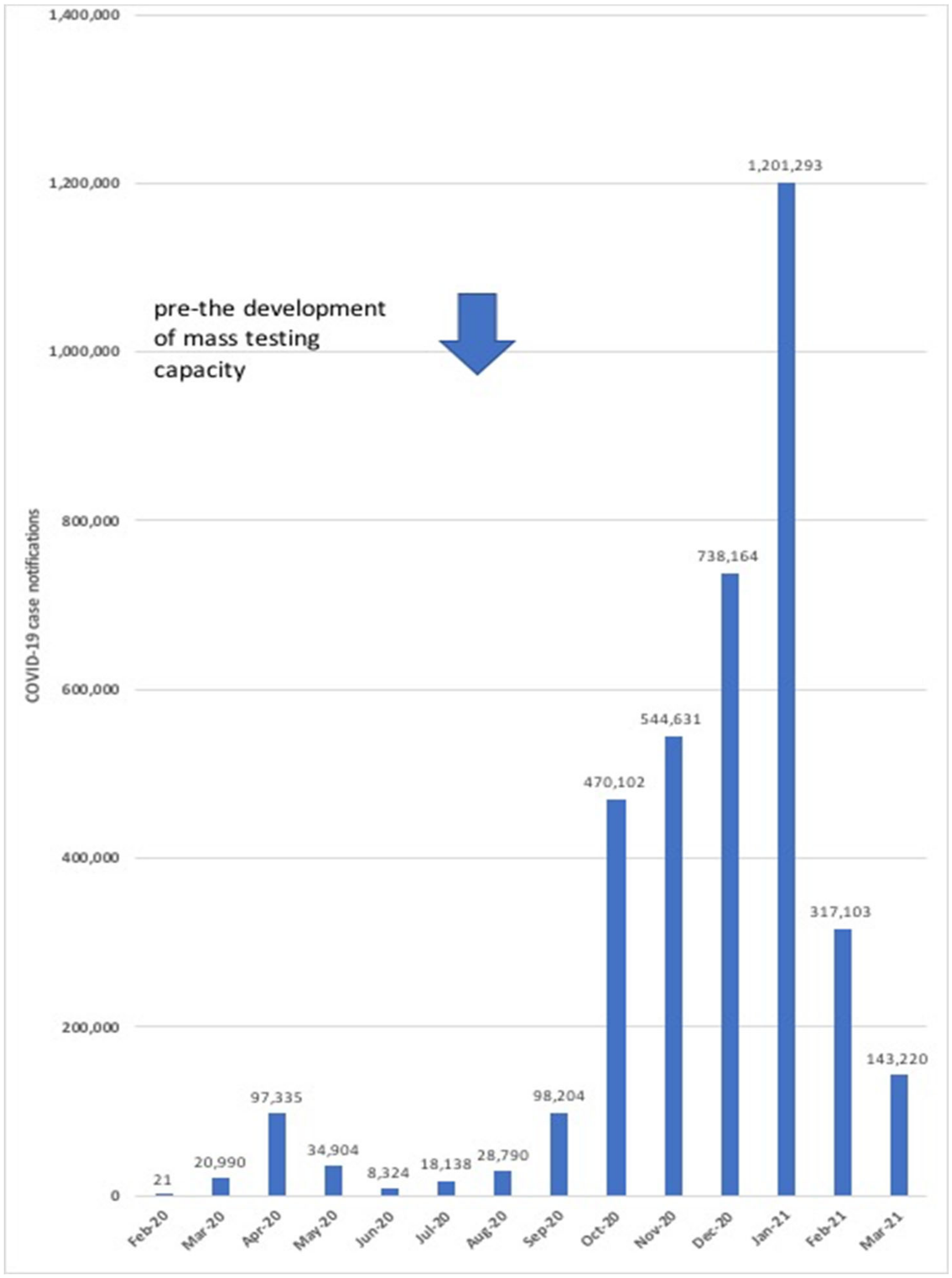
3. Evolution of the COVID-19 Epidemic and Testing Strategies in England
4. Virological Properties of SARS-CoV-2
4.1. SARS-CoV-2 Infection and the Immune Response
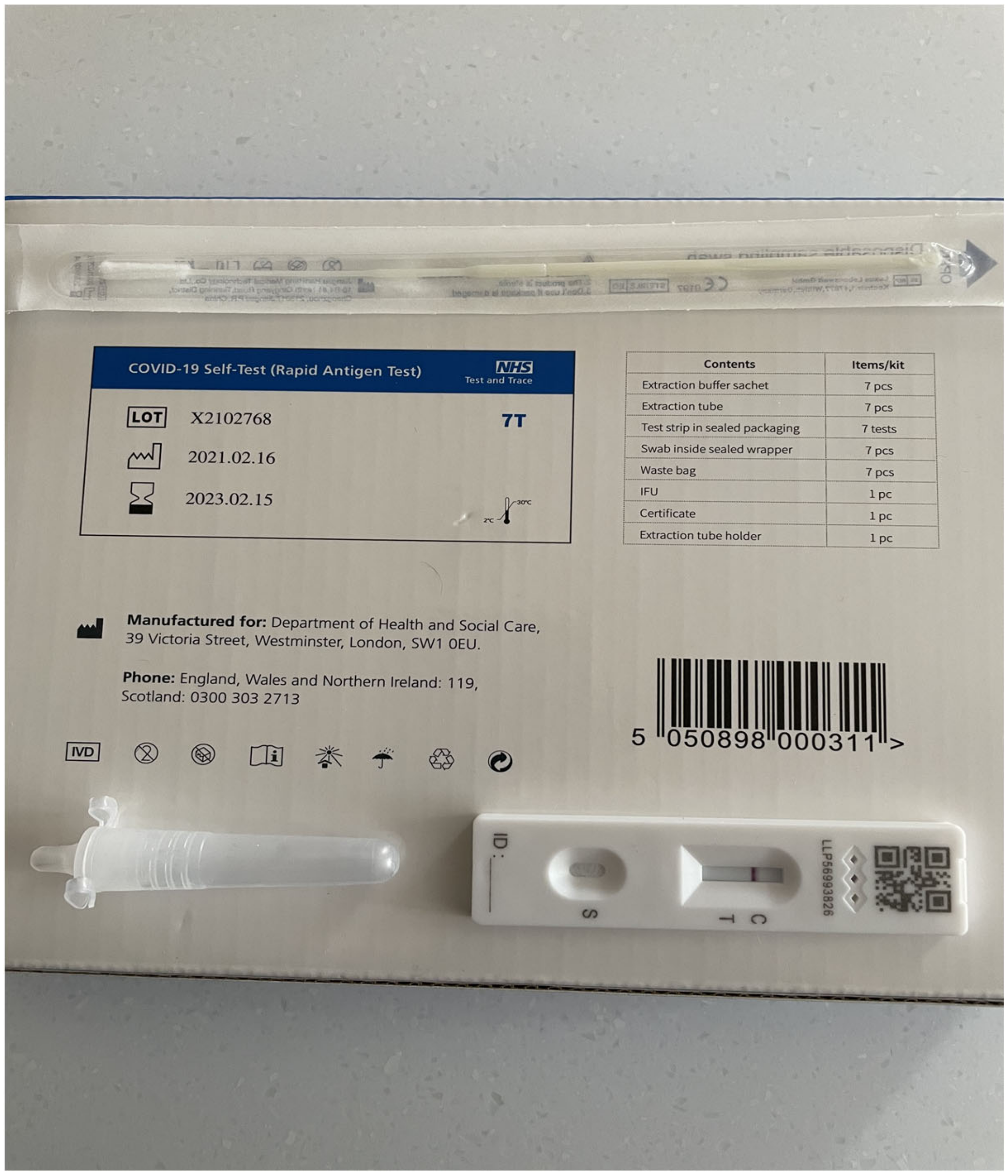
4.2. Diagnostic and Seroprevalence Testing for SARS-CoV-2 Infection
5. Population Antibody Screening for SARS-CoV-2 Infection Undertaken by the UK Government in England
6. The Challenges of SARS-CoV-2 Population Antibody Testing
7. Final Comments
Funding
Data Availability Statement
Conflicts of Interest
References
- Li, Q.; Guan, X.; Wu, P.; Wang, X.; Zhou, L.; Tong, Y.; Ren, R.; Leung, K.S.M.; Lau, E.H.Y.; Wong, J.Y.; et al. Early transmission dynamics in Wuhan, China, of Novel Coronavirus—Infected pneumonia. N. Engl. J. Med. 2020, 382, 1199–1207. [Google Scholar] [CrossRef] [PubMed]
- Zhu, N.; Zhang, D.; Wang, W.; Li, X.; Yang, B.; Song, J.; Zhao, X.; Huang, B.; Shi, W.; Lu, R.; et al. A Novel Coronavirus from Patients with Pneumonia in China, 2019. N. Engl. J. Med. 2020, 382, 727–733. [Google Scholar] [CrossRef] [PubMed]
- Coronaviridae Study Group of the International Committee on Taxonomy of Viruses. The species Severe acute respiratory syndrome-related coronavirus: Classifying 2019-nCoV and naming it SARS-CoV-2. Nat. Microbiol. 2020, 5, 536–544. [Google Scholar] [CrossRef]
- Shultz, J.M.; Perlin, A.; Saltzman, R.G.; Espinel, Z.; Galea, S. Pandemic March: 2019 Coronavirus Disease’s First Wave Circumnavigates the Globe. Disaster Med. Public Heal. Prep. 2020, 14, e28–e32. [Google Scholar] [CrossRef] [PubMed]
- Docherty, A.B.; Harrison, E.M.; Green, C.A.; Hardwick, H.E.; Pius, R.; Norman, L.; Holden, K.A.; Read, J.M.; Dondelinger, F.; Carson, G.; et al. Features of 20 133 UK patients in hospital with covid-19 using the ISARIC WHO Clinical Characterisation Protocol: Prospective observational cohort study. BMJ 2020, 369, m1985. [Google Scholar] [CrossRef]
- Lillie, P.J.; Samson, A.; Li, A.; Adams, K.; Capstick, R.; Barlow, G.D.; Easom, N.; Hamilton, E.; Moss, P.J.; Evans, A.; et al. Novel coronavirus disease (Covid-19): The first two patients in the UK with person to person transmission. J. Infect. 2020, 80, 578–606. [Google Scholar] [CrossRef]
- GOV.UK Coronavirus (COVID-19) in the UK. The official UK Government website for data and insights on Coronavirus (COVID-19). Available online: https://coronavirus.data.gov.uk/details/ (accessed on 2 April 2021).
- Hanson, K.E.; Caliendo, A.M.; Arias, C.A.; Englund, J.A.; Lee, M.J.; Loeb, M.; Patel, R.; El Alayli, A.; Kalot, M.A.; Falck-Ytter, Y.; et al. Infectious Diseases Society of America Guidelines on the Diagnosis of COVID-19. Clin. Infect. Dis. 2020, 16, 760. [Google Scholar] [CrossRef] [PubMed]
- Machado, B.A.S.; Hodel, K.V.S.; Barbosa-Júnior, V.G.; Soares, M.B.P.; Badaró, R. The Main Molecular and Serological Methods for Diagnosing COVID-19: An Overview Based on the Literature. Viruses 2020, 13, 40. [Google Scholar] [CrossRef]
- Garneret, P.; Coz, E.; Martin, E.; Manuguerra, J.C.; Brient-Litzler, E.; Enouf, V.; Obando, D.F.G.; Olivo-Marin, J.C.; Monti, F.; van der Werf, S.; et al. Performing point-of-care molecular testing for SARS-CoV-2 with RNA extraction and isothermal amplification. PLoS ONE 2021, 16, e0243712. [Google Scholar] [CrossRef]
- Mak, G.C.; Lau, S.S.; Wong, K.K.; Chow, N.L.; Lau, C.; Lam, E.T.; Chan, R.C.; Tsang, D.N. Evaluation of rapid antigen detection kit from the WHO Emergency Use List for detecting SARS-CoV-2. J. Clin. Virol. 2021, 134, 104712. [Google Scholar] [CrossRef]
- Toptan, T.; Eckermann, L.; Pfeiffer, A.E.; Hoehl, S.; Ciesek, S.; Drosten, C.; Corman, V.M. Evaluation of a SARS-CoV-2 rapid antigen test: Potential to help reduce community spread? J. Clin. Virol. 2021, 135, 104713. [Google Scholar] [CrossRef]
- Mak, G.C.; Lau, S.S.; Wong, K.K.; Chow, N.L.; Lau, C.; Lam, E.T.; Chan, R.C.; Tsang, D.N. Analytical sensitivity and clinical sensitivity of the three rapid antigen detection kits for detection of SARS-CoV-2 virus. J. Clin. Virol. 2020, 133, 104684. [Google Scholar] [CrossRef]
- Kohmer, N.; Toptan, T.; Pallas, C.; Karaca, O.; Pfeiffer, A.; Westhaus, S.; Widera, M.; Berger, A.; Hoehl, S.; Kammel, M.; et al. The Comparative Clinical Performance of Four SARS-CoV-2 Rapid Antigen Tests and Their Correlation to Infectivity In Vitro. J. Clin. Med. 2021, 10, 328. [Google Scholar] [CrossRef]
- Chen, P.F.; Yu, X.X.; Liu, Y.P.; Ren, D.; Shen, M.; Huang, B.S.; Gao, J.L.; Huang, Z.Y.; Wu, M.; Wang, W.-Y.; et al. Virus load and virus shedding of SARS-CoV-2 and their impact on patient outcomes. World J. Clin. Cases 2020, 8, 6252–6263. [Google Scholar] [CrossRef]
- Zhou, R.; Li, F.; Chen, F.; Liu, H.; Zheng, J.; Lei, C.; Wu, X. Viral dynamics in asymptomatic patients with COVID-19. Int. J. Infect. Dis. 2020, 96, 288–290. [Google Scholar] [CrossRef]
- Zhang, X.; Lu, S.; Li, H.; Wang, Y.; Lu, Z.; Liu, Z.; Lai, Q.; Ji, Y.; Huang, X.; Li, Y.; et al. Viral and antibody kinetics of COVID-19 patients with different disease severities in acute and convalescent phases: A 6-month follow-up study. Virol. Sin. 2020, 35, 820–829. [Google Scholar] [CrossRef]
- Iacobucci, G. Covid-19: What is the UK’s testing strategy? BMJ 2020, 368, m1222. [Google Scholar] [CrossRef]
- Department of Health and Social Care. Coronavirus (COVID-19) Scaling up Our Testing Programmes. Policy Paper Published 4th April 2020. Available online: www.gov.uk/government/publications/coronavirus-covid-19-scaling-up-testing-programmes (accessed on 9 February 2021).
- Ogimi, C.; Kim, Y.J.; Martin, E.T.; Huh, H.J.; Chiu, C.H.; Englund, J.A. What’s new with the old coronaviruses? J. Pediatric. Infect. Dis. Soc. 2020, 9, 210–217. [Google Scholar] [CrossRef]
- Corman, V.M.; Muth, D.; Niemeyer, D.; Drosten, C. Hosts and Sources of Endemic Human Coronaviruses. Adv. Applied Microbiol. 2018, 100, 163–188. [Google Scholar] [CrossRef]
- Almeida, J.D.; Berry, D.M.; Cunningham, C.H.; Hamre, D.; Hofstad, M.S.; Mallucci, L.; McIntosh, K.; Tyrrrell, D.A. Coronaviruses. Nature 1968, 220, 650. [Google Scholar]
- Wu, A.; Peng, Y.; Huang, B.; Ding, X.; Wang, X.; Niu, P.; Meng, J.; Zhu, Z.; Zhang, Z.; Wang, J.; et al. Genome Composition and Divergence of the Novel Coronavirus (2019-nCoV) Originating in China. Cell Host Microbe 2020, 27, 325–328. [Google Scholar] [CrossRef]
- Shang, J.; Wan, Y.; Luo, C.; Ye, G.; Geng, Q.; Auerbach, A.; Li, F. Cell entry mechanisms of SARS-CoV-2. Proc. Natl. Acad. Sci. USA 2020, 117, 1127–1134. [Google Scholar] [CrossRef] [PubMed]
- Chen, Y.; Guo, Y.; Pan, Y.; Zhao, Z.J. Structure analysis of the receptor binding of 2019-nCoV. Biochem. Biophys. Res. Commun. 2020, 525, 135–140. [Google Scholar] [CrossRef]
- Xia, X. Domains and Functions of Spike Protein in Sars-Cov-2 in the Context of Vaccine Design. Viruses 2021, 13, 109. [Google Scholar] [CrossRef]
- Alipoor, S.D.; Mortaz, E.; Jamaati, H.; Tabarsi, P.; Bayram, H.; Varahram, M.; Adcock, I.M. COVID-19: Molecular and Cellular Response. Front. Cell. Infect. Microbiol. 2021, 11, 563085. [Google Scholar] [CrossRef]
- McBride, R.; Van Zyl, M.; Fielding, B.C. The Coronavirus Nucleocapsid Is a Multifunctional Protein. Viruses 2014, 6, 2991–3018. [Google Scholar] [CrossRef]
- Lu, S.; Ye, Q.; Singh, D.; Cao, Y.; Diedrich, J.K.; Yates, J.R.; Villa, E.; Cleveland, D.W.; Corbett, K.D. The SARS-CoV-2 nucleocapsid phosphoprotein forms mutually exclusive condensates with RNA and the membrane-associated M protein. Nat. Commun. 2021, 12, 1–15. [Google Scholar] [CrossRef]
- Lam, T.T.; Jia, N.; Zhang, Y.W.; Shum, M.H.; Jiang, J.F.; Zhu, H.C.; Tong, Y.G.; Shi, Y.X.; Ni, X.B.; Liao, Y.S.; et al. Identifying SARS-CoV-2-related coronaviruses in Malayan pangolins. Nature 2020, 583, 282–285. [Google Scholar] [CrossRef]
- Cui, J.; Li, F.; Shi, Z.-L. Origin and evolution of pathogenic coronaviruses. Nat. Rev. Microbiol. 2019, 17, 181–192. [Google Scholar] [CrossRef]
- Nakagawa, S.; Miyazawa, T. Genome evolution of SARS-CoV-2 and its virological characteristics. Inflamm. Regen. 2020, 40, 1–7. [Google Scholar] [CrossRef] [PubMed]
- Sallard, E.; Halloy, J.; Casane, D.; Decroly, E.; van Helden, J. Tracing the origins of SARS-COV-2 in coronavirus phylogenies: A review. Environ. Chem. Lett. 2021, 19, 769–785. [Google Scholar] [CrossRef] [PubMed]
- Van Dorp, L.; Acman, M.; Richard, D.; Shaw, L.P.; Ford, C.E.; Ormond, L.; Owen, C.J.; Pang, J.; Tan, C.C.; Boshier, F.A.; et al. Emergence of genomic diversity and recurrent mutations in SARS-CoV-2. Infect. Genet. Evol. 2020, 83, 104351. [Google Scholar] [CrossRef] [PubMed]
- Lauring, A.S.; Hodcroft, E.B. Genetic variants of SARS-CoV-2—what do they mean? JAMA 2021, 325, 529–531. [Google Scholar] [CrossRef] [PubMed]
- Grubaugh, N.D.; Hodcroft, E.B.; Fauver, J.R.; Phelan, A.L.; Cevik, M. Public health actions to control new SARS-CoV-2 variants. Cell 2021, 184, 1127–1132. [Google Scholar] [CrossRef]
- Zhou, X.; Li, Y.; Li, T.; Zhang, W. Follow-up of asymptomatic patients with SARS-CoV-2 infection. Clin. Microbiol. Infect. 2020, 26, 957–959. [Google Scholar] [CrossRef] [PubMed]
- Yang, X.; Yu, Y.; Shu, H.; Xia, J.; Liu, H.; Wu, Y.; Zhang, L.; Yu, Z.; Fang, M.; Yu, T.; et al. Clinical course and outcomes of critically ill patients with SARS-CoV-2 pneumonia in Wuhan, China: A single-centred, retrospective, observational study. Lancet Resp. Med. 2020, 8, 475–481. [Google Scholar] [CrossRef]
- Nagu, P.; Parashar, A.; Behl, T.; Mehta, V. CNS implications of COVID-19: A comprehensive review. Rev. Neurosci. 2021, 32, 219–234. [Google Scholar] [CrossRef] [PubMed]
- Long, Q.X.; Tang, X.J.; Shi, Q.L.; Li, Q.; Deng, H.J.; Yuan, J.; Hu, J.L.; Xu, W.; Zhang, Y.; Lv, F.J.; et al. Clinical and immunological assessment of asymptomatic SARS-CoV-2 infections. Nat. Med. 2020, 26, 1200–1204. [Google Scholar] [CrossRef]
- Blanco-Melo, D.; Nilsson-Payant, B.E.; Liu, W.C.; Uhl, S.; Hoagland, D.; Møller, R.; Jordan, T.X.; Oishi, K.; Panis, M.; Sachs, D.; et al. Imbalanced host response to SARS-CoV-2 drives development of COVID-19. Cell 2020, 181, 1036–1045. [Google Scholar] [CrossRef]
- Wang, X.; Gui, J. Cell-mediated immunity to SARS-CoV-2. Pediatr. Investig. 2020, 4, 281–291. [Google Scholar] [CrossRef]
- Sette, A.; Crotty, S. Adaptive immunity to SARS-CoV-2 and COVID-19. Cell 2021, 184, 861–880. [Google Scholar] [CrossRef] [PubMed]
- Galipeau, Y.; Greig, M.; Liu, G.; Driedger, M.; Langlois, M.-A. Humoral Responses and Serological Assays in SARS-CoV-2 Infections. Front. Immunol. 2020, 11, 610688. [Google Scholar] [CrossRef] [PubMed]
- Long, Q.X.; Liu, B.Z.; Deng, H.L.; Wu, G.C.; Deng, K.; Chen, Y.K.; Liao, P.; Qiu, J.F.; Lin, Y.; Cai, X.F.; et al. Antibody responses to SARS-CoV-2 in patients with COVID-19. Nat. Med. 2020, 26, 845–848. [Google Scholar] [CrossRef] [PubMed]
- Peng, Y.; Mentzer, A.J.; Liu, G.; Yao, X.; Yin, Z.; Dong, D.; Dejnirattsai, W.; Rostron, T.; Supasa, P.; Liu, C.; et al. Broad and strong memory CD4+ and CD8+ T cells induced by SARS-CoV-2 in UK convalescent individuals following COVID-19. Nat. Immunol. 2020, 21, 1336–1345. [Google Scholar] [CrossRef]
- Sherina, N.; Piralla, A.; Du, L.; Wan, H.; Kumagai-Braesch, M.; Andréll, J.; Braesch-Andersen, S.; Cassaniti, I.; Percivalle, E.; Sarasini, A.; et al. Persistence of SARS-CoV-2-specific B and T cell responses in convalescent COVID-19 patients 6–8 months after the infection. Med 2021, 2, 281–295.e4. [Google Scholar] [CrossRef] [PubMed]
- Sui, Y.; Bekele, Y.; Berzofsky, J.A. Potential SARS-CoV-2 immune correlates of protection in infection and vaccine immunization. Pathogens 2021, 10, 138. [Google Scholar] [CrossRef]
- Dutta, N.K.; Mazumdar, K.; Gordy, J.T. The Nucleocapsid Protein of SARS–CoV-2: A Target for Vaccine Development. J. Virol. 2020, 94, e00647-20. [Google Scholar] [CrossRef] [PubMed]
- Ju, B.; Zhang, Q.I.; Ge, J.; Wang, R.; Sun, J.; Ge, X.; Yu, J.; Shan, S.; Zhou, B.; Song, S.; et al. Human neutralizing antibodies elicited by SARS-CoV-2 infection. Nature 2020, 584, 115–119. [Google Scholar] [CrossRef]
- Theel, E.S.; Couturier, M.R.; Filkins, L.; Palavecino, E.; Mitchell, S.; Campbell, S.; Pentella, M.; Butler-Wu, S.; Jerke, K.; Dharmarha, V.; et al. Application, Verification, and Implementation of SARS-CoV-2 Serologic Assays with Emergency Use Authorization. J. Clin. Microbiol. 2020, 59, e02148-20. [Google Scholar] [CrossRef]
- Public Health England. COVID-19: Laboratory Evaluations of Serological Assays. Reports published 19th May 2020. Available online: www.gov.uk/government/publications/covid-19-laboratory-evaluations-of-serological-assays#history (accessed on 10 March 2021).
- Mulchandani, R.; Jones, H.E.; Taylor-Phillips, S.; Shute, J.; Perry, K.; Jamarani, S.; Brooks, T.; Charlett, A.; Hickman, M.; Oliver, I.; et al. Accuracy of UK rapid test consortium (UK-RTC) “AbC-19 rapid test” for detection of previous SARS-CoV-2 infection in key workers: Test accuracy study. BMJ 2020, 371, m4262. [Google Scholar] [CrossRef] [PubMed]
- Almahboub, S.A.; Algaissi, A.; AlFaleh, M.A.; ElAssouli, M.-Z.; Hashem, A.M. Evaluation of Neutralizing Antibodies Against Highly Pathogenic Coronaviruses: A Detailed Protocol for a Rapid Evaluation of Neutralizing Antibodies Using Vesicular Stomatitis Virus Pseudovirus-Based Assay. Front. Microbiol. 2020, 11. [Google Scholar] [CrossRef] [PubMed]
- Bahadir, E.B.; Sezgintürk, M.K. Lateral flow assays: Principles, designs and labels. Trends Analyt. Chem. 2016, 82, 286–306. [Google Scholar] [CrossRef]
- Maple, P.; Sikora, K. How Useful is COVID-19 Antibody Testing—A Current Assessment for Oncologists. Clin. Oncol. 2021, 33, e73–e81. [Google Scholar] [CrossRef]
- Atchison, C.; Pristerà, P.; Cooper, E.; Papageorgiou, V.; Redd, R.; Piggin, M.; Flower, B.; Fontana, G.; Satkunarajah, S.; Ashrafian, H.; et al. Usability and Acceptability of Home-based Self-testing for Severe Acute Respiratory Syndrome Coronavirus 2 (SARS-CoV-2) Antibodies for Population Surveillance. Clin. Infect. Dis. 2021, 72, e384–e393. [Google Scholar] [CrossRef] [PubMed]
- Moshe, M.; Daunt, A.; Flower, B.; Simmons, B.; Brown, J.C.; Frise, R.; Penn, R.; Kugathasan, R.; Petersen, C.; Stockmann, H.; et al. SARS-CoV-2 lateral flow assays for possible use in national covid-19 seroprevalence surveys (React 2): Diagnostic accuracy study. BMJ 2021, 372. [Google Scholar] [CrossRef]
- Whitman, J.D.; Hiatt, J.; Mowery, C.T.; Shy, B.R.; Yu, R.; Yamamoto, T.N.; Rathore, U.; Goldgof, G.M.; Whitty, C.; Woo, J.M.; et al. Evaluation of SARS-CoV-2 serology assays reveals a range of test performance. Nat. Biotechnol. 2020, 38, 1174–1183. [Google Scholar] [CrossRef] [PubMed]
- Venter, M.; Richter, K. Towards effective diagnostic assays for COVID-19: A review. J. Clin. Pathol. 2020, 73, 370–377. [Google Scholar] [CrossRef]
- Sethuraman, N.; Jeremiah, S.S.; Ryo, A. Interpreting diagnostic tests for SARS-CoV-2. JAMA 2020, 323, 2249–2251. [Google Scholar] [CrossRef] [PubMed]
- Manisty, C.; Treibel, T.A.; Jensen, M.; Semper, A.; Joy, G.; Gupta, R.K.; Cutino-Moguel, T.; Andiapen, M.; Jones, J.; Taylor, S.; et al. Time series analysis and mechanistic modelling of heterogeneity and sero-reversion in antibody responses to mild SARS-CoV-2 infection. EBioMedicine 2021, 65, 103259. [Google Scholar] [CrossRef]
- Seow, J.; Graham, C.; Merrick, B.; Acors, S.; Pickering, S.; Steel, K.J.A.; Hemmings, O.; O’Byrne, A.; Kouphou, N.; Galao, R.P.; et al. Longitudinal observation and decline of neutralizing antibody responses in the three months following SARS-CoV-2 infection in humans. Nat. Microbiol. 2020, 5, 1598–1607. [Google Scholar] [CrossRef]
- Dan, J.M.; Mateus, J.; Kato, Y.; Hastie, K.M.; Yu, E.D.; Faliti, C.E.; Grifoni, A.; Ramirez, S.I.; Haupt, S.; Frazier, A.; et al. Immunological memory to SARS-CoV-2 assessed for up to 8 months after infection. Science 2021, 371. [Google Scholar] [CrossRef]
- Hartog, G.D.; A Vos, E.R.; Hoogen, L.L.V.D.; van Boven, M.; Schepp, R.M.; Smits, G.; van Vliet, J.; Woudstra, L.; Wijmenga-Monsuur, A.J.; E van Hagen, C.C.; et al. Persistence of antibodies to SARS-CoV-2 in relation to symptoms in a nationwide prospective study. Clin. Infect. Dis. 2021. [Google Scholar] [CrossRef]
- Pradenas, E.; Trinité, B.; Urrea, V.; Marfil, S.; Ávila-Nieto, C.; de la Concepción, M.L.R.; Tarrés-Freixas, F.; Pérez-Yanes, S.; Rovirosa, C.; Ainsua-Enrich, E.; et al. Stable neutralizing antibody levels 6 months after mild and severe COVID-19 episodes. Med 2021, 2, 313–320.e4. [Google Scholar] [CrossRef]
- Department of Health and Social Care. COVID-19 Surveillance and Immunity Studies. Available online: https://www.gov.uk/government/publications/covid-19-surveillance (accessed on 26 March 2021).
- Offices of the Nuffield Professor of Medicine, University of Oxford. COVID-19 Infection Survey. Available online: https://www.ndm.ox.ac.uk/covid-19/covid-19-infection-survey (accessed on 26 March 2021).
- National SARS-CoV-2 Serology Assay Evaluation Group. Performance characteristics of five immunoassays for SARS-CoV-2: A head-to-head benchmark comparison. Lancet Infect. Dis. 2020, 20, 1390–1400. [Google Scholar] [CrossRef]
- Office for National Statistics. Coronavirus (COVID-19) Infection Survey, Antibody Data for the UK: 16 March 2021. Available online: https://www.ons.gov.uk/peoplepopulationandcommunity/healthandsocialcare/conditionsanddiseases/articles/coronaviruscovid19infectionsurveyantibodydatafortheuk/16march2021 (accessed on 26 March 2021).
- Ward, H.; Atchison, C.; Whitaker, M.; Ainslie, K.E.C.; Elliott, J.; Okell, L.; Redd, R.; Ashby, D.; Donnelly, C.A.; Barclay, W.; et al. SARS-CoV-2 antibody prevalence in England following the first peak of the pandemic. Nat. Commun. 2021, 12, 1–8. [Google Scholar] [CrossRef] [PubMed]
- Ward, H.; Cooke, G.; Whitaker, M.; Redd, R.; Eales, O.; Brown, J.C.; Collet, K.; Cooper, E.; Daunt, A.; Jones, K.; et al. REACT-2 Round 5: Increasing prevalence of SARS-CoV-2 antibodies demonstrate impact of the second wave and of vaccine roll-out in England. MedRxiv 2021. [Google Scholar] [CrossRef]
- Wallace, S.; Hall, V.; Charlett, A.; Kirwan, P.D.; Cole, M.J.; Shroti, M.; Rokadiya, S.; Oguti, B.; Vusurikaia, A.; Zambon, M.; et al. SIREN protocol: Impact of detectable anti-SARS-CoV-2 on the subsequent incidence of COVID-19 in 100,000 healthcare workers: Do antibody positive healthcare workers have less reinfection than antibody negative healthcare workers? MedRxiv 2021. [Google Scholar] [CrossRef]
- Hall, V.J.; Foulkes, S.; Charlett, A.; Atti, A.; Monk, E.J.M.; Simmons, R.; Wellington, E.; Cole, M.J.; Saei, A.; Oguti, B.; et al. SARS-CoV-2 infection rates of antibody-positive compared with antibody-negative health-care workers in England: A large, multicentre, prospective cohort study (SIREN). Lancet 2021, 397, 1459–1469. [Google Scholar] [CrossRef]
- Krutikov, M.; Palmer, T.; Donaldson, A.; Lorencatto, F.; Forbes, G.; Copas, A.; Robson, J.; Hopkins, S.; Moss, P.; Farrar, J.; et al. Study Protocol: Understanding SARS-Cov-2 infection, immunity and its duration in care home residents and staff in England (VIVALDI). Wellcome Open Res. 2021, 5, 232. [Google Scholar] [CrossRef] [PubMed]
- Krutikov, M.; Palmer, T.; Tut, G.; Fuller, C.; Shroti, M.; Williams, H.; Davies, D.; Irwin-Singer, A.; Robson, J.; Hayward, A.; et al. Incidence of SARS-CoV-2 infection according to baseline antibody status in staff and residents of 100 long term care facilities (VIVALDI study). MedRxiv 2021. [Google Scholar] [CrossRef]
- Office for National Statistics. COVID-19 Schools Infection Survey: Methods and Further Information. Available online: https://www.ons.gov.uk/peoplepopulationandcommunity/healthandsocialcare/conditionsanddiseases/methodologies/covid19schoolsinfectionsurveymethodsandfurtherinformation (accessed on 1 April 2021).
- Office for National Statistics. COVID-19 Schools Infection Survey Round 2, England: December 2020. Available online: https://www.ons.gov.uk/peoplepopulationandcommunity/healthandsocialcare/conditionsanddiseases/bulletins/covid19schoolsinfectionsurveyround2england/december2020 (accessed on 1 April 2021).
- Department of Health and Social Care. Protective Immunity from T Cells in Healthcare Workers. Available online: http://www.pitch-study.org/index.html (accessed on 2 April 2021).
- Angyal, A.; Longet, S.; Moore, S.; Payne, R.P.; Harding, A.; Tipton, T.; Rongkard, P.; Ali, M.; Hering, L.M.; Meardon, N.; et al. T-cell and antibody responses to first BNT 162b2 vaccine dose in previously SARS-CoV-2 infected and infection- naïve UK healthcare workers: A multicentre, prospective, observational cohort study. Available online: https://ssrn.com/abstract=3812375 (accessed on 2 April 2021).
- Osborne, K.; Gay, N.; Hesketh, L.; Morgan-Capner, P.; Miller, E. Ten years of serological surveillance in England and Wales: Methods, results, implications and action. Int. J. Epidemiol. 2000, 29, 362–368. [Google Scholar] [CrossRef]
- Maple, P.; Jones, C.S.; Wall, E.; Vyse, A.; Edmunds, W.; Andrews, N.J.; Miller, E. Immunity to diphtheria and tetanus in England and Wales. Vaccine 2000, 19, 167–173. [Google Scholar] [CrossRef]
- Nardone, A.; Pebody, R.; Maple, P.; Andrews, N.; Gay, N.; Miller, E. Sero-epidemiology of Bordetella pertussis in England and Wales. Vaccine 2004, 22, 1314–1319. [Google Scholar] [CrossRef]
- Vyse, A.J.; Gay, N.J.; Hesketh, L.M.; Pebody, R.; Morgan-Capner, P.; Miller, E. Interpreting serological surveys using mixture models: The seroepidemiology of measles, mumps and rubella in England and Wales at the beginning of the 21st century. Epidemiol. Infect. 2006, 134, 1303–1312. [Google Scholar] [CrossRef]
- Balogun, M.; Ramsay, M.; Hesketh, L.; Andrews, N.; Osborne, K.; Gay, N.; Morgan-Capner, P. The Prevalence of Hepatitis C in England and Wales. J. Infect. 2002, 45, 219–226. [Google Scholar] [CrossRef][Green Version]
- Bundle, N.; Balasegaram, S.; Parry, S.; Ullah, S.; Harris, R.J.; Ahmad, K.; Foster, G.R.; Tong, C.Y.; Orkin, C. Seroprevalence and demographic factors associated with hepatitis B, hepatitis C and HIV infection from a hospital emergency department testing programme, London, United Kingdom, 2015 to 2016. Eurosurveillance 2019, 24, 1800377. [Google Scholar] [CrossRef] [PubMed]
- Harris, R.J.; Whitaker, H.J.; Andrews, N.J.; Aiano, F.; Amin-Chowdhury, Z.; Flood, J.; Borrow, R.; Linley, E.; Ahmad, S.; Stapley, L.; et al. Serological surveillance of SARS-CoV-2: Six-month trends and antibody response in a cohort of public health workers. J. Infect. 2021, 82, 162–169. [Google Scholar] [CrossRef]
- Groves, D.C.; Rowland-Jones, S.L.; Angyal, A. The D614G mutations in the SARS-CoV-2 spike protein: Implications for viral infectivity, disease severity and vaccine design. Biochem. Biophys. Res. Commun. 2021, 538, 104–107. [Google Scholar] [CrossRef]
- Padma, T.V. Indian government should heed its scientists on COVID. Nat. Cell Biol. 2021, 593, 9. [Google Scholar] [CrossRef]
- Prévost, J.; Finzi, A. The great escape? SARS-CoV-2 variants evading neutralizing responses. Cell Host Microbe 2021, 29, 322–324. [Google Scholar] [CrossRef] [PubMed]
- Wang, Z.; Schmidt, F.; Weisblum, Y.; Muecksch, F.; Barnes, C.O.; Finkin, S.; Schaefer-Babajew, D.; Cipolla, M.; Gaebler, C.; Lieberman, J.A.; et al. mRNA vaccine-elicited antibodies to SARS-CoV-2 and circulating variants. Nature 2021, 592, 616–622. [Google Scholar] [CrossRef]
- Gale, C.; Knight, M.; Ladhani, S.; Draper, E.S.; Sharkey, D.; Doherty, C.; Mactier, H.; Kurinzcuk, J.J. National active surveillance to understand and inform neonatal care in COVID-19. Arch. Dis. Child Fetal. Neonatal. Ed. 2020, 105, 346–347. [Google Scholar] [CrossRef]
- Ladhani, S.N.; Baawuah, F.; Beckmann, J.; Okike, I.O.; Ahmad, S.; Garstang, J.; Brent, A.J.; Brent, B.; Walker, J.; Andrews, N.; et al. SARS-CoV-2 infection and transmission in primary schools in England in June-December 2020 (sKIDs): An active, prospective surveillance study. Lancet Child Adolesc. Health 2021. [Google Scholar] [CrossRef]
- Ismail, S.A.; Saliba, V.; Bernal, J.L.; Ramsay, M.E.; Ladhani, S.N. SARS-CoV-2 infection and transmission in educational settings: A prospective, cross-sectional analysis of infection clusters and outbreaks in England. Lancet Infect Dis. 2021, 21, 344–353. [Google Scholar] [CrossRef]
- Public Health England. COVID-19: Paediatric Surveillance. Available online: https://www.gov.uk/guidance/covid-19-paediatric-surveillance (accessed on 10 May 2021).
- Bian, L.; Gao, F.; Zhang, J.; He, Q.; Mao, Q.; Xu, M.; Liang, Z. Effects of SARS-CoV-2 variants on vaccine efficacy and response strategies. Expert Rev. Vaccines 2021, 14, 1–9. [Google Scholar] [CrossRef] [PubMed]
- Kumar, A.; Dowling, W.E.; Román, R.G.; Chaudhari, A.; Gurry, C.; Le, T.T.; Tollefson, S.; E Clark, C.; Bernasconi, V.; A Kristiansen, P. Status Report on COVID-19 Vaccines Development. Curr. Infect. Dis. Rep. 2021, 23, 1–12. [Google Scholar] [CrossRef]
- Dropkin, G. COVID-19 UK Lockdown Forecasts and R0. Front Public Health 2020, 8, 256. [Google Scholar] [CrossRef]


| Testing Programme | Target | Delivery Mechanism |
|---|---|---|
| Pillar one | Scaling up National Health Service virus swab testing for those with a medical need and, where possible, the most critical key workers. | Testing to be performed by National Health Service and Public Health England laboratories. |
| Pillar two | Mass-virus swab testing for critical key workers in the National Health Service, social care and other sectors. | Commercial/private partnerships initiated to deliver a mass testing capability. |
| Pillar three | Mass antibody testing to help determine if people have immunity to coronavirus. | Once satisfactory commercial tests identified proposed home testing using finger prick samples. |
| Pillar four | Surveillance testing to learn more about the disease and help develop new tests and treatments. | National surveillance programmes for population blood testing, using high accuracy antibody testing delivered by designated laboratories. |
| Pillar five | Spearheading a diagnostics national effort to build a mass-testing capacity at a completely new scale. | Working with commercial/industrial partners to deliver high volume testing capacity as part of a resilient, diagnostic capability. |
| Commercial Assay | Samples Tested * | Sensitivity (95% CI) | Specificity (95% CI) |
|---|---|---|---|
| Beckman Coulter Access Anti-SARS-CoV-2 IgG assay | 100 pos, 499 neg | 76.5% (66.0–85.0) | 99.3% (97.8–99.8) |
| Siemens Atellica-IM SARS-CoV-2 Total (COV2T) assay | 100 pos, 499 neg | 89.4% (80.8–95.0) | 100% (99.1–100) |
| Ortho Clinical Diagnostics Vitros Anti-SARS -CoV-2 assay | 100 pos, 491 neg | 91.8% (83.8–96.6) | 99.5% (98.2–99.9) |
| DiaSorin LIAISON SARS-CoV-2 S1/S2 IgG assay | 100 pos, 472 neg | 69.4% (58.5–79.0) | 97.7% (95.8–99.0) |
| Euroimmun Anti-SARS-CoV-2 ELISA (IgG) assay | 93 pos, 499 neg | 73.4% (62.3–82.7) | 99.0% (97.5–99.7) |
| Abbott SARS-CoV-2 IgG assay | 96 pos, 760 neg | 93.9% (86.3–98.0) | 100% (99.1–100) |
| Roche Elecsys Anti-SARS-CoV-2 EIA | 93 pos, 472 neg | 86.1% (76.5–92.8) | 100% (99.1–100) |
| Siemens Atellica-IM SARS-CoV-2 (sCOVG) assay | 115 pos, 500 neg | 72.5% (61.4–81.9) | 100% (98.9–100) |
| Study Protocol | Assay Used/Samples Tested | Observations |
|---|---|---|
| Office for National Statistics COVID-19 Infection Survey. Commenced April 2020 [68]. | Commercial and in-house assays used [69]. Target of testing 150,000 people in two-week periods from October 2020. | In England, as of March 2021, it is estimated that 34.6% (95%CI: 34.0–35.3) of individuals have antibodies against SARS-CoV-2 [70]. |
| Imperial College. Real-time assessment of community transmission (REACT) study—2. Commenced May 2020 [71]. | Lateral flow assays for home—based testing [58]. Approximately 150,000 volunteers tested every six weeks. | In round 5 [72] conducted between 26 January 2021 and 8 February 2021, sera were tested from 155,172 people and the overall SARS-CoV-2 seroprevalence was 13.9%. |
| Public Health England. Sarscov2 immunity and Reinfection Evaluation (SIREN) study [73]. | Commercial and in-house assays used. Target of recruiting 100,000 healthcare workers with samples collected over a 12-month period. | A prior history of SARS-CoV-2 infection was associated with an 84% lower risk of reinfection [74]. |
| COVID-19 surveillance study in care homes (Vivaldi 1 and Vivaldi 2)—University College London and University of Birmingham and others [75]. | Abbott ARCHITECT SARS-CoV-2 IgG (nucleoprotein) and MSD V-Plex COVID-19 assay (76). Staff and residents of 105 care homes. Target 5000 residents and 6500 staff. | In a sample of 682 residents and 1429 staff, the presence of IgG antibodies to nucleocapsid was associated with a substantially reduced risk of reinfection for up to 10 months following primary infection [76]. |
| Schools Infection Survey -the London School of Hygiene and Tropical Medicine, Public Health England, and the Office for National Statistics | Finger prick and oral fluid samples tested for antibodies using commercial (Roche cobas Elecsys anti-SARS-CoV-2 assay) and in-house assays [77]. | In the second round of testing, 7089 pupils and 5114 staff in 121 schools were tested. Approximately, 15% of staff were SARS-CoV-2 antibody positive [78]. |
| Protective immunity from T-cells in healthcare workers (PITCH)—Several UK universities [79]. | Peripheral blood mononuclear cells tested by IFNgamma ELIspot assay. Target of recruiting 2000 healthcare workers. | In a study of 237 healthcare workers following a single dose of the Pfizer BN2162b2 vaccine T cell responses were six-fold higher in vaccine recipients at 28 days post vaccination compared to infection naïve participants [80]. |
Publisher’s Note: MDPI stays neutral with regard to jurisdictional claims in published maps and institutional affiliations. |
© 2021 by the author. Licensee MDPI, Basel, Switzerland. This article is an open access article distributed under the terms and conditions of the Creative Commons Attribution (CC BY) license (https://creativecommons.org/licenses/by/4.0/).
Share and Cite
Maple, P.A.C. Population (Antibody) Testing for COVID-19—Technical Challenges, Application and Relevance, an English Perspective. Vaccines 2021, 9, 550. https://doi.org/10.3390/vaccines9060550
Maple PAC. Population (Antibody) Testing for COVID-19—Technical Challenges, Application and Relevance, an English Perspective. Vaccines. 2021; 9(6):550. https://doi.org/10.3390/vaccines9060550
Chicago/Turabian StyleMaple, Peter A. C. 2021. "Population (Antibody) Testing for COVID-19—Technical Challenges, Application and Relevance, an English Perspective" Vaccines 9, no. 6: 550. https://doi.org/10.3390/vaccines9060550
APA StyleMaple, P. A. C. (2021). Population (Antibody) Testing for COVID-19—Technical Challenges, Application and Relevance, an English Perspective. Vaccines, 9(6), 550. https://doi.org/10.3390/vaccines9060550