Communication

Antibody Response to a Live-Modified Virus Vaccine against Bovine Viral Diarrhoea in Dairy Cattle in a Field Trial

by
Małgorzata D. Klimowicz-Bodys
1,
Katarzyna Płoneczka-Janeczko
1,
Michał Czopowicz
2,
Mirosław Paweł Polak
3,
Agnieszka Lachowicz-Wolak
1 and
Krzysztof Rypuła
1,*
1
Division of Infectious Diseases of Animals and Veterinary Administration, Department of Epizootiology and Clinic of Birds and Exotic Animals, Faculty of Veterinary Medicine, Wroclaw University of Environmental and Life Sciences, pl. Grunwaldzki 45, 50-366 Wroclaw, Poland
2
Division of Veterinary Epidemiology and Economics, Institute of Veterinary Medicine, Warsaw University of Life Sciences–SGGW, ul. Nowoursynowska 159c, 02-776 Warsaw, Poland
3
Department of Virology, National Veterinary Research Institute, al. Partyzantów 57, 24-100 Puławy, Poland
*
Author to whom correspondence should be addressed.
Vaccines 2021, 9(3), 259; https://doi.org/10.3390/vaccines9030259
Submission received: 24 February 2021 / Revised: 11 March 2021 / Accepted: 11 March 2021 / Published: 15 March 2021
(This article belongs to the Special Issue Bovine Viral Diarrhea/Mucosal Disease)

Abstract

:
(1) Background: The objective of the study was to evaluate the long-term antibody response of dairy cows to a single dose of a commercial modified-live virus (MLV) vaccine against bovine viral diarrhea (Mucosiffa® CEVA Sante Animale, Liburne, France). (2) Methods: The study was carried out in a dairy cattle herd counting 290 animals negative for bovine viral diarrhoea virus (BVDV). The vaccination was implemented following the manufacturer’s instructions. Twelve dairy cows were randomly selected before the study, and blood samples were collected right before the vaccination and then 12 times at 1-month intervals. The serum samples were screened using a virus neutralization test (VNT) and ELISA. (3) Results: Both tests showed that antibody titers increased significantly in all animals within the first month post-vaccination, and continued to increase significantly until the second (VNT) and third (ELISA) month post-vaccination. Antibody titers remained high and stable until the end of the study. Moreover, cows did not show any adverse reactions or clinical symptoms of the disease. (4) Conclusion: The results of this study indicated that the administration of one dose MLV vaccine was able to stimulate long-lasting (12-months) and strong antibody response in all vaccinated cows.

1. Introduction

Bovine viral diarrhea virus 1 (BVDV-1) and bovine viral diarrhea virus 2 (BVDV-2), (according to the new taxonomic classification referred to as Pestivitus A and Pestivius B, respectively) are small single-stranded positive-sense RNA viruses, classified in the Pestivirus genus and the Flaviviridae family [1]. These viruses cause an infectious and contagious disease in cattle and wild ruminants, bovine viral diarrhea (BVD), characterized by various clinical signs relating to the gastrointestinal and respiratory systems. Viral infection induces immunosuppression and reproductive disorders as well. BVD causes large economic losses due to forced culling, morbidity and mortality [2,3]. The impact of BVDV depends on its virulence, the time of infection, herd immunity, level of disease prevalence, herd production level and concomitant infections [4,5].
BVDV-1 is endemic in many regions of the world including Poland [6,7]. BVDV infection has been found in 30–70% of cattle dairy herds [8,9] with a high percentage of BVDV-positive animals, especially in large dairy herds [10] in Poland. The vaccination with a modified-live virus (MLV) is currently a well-accepted procedure in the BVD control programs in most of the European countries and is considered a complementary biosecurity tool in countries with high BVD prevalence, in order to prevent infection and re-infections in cattle herds [11].
The vaccination has provided protection against BVDV that has been effective from a herd perspective, reduced clinical signs of disease and economic losses [4,5] although vaccines could have limited durations of immunity [12]. The value of vaccination is that it increases herd immunity so that the incidence of BVDV infection and the percentage of calves born as persistently infected (PI) are reduced in the herd as a whole [13]. For many years, the vaccination with MLV vaccines has been limited in pregnant cows [14], but now several MLV vaccines against BVD are approved for use during pregnancy and lactation, one of them being Mucosiffa® (CEVA Sante Animale, Liburne, France). However, detailed data on the long-term antibody response to a single vaccination with a commercial MLV vaccine are scarce.
This study aimed to evaluate the long-term antibody response of dairy cows to a single dose of a commercial modified-life virus (MLV) vaccine against BVD.

2. Materials and Methods

2.1. Animals

The study was carried out on a dairy farm in Greater Poland voivodeship, between February 2018 and May 2019. The herd counted 290 heads. Infections with rotavirus, coronavirus and Cryptosporidium parvum in calves and Streptococcus uberis in adult cows had been previously detected in the herd. All animals were free from bovine herpesvirus type 1 (BHV-1). This herd had been involved in a voluntary BVD control program and had all of their animals previously tested for BVDV. All animals were analyzed with two commercial ELISAs: for BVDV antibody detection—IDEXX BVDV Total Ab Test (Scandinavia AB, Sweden) and for BVDV antigen detection—IDEXX BVDV Ag/Serum Plus ( Switzerland GmbH, Switzerland). The samples were tested individually. All tests were performed at the Diagnostic Laboratory EPI-VET of the Faculty of Veterinary Medicine, Wroclaw, Poland. The cattle were inseminated within 4–6 weeks after vaccination. Cows and their offspring were monitored after vaccination. The study was field research. No animals were introduced into the herd during the study.

2.2. Vaccination

The monovalent commercial vaccine Mucosiffa® (CEVA Sante Animale, Liburne, France) containing live-attenuated, cytopathic BVDV C24V strain, subgenotype 1a at a minimum titer of 103.5 TCID50 and maximum titer of 106 TCID50 per 2 ml dose was used. The vaccination was started in March 2018. All animals were vaccinated following the manufacturer’s instructions.

2.3. Samples Collection

Twelve adult cows (1st, 2nd and 3rd lactation) from this herd were randomly selected for the study using a simple random method. This sample size was chosen to ensure at least 95% certainty (level of confidence) that at least one seronegative cow would have been detected if less than 75% of the vaccinated cows had seroconverted after vaccination. The following formula was used for the calculation of the sample size [15]:
n = ( 1 ( 1 P ) 1 / d ) × ( N d 2 ) + 1
where:
  • n—required sample size
  • N—herd size (290)
  • d—minimum expected number of animals that did not seroconvert after vaccination (25%)
  • P—the probability of finding at least one seronegative animal in the sample (i.e., a level of confidence = 95%)
Blood samples were taken from the jugular vein right before the vaccination (day 0) and then 12 times at one-month intervals. Blood was left at room temperature for 8–12 h after collection and centrifuged. Then, serum samples were frozen at −80 °C and transported directly to the Diagnostic Laboratory of Department of Virology of National Veterinary Research Institute, Pulawy, Poland, for virus neutralization test (VNT) and ELISA.

2.4. ELISA

Serum samples were tested with Erns Ab ELISA (BVDV Total Ab Test, IDEXX, Switzerland). Samples with sample-to-positive control ratio (S/P%) values higher than or equal to 30% were classified as positive, those with values less than 20% were considered negative and samples with values ranging from 20% to 29% were considered inconclusive. ELISA was performed according to the manufacturer’s manual at an absorbance of 450 nm wavelength. The test has been shown to be 96.7% sensitive and 97.1% specific compared with VNT [16].

2.5. Virus Neutralization Test (VNT)

VNT was done in two-fold serial dilutions (from 1:5 to 1:640) of serum samples from all the cows. All serum samples were tested for neutralizing antibodies against cytopathic (cp) BVDV-1a strain Singer used at the concentration of 100 TCID50. Viral controls included 100, 10, 1 and 0.1 TCID50. The cell culture control was Madin–Darby Bovine Kidney (MDBK) without BVDV infection. The antibody titer was read after 4 days of incubation at 37 °C and 5% CO2 atmosphere and expressed as the reciprocal of the highest dilution inhibiting cytopathic effect in 50% of dilution wells. The titer was presented as a binary logarithm (log2) value.

2.6. Statistical Analysis

Antibody titers were presented as the median, interquartile range (IQR) and range, and compared between consecutive months using the Friedman test and Wilcoxon signed-rank test with Bonferroni correction for familywise error. Correlation between VNT titers and ELISA S/P% was determined using Spearman’s rank correlation coefficient (Rs). A significance level (α) was set at 0.05. Statistical analysis was performed in TIBCO Statistica 13.3.0 (TIBCO Software Inc., Palo Alto, CA, USA).

3. Results

At the beginning of the study, all animals were seronegative for antibodies against BVDV and negative for BVDV antigen. All newborn calves subsequently tested (as part of the BVDV monitoring program) were negative.
All cows showed a serological response to vaccination in both VNT and ELISA one month after administration of one dose of the live-modified vaccine. Then, antibody titers tended to increase significantly compared to the previous measurement on the 2nd month in VNT (Figure 1) and the 2nd and 3rd month in ELISA (Figure 2). The antibody titers remained high and stable until the end of the study. VNT titers were significantly positively correlated to ELISA results at all time-points except for the 9th month (Table 1).
Animals remained healthy throughout the study and no vaccinated cows aborted. No adverse reactions were observed following vaccination and no clinical signs of BVD disease were noted.

4. Discussion

The basis of successful BVDV eradication and control are: eradication efforts (identification and removing of PI animals), biosecurity and husbandry procedures to prevent the infections from being reintroduced as well as vaccination, or a combination of these factors. One of the main goals of vaccination against BVDV is to prevent persistent infection [17].
The aim of this study was to investigate the effectiveness of the live-attenuated (MLV) monovalent vaccine against BVDV, licensed for sale in Poland. Our study showed that vaccinated animals responded serologically to vaccination and had detectable VNT antibody titers to BVDV for at least one year following vaccination. Moreover, one dose of the commercial MLV vaccine ensures a protective level of antibodies in all vaccinated animals, which emerges as soon as one month after vaccination and remains stable for at least one year.
In our study, neutralizing antibody response was detected in vaccinated animals one month after vaccination, reaching a peak of 4.3 log2 titers. The antibody titer was significantly higher (5.8 log2, p < 0.05) a month later. Our results agree with those of Meyer et al. [18] who reported a similarly strong and durable neutralizing antibody response two months after Mucosiffa® (CEVA Sante Animale, Liburne, France) vaccine administration. We also observed an increase of antibody titers after two months for VNT, although they were not statistically significant.
The VNT titer ≥ 1/20 (4.32 log2) which is considered as protective [19] was reached in all tested cows 2 months after vaccination and was maintained for the entire study period. The results of VNT were strongly positively correlated with the results of ELISA, which indicates that both tests yield consistent results.
Exposure of naïve cattle to the virus near the time of insemination could result in a reduction of pregnancy rates due to decreased conception rates and early embryonic death [17]. However, in our study, no reproduction disorders were observed. The calves were born healthy and negative for the BVDV antigen. Moreover, several commercial MLV BVDV vaccines have been validated for fetal protection when females were immunized prior to breeding [18,20]. It has been proven that a 3–6 weeks interval between vaccination and breeding or insemination is safe for both mothers and developing fetuses. It is probably safe to give a second dose of the MLV BVDV vaccination during the second half of gestation. However, there is a risk of fetal infection by the vaccine virus [21]. In our study, we used only one dose of vaccine 4 weeks before insemination and all tested animals responded positively to vaccination, so there was no need for a second vaccination. Griebel [21] concluded that there was no need for a booster vaccination during pregnancy which minimizes potential risk to the fetus and reduces cost for the producer.
Although vaccination does not always induce measurable antibodies in all individuals, BVDV vaccination increases herd immunity, so that the incidence of clinical disease and the percentage of PI calves born are reduced. The number of cows that were enrolled in our study provides very trustworthy information that at least three-fourths of cows in this herd mounted a similar serological response to the vaccination. Moreover, if exposure occurs, vaccination will also reduce the spread of the BVD virus in the herd [13]. Independently from the vaccination, regular monitoring of the farms should be undertaken as part of the BVDV control program.
The ability of MLV vaccines to induce an adequate neutralizing antibody titer is mainly associated with a reduction of viremia and viral shedding. However, the use of live-attenuated vaccines has been limited due to the use of BVDV contaminated fetal bovine sera (FBS). These live vaccines have the potential to increase the transmission of the virus and cause disease outbreaks in susceptible animals [13]. Raw FBS testing for the presence of BVDV before Gamma irradiation is demanded by most biopharmaceutical companies, which is required by the EU regulation Directive for Pharmaceutical Raw Material of Bovine Origin (EMEA-CPMP-BWP-1793-02). However, some FBS producers who want to avoid the risk that their products will not be approved for use in the production of bioproducts irradiate raw FBS prior to testing for BVDV, without disclosing this information to the biopharmaceutical companies [22]. Antos et al. [23] examined commercial FBS and vaccines available on the Polish market for immunprophylaxis and diagnostics. Only one serum showed the presence of an infectious virus and it was contaminated with two species of BVDV. Additionally, one inactivated vaccine against infectious bovine rhinotracheitis (IBR) was contaminated with BVDV-1.

5. Conclusions

BVDV vaccination increases herd immunity so that the risk of the incidence of animal acute disease and the percentage of PI calves are reduced. Given these promising results, we consider that the use of an MLV Mucosiffa® (CEVA Sante Animale, Liburne, France) vaccine in a combination with a comprehensive BVDV control program should reduce the risk of BVDV infections in cattle herds.

Author Contributions

All authors of this research paper have directly participated in the planning and/or execution and/or analysis of this study. M.D.K.-B. wrote the manuscript and analyzed data; K.P.-J. supervised the findings of this work; M.C. provided statistical analysis and designed the table and figures; M.P.P. performed and developed VNT and ELISA tests; A.L.-W. edited the manuscript; K.R. contributed to the design of the research, analyzed data and supervised the findings of this work. All authors have read and agreed to the published version of the manuscript.

Funding

This research received no external funding.

Institutional Review Board Statement

The samples used in this study originally came from the samples used as material for routine diagnostics of the health status of dairy cattle herds and the owner provided informed consent prior to initiation of the study. The II Local Ethical Committee for Animal Experiments, Wroclaw, Poland, qualified the study as research that did not require ethics committee approval.

Informed Consent Statement

The study did not involve humans.

Data Availability Statement

The datasets used and/or analyzed during the current study are available via e-mail from the corresponding author on reasonable request.

Acknowledgments

The research was co-financed under the Leading Research Groups support project from the subsidy increased for the period 2020–2025 in the amount of 2% of the subsidy referred to Art. 387 (3) of the Law of 20 July 2018 on Higher Education and Science, obtained in 2019. The authors want to thank the technicians from the Diagnostic Laboratory EPI-VET of Faculty of Veterinary Medicine in Wroclaw, and the Diagnostic Laboratory of Department of Virology of National Veterinary Research Institute in Pulawy who performed the laboratory work and Radoslaw Proban DVM for technical support and help with vaccination and blood collection.

Conflicts of Interest

The authors declare no conflict of interest. The funders had no role in the design of the study; in the collection, analyses, or interpretation of data; in the writing of the manuscript, or in the decision to publish the results.

References

  1. International Committee on Taxonomy of Viruses (ICTV). Flaviviridae. In Virus Taxonomy: The Classification and Nomenclature of Viruses; Report of ICTV; Berlin, Germany, 2019; Available online: www.ictv.global/report/flaviviridae (accessed on July 2019).
  2. Simmonds, P.; Becher, P.; Bukh, J.; Gould, E.A.; Meyers, G.; Monath, T.; Muerhoff, S.; Pletnev, A.; Rico-Hesse, R.; Smith, D.B.; et al. ICTV Report Consortium. ICTV Virus Taxonomy Profile, Flaviviridae. J. Gen. Virol. 2017, 98, 2–3. [Google Scholar] [CrossRef] [PubMed]
  3. Houe, H. Economic impact of BVDV infection in dairies. Biologicals 2003, 31, 137–143. [Google Scholar] [CrossRef]
  4. Rodning, P.S.; Givens, D.M.; Marley, D.S.M.; Zhang, Y.; Riddell, P.K.; Galik, K.P.; Hathcock, T.L.; Gard, J.A.; Prevatt, J.W.; Owsley, F.W. Reproductive and economic impact following controlled introduction of cattle persistently infected with bovine viral diarrhea virus into a naïve group of heifers. Therio 2012, 78, 1508–1516. [Google Scholar] [CrossRef] [PubMed]
  5. Stott, A.W.; Humphry, R.W.; Gunn, G.J. Modelling the effects of previous infection and re-infection on the costs of bovine viral diarrhoea outbreaks in beef herds. Vet. J. 2010, 185, 138–143. [Google Scholar] [CrossRef]
  6. Kuta, A.; Polak, M.P.; Larska, M.; Żmudziński, J.F. Predominance of bovine viral diarrhea virus 1b and 1d subtypes during eight years of survey in Poland. Vet. Microbiol. 2013, 166, 639–644. [Google Scholar] [CrossRef] [PubMed]
  7. Mirosław, P.; Polak, M.P. Increased genetic variation of bovine viral diarrhea virus in dairy cattle in Poland. BMC Vet. Res. 2019, 15, 278. [Google Scholar] [CrossRef] [Green Version]
  8. Kuta, A.; Polak, M.P.; Larska, M.; Żmudziński, J.F. Monitoring of Bovine Viral Diarrhoea Virus (BVDV) infection in Polish dairy herds using bulk tank milk samples. Bull. Vet. Inst. Pulawy 2013, 57, 149–156. [Google Scholar] [CrossRef] [Green Version]
  9. Rypuła, K.; Płoneczka-Janeczko, K.; Czopowicz, M.; Klimowicz-Bodys, M.D.; Shabunin, S.; Siegwalt, G. Occurrence of BVDV infection and the presence of potential risk factors in dairy cattle herds in Poland. Animals 2020, 10, 230. [Google Scholar] [CrossRef] [PubMed] [Green Version]
  10. Rypuła, K.; Kumala, A.; Kaba, J.; Płoneczko-Janeczko, K.; Wojewoda-Kotwica, B.; Mazurkiewicz, M. Epidemiological aspects of BVDV infections in dairy cattle herds in Poland. Med. Wet. 2010, 66, 684–687. (In Polish) [Google Scholar]
  11. Moennig, V.; Houe, H.; Lindberg, A. BVD control in Europe: Current status and perspectives. Anim. Health Res. Rev. 2005, 6, 63–74. [Google Scholar] [CrossRef] [PubMed]
  12. Fulton, R.W. Impact of species and subgenotypes of bovine viral diarrhea virus on control by vaccination. Anim. Health Res. Rev. 2015, 16, 40–54. [Google Scholar] [CrossRef] [PubMed]
  13. Ridpath, J.F. Immunology of BVDV vaccines. Biologicals 2013, 41, 14–19. [Google Scholar] [CrossRef]
  14. Newcomer, B.W.; Chamorro, M.F.; Walz, P.H. Vaccination of cattle against bovine viral diarrhea virus. Vet. Microbiol. 2017, 206, 78–83. [Google Scholar] [CrossRef]
  15. Surveys. Veterinary Epidemiology, 4th ed.; Thrusfield, M.; Christley, R. (Eds.) John Wiley & Sons Ltd.: Oxford, UK, 2018; pp. 270–296. [Google Scholar]
  16. Lanyon, S.R.; Anderson, M.L.; Bergman, E.; Reichel, M.P. Validation and evaluation of a commercially available ELISA for the detection of antibodies specific to bovine viral diarrhoea virus (bovine pestivirus). Aust. Vet. J. 2013, 91, 52–56. [Google Scholar] [CrossRef]
  17. Newcomer, B.W.; Walz, P.H.; Givens, M.D. Efficacy of bovine viral diarrhea virus vaccination to prevent reproductive disease: A meta-analysis. Therio 2015, 83, 360–365. [Google Scholar] [CrossRef] [PubMed]
  18. Meyer, G.; Deplanche, M.; Roux, D.; Moulignie, M.; Picard-Hagen, N.; Lyazrhi, F.; Raboisson, D.; Mathevet, P.; Schelcher, F. Fetal protection against bovine viral diarrhoea type 1 virus infection after one administration of a live-attenuated vaccine. Vet. J. 2012, 192, 242–245. [Google Scholar] [CrossRef]
  19. Sozzi, E.; Righi, C.; Boldini, M.; Bazzucchi, M.; Pezzoni, G.; Gradassi, M.; Petrini, S.; Lelli, D.; Ventura, G.; Pierini, I.; et al. Cross-Reactivity Antibody Response after Vaccination with Modified Live and Killed Bovine Viral Diarrhoea Virus (BVD). Vaccines 2020, 8, 374. [Google Scholar] [CrossRef]
  20. Rodning, S.P.; Marley, M.S.; Zhang, Y.; Eason, A.B.; Nunley, C.L.; Walz, P.H.; Riddell, K.P.; Galik, P.K.; Brodersen, B.W.; Givens, M.D. Comparison of three commercial vaccines for preventing persistent infection with bovine viral diarrhea virus. Therio 2010, 73, 1154–1163. [Google Scholar] [CrossRef] [PubMed]
  21. Griebel, P.J. BVDV vaccination in North America: Risk versus benefits. Anim. Health Res. Rev. 2015, 16, 27–32. [Google Scholar] [CrossRef]
  22. Hawkes, P.W.; Fetal Bovine Serum (FBS) Irradiation Working Group. The Harmonization and Standardization of Irradiation Procedures for Fetal Bovine Serum (FBS). Available online: https://www.biowest.net (accessed on 20 May 2016).
  23. Antos, A.; Rola, J.; Bednarski, M.; Krzysiak, M.K.; Kęsik-Maliszewska, J.; Larska, M. Is contamination of bovine-sourced material with bovine viral diarrhea virus still a problem in countries with ongoing eradication campaigns? Ann. Anim. Sci. 2020. [Google Scholar] [CrossRef]
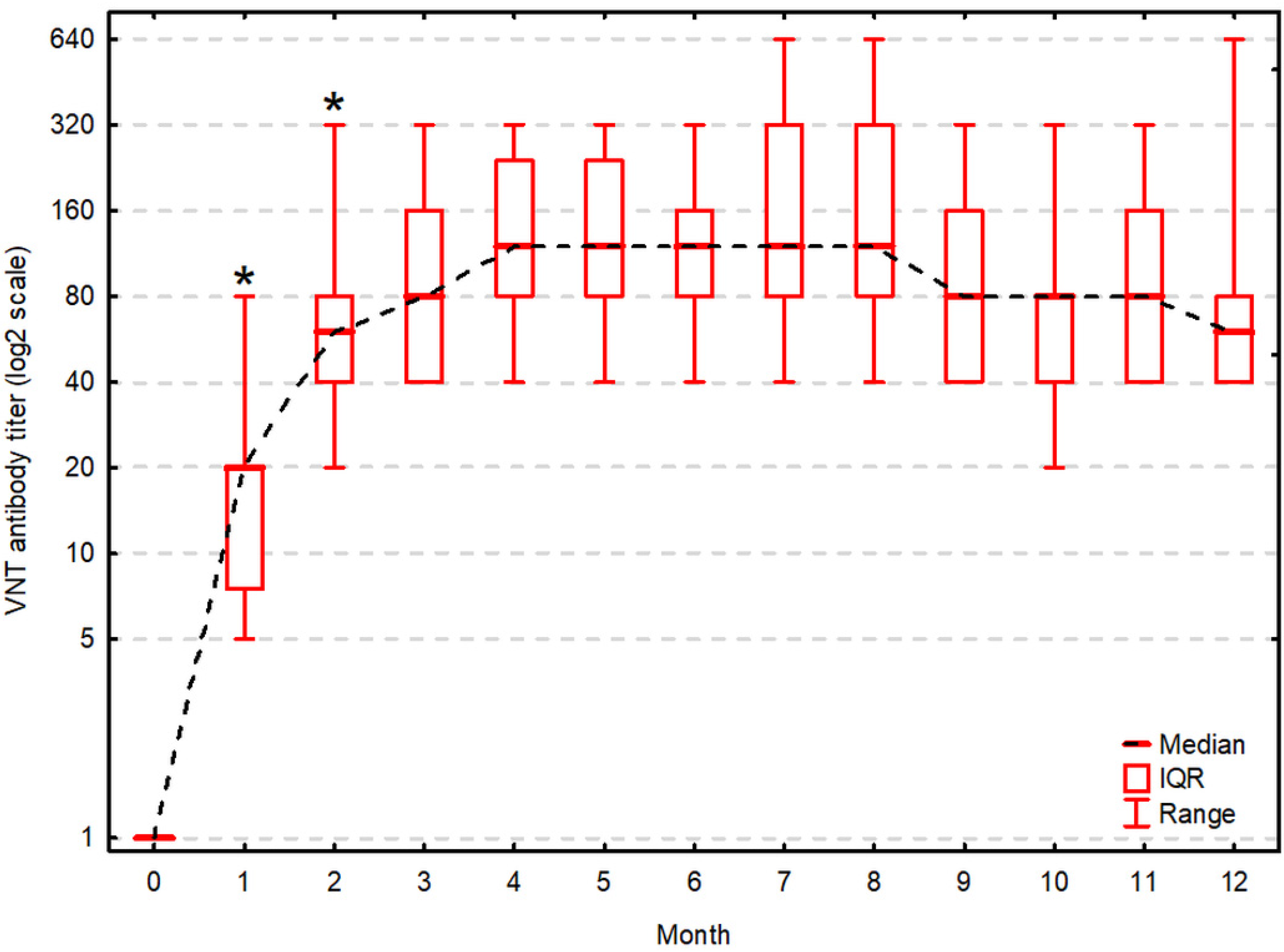
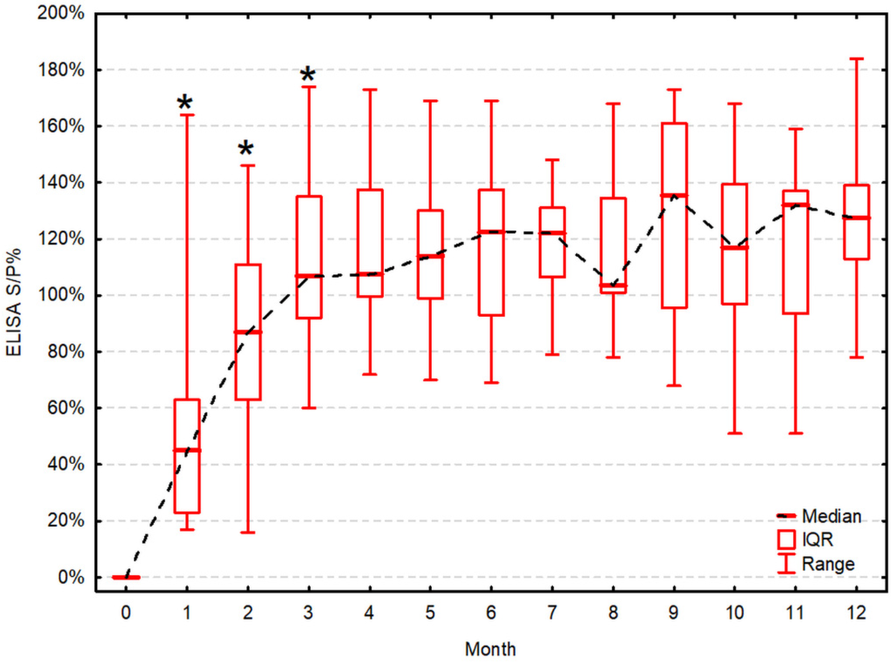
Figure 1. Antibody titers in virus neutralization test during 12 months post-vaccination. Antibody titers (on log base 2 transformed scale) are presented as the median, interquartile range (IQR) and range. Asterisk (*) indicates a statistically significant increase of the titer compared to the previous one (α = 0.05).
Vaccines 09 00259 g001
Figure 2. Sample-to-positive control ratio (S/P%) in ELISA during 12 months post-vaccination. S/P% in ELISA are presented as the median, IQR and range. Asterisk (*) indicates a statistically significant increase of the titer compared to the previous one (α = 0.05).
Vaccines 09 00259 g002
Table 1. Antibody titers in virus neutralization test (VNT) and sample-to-positive control ratio in ELISA during 12 months post-vaccination.
Test (Month)VNT [log2 Transformed Titer]Significance of an Increment Compared to the Previous Testing (p)ELISA [S/P%]Significance of an Increment Compared to the Previous Testing (p)Correlation between VNT and ELISA (Rs, p-Value)
00-0--
14.3, 2.8–4.3 (2.3–6.3)0.009 145, 23–63 (17–164)0.009 10.91, p < 0.001 1
25.8, 5.3–6.3 (4.3–8.3)0.014 187, 63–111 (16–146)0.030 10.88, p < 0.001 1
36.3, 5.3–7.3 (5.3–8.3)ns107, 92–135 (60–174)0.030 10.90, p < 0.001 1
46.8, 6.3–7.8 (5.3–8.3)ns108, 100–138 (72–173)ns0.79, p = 0.003 1
56.8, 6.3–7.8 (5.3–8.3)ns114, 99–130 (70–169)ns0.74, p = 0.006 1
66.8, 6.3–7.3 (5.3–8.3)ns123, 93–138 (69–169)ns0.84, p = 0.001 1
76.8, 6.3–8.3 (5.3–9.3)ns122, 107–131 (79–148)ns0.82, p = 0.001 1
86.8, 6.3–8.3 (5.3–9.3)ns104, 101–135 (78–168)ns0.91, p < 0.001 1
96.3, 5.3–7.3 (5.3–8.3)ns136, 96–161 (68–173)ns0.30, p = 0.353
106.3, 5.3–6.3 (4.3–8.3)ns117, 97–140 (51–168)ns0.90, p < 0.001 1
116.3, 5.3–7.3 (5.3–8.3)ns132, 94–137 (51–159)ns0.70, p = 0.011 1
125.8, 5.3–6.3 (5.3–9.3)ns128, 113–139 (78–184)ns0.92, p < 0.001 1
Antibody titers in VNT (presented as binary logarithm) and sample-to-positive control ratio (S/P%) in indirect ELISA are presented as the median, interquartile range (IQR) and range. 1—statistically significant at α = 0.05. ns—non-significant.
Publisher’s Note: MDPI stays neutral with regard to jurisdictional claims in published maps and institutional affiliations.

