
Kikuchi–Fujimoto Disease Post COVID-19 Vaccination: Case Report and Review of Literature


Abstract
:1. Introduction
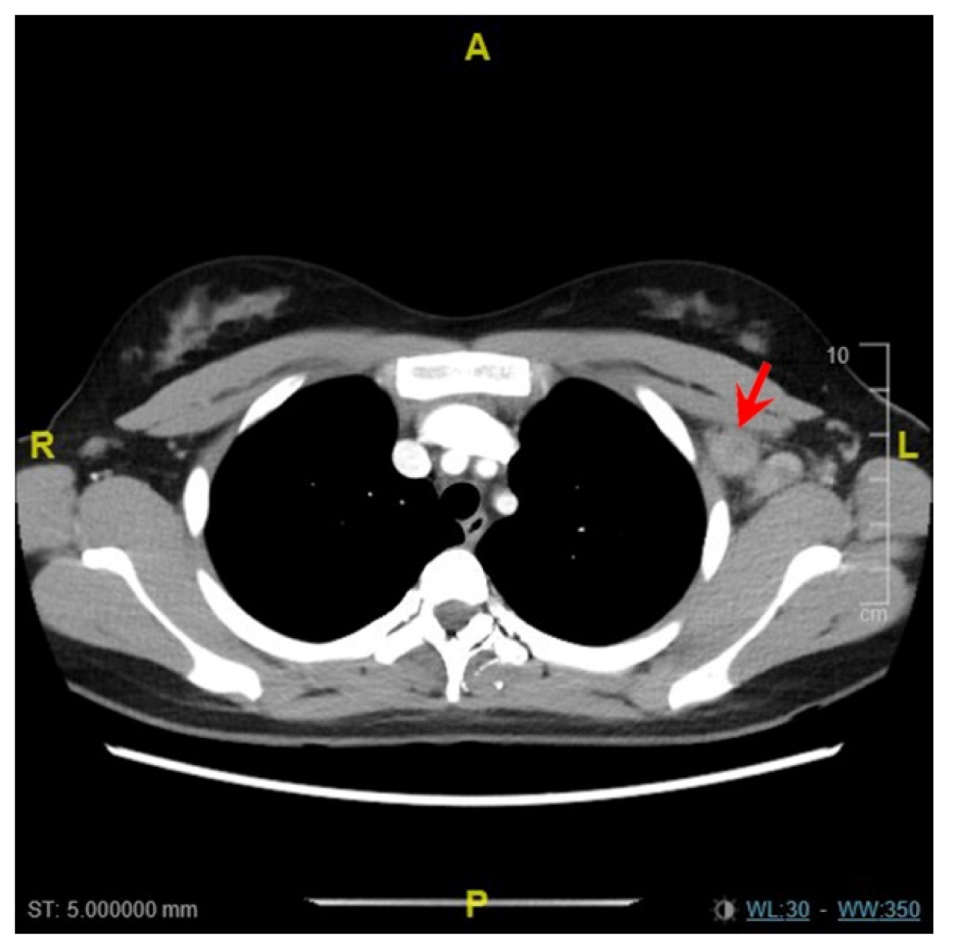
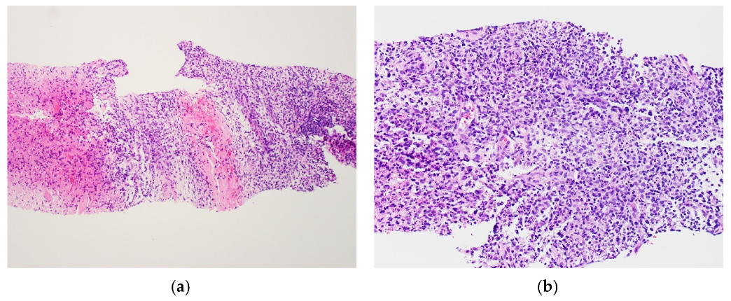
2. Case Report
3. Discussion
4. Conclusions
Author Contributions
Funding
Institutional Review Board Statement
Informed Consent Statement
Data Availability Statement
Conflicts of Interest
References
- Hartsock, R.J. Postvaccinial lymphadenitis. Hyperplasia of lymphoid tissue that simulates malignant lymphomas. Cancer 1968, 21, 632–649. [Google Scholar] [CrossRef]
- Lehman, C.D.; D’Alessandro, H.A.; Mendoza, D.P.; Succi, M.D.; Kambadakone, A.; Lamb, L.R. Unilateral Lymphadenopathy After COVID-19 Vaccination: A Practical Management Plan for Radiologists Across Specialties. J. Am. Coll. Radiol. 2021, 18, 843–852. [Google Scholar] [CrossRef] [PubMed]
- Soub, H.A.; Ibrahim, W.; Maslamani, M.A.; AAli, G.; Ummer, W.; Abu-Dayeh, A. Kikuchi-Fujimoto disease following SARS CoV2 vaccination: Case report. IDCases 2021, 25, e01253. [Google Scholar] [CrossRef]
- Asano, S.; Akaike, Y.; Jinnouchi, H.; Muramatsu, T.; Wakasa, H. Necrotizing lymphadenitis: A review of clinicopathological, immunohistochemical and ultrastructural studies. Hematol. Oncol. 1990, 8, 251–260. [Google Scholar] [CrossRef] [PubMed]
- Bosch, X.; Guilabert, A.; Miquel, R.; Campo, E. Enigmatic Kikuchi-Fujimoto Disease: A Comprehensive Review. Am. J. Clin. Pathol. 2004, 122, 141–152. [Google Scholar] [CrossRef] [PubMed]
- Perry, A.M.; Choi, S.M. Kikuchi-Fujimoto Disease: A Review. Arch. Pathol. Lab. Med. 2018, 142, 1341–1346. [Google Scholar] [CrossRef] [PubMed] [Green Version]
- Podugu, A.; Kobe, M. Kikuchi-Fujimoto Disease (KFD): A Rare Cause of Fever and Lymphadenopathy Following Influenza Vaccination. Chest 2013, 144, 230A. [Google Scholar] [CrossRef]
- Watanabe, T.; Hashidate, H.; Hirayama, Y.; Iinuma, Y. Kikuchi–Fujimoto disease following vaccination against human papilloma virus infection and Japanese encephalitis. Eur. J. Pediatr. 2012, 171, 1409–1411. [Google Scholar] [CrossRef] [PubMed]
- Özütemiz, C.; Krystosek, L.A.; Church, A.L.; Chauhan, A.; Ellermann, J.M.; Domingo-Musibay, E.; Steinberger, D. Lymphadenopathy in COVID-19 Vaccine Recipients: Diagnostic Dilemma in Oncologic Patients. Radiology 2021, 300, E296–E300. [Google Scholar] [CrossRef] [PubMed]
- Cardoso, F.; Reis, A.; Osório, C.; Scigliano, H.; Nora, M. A Case of Cervical Lymphadenopathy after Vaccination Against COVID-19. Cureus 2021. [Google Scholar] [CrossRef] [PubMed]
- Placke, J.-M.; Reis, H.; Hadaschik, E.; Roesch, A.; Schadendorf, D.; Stoffels, I.; Klode, J. Coronavirus disease 2019 vaccine mimics lymph node metastases in patients undergoing skin cancer follow-up: A monocentre study. Eur. J. Cancer 2021, 154, 167–174. [Google Scholar] [CrossRef]
- Hagen, C.; Nowack, M.; Messerli, M.; Saro, F.; Mangold, F.; Bode, P.K. Fine needle aspiration in COVID-19 vaccine-associated lymphadenopathy. Swiss Med. Wkly. 2021. [Google Scholar] [CrossRef]
- Tintle, S.; Chen, M. Lymphadenopathy with florid lymphoid and Langerhans cell hyperplasia and hemophagocytosis mimicking lymphoma after COVID-19 mRNA vaccination. eJHaem 2021, jha2.265. [Google Scholar] [CrossRef]
- Tan, N.J.H.; Tay, K.X.J.; Wong, S.B.J.; Nga, M.E. COVID-19 post-vaccination lymphadenopathy: Report of cytological findings from fine needle aspiration biopsy. Diagn. Cytopathol. 2021, dc.24863. [Google Scholar] [CrossRef] [PubMed]
- Jaseb, K.; Nameh Goshay Fard, N.; Rezaei, N.; Sadeghian, S.; Sadeghian, S. COVID-19 in a case with Kikuchi-Fujimoto disease. Clin. Case Rep. 2021, 9, 1279–1282. [Google Scholar] [CrossRef]
- Stimson, L.; Stitson, R.; Bahhadi-Hardo, M.; Renaudon-Smith, E. COVID-19 associated Kikuchi-Fujimoto disease. Br. J. Haematol. 2021, 192. [Google Scholar] [CrossRef]


| Author | Age | Gender | Vaccine Received | Interval between Vaccination and LAD 1 or First Symptom | Site(s) of LAD * | Type of Sampling † | Diagnosis |
|---|---|---|---|---|---|---|---|
| Our case, patient A | 18 | Female | Pfizer- BioNTech | 35 days | Supraclavicular, subpectoral and axillary | Core biopsy | KFD 2 |
| Our case, patient B | 34 | Male | Pfizer- BioNTech | 17 days | Axillary | Core biopsy | KFD |
| Soub et al. (August 2021) [3] | 18 | Male | Pfizer- BioNTech | 10 days | Supraclavicular, cervical and axillary | Excision biopsy | KFD |
| Özütemiz et al. (February 2021) [9]; oncologic patients | 32 | Female | Pfizer- BioNTech | 6 days | Axillary | Nil | – |
| 57 | Female | Pfizer- BioNTech | 5 days | Axillary | Nil | – | |
| 41 | Male | Pfizer- BioNTech | 4 days | Axillary | Nil | – | |
| 46 | Female | Pfizer- BioNTech | 15 days | Axillary, supraclavicular | Nil | Reactive lymph node | |
| 38 | Female | Pfizer- BioNTech | 8 days | Axillary | Core biopsy | Reactive follicular hyperplasia | |
| Cardoso et al. (May 2021) [10] | 48 | Female | Pfizer- BioNTech | 2 weeks | Cervical | Excision biopsy | Reactive follicular hyperplasia |
| Placke et al. (June 2021) [11]; patients with previous skin cancer | 54 | Female | CureVac | 30 days | Axillary | Excision biopsy (sentinel node) | Lymphofollicular hyperplasia |
| 28 | Female | Pfizer- BioNTech | 28 days | Axillary | Excision biopsy (selective) | Sarcoid-like reaction | |
| 58 | Male | Pfizer- BioNTech | 7 days | Axillary | Excision biopsy (selective) | Not mentioned, but non-malignant | |
| 77 | Male | Pfizer- BioNTech | 11 days | Axillary | Excision biopsy (sentinel node) | Not mentioned, but non-malignant | |
| 91 | Male | Pfizer- BioNTech | 16 days | Axillary | Excision biopsy (sentinel node) | Not mentioned, but non-malignant | |
| 44 | Male | Pfizer- BioNTech | 15 days | Axillary | Excision biopsy (sentinel node) | Not mentioned, but non-malignant | |
| 43 | Female | Pfizer- BioNTech | 50 days | Axillary | Lymphadenectomy | Not mentioned, but non-malignant | |
| 84 | Female | Pfizer- BioNTech | 12 days | Axillary | Excision biopsy (sentinel node) | Not mentioned, but non-malignant | |
| Hagen et al. (July 2021) [12] | 66 | Male | Moderna | 22 days | Axillary | FNAC 3 | Reactive lymphadenopathy |
| 41 | Female | Moderna | 3 days | Infraclavicular | FNAC | Reactive lymphadenopathy | |
| 47 | Female | Pfizer- BioNTech | 19 days | Supraclavicular | FNAC | Reactive lymphadenopathy | |
| 47 | Female | Moderna | 8 days | Cervical | FNAC | Reactive lymphadenopathy | |
| 52 | Male | Pfizer- BioNTech | 12 days | Retroclavicular (contralateral) | FNAC | Negative for malignancy | |
| Tintle and Chen (July 2021) [13] | 23 | Female | Moderna | 1 week | Axillary, intra-abdominal | Excision biopsy | Lymphoid and Langerhan cell hyperplasia, hemophagocytosis |
| Tan et al. (August 2021) [14] | 34 | Male | Pfizer- BioNTech | 1 day | Supraclavicular | FNAC | Reactive lymphadenopathy |
Publisher’s Note: MDPI stays neutral with regard to jurisdictional claims in published maps and institutional affiliations. |
© 2021 by the authors. Licensee MDPI, Basel, Switzerland. This article is an open access article distributed under the terms and conditions of the Creative Commons Attribution (CC BY) license (https://creativecommons.org/licenses/by/4.0/).
Share and Cite
Tan, H.M.; Hue, S.S.-S.; Wee, A.; See, K.C. Kikuchi–Fujimoto Disease Post COVID-19 Vaccination: Case Report and Review of Literature. Vaccines 2021, 9, 1251. https://doi.org/10.3390/vaccines9111251
Tan HM, Hue SS-S, Wee A, See KC. Kikuchi–Fujimoto Disease Post COVID-19 Vaccination: Case Report and Review of Literature. Vaccines. 2021; 9(11):1251. https://doi.org/10.3390/vaccines9111251
Chicago/Turabian StyleTan, Hui Min, Susan Swee-Shan Hue, Aileen Wee, and Kay Choong See. 2021. "Kikuchi–Fujimoto Disease Post COVID-19 Vaccination: Case Report and Review of Literature" Vaccines 9, no. 11: 1251. https://doi.org/10.3390/vaccines9111251
APA StyleTan, H. M., Hue, S. S.-S., Wee, A., & See, K. C. (2021). Kikuchi–Fujimoto Disease Post COVID-19 Vaccination: Case Report and Review of Literature. Vaccines, 9(11), 1251. https://doi.org/10.3390/vaccines9111251