Abstract
The thymus is the main lymphoid organ that regulates the immune and endocrine systems by controlling thymic cell proliferation and differentiation. The gland is a primary lymphoid organ responsible for generating mature T cells into CD4+ or CD8+ single-positive (SP) T cells, contributing to cellular immunity. Regarding humoral immunity, the thymic plasma cells almost exclusively secrete IgG1 and IgG3, the two main complement-fixing effector IgG subclasses. Deformity in the thymus can lead to inflammatory diseases. Hassall’s corpuscles’ epithelial lining produces thymic stromal lymphopoietin, which induces differentiation of CDs thymocytes into regulatory T cells within the thymus medulla. Thymic B lymphocytes produce immunoglobulins and immunoregulating hormones, including thymosin. Modulation in T cell and naive T cells decrement due to thymus deformity induce alteration in the secretion of various inflammatory factors, resulting in multiple diseases. Influenza virus activates thymic CD4+ CD8+ thymocytes and a large amount of IFNγ. IFNs limit virus spread, enhance macrophages’ phagocytosis, and promote the natural killer cell restriction activity against infected cells. Th2 lymphocytes-produced cytokine IL-4 can bind to antiviral INFγ, decreasing the cell susceptibility and downregulating viral receptors. COVID-19 epitopes (S, M, and N proteins) with ≥90% identity to the SARS-CoV sequence have been predicted. These epitopes trigger immunity for antibodies production. Boosting the immune system by improving thymus function can be a therapeutic strategy for preventing virus-related diseases. This review aims to summarize the endocrine-immunoregulatory functions of the thymus and the underlying mechanisms in the prevention of COVID-19.
1. Introduction
The coronavirus family was first identified in the late []. In the decades since, the world has experienced many lethal episodes of the coronavirus family. Coronavirus diseases were noted with mild or severe infections in the respiratory tract []. In 2002, severe acute respiratory syndrome coronavirus (SARS-CoV) emerged and infected many populations worldwide []. In 2012, the Middle East respiratory syndrome (MERS-CoV) outbreak infected Middle Eastern countries, with symptoms of chronic respiratory syndrome []. In 2019, SARS-CoV-2 was identified in Wuhan, China, which affected a total of 30,524,214 people with a mortality of 952,240 at the time of drafting this paper. SARS-CoV, MERS-CoV, and SARS-CoV-2 involve serious respiratory tract infections followed by fever, cough, dyspnea, and fatigue [].
The thymus gland is the chief lymphoid organ that regulates the functions of the immune and endocrine systems by controlling the levels of hormones and cytokines. The thymus gland protects against various internal and external stresses through immunoregulatory properties, nerve systems, and endocrine pathways. The thymus gland controls cell proliferation, apoptosis, hormones, and neuropeptides, as well as regulating intrathymic T cell differentiation and production of a repertoire of the T cell. The thymus is located in front of the heart behind the sternum. It has two identical thymic lobes on each side, made up of the cortex and the central medulla, surrounded by an outer capsule []. The thymus gland is most actively functioning in fetal and neonatal life and starts shrinking in tissue mass and is replaced with fat during thymic involution []. In 1961, Jacques Miller discovered the immunoregulatory role of the thymus in newborn mice by studying involvement in a lymphocyte population [].
The reason for children being less exposed to SARS-CoV-2 could be attributed to the significant capacity of children for maintaining the availability of rare T cell clonotypes that originate in the thymus and the variety of the T cell populations, which supposes a causative connection in the rising tendency of infection with age [,]. This clonotype is rare if it is not utilized, which leads to less proliferation and postpones presenting viral antigens to SARS-CoV-2-specific T cells, permitting more effects of virus damage and breakout. This late activation of the adaptive power in the immune system appears as lymphopenia in lack of effective virus-specific clonal growth specific to epitopes of the virus presented by the lymphatic nodes. It was suggested by Rousseau et al. (2020) [] that stimulating the same approach will help in reducing the severity of this virus. Thymic hormones such as thymosin-α-1-Fc (TA-1) have been shown to increase the naive CD8 and CD4 cells that have recombined in the blood and stimulate thymopoiesis, then TA-1 adjusts a hyperinflammatory response via dendritic cells (DCs) for immunosuppressing and activating natural killer (NK) cell function. The absence of type 1 interferon (IFN) in alveolar cells and the presence of a lymphopenia response in SARS-CoV-2 diseases propose that incorporation of αβ-IFN and TA-1 may present the synergistic action to attract the adaptive immunity that helps significantly in a much-needed response.
The immune system is generally classified into innate immunity which provides the first line of defense against different stimuli, such as antigens and chemical, biochemical, physiological, and physical stresses. Cellular innate immunity mediates their actions through macrophages, granulocytes, NK cells, and DCs, either by engulfing the antigens in a process called phagocytosis or by acting as antigen-presenting cells to expose the antigens to the cells of the acquired immunity, which is a more specific type of immunity. B lymphocytes and T lymphocytes are interrelated cellular counterparts of the acquired immune system, expressed as surface receptors that recognize specific antigens and have the potential for long-term immunological memory. Lymphocytes are generated in the bone marrow, where only B lymphocytes mature and are exported to the periphery. However, T lymphocytes, as hematopoietic precursors, migrate to the thymus to grow, develop, and differentiate [].
The thymus offers specialized conditions for developing various functional and self-tolerant T cells. Once in the thymus, precursor cells enter the subcapsular cortical region, undergoing several developmental stages to become thymocytes. In the differentiation process, thymocytes move from the cortex area to the medulla for negative and positive thymic selection []. Thymocytes interact with the thymic epithelial cells and trigger their differentiation into mature clusters of differentiation 4 (CD4+) and CD8+. Cytotoxic T lymphocytes (CTLs) undergo a process known as the positive selection, where thymocytes recognize and bind to self-peptides of the major histocompatibility complex (MHC). This process determines whether the T lymphocyte is CD4+ (helper) or CD8+ (cytotoxic/killer), depending on bonding to the type of MHC (class I or class II). When self-reactive thymocytes fail to recognize self-antigens and strongly bind to the self-peptide MHC, they undergo negative selection and are eliminated by the process of apoptosis []. The naive T lymphocytes disembark the thymus into the secondary lymphoid organs such as lymph nodes, where they are activated by foreign peptides of MHC that are found on the surfaces of the antigen-presenting cells (APCs). This type of activation results in the proliferation and differentiation of effector T lymphocytes into four types that can produce cytokines and respond to different pathogens. The thymus gland also secretes the thymosin hormone, which has a functional role in T lymphocyte differentiation and maturation to mediate immunological response []. T lymphocytes are the most fundamental components of cellular immunity. Besides humoral immunity, they also have featured roles as a complementary component to innate immunity, which cannot efficiently defend against all pathogens.
Some immunodeficiency viral diseases such as thymic lymphoid hyperplasia (thymitis), loss of Hassall’s corpuscles, and dysinvolution have been associated with the malfunctioning of thymus or viral infections of Hassall’s corpuscles []. Hassall’s corpuscles were also found to be severely damaged upon infection with herpes simplex virus pneumonia and ependymoma []. SARS-CoV induces immune-mediated lymphocyte damage, bone marrow or thymus suppression, or cell programming death []. The SARS-CoV-2 infection causes lymphopenia in peripheral circulation, which is counterbalanced by the thymus to enhance lymphocyte recirculation between peripheral blood and SARS-specific IgG immunoglobulins release, with no elevation in the levels of interleukin (IL) 8 and tumor necrosis factor (TNF-α) [,]. The aim of the current study is to support the notion that a defect in thymic tolerogenic function is implicated as an essential factor in the pathophysiology of autoimmunity and virus-related diseases, including COVID-19. This piece of work underpins the reported literature on the physiology of the thymus (Figure 1) and the biological role of different thymic hormones with regard to the modulation of inflammatory responses and involvement in the maturation and differentiation of immune cells, as well as advocating the clinical and biological application in the treatment of inflammatory disorders, including viral diseases.
Figure 1.
Overall role of the thymus gland in the development of T cells.
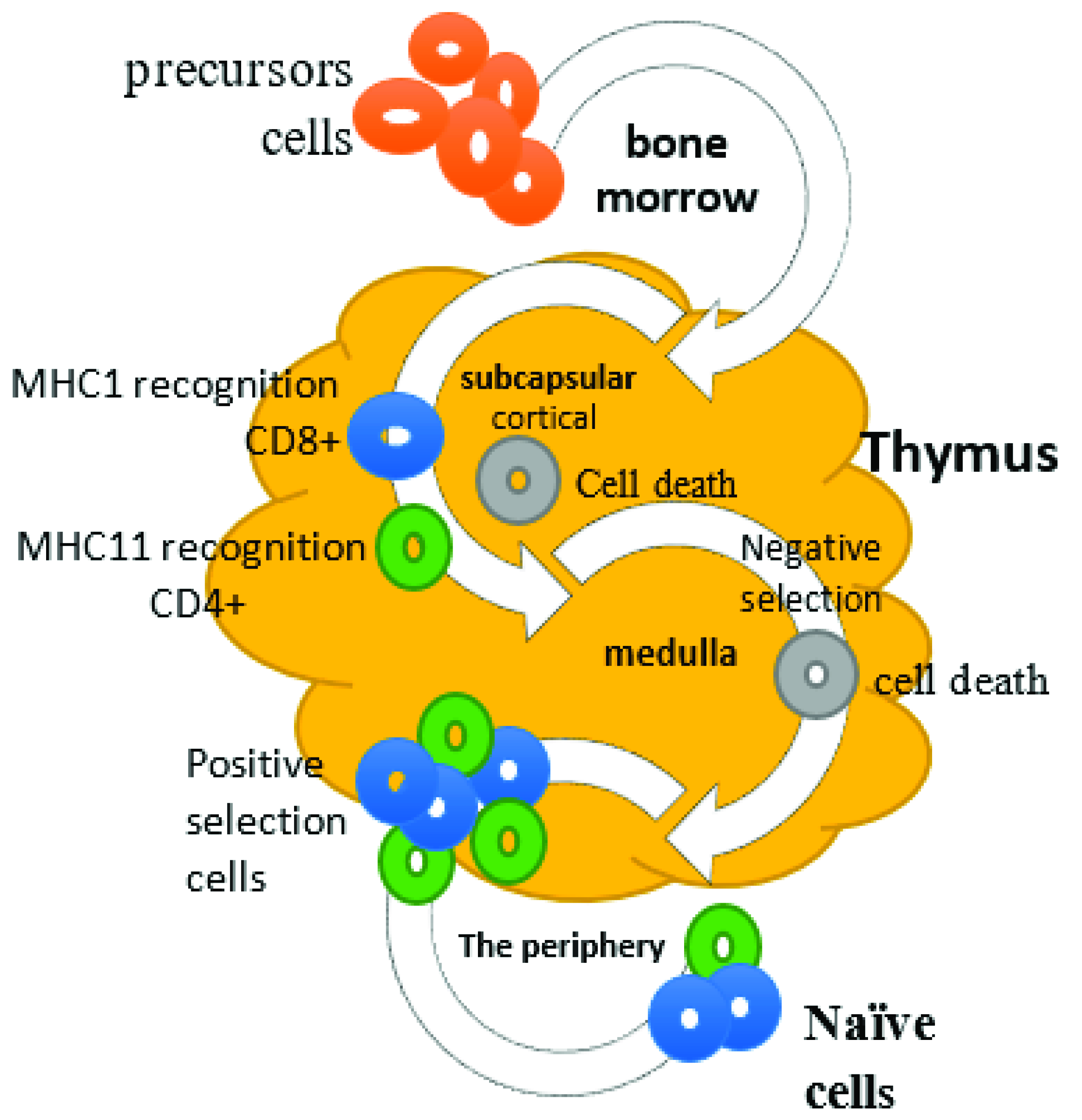
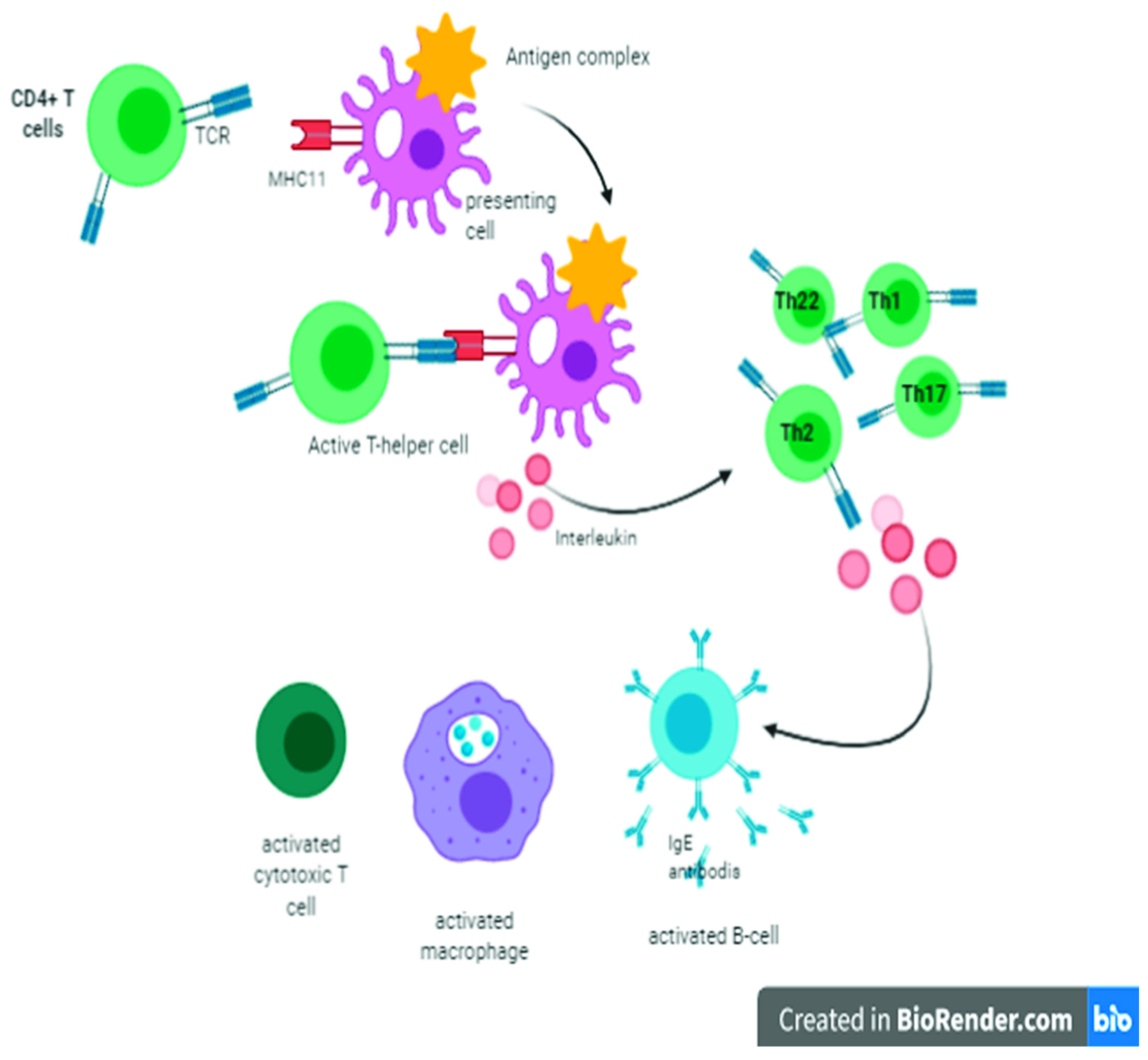
2. Cellular Immunity and Role of T Lymphocytes
There is compelling evidence that the thymus is responsible for the development and differentiation of T lymphocytes. T lymphocytes mediate cellular immunity, providing a defense mechanism against intracellular microorganisms either through the direct killing of the cells that host microorganisms in their cytoplasm or the activation of other immune cells to destroy ingested microorganisms by the process of phagocytosis and productions of antibodies against specific antigens [,]. It has been reported that short chains of peptides that bind to MHC-I molecules are derived from degraded intracellular cytosolic proteins of microorganisms, including virus-mediated infections. On the other hand, peptides that bind to MHC-II molecules are derived from extracellular proteins of infectious agents []. The CD4+ T lymphocytes recognize antigens presented by MHC-II molecules, readily expressed on DCs, macrophages, and B lymphocytes []. Consequently, CD4+ T lymphocytes are activated and differentiated into one of several subsets of effector T helper (Th) lymphocytes, including, Th1, Th2, Th9, Th17, and Th22, which regulate the immune responses (Figure 2) by secreting cytokines []. Th2 lymphocytes also regulate the production of antibodies from B lymphocytes by secreting ILs such as IL-3, IL-4, and IL-5 and differentiation to antibody-secreting plasma cells []. Th17 has been reported to prevent mucosal respiratory viral infections in the lungs through recruiting macrophages and neutrophils, which ultimately clears pathogens, mediating inflammation, and maintaining tissue integrity []. Th17 lymphocytes have suppressed the detrimental tissue inflammations in viral infections. The exact underlying defensive mechanism of Th17 lymphocytes remains to be elucidated because lymphocytes-associated viral lung pathologies have been reported previously [].
Figure 2.
Antigen-presenting cells present an antigen complex with a major histocompatibility complex (MHC) to stimulate immature T cells to become either cytotoxic cells (CD8), when the T cell receptor binds to MHC class I, or Th cells (CD4+), when it binds to MHC class II. Once CD4 cells are activated, they will begin proliferation or clonal expansion and differentiate into Th17, Th9, Th1, Th2, and Th22 at the same time; they will secret interleukins that will stimulate a humoral immune response to produce antibodies (IgE), as well as cellular immune response and nonspecific defense by activated cytotoxic T cells and macrophages.
It is well known that inflammation is a central player in the pathogenesis of SARS-CoV pneumonia and edema []. Moreover, Th1 lymphocytes are responsible for the proliferation and differentiation of cytotoxic CD8+ T lymphocytes and other cells in response to intracellular pathogens, as well as latent viral infections and tumors, which are all weak innate immune response inducers. Various cytokines such as IL-2, IL-12, IL-15, and IL-21 are involved in CD8+ T lymphocyte differentiation and the generation of effector and memory lymphocytes. CD4+ Th lymphocytes promote CD8+ T lymphocyte activation, either directly by cytokine production or indirectly by enhancing the ability of APCs to stimulate the activation process []. CTLs recognize the cells presenting MHC-I molecules, which serve as legends to the T-cell receptors (TCRs) on their surfaces, targeting them for destruction. In this process, only antigen-expressing cells are affected and destroyed. CTLs activate macrophages through interferon gamma (IFNγ) production, which can phagocytose microorganisms []. The activity of Th lymphocytes and CTLs is regulated by the regulatory T lymphocytes (Treg), known as suppressor T lymphocytes, and a subtype of CD4+ T lymphocyte. Treg lymphocytes account for 5–10% of CD4+ T lymphocytes in the periphery and play an essential role in inhibiting autoimmune and chronic inflammatory diseases. Treg cells also eliminate self-reactive T lymphocytes that have escaped central tolerance (negative thymic selection) by the mechanism of peripheral tolerance []. Once a pathogen or a disease-causing agent is identified, naive T lymphocytes proliferate and differentiate into effector T lymphocytes, ultimately targeting and eliminating the foreign invaders. Effector T lymphocytes also serve as memory T lymphocytes, such as stem cell memory T lymphocytes (Tscm), central memory T lymphocytes (Tcm), and effector memory T lymphocytes (Tem), as well as terminal effector T lymphocytes and tissue-resident memory T lymphocytes, which can respond faster and more efficiently in the future against the same infection []. Compared with naive T lymphocytes, Tscm cells reveal higher expression levels of C-X-C motif chemokine receptor 3 (CXCR3), apoptosis antigen-1 (APO-1), IL-2 receptor (IL-2Rβ), and leukocyte function-associated antigen-1 (LFA-1). Tem and Tcm cells are differentiated by the function and expression of the C-C chemokine receptor type-7 (CCR7) protein. Tcm cells are occupied in the lymphoid system and have no direct role, while Tem cells are found in the non-organ lymphatic tissue and have a rapid and more significant function than Tcm cells [].
3. Function of Hassall’s Corpuscles in Viral Infection
There is compelling evidence that thymic stromal lymphopoietin (TSLP) is a cytokine, which stimulates B lymphocytes development derived from thymic stromal cell line Z210R.1. It has been reported that Hassall’s corpuscles’ epithelial cells produced TSLP, responsible for the activation of thymic DCs to induce and generate CD4+ CD25+ regulatory T cells within the thymus. In addition, Hassall’s corpuscles are accountable for thymocyte development and removal of apoptotic thymocytes inside the thymus []. The primary extravillous trophoblast (EVT) expressed the cytokine TSLP and TSLP receptors. Studies have shown that TNF-α and IL-4 or pregnancy-associated hormones lead to a substantial rise in TSLP-mediated primary human EVT propagation and invasion in vitro. TSLP has a crucial role in human EVT invasion and regulation of the placenta in the first trimester of pregnancy [].
Transforming growth factor alpha (TGF-α) is associated with medullary human thymic epithelial cells (TECs) and thymic Hassall’s corpuscles, whereas epidermal growth factor (EGF) receptor was concentrated only in TEC cells through the thymus tissue []. Both TGF-α and EGF are crucial regulatory precursors for synthesizing TEC-derived cytokines in the thymus and act as essential modulators for developing T lymphocyte proliferation in humans []. TGFβRII was found to mediate TEC signaling and reduce their improvement in Hassall’s corpuscles in mice [].
Hassall’s corpuscles are composed of terminally differentiated medullary TECs with properties of cellular senescence and release inflammatory cytokines and chemokines, such as CXCL5, that employ and activate neutrophils to release IL-23 in the thymic medulla. Thymic plasmacytoid DCs express IL-23 receptors essentially produce IFNγ, which functions in cell maturation []. The human thymus expresses antibodies IgG, IgA, IgM, IgD, and IgE, and light chains, in the cells of Hassall’s corpuscles. In the thymic medulla, the production of IgG, IgA, and IgM by plasma cells is controlled. (Figure 3) [].
Figure 3.
Functions of thymic Hassall’s corpuscles in normal state and after infection with influenza virus. Thymic stromal lymphopoietin (TSLP), a cytokine that is the primary hormone produced by the epithelial cells of Hassall’s corpuscles, was found in the thymus and was responsible for the activation of thymic dendritic cells. Hassall’s corpuscles secrete TSLP-inducing dendritic cells to induce and generate CD4+ CD8+ CD25+ regulatory T cells within the thymus. Thymic plasmacytoid dendritic cells express IL-23 receptors. Transforming growth factor alpha (TGF-α) was identified in medullary human thymic epithelial (TE) cells and thymic Hassall’s corpuscles while epidermal growth factor receptor (EGF-R) was concentrated to TE cells through the thymus tissue. Hence, TGF-α and EGF are crucial regulatory precursors for the synthesis of TE cell-derived cytokines in the thymus. The human thymus shows the existence of antibodies IgG, IgA, IgM, IgD, and IgE, which are secretory constituents in Hassall’s corpuscles. There is a strong connection between the amounts of IgA and secretory components in the cells of Hassall’s corpuscles, and the thymus may have to be considered as an active portion of the secretory-IgA system of Hassall’s corpuscles. The influenza virus activates thymic CD4+ CD8+ thymocytes, leading to the secretion of a large amount of interferon IFNγ.
IL-1α/β enhances IgM and recruitment of CD4+ T cells at the site of infection but does not contribute to killing the virus-infected cell []. Additionally, CD4+ but not CD8+ Treg cells were suppressed by IL-6, allowing pathogen clearance and host survival in virus-induced infection []. In H1N1 influenza, IL-6 levels elevated in severe cases of virally infected patients [] and activated CD4+ CD8+ thymocytes and a large amount of IFNγ []. While IL-32 is a part of a negative feedback loop, inhibiting sIL-6R and upregulating IL-6 is essential for the survival of an influenza A virus infection [,]. Few immunodeficiency viral diseases have been linked with the thymus in childhood, including thymic lymphoid hyperplasia (thymitis), loss of Hassall’s corpuscles, and dysinvolution []. Hassall’s corpuscles were found to be altered and damaged in a 4-year-old boy infected with herpes simplex virus pneumonia and ependymoma []. Modulation in the size of the thymus has been associated with hyperactivation of dystrophic calcification of Hassall bodies, reflecting the decrease in the number of CD4+ cells in drug-addicted patients []. Lymphocytopenia is a noticeable portion of SARS-CoV contagion. It may be immune-mediated lymphocyte damage, bone marrow or thymus suppression, or cell programming death []. After a viral infection, lymphopenia is noticed in peripheral circulation [].
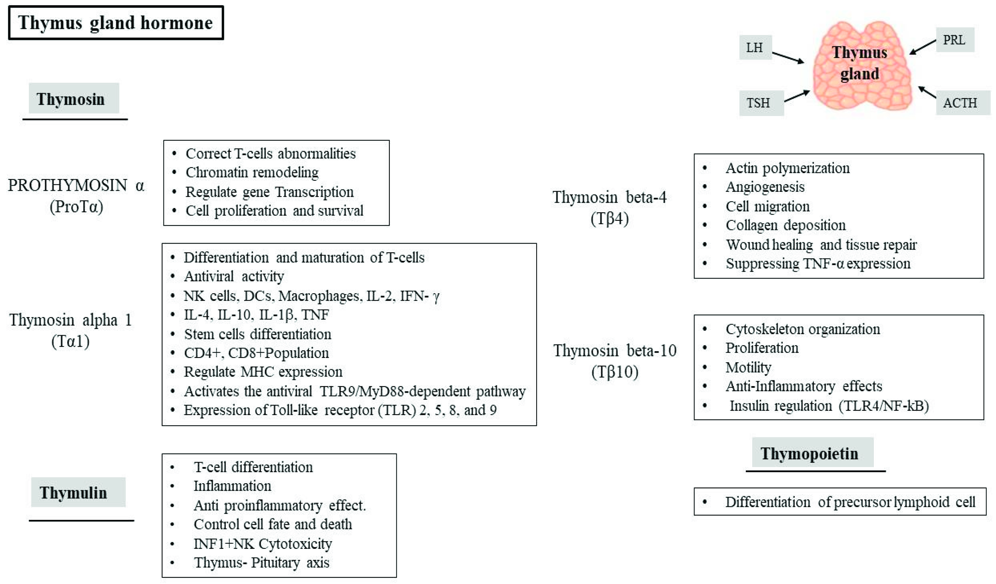
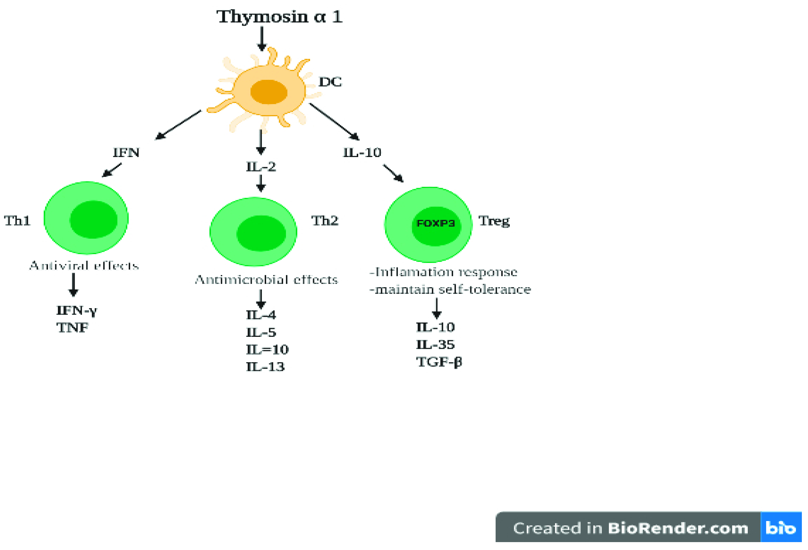
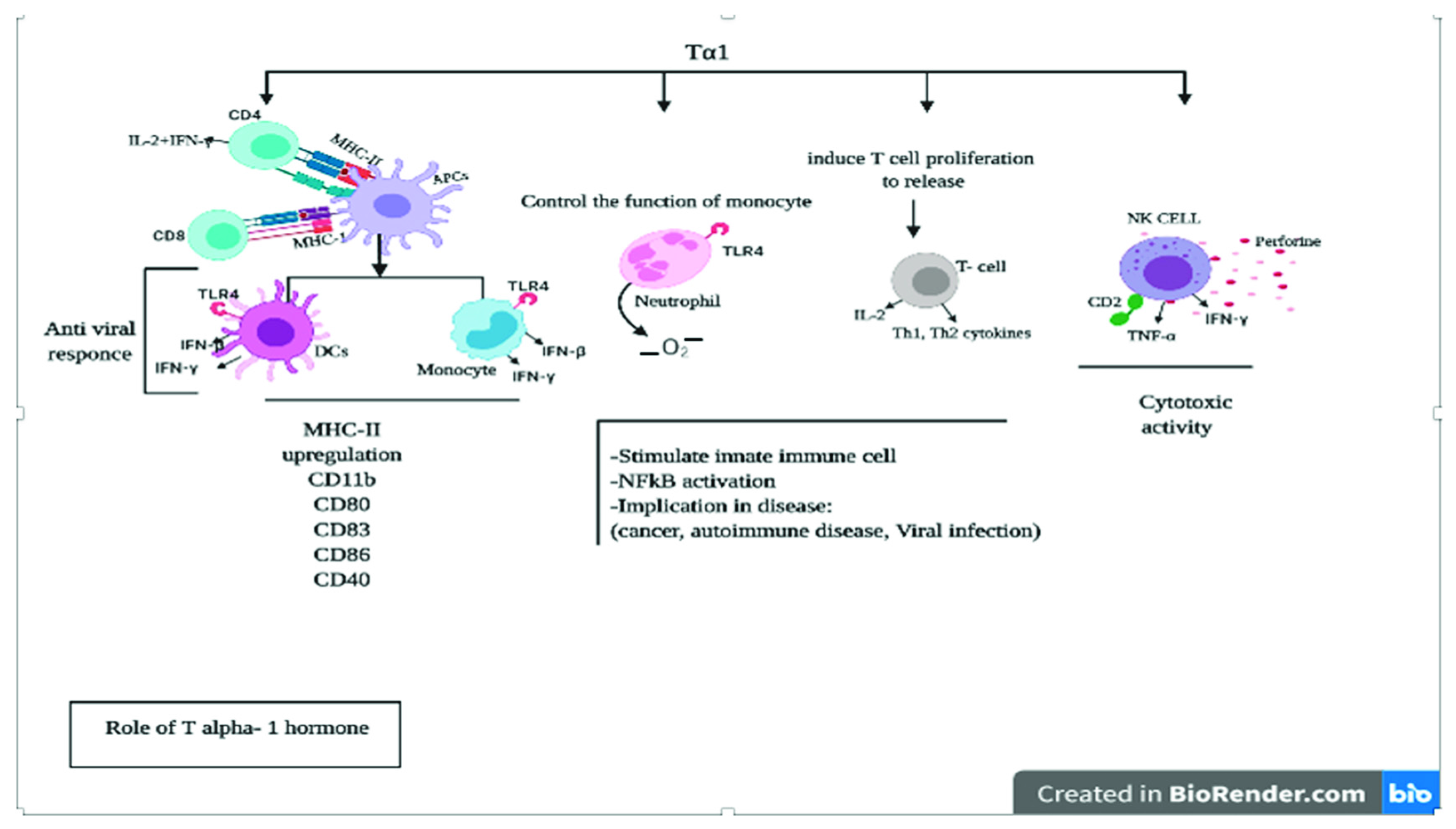
4. Role of Thymic Hormones in Viral Infection
The thymus is a lymphoid organ involved in T lymphocyte maturation and differentiation. It is known that TECs can secrete IL-6 and granulocyte-macrophage colony-stimulating factor (GM-CSF) and thymic hormones in circulation, which promote thymocyte differentiation and proliferation and have anti-inflammatory effects. Thymic hormones (Figure 4) such as thymopoietin, thymosin alpha 1 (Tα1), and thymuline have a potential role in the differentiation and functions of lymphocytes, thus they may have the potential for T lymphocyte-related diseases. In addition, the thymus mediates neuroendocrine interactions directly affected by pituitary hormones, consequently affecting the neuroendocrine function of the thymus []. The active biological thymic peptides have been extracted and purified in a process called Thymosin Fraction V, along with several main peptides such as prothymosin α (ProTα), Tα1, thymosin beta-4 (Tβ4), thymosin beta-10 (Tβ10), and thymuline, for the maturation and differentiation of immature thymocytes [,,]. These peptides are biologically essential and known to activate the immune system through several mechanisms and signaling pathways, including stimulation of T cell differentiation and maturation, activation of NK cells, DCs, and induction of proinflammatory cytokine release []. The previous description indicates that thymic hormones can mediate anti-inflammatory effects, and future clinical trials are needed to translate them against inflammatory disorders and viral diseases []. In an experimental model of allergic asthma, a dose of DNA nanoparticles, including thymuline plasmids, could protect the lungs from some injurious inflammation and muscular hypertrophy, which recovered respiratory mechanical functions []. Mice were treated through intratracheal administration with a dose of thymuline-expressing plasmids administrated with nanoparticles to enable the thymuline to infiltrate the mucus barrier of the respiratory system [].
Figure 4.
Types and roles of thymus gland hormones. The thymus produces immunoregulating hormones such as thymosin, and its family includes prothymosin alpha, thymosin alpha 1, thymosin beta-4 and thymosin beta-10, thymuline, and thymopoietin.
The most important member of the thymosin family is Tα1 (Figure 5) and its precursor ProTα [].
Figure 5.
T alpha 1 cells and functional outcomes by targeting dendritic cells. Tα1 cells can modulate dendritic cell (DC) function. DCs express variable receptors for communications to induce T helper type 1 (Th1), T helper type2 (Th2), and regulatory T cells (Treg) priming antigen-specific T cell activation. Tα1 cells convert resting DCs into cells capable of promoting the polarization and differentiation of naive T cells.
Tα1 is highly expressed in the thymus and peripheral tissues and produced through cleavage of ProTα in the thymus, pituitary, and brain []. Thymus hormones are targeted to control viral infectious diseases and inflammatory and autoimmune diseases [,]. Tα1, Tβ4, and Tβ10 displayed positive immunomodulatory effects by inducing Th lymphocytes (CD4+) and activating cytotoxic T lymphocytes (CD8+), maintaining immune homeostasis in viral infection [].
Tα1 boosts immunity through the differentiation and maturation of T cells, and the activation of NK cells, DCs, and release of proinflammatory cytokines [] (Figure 6). The pharmacological and immunomodulatory effects of Tα1 have been investigated in various animal and human studies, including the treatment of chronic hepatitis B and C, cytomegalovirus infection, sepsis, a chronic obstructive pulmonary disorder, HIV/AIDS, and SARS-CoV [,,]. The principal effector cells in the innate immune system are DCs, NK cells, and NK T cells, with monocytes, macrophages, and Tα1 as a biological response modifier, which regulates the differentiation and maturation in viral infections []. Tα1 modulates T lymphocytes and was used as a prophylactic agent against the SARS virus throughout the 2003 pandemic. Therefore, it can control immunity, inflammation, and the development of the disease []. Tα1 is widely used as a therapeutic agent in viral, fungal, and bacterial infectious diseases, either as a monotherapy or a vaccine enhancer, as an adjuvant with IFNα [,]. Under normal conditions, transformed cells can upregulate and increase the expression of MHC-I, MHC-II, and macroglobulin (B2) [], generally required for recognition by the immune system of the virally infected cells, and thus can directly blunt the growth of viruses []. Additionally, when mononuclear cells are treated with ProTα, there is an increased expression of IFNα-inducible protein, which possesses significant antiviral activity []. Mujtaba et al. recommended using re-designing antivirals and important potential inhibitors against COVID 19 []. Tα1 can modulate both T lymphocyte maturation and NK cell-mediated cytotoxicity, as well as stimulation of lymphokine and cytokines production by peripheral blood lymphocytes, which include macrophage migration inhibitory factor, CSF, GM-CSF, B lymphocyte growth factor, IFNα, IFNγ cascade, and IL-2 by activated lymphocytes. Tα1 also regulates transcriptional factors involved in the immune response and influencing protein/antigen expression []. Tα1 regulates Th1-type cytokines that may affect thymocytes by stimulating their differentiation, converting them to active T lymphocytes, and enhancing NK cell activity []. When immature DCs (iDCs) in the periphery blood are activated by pathogenic determinants known as pathogen-associated molecular patterns or by immune cytokines (TNF-α and IL-1β), iDCs become activated and transformed into fully mature DCs (mDCs) by upregulating co-stimulatory molecules (CD-40, CD-86, CD-80, and CD-83) followed by overexpression of IL-12 []. There is compelling evidence that Tα1 modulates mitogen-activated protein kinases (MAPKs) and signals the transduction pathway to activate bone marrow-derived macrophages [] and mDCs through interaction with Toll-like receptors (TLR) by the MyD88-dependent pathway, in particular TLR2, TLR5, TLR8, and TLR9. It is known that mDCs migrate to lymph nodes and macrophages, where they interact with numbers of naive T lymphocytes. DCs increase antigen presentation and synapses with CD4+ helper T lymphocytes and Th cytokines (e.g., IL-2, IFNγ) []. Therefore, activating some intracellular signaling pathways such as NF-kB and p38 MAPK are required for the therapeutic efficacy of thymus hormones [,,]. IFN-I plays a vital role in mediating T cell response at the site of viral infection. Moreover, IFN-I can stimulate genes that promote autophagy and boost immunity against viral infections []. Studies have shown that in MERS-CoV, macrophages are primary effectors of the innate immune system and DCs are present abundantly in infected lungs. They control inflammatory cytokine producers and APCs by direct interaction with the antigen and surface and the intracellular receptors []. This causes the production of interferon gamma-induced protein 10 (CXCL10), mRNAs, and IFN (IFNλ1, IFNα/β expression), which can enhance the production of CD8+ T lymphocytes and cytolytic functions by producing perforin and granzyme []. This inhibits viral infection through direct cytotoxicity or by proinflammatory cytokine productions [,].
Figure 6.
Effect of Tα1 on cells and pathways of the immune system. Tα1 can trigger multiple downstream pathways with distinct types of immune cells, pro/anti-inflammatory cytokine, and induce an immune response by Toll-like receptor (TLR) expression and cytokine production, leading to the initiation of a subsequent phase of immunity.
7. Conclusions
The thymus is a primary lymphoid organ responsible for generating mature T cells into CD4+ or CD8+ single-positive (SP) T cells, contributing to cellular immunity. Regarding humoral immunity, the thymic plasma cells secrete IgG1 and IgG3 almost exclusively, the two main complement-fixing effector IgG subclasses. Hassall’s corpuscles perform an admirable function. Understanding the endocrine and immune structure and part of the thymus in thymocyte differentiation gives us deep insight into immature T cells, TECs, macrophages, DCs, and thymic B lymphocytes producing humoral immunity and specific cytokine-governing T cell maturation within the thymus. SARS-CoV negatively affects thymocyte development and migration to other lymphoid tissues, followed by cytokine storm-induced chronic inflammation. Understanding the structure and function of SARS-CoV2 helps to develop insight into viral infection with host cells. Thymic hormones have shown promising effects against inflammatory diseases, including SARS-CoV, by promoting the phagocytosis of macrophages and the secretion of antibodies. Recently, various approaches have been adopted to efficiently deliver thymic hormones, such as nanoparticles containing thymus peptides and gene therapy. The immunoregulatory and endocrine function of the thymus activates the immune system by inducing several T lymphocytes and can be effective in preventing inflammatory and virus-related diseases.
Author Contributions
E.A.A.-S. and M.A.A. equally contributed, designed, drafted, and reviewed the manuscript; H.A.A., F.M.A., T.A., A.A.-Q., R.A. and Z.A. drafted the manuscript. A.S., Z.Z.G. and A.M.H. reviewed and edited it. Each author contributed to designing and/or reviewing the figures. All authors approved the manuscript. All authors have read and agreed to the published version of the manuscript.
Funding
This research received no external funding.
Institutional Review Board Statement
Not applicable.
Informed Consent Statement
Not applicable.
Data Availability Statement
Not applicable.
Conflicts of Interest
The authors declare no conflict of interest.
References
- Principi, N.; Bosis, S.; Esposito, S. Effects of Coronavirus Infections in Children. Emerg. Infect. Dis. 2010, 16, 183–188. [Google Scholar] [CrossRef] [PubMed]
- Abdul-Rasool, S.; Fielding, B.C. Understanding human coronavirus HCoV-NL63. Open Virol. J. 2010, 4, 76. [Google Scholar] [CrossRef] [PubMed]
- Meo, S.A.; Alhowikan, A.M.; Al-Khlaiwi, T.; Meo, I.M.; Halepoto, D.M.; Iqbal, M.; Usmani, A.M.; Hajjar, W.; Ahmed, N. Novel coronavirus 2019-nCoV: Prevalence, biological and clinical characteristics comparison with SARS-CoV and MERS-CoV. Eur. Rev. Med. Pharmacol. Sci. 2020, 24, 2012–2019. [Google Scholar] [PubMed]
- Prompetchara, E.; Ketloy, C.; Palaga, T. Immune responses in COVID-19 and potential vaccines: Lessons learned from SARS and MERS epidemic. Asian Pac. J. Allergy Immunol. 2020, 38, 1–9. [Google Scholar]
- Cao, M.; Zhang, D.; Wang, Y.; Lu, Y.; Zhu, X.; Li, Y.; Xue, H.; Lin, Y.; Zhang, M.; Sun, Y.; et al. Clinical features of patients infected with 2019 novel coronavirus in Wuhan, China. Lancet 2020, 395, 497–506. [Google Scholar]
- Drljača, J.N.; Vejnović, A.-M.T.; Miljković, D.M.; Popović, M.J.; Rakić, D.B.; Sekulić, S.R.; Čapo, I.Đ.; Petković, B.B. Changes in mouse thymus after exposure to tube-restraint stress. Arch. Biol. Sci. 2020, 72, 5–11. [Google Scholar] [CrossRef]
- Young, B.; O’Dowd, G.; Woodford, P. Wheater’s Functional Histology: A Text and Colour Atlas, 6th ed.; Elsevier: Philadelphia, PA, USA, 2013; pp. 204–206. ISBN 9780702047473. [Google Scholar]
- Miller, J. The early work on the discovery of the function of the thymus, an interview with Jacques Miller. Cell Death Differ. 2019, 27, 396–401. [Google Scholar] [CrossRef]
- Rousseau, M.A.; Chindelevitch, L.; An, G.; Hu, L.; Thareja, R.; Stephens, D.; Rish, i. Understanding Thethymus with Applications to Covid-19 Pathophysiology and Susceptibility with Potential Therapeutics. 2020. Available online: https://www.researchgate.net/publication/342882478_UNDERSTANDING_THE_THYMUS_WITH_APPLICATIONS_TO_SARS-COV-2_PATHOPHYSIOLOGY_AND_SUSCEPTIBILITY_WITH_POTENTIAL_THERAPEUTICS_A_PREPRINT#fullTextFileContent (accessed on 29 July 2020).
- Rehman, S.; Majeed, T.; Ansari, M.A.; Ali, U.; Sabit, H.; Al-Suhaimi, E.A. Current scenario of COVID-19 in pediatric age group and physiology of immune and thymus response. Saudi J. Biol. Sci. 2020, 27, 2567–2573. [Google Scholar] [CrossRef]
- Thapa, P.; Farber, D.L. The Role of the Thymus in the Immune Response. Thorac. Surg. Clin. 2019, 29, 123–131. [Google Scholar] [CrossRef]
- Naparstek, Y.; Holoshitz, J.; Eisenstein, S.; Reshef, T.; Rappaport, S.; Chemke, J.; Ben-Nun, A.; Cohen, I.R. Effector T lymphocyte line cells migrate to the thymus and persist there. Nature 1982, 300, 262–264. [Google Scholar] [CrossRef]
- Dembic, Z. The Cytokines of the Immune System: The Role of Cytokines in Disease Related to Immune Response; Academic Press: Cambridge, MA, USA, 2015. [Google Scholar]
- Mishalani, S.H.; Lones, M.A.; Said, J.W. Multilocular thymic cyst. A novel thymic lesion associated with human immunodeficiency virus infection. Arch. Pathol. Lab. Med. 1995, 119, 467. [Google Scholar]
- Inaba, H.; Rabah, R.; Meert, K.L.; Bhambhani, K. Herpes Simplex Virus Pneumonia in a Patient with Ependymoma. J. Pediatr. Hematol. 2004, 26, 108–111. [Google Scholar] [CrossRef] [PubMed]
- He, Z.; Zhao, C.; Dong, Q.; Zhuang, H.; Song, S.; Peng, G.; Dwyer, D.E. Effects of severe acute respiratory syndrome (SARS) coronavirus infection on peripheral blood lymphocytes and their subsets. Int. J. Infect. Dis. 2005, 9, 323–330. [Google Scholar] [CrossRef] [PubMed]
- Da Guan, W.; Mok, C.K.P.; Chen, Z.L.; Feng, L.Q.; Li, Z.T.; Huang, J.C.; Ke, C.W.; Deng, X.; Ling, Y.; Wu, S.G.; et al. Characteristics of Traveler with Middle East Respiratory Syndrome, China, 2015. Emerg. Infect. Dis. 2015, 21, 2278–2280. [Google Scholar] [CrossRef] [PubMed]
- Alberola-Ila, J.; Hogquist, K.; Swan, K.A.; Bevan, M.J.; Perlmutter, R.M. Positive and negative selection invoke distinct signaling pathways. J. Exp. Med. 1996, 184, 9–18. [Google Scholar] [CrossRef]
- Abbas, A.K.; Lichtman, A.H.; Pillai, S. Cellular and Molecular Immunology; Elsevier Health Sciences: Amsterdam, The Netherlands, 2017. [Google Scholar]
- Budd, R.C.; Fortner, K.A. T lymphocytes. In Kelley and Firestein’s Textbook of Rheumatology; Elsevier: Amsterdam, The Netherlands, 2017; pp. 189–206. [Google Scholar]
- Rich, R.R.; Fleisher, T.A.; Shearer, W.T.; Schroeder, H.W., Jr.; Frew, A.J.; Weyand, C.M. Clinical Immunology E-Book: Principles and Practice; Elsevier Health Sciences: Amsterdam, The Netherlands, 2019. [Google Scholar]
- Maddur, M.S.; Bayry, J. B cells drive Th2 responses by instructing human dendritic cell maturation. OncoImmunology 2015, 4, e1005508. [Google Scholar] [CrossRef]
- Rathore, J.S.; Wang, Y. Protective role of Th17 cells in pulmonary infection. Vaccine 2016, 34, 1504–1514. [Google Scholar] [CrossRef]
- Le Rouzic, O.; Pichavant, M.; Frealle, E.; Guillon, A.; Si-Tahar, M.; Gosset, P. Th17 cytokines: Novel potential therapeutic targets for COPD pathogenesis and exacerbations. Eur. Respir. J. 2017, 50, 1602434. [Google Scholar] [CrossRef]
- Hotez, P.J.; Bottazzi, M.E.; Corry, D.B. The potential role of Th17 immune responses in coronavirus immunopathology and vaccine-induced immune enhancement. Microbes Infect. 2020, 22, 165–167. [Google Scholar] [CrossRef]
- Chen, X.; Du, Y.; Lin, X.; Qian, Y.; Zhou, T.; Huang, Z. CD4+ CD25+ regulatory T cells in tumor immunity. Int. Immunopharmacol. 2016, 34, 244–249. [Google Scholar] [CrossRef]
- Swerdlow, S.H.; Jaffe, E.S.; Brousset, P.; Chan, J.K.; de Leval, L.; Gaulard, P.; Harris, N.L.; Pileri, S.; Weiss, L.M. Cytotoxic T-cell and NK-cell Lymphomas. Am. J. Surg. Pathol. 2014, 38, e60–e71. [Google Scholar] [CrossRef]
- Liu, Q.; Sun, Z.; Chen, L. Memory T cells: Strategies for optimizing tumor immunotherapy. Protein Cell 2020, 11, 549–564. [Google Scholar] [CrossRef] [PubMed]
- Zhou, Y.; Shi, X.; Chen, H.; Zhang, S.; Salker, M.S.; Mack, A.F.; Föller, M.; Mak, T.W.; Singh, Y.; Lang, F. DJ-1/Park7 Sensitive Na+/H+Exchanger 1 (NHE1) in CD4+T Cells. J. Cell. Physiol. 2016, 232, 3050–3059. [Google Scholar] [CrossRef]
- Fitzgerald, K.A.; O’Neill, L.A.; Gearing, A.J.; Callard, R.E. The Cytokine FactsBook and Webfacts, 2nd ed.; Academic Press: Cambridge, MA, USA, 2001. [Google Scholar] [CrossRef]
- Wu, H.-X.; Guo, P.-F.; Jin, L.-P.; Liang, S.-S.; Li, D.-J. Functional regulation of thymic stromal lymphopoietin on proliferation and invasion of trophoblasts in human first-trimester pregnancy. Hum. Reprod. 2010, 25, 1146–1152. [Google Scholar] [CrossRef] [PubMed][Green Version]
- Sakaguchi, S.; Miyara, M.; Costantino, C.; Hafler, D.A. FOXP3+ regulatory T cells in the human immune system. Nat. Rev. Immunol. 2010, 10, 490–500. [Google Scholar] [CrossRef] [PubMed]
- Le, P.T.; Lazorick, S.; Whichard, L.P.; Haynes, B.F.; Singer, K.H. Regulation of cytokine production in the human thymus: Epidermal growth factor and transforming growth factor alpha regulate mRNA levels of interleukin 1 alpha (IL-1 alpha), IL-1 beta, and IL-6 in human thymic epithelial cells at a post-transcriptional level. J. Exp. Med. 1991, 174, 1147–1157. [Google Scholar] [CrossRef] [PubMed]
- Odaka, C.; Hauri-Hohl, M.; Takizawa, K.; Nishikawa, Y.; Yano, M.; Matsumoto, M.; Boyd, R.; Holländer, G.A. TGF-β type II receptor expression in thymic epithelial cells inhibits the development of Hassall’s corpuscles in mice. Int. Immunol. 2013, 25, 633–642. [Google Scholar] [CrossRef][Green Version]
- Wang, J.; Wang, Q.; Han, T.; Li, Y.-K.; Zhu, S.-L.; Ao, F.; Feng, J.; Jing, M.-Z.; Wang, L.; Ye, L.-B.; et al. Soluble interleukin-6 receptor is elevated during influenza A virus infection and mediates the IL-6 and IL-32 inflammatory cytokine burst. Cell. Mol. Immunol. 2014, 12, 633–644. [Google Scholar] [CrossRef] [PubMed]
- Henry, L.; Anderson, G. Immunoglobulins in Hassall’s corpuscles of the human thymus. J. Anat. 1990, 168, 185–197. [Google Scholar] [PubMed]
- Schmitz, N.; Kurrer, M.; Bachmann, M.F.; Kopf, M. Interleukin-1 Is Responsible for Acute Lung Immunopathology but Increases Survival of Respiratory Influenza Virus Infection. J. Virol. 2005, 79, 6441–6448. [Google Scholar] [CrossRef]
- Longhi, P.; Wright, K.; Lauder, S.N.; Nowell, M.; Jones, G.; Godkin, A.; Jones, S.A.; Gallimore, A.M. Interleukin-6 Is Crucial for Recall of Influenza-Specific Memory CD4+ T Cells. PLoS Pathog. 2008, 4, e1000006. [Google Scholar] [CrossRef] [PubMed]
- Paquette, S.G.; Banner, D.; Zhao, Z.; Fang, Y.; Huang, S.S.H.; Leόn, A.J.; Ng, D.C.K.; Almansa, R.; Martin-Loeches, I.; Ramírez, P.; et al. Interleukin-6 Is a Potential Biomarker for Severe Pandemic H1N1 Influenza A Infection. PLoS ONE 2012, 7, e38214. [Google Scholar] [CrossRef] [PubMed]
- Liu, B.; Zhang, X.; Deng, W.; Liu, J.; Li, H.; Wen, M.; Bao, L.; Qu, J.; Liu, Y.; Li, F.; et al. Severe influenza A(H1N1)pdm09 infection induces thymic atrophy through activating innate CD8+CD44hi T cells by upregulating IFN-γ. Cell Death Dis. 2014, 5, e1440. [Google Scholar] [CrossRef]
- Velazquez-Salinas, L.; Verdugo-Rodriguez, A.; Rodriguez, L.L.; Borca, M.V. The Role of Interleukin 6 During Viral Infections. Front. Microbiol. 2019, 10, 1057. [Google Scholar] [CrossRef] [PubMed]
- Edston, E. Thymus Involution and Intravenous Drug Abuse. Am. J. Forensic Med. Pathol. 2020, 41, 32–34. [Google Scholar] [CrossRef]
- Lunin, S.M.; Novoselova, E.G. Thymus hormones as prospective anti-inflammatory agents. Expert Opin. Ther. Targets 2010, 14, 775–786. [Google Scholar] [CrossRef]
- Goldstein, A.L. History of the Discovery of the Thymosins. Ann. N. Y. Acad. Sci. 2007, 1112, 1–13. [Google Scholar] [CrossRef]
- Rafie, C.; Campa, A.; Smith, S.; Huffman, F.; Newman, F.; Baum, M.K. Cocaine Reduces Thymic Endocrine Function: Another Mechanism for Accelerated HIV Disease Progression. AIDS Res. Hum. Retrovir. 2011, 27, 815–822. [Google Scholar] [CrossRef]
- Severa, M.; Zhang, J.; Giacomini, E.; Rizzo, F.; Etna, M.P.; Cruciani, M.; Garaci, E.; Chopp, M.; Coccia, E.M. Thymosins in multiple sclerosis and its experimental models: Moving from basic to clinical application. Mult. Scler. Relat. Disord. 2018, 27, 52–60. [Google Scholar] [CrossRef]
- Romani, L.; Bistoni, F.; Gaziano, R.; Bozza, S.; Montagnoli, C.; Perruccio, K.; Pitzurra, L.; Bellocchio, S.; Velardi, A.; Rasi, G.; et al. Thymosin α 1 activates dendritic cells for antifungal Th1 resistance through Toll-like receptor signaling. Blood 2004, 103, 4232–4239. [Google Scholar] [CrossRef]
- Goldstein, A.L.; Badamchian, M. Thymosins: Chemistry and biological properties in health and disease. Expert Opin. Biol. Ther. 2004, 4, 559–573. [Google Scholar] [CrossRef]
- da Silva, A.L.; Martini, S.V.; Abreu, S.C.; Samary, C.d.S.; Diaz, B.L.; Fernezlian, S.; de Sá, V.K.; Capelozzi, V.L.; Boylan, N.J.; Goya, R.G.; et al. DNA nanoparticle-mediated thymulin gene therapy prevents airway remodeling in experimental allergic asthma. J. Control Release 2014, 180, 125–133. [Google Scholar] [CrossRef]
- Da Silva, A.L.; De Oliveira, G.P.; Kim, N.; Cruz, F.F.; Kitoko, J.Z.; Blanco, N.G.; Martini, S.V.; Hanes, J.; Rocco, P.R.M.; Suk, J.S.; et al. Nanoparticle-based thymulin gene therapy therapeutically reverses key pathology of experimental allergic asthma. Sci. Adv. 2020, 6, eaay7973. [Google Scholar] [CrossRef]
- Palaszynski, E.; Moody, T.; O’Donohue, T.; Goldstein, A. Thymosin α1-like peptides: Localization and biochemical characterization in the rat brain and pituitary gland. Peptides 1983, 4, 463–467. [Google Scholar] [CrossRef]
- Camerini, R.; Garaci, E. Historical review of thymosin α 1 in infectious diseases. Expert Opin. Biol. Ther. 2015, 15 (Suppl. 1), S117–S127. [Google Scholar] [CrossRef]
- Jia, R.; Liu, S.; Xu, J.; Liang, X. IL16 deficiency enhances Th1 and cytotoxic T lymphocyte response against influenza A virus infection. Biosci. Trends 2019, 13, 516–522. [Google Scholar] [CrossRef]
- Xie, M.; Chen, Q. Insight into 2019 novel coronavirus—An updated interim review and lessons from SARS-CoV and MERS-CoV. Int. J. Infect. Dis. 2020, 94, 119–124. [Google Scholar] [CrossRef] [PubMed]
- You, J.; Zhuang, L.; Cheng, H.-Y.; Yan, S.-M.; Yu, L.; Huang, J.-H.; Tang, B.-Z.; Huang, M.-L.; Ma, Y.-L.; Chongsuvivatwong, V.; et al. Efficacy of thymosin alpha-1 and interferon alpha in treatment of chronic viral hepatitis B: A randomized controlled study. World J. Gastroenterol. 2006, 12, 6715–6721. [Google Scholar] [CrossRef] [PubMed]
- Garaci, E.; Favalli, C.; Pica, F.; Vallebona, P.S.; Palamara, A.T.; Matteucci, C.; Pierimarchi, P.; Serafino, A.; Mastino, A.; Bistoni, F.; et al. Thymosin Alpha 1: From Bench to Bedside. Ann. N. Y. Acad. Sci. 2007, 1112, 225–234. [Google Scholar] [CrossRef] [PubMed]
- Palamara, A.; Bue, M.; Savini, P. Thymosin alpha 1 inhibits Sendai virus replication: Involvement of intracellular redox state. In Proceedings of the 6th International Expert Forum of Immunotherapy and Gene Therapy, Florence, Italy, 6–8 May 1998; International Medical Press: London, UK, 1998. [Google Scholar]
- Skopeliti, M.; Iconomidou, V.A.; Derhovanessian, E.; Pawelec, G.; Voelter, W.; Kalbacher, H.; Tsitsilonis, O.E. Prothymosin α immunoactive carboxyl-terminal peptide TKKQKTDEDD stimulates lymphocyte reactions, induces dendritic cell maturation and adopts a β-sheet conformation in a sequence-specific manner. Mol. Immunol. 2009, 46, 784–792. [Google Scholar] [CrossRef]
- Bhat, M.A.; Rahman, S.; Rather, I.A.; Banday, I.; Syed, S.; Koser, H.; Kamal, M.A.; Minakshi, R.; Jan, A.T. Coronavirus Disease-2019 (COVID-19) in 2020: A Perspective Study of a Global Pandemic. Curr. Pharm. Des. 2020, 27. [Google Scholar] [CrossRef]
- Garaci, E.; Pica, F.; Serafino, A.; Balestrieri, E.; Matteucci, C.; Moroni, G.; Sorrentino, R.; Zonfrillo, M.; Pierimarchi, P.; Vallebona, P.S. Thymosin α1 and cancer: Action on immune effector and tumor target cells. Ann. N. Y. Acad. Sci. 2012, 1269, 26–33. [Google Scholar] [CrossRef] [PubMed]
- Pica, F.; Chimenti, M.S.; Gaziano, R.; Buè, C.; Casalinuovo, I.A.; Triggianese, P.; Conigliaro, P.; Di Carlo, D.; Cordero, V.; Adorno, G.; et al. Serum thymosin α 1 levels in patients with chronic inflammatory autoimmune diseases. Clin. Exp. Immunol. 2016, 186, 39–45. [Google Scholar] [CrossRef] [PubMed]
- Banchereau, J.; Steinman, R.M. Dendritic cells and the control of immunity. Nature 1998, 392, 245–252. [Google Scholar] [CrossRef]
- Sodhi, A.; Paul, S. Involvement of mitogen-activated protein kinases in the signal transduction pathway of bone marrow-derived macrophage activation in response to in vitro treatment with thymosin alpha 1. Int. Immunopharmacol. 2001, 2, 47–58. [Google Scholar] [CrossRef]
- Samara, P.; Ioannou, K.; Tsitsilonis, O.E. Prothymosin alpha and immune responses: Are we close to potential clinical applications? In Vitamins and Hormones; Academic Press: Cambridge, MA, USA, 2016; Volume 102, pp. 179–207. [Google Scholar]
- Yao, Q.; Doan, L.X.; Zhang, R.; Bharadwaj, U.; Li, M.; Chen, C. Thymosin-α1 modulates dendritic cell differentiation and functional maturation from human peripheral blood CD14+ monocytes. Immunol. Lett. 2007, 110, 110–120. [Google Scholar] [CrossRef]
- Zhang, P.; Chan, J.; Dragoi, A.-M.; Gong, X.; Ivanov, S.; Li, Z.-W.; Chuang, T.; Tuthill, C.; Wan, Y.; Karin, M.; et al. Activation of IKK by thymosin α1 requires the TRAF6 signalling pathway. EMBO Rep. 2005, 6, 531–537. [Google Scholar] [CrossRef]
- Tian, Y.; Grifoni, A.; Sette, A.; Weiskopf, D. Human T Cell Response to Dengue Virus Infection. Front. Immunol. 2019, 10, 2125. [Google Scholar] [CrossRef]
- Plüddemann, A.; Mukhopadhyay, S.; Gordon, S. Innate immunity to intracellular pathogens: Macrophage receptors and responses to microbial entry. Immunol. Rev. 2011, 240, 11–24. [Google Scholar] [CrossRef]
- Nowacki, T.M.; Kuerten, S.; Zhang, W.; Shive, C.L.; Kreher, C.R.; Boehm, B.O.; Lehmann, P.V.; Tary-Lehmann, M. Granzyme B production distinguishes recently activated CD8+ memory cells from resting memory cells. Cell. Immunol. 2007, 247, 36–48. [Google Scholar] [CrossRef]
- Shibabaw, T.; Molla, M.D.; Teferi, B.; Ayelign, B. Role of IFN and Complements System: Innate Immunity in SARS-CoV-2. J. Inflamm. Res. 2020, 13, 507–518. [Google Scholar] [CrossRef]
- Xia, H.; Shi, P.-Y. Antagonism of Type I Interferon by Severe Acute Respiratory Syndrome Coronavirus 2. J. Interf. Cytokine Res. 2020, 40, 543–548. [Google Scholar] [CrossRef]
- Nakhlband, A.; Fakhari, A.; Azizi, H. Interferon-beta offers promising avenues to COVID-19 treatment: A systematic review and meta-analysis of clinical trial studies. Naunyn-Schmiedeberg Arch. Pharmacol. 2021, 394, 829–838. [Google Scholar] [CrossRef]
- Aslam, M.; Zaidi, S.; Toor, R.; Gull, I.; Iqbal, M.; Abbas, Z.; Tipu, I.; Ahmed, A.; Athar, M.; Harito, C.; et al. Interferon α2–Thymosin α1 Fusion Protein (IFNα2–Tα1): A Genetically Engineered Fusion Protein with Enhanced Anticancer and Antiviral Effect. Materials 2021, 14, 3318. [Google Scholar] [CrossRef] [PubMed]
- Li, B.; Zhang, K.; Ye, Y.; Xing, J.; Wu, Y.; Ma, Y.; Li, Y. Li Effects of Castration on miRNA, lncRNA, and mRNA Profiles in Mice Thymus. Genes 2020, 11, 147. [Google Scholar] [CrossRef]
- Yan, F.; Mo, X.; Liu, J.; Ye, S.; Zeng, X.; Chen, D. Thymic function in the regulation of T cells, and molecular mechanisms underlying the modulation of cytokines and stress signaling. Mol. Med. Rep. 2017, 16, 7175–7184. [Google Scholar] [CrossRef]
- Ströher, U.; DiCaro, A.; Li, Y.; Strong, J.E.; Aoki, F.; Plummer, F.; Jones, S.M.; Feldmann, H. Severe Acute Respiratory Syndrome–Related Coronavirus Is Inhibited by Interferon-α. J. Infect. Dis. 2004, 189, 1164–1167. [Google Scholar] [CrossRef] [PubMed]
- Lu, C.-C.; Chen, M.-Y.; Lee, W.-S.; Chang, Y.-L. Potential therapeutic agents against COVID-19: What we know so far. J. Chin. Med. Assoc. 2020, 83, 534–536. [Google Scholar] [CrossRef] [PubMed]
- Holdsworth, S.R.; Gan, P.Y. Cytokines: Names and numbers you should care about. Clin. J. Am. Soc. Nephrol. 2015, 10, 2243–2254. [Google Scholar] [CrossRef] [PubMed]
- Sainz, B., Jr.; Mossel, E.C.; Peters, C.J.; Garry, R.F. Interferon-beta and interferon-gamma synergistically inhibit the replication of severe acute respiratory syndrome-associated coronavirus (SARS-CoV). Virology 2004, 329, 11–17. [Google Scholar] [CrossRef] [PubMed]
- de Lang, A.; Osterhaus, A.; Haagmans, B.L. Interferon-γ and interleukin-4 downregulate expression of the SARS coronavirus receptor ACE2 in Vero E6 cells. Virology 2006, 353, 474–481. [Google Scholar] [CrossRef]
- Hofmann, H.; Pyrc, K.; van der Hoek, L.; Geier, M.; Berkhout, B.; Pohlmann, S. Human coronavirus NL63 employs the severe acute respiratory syndrome coronavirus receptor for cellular entry. Proc. Natl. Acad. Sci. USA 2005, 102, 7988–7993. [Google Scholar] [CrossRef] [PubMed]
- Sarzi-Puttini, P.; Giorgi, V.; Sirotti, S.; Marotto, D.; Ardizzone, S.; Rizzardini, G.; Antinori, S.; Galli, M. COVID-19, cytokines and immunosuppression: What can we learn from what can we learn from severe acute respiratory syndrome? Clin. Exp. Rheumatol. 2020, 38, 337–342. [Google Scholar] [PubMed]
- Yoshikawa, T.; Hill, T.; Li, K.; Peters, C.J.; Tseng, C.-T.K. Severe Acute Respiratory Syndrome (SARS) Coronavirus-Induced Lung Epithelial Cytokines Exacerbate SARS Pathogenesis by Modulating Intrinsic Functions of Monocyte-Derived Macrophages and Dendritic Cells. J. Virol. 2009, 83, 3039–3048. [Google Scholar] [CrossRef]
- Lo, A.W.; Tang, N.L.; To, K.-F. How the SARS coronavirus causes disease: Host or organism? J. Pathol. 2005, 208, 142–151. [Google Scholar] [CrossRef]
- Okabayashi, T.; Kariwa, H.; Yokota, S.-I.; Iki, S.; Indoh, T.; Yokosawa, N.; Takashima, I.; Tsutsumi, H.; Fujii, N. Cytokine regulation in SARS coronavirus infection compared to other respiratory virus infections. J. Med. Virol. 2006, 78, 417–424. [Google Scholar] [CrossRef]
- Law, H.K.-W.; Cheung, C.Y.; Ng, H.Y.; Sia, S.F.; Chan, Y.O.; Luk, W.; Nicholls, J.M.; Peiris, J.S.M.; Lau, Y.L. Chemokine up-regulation in SARS-coronavirus–infected, monocyte-derived human dendritic cells. Blood 2005, 106, 2366–2374. [Google Scholar] [CrossRef]
- Spencer, J.; Choy, M.; Hussell, T.; Papadaki, L.; Kington, J.P.; Isaacson, P.G. Properties of human thymic B cells. Immunology 1992, 75, 596–600. [Google Scholar]
- Isaacson, P.G.; Norton, A.J.; Addis, B.J. The human thymus contains a novel population of B lymphocytes. Lancet 1987, 2, 1488–1491. [Google Scholar] [CrossRef]
- Perera, J.; Meng, L.; Meng, F.; Huang, H. Autoreactive thymic B cells are efficient antigen-presenting cells of cognate self-antigens for T cell negative selection. Proc. Natl. Acad. Sci. USA 2013, 110, 17011–17016. [Google Scholar] [CrossRef] [PubMed]
- Yamano, T.; Nedjic, J.; Hinterberger, M.; Steinert, M.; Koser, S.; Pinto, S.; Gerdes, N.; Lutgens, E.; Ishimaru, N.; Busslinger, M.; et al. Thymic B Cells Are Licensed to Present Self Antigens for Central T Cell Tolerance Induction. Immunity 2015, 42, 1048–1061. [Google Scholar] [CrossRef]
- Lalle, M.; Minellli, M.; Tarantini, P.; Marino, M.; Cerasoli, V. Cellular and humoral immune alterations in thymectomized patients for thymoma. Ann. Hematol. Springer Verl. 2009, 88, 847–853. [Google Scholar] [CrossRef]
- Vidarsson, G.; Dekkers, G.; Rispens, T. IgG subclasses and allotypes: From structure to effector functions. Front. Immunol. 2014, 5, 520. [Google Scholar] [CrossRef]
- Frasca, D.; Diaz, A.; Romero, M.; Mendez, N.V.; Landin, A.M.; Blomberg, B.B. Effects of age on H1N1-specific serum IgG1 and IgG3 levels evaluated during the 2011–2012 influenza vaccine season. Immun. Ageing 2013, 10, 14. [Google Scholar] [CrossRef]
- Nutt, S.L.; Taubenheim, N.; Hasbold, J.; Corcoran, L.M.; Hodgkin, P.D. The genetic network controlling plasma cell differentiation. Semin. Immunol. 2011, 23, 341–349. [Google Scholar] [CrossRef]
- Reinholdt, J.; Husby, S. IgA and Mucosal Homeostasis. In Madame Curie BioScience Database; Landes Bioscience: Austin, TX, USA, 2013. [Google Scholar]
- Klimpel, G.R. Immune defenses. In Medical Microbiology, 4th ed.; University of Texas Medical Branch at Galveston: Galveston, TX, USA, 1996. [Google Scholar]
- Schroeder, H.W., Jr.; Cavacini, L. Structure and function of immunoglobulins. J. Allergy Clin. Immunol. 2010, 125, S41–S52. [Google Scholar] [CrossRef]
- Janeway, C.A.T.; Walport, M.; Shlomchik, M. Immunobiology, Part III, The Development of Mature Lymphocyte Receptor Repertoires; Garland Science: New York, NY, USA, 2001. [Google Scholar]
- Perera, J.; Huang, H. The development and function of thymic B cells. Experientia 2015, 72, 2657–2663. [Google Scholar] [CrossRef] [PubMed]
- Nuñez, S.; Moore, C.; Gao, B.; Rogers, K.; Hidalgo, Y.; del Nido, P.J.; Restaino, S.; Naka, Y.; Bhagat, G.; Madsen, J.C.; et al. The human thymus perivascular space is a functional niche for viral-specific plasma cells. Sci. Immunol. 2016, 1, eaah4447. [Google Scholar] [CrossRef] [PubMed]
- Frieman, M.; Heise, M.; Baric, R. SARS coronavirus and innate immunity. Virus Res. 2008, 133, 101–112. [Google Scholar] [CrossRef] [PubMed]
- Iwasaki, A.; Medzhitov, R. Control of adaptive immunity by the innate immune system. Nat. Immunol. 2015, 16, 343–353. [Google Scholar] [CrossRef]
- Qin, C.; Zhou, L.; Hu, Z.; Zhang, S.; Yang, S.; Tao, Y.; Xie, C.; Ma, K.; Shang, K.; Wang, W.; et al. Dysregulation of immune response in patients with COVID-19 in Wuhan, China. Clin. Infect. Dis. 2020, 71, 762–768. [Google Scholar] [CrossRef]
- Cuvelier, P.; Roux, H.; Couëdel-Courteille, A.; Dutrieux, J.; Naudin, C.; de Muylder, B.C.; Cheynier, R.; Squara, P.; Marullo, S. Protective reactive thymus hyperplasia in COVID-19 acute respiratory distress syndrome. Crit. Care 2021, 25, 4. [Google Scholar] [CrossRef]
- Minakshi, R.; Jan, A.T.; Rahman, S.; Kim, J. A Testimony of the Surgent SARS-CoV-2 in the Immunological Panorama of the Human Host. Front. Cell. Infect. Microbiol. 2020, 10, 575404. [Google Scholar] [CrossRef] [PubMed]
- Fan, L.; Li, D.; Xue, H.; Zhang, L.; Liu, Z.; Zhang, B.; Zhang, L.; Yang, W.; Xie, B.; Duan, X.; et al. Progress and prospect on imaging diagnosis of COVID-19. Chin. J. Acad. Radiol. 2020, 3, 4–13. [Google Scholar] [CrossRef]
- Fisher, D.; Heymann, D. Q&A: The novel coronavirus outbreak causing COVID-19. BMC Med. 2020, 18, 57. [Google Scholar]
Publisher’s Note: MDPI stays neutral with regard to jurisdictional claims in published maps and institutional affiliations. |
© 2021 by the authors. Licensee MDPI, Basel, Switzerland. This article is an open access article distributed under the terms and conditions of the Creative Commons Attribution (CC BY) license (https://creativecommons.org/licenses/by/4.0/).