Article

Immunogenicity and Protective Ability of Genotype I-Based Recombinant Japanese Encephalitis Virus (JEV) with Attenuation Mutations in E Protein against Genotype V JEV

Department of Virology I, National Institute of Infectious Diseases, 1-23-1 Toyama, Shinjuku, Tokyo 162-8640, Japan
*
Author to whom correspondence should be addressed.
Vaccines 2021, 9(10), 1077; https://doi.org/10.3390/vaccines9101077
Submission received: 28 August 2021 / Revised: 21 September 2021 / Accepted: 22 September 2021 / Published: 25 September 2021
(This article belongs to the Section Attenuated/Inactivated/Live and Vectored Vaccines)

Abstract

:
Genotype V (GV) Japanese encephalitis virus (JEV) has emerged in Korea and China since 2009. Recent findings suggest that current Japanese encephalitis (JE) vaccines may reduce the ability to induce neutralizing antibodies against GV JEV compared to other genotypes. This study sought to produce a novel live attenuated JE vaccine with a high efficacy against GV JEV. Genotype I (GI)-GV intertypic recombinant strain rJEV-EXZ0934-M41 (EXZ0934), in which the E region of the GI Mie/41/2002 strain was replaced with that of GV strain XZ0934, was introduced with the same 10 attenuation substitutions in the E region found in the live attenuated JE vaccine strain SA 14-14-2 to produce a novel mutant virus rJEV-EXZ/SA14142m-M41 (EXZ/SA14142m). In addition, another mutant rJEV-EM41/SA14142m-M41 (EM41/SA14142m), which has the same substitutions in the Mie/41/2002, was also produced. The neuroinvasiveness and neurovirulence of the two mutant viruses were significantly reduced in mice. The mutant viruses induced neutralizing antibodies against GV JEV in mice. The growth of EXZ/SA14142m was lower than that of EM41/SA14142m. In mouse challenge tests, a single inoculation with a high dose of the mutants blocked lethal GV JEV infections; however, the protective efficacy of EXZ/SA14142m was weaker than that of EM41/SA14142m in low-dose inoculations. The lower protection potency of EXZ/SA14142m may be ascribed to the reduced growth ability caused by the attenuation mutations.

1. Introduction

Japanese encephalitis (JE) is a severe neurological disorder caused by infection with a mosquito-borne arbovirus. Japanese encephalitis virus (JEV) is a critical public health problem in Asian countries. There are an estimated 68,000 cases of JE per year, occurring mainly in China, India, and Southeast Asian countries, resulting in 15,000 fatalities, mostly in children [1,2,3].
JEV belongs to the genus Flavivirus in the family Flaviviridae and is amplified in a bird/pig-mosquito transmission cycle [4]. The mosquitoes also transmit JEV to humans and horses, which are ‘dead-end’ hosts. JEV has a single-stranded positive-sense RNA genome with a single open reading frame that encodes three structural proteins (C, prM, and E) and seven non-structural proteins (NS1, NS2A, NS2B, NS3, NS4A, NS4B, and NS5). The genome also contains non-coding regions (NCRs) at its 5′- and 3′-terminal ends. JEV is classified into five genotypes (GI, GII, GIII, GIV, and GV) based on genome sequence [5,6]. GIII strains were widely distributed and were most frequently identified in JE endemic areas until the 1990s. However, the major genotype has begun to change from the GIII to GI strain since the early 1990s in most JE endemic areas [7,8,9]. Although the reason for the broad shift from GIII to GI remains unclear, current findings suggest that GI strains circulating in recent years replicate more efficiently in birds and mosquitoes [8,10]. GII strains have been detected in Indonesia, Singapore, Korea, Malaysia, and Australia, whereas GIV strains have been isolated only in Indonesia [11,12].
The first GV JEV (Muar strain) was isolated from a patient with encephalitis in Malaysia in 1952; however, no other GV strain has been identified for >50 years [6]. In 2009, a GV strain was isolated from a Culex mosquito pool in China [13]. Subsequently, genomes of GV JEVs were detected in mosquitoes in 2010 and 2012 in Korea [14,15]. Currently, GV is the majority of JEV strains detected in Korea, although GI and GIII strains have also been identified [14,16]. A group in Korea succeeded in isolating GV JEV from a patient with JE [17]. GV JEV endemic areas may spread to other regions in the future. More attention needs to be focused on monitoring the dynamics of circulating JEV strains in JE endemic areas. To understand the characteristics of GV JEV, it is essential to consider the response to emerging GV JEV. In recent years, several groups have attempted to elucidate the growth and pathogenic properties of GV JEV [18,19,20,21,22]. We and the French group showed that GV strains are highly pathogenic in mice compared with the GI and GIII strains, and those structural proteins are involved in viral virulence [18,21]. The growth ability of the GV Muar strain is clearly lower than that of the GI and GIII strains in mouse neuroblastoma cells, and NS2A is associated with the growth characteristics of the Muar strain [22]. Thus, previous reports suggest that GV JEV has different growth and pathogenic characteristics compared to GI and GIII JEV. Our serological analysis showed that the ratio of the neutralization titer against GV Muar to that against GI Mie/41/2002 was less than 1:2 in most JE patient sera in Vietnam and Japan, in which GV JEV has not been identified [23,24].
Live attenuated and inactivated JE vaccines currently used are produced from GIII strains [25,26]. A live attenuated JE-yellow fever chimeric vaccine has also been licensed in certain countries, and the vaccine contains prM-E region of an attenuated GIII SA 14-14-2 strain [27]. We previously examined the neutralization efficacy of inactivated GIII Beijing-1-derived JE vaccine against the GV Muar strain in a mouse model; the neutralizing ability of the vaccine against Muar was reduced compared with GI and GIII strains [19]. The Chinese group also indicated that low levels of neutralizing and protective antibodies induced by the SA 14-14-2-derived live attenuated vaccine were observed against the Chinese GV isolate XZ0934 strain [20]. Furthermore, IgGs induced after infection by XZ0934 exhibit poor neutralizing ability against GIII strains [18]. A comparative analysis of E protein amino acid sequences (500 residues) among GI, GIII, and GV JEV strains revealed a higher homology between GI and GIII strains than between GI and GV, or between GIII and GV isolates. There were 40 residues (8%) that differed between the GIII Beijing-1 and GV Muar strains, but fewer than 12 residues (2.4%) and 8 (1.6%) residues were different between the Beijing-1 strain and the GI strains and between the Beijing-1 strain and the GIII strains, respectively [19]. These findings suggest that GV JEV may be distinct from GI and GIII JEV in terms of antigenicity. The lower homology between the GIII vaccine strains and GV isolates may be responsible for the weak efficacy of the vaccines against GV strains.
The SA 14-14-2-based live JE vaccine, which was licensed for commercial application in China in 1989, and is now widely used in China and several other Asian countries, was developed by serial passages of the parent GIII JEV SA 14 strain in mouse brain and primary hamster kidney cells [28]. In the attenuation process, many mutations accumulated in the SA 14-14-2 genome. Ten amino acid differences in the E protein were confirmed between SA 14 and SA 14-14-2, and amino acid residues at these positions were conserved in most JEV isolates, including GV JEV strains (Table S1). Previous reports have demonstrated that mutations in the E protein are mainly responsible for the attenuation of virulence in the SA 14 strain and other virulent strains [28,29,30,31,32,33,34,35,36,37]. These findings raise the possibility that introducing all ten amino acid substitutions found in the E region of SA 14-14-2 strain into that of GV JEV may reduce its virulence and that the attenuated GV JEV could be used as a novel live vaccine that is highly effective against GV infections.
We previously produced an intertypic recombinant JEV strain, rJEV-EXZ0934-M41 (EXZ0934), in which the E region sequence of the GI Mie/41/2002 strain was replaced with that of the emerging GV strain XZ0934 [19,21]. In this study, we sought to produce a novel live attenuated JE vaccine with high efficacy against GV JEV using the intertypic recombinant strain and by introducing the ten amino acid substitutions in the E region of EXZ0934, examined the properties of the strain in vitro and in vivo, and evaluated the efficacy of the recombinant virus as a candidate vaccine against GV JEV infections.

2. Materials and Methods

2.1. Cell Culture

African green monkey kidney Vero cells (strain 9013), mouse neuroblastoma Neuro-2a cells, and human neuroblastoma IMR-32 cells were cultured at 37 °C in 5% CO2 in Eagle’s minimal essential medium (MEM) (Sigma-Aldrich, St. Louis, MO, USA), supplemented with 10% heat-inactivated fetal bovine serum (FBS) (Sigma-Aldrich, St. Louis, MO, USA) and 100 U/mL penicillin-streptomycin (Thermo Fisher Scientific, Waltham, MA, USA).

2.2. Viruses

The GI JEV strain Mie/41/2002 (GenBank accession No. AB241119), which was isolated from pig serum in Japan in 2002 [38,39], and the GV JEV strain Muar (GenBank accession no. HM59272), which was isolated from a patient with encephalitis in Malaysia in 1952, were used [19]. The working virus stocks were prepared through amplification in Vero cells.

2.3. Recombinant Viruses

The GI-GV intertypic virus EXZ0934 was used [19]. In addition, we constructed additional mutant viruses, rJEV-EM41/SA14142m-M41 (EM41/SA14142m) and rJEV-EXZ/SA14142m-M41 (EXZ/SA14142m) in the Mie/41/2002 backbone, as described previously [19,21]. Briefly, the E region of the infectious cDNA clone rJEV(Mie/41/2002)/pMW119 was replaced with the in vitro-synthesized E region DNA of Mie/41/2002 and XZ0934 strains with ten amino acid mutations (Eurofins Genomics, Tokyo, Japan) (Figure 1 and Figure S1). Each synthesized DNA fragment was inserted into the corresponding region using conventional molecular cloning methods and the In-Fusion Cloning system (Takara Bio, Shiga, Japan). The nucleotide sequences of the viral genome regions of the recombinant clones were determined after amplification of the plasmids in Escherichia coli STBL2 (Thermo Fisher Scientific, Waltham, MA, USA). Recombinant viruses were recovered by transfecting Vero cells with in vitro-transcribed recombinant viral RNA, as previously described [39]. An aliquot of the culture supernatant of the transfected Vero cells was passaged once in Vero cells, and the culture fluid was used as the recombinant virus solution (v1). The nucleotide sequences of the recombinant viruses were also determined, and no additional nucleotide mutations were detected.

2.4. Analysis of Plaque Morphology and Growth Kinetics

Infectious viral titers for each sample were determined using plaque-formation assays. Vero cells (~3 × 10⁵/well) were plated in 12-well culture plates and inoculated with each virus for 1 h at 35–37 °C. Next, a MEM-based overlay medium containing 1% methylcellulose and 2% FBS was added to the wells, and the cells were incubated for 4–5 days at 35–37 °C, after which the cells were fixed using a 10% formalin-PBS solution and stained with methylene blue, as described previously [39].
The in vitro growth ability of the JEV strains was analyzed as described previously [39]. Briefly, cells were cultured in 6-well culture plates and infected with each JEV strain in 3 mL MEM supplemented with 2% FBS (2F/MEM) at a multiplicity of infection (MOI) of 0.05 plaque-forming units (PFU)/cell. Small aliquots (200 μL) of the media were collected at one-day intervals, and infectious viral titers were determined using plaque-formation assays in Vero cells, as described above. Infectious titers of the parental and mutant viruses at 3 days post infection were statistically compared using BellCurve for Excel (Social Survey Research Information, Tokyo, Japan), using student’s t-test. The statistical significance was set at p < 0.05.

2.5. Mouse Challenge Experiment and Sample Collection

Female ddY mice (Japan SLC, Inc., Shizuoka, Japan) were used for mouse challenge tests. For the neuroinvasiveness analysis, groups of mice (3 weeks old, n = 10) were inoculated intraperitoneally (i.p.) with 100 μL (1 × 10⁵ PFU) of virus solution diluted in 0.9% NaCl solution. The mice were observed, and the bodyweight of the mice was measured every day for 20 days after inoculation to determine survival rates. Survival curves were compared using BellCurve for Excel and the log-rank (Mantel–Cox) test. Statistical significance was set at p < 0.05. The surviving mice were sacrificed, and their sera were collected for further immunological analyses, as described below. For neurovirulence analysis, groups of mice (4 weeks old, n = 10 or 5) were inoculated intracerebrally (i.c.) with 30 μL (3 × 10³ PFU) of virus solution, and then the mice were observed for 18 days to determine survival rates, as described above.
For protection ability analysis, groups of mice (3 weeks old, n = 10) were inoculated i.p. with 100 μL (1 × 10⁵, 1 × 10⁴, or 1 × 10³ PFU) of virus (EM41/SA14142m or EXZ/SA14142m) solution. In some groups, mice were inoculated again with the recombinant virus solution two weeks after the initial infection. Two or five weeks after the initial inoculation, blood samples of the inoculated mice were collected, and two or three days after collection, the mice were inoculated i.p. with 100 μL (1 × 10⁴ PFU) of virus (Muar) solution. The mice were observed, and the bodyweight of mice was measured every day for 21 days after inoculation to determine survival rates, as described above.
For growth analysis in mice, groups of mice (n = 5) were inoculated i.p. with 100 μL (1 × 10⁴ PFU) JEV solution. Serum, brain, and spleen were collected from the mice at two and five days post-infection, and the titer and RNA levels of the infectious virus in the samples were measured, as described below. Tissue weights were determined, and the tissues were homogenized in 500 μL of MEM with 2% FBS, and the homogenate was used for further analyses.

2.6. Measurement of Infectious Viral Titer

Viral titers for each sample were determined by plaque-formation assays, as described above, and then statistically compared using BellCurve for Excel, employing the Kruskal-Wallis (Steel-Dwass) test. The statistical significance was set at p < 0.05.

2.7. Measurement of Viral-Genome Copy Number

The total RNA was extracted from serum and spleen/brain samples using a High Pure Viral RNA Purification Kit (Roche Diagnostics, Mannheim, Germany) and TRIzol LS reagent (Thermo Fisher Scientific, Waltham, MA, USA), respectively. JEV RNA was quantified by RT-qPCR using Fast Virus One-Step Master Mix (Thermo Fisher Scientific, Waltham, MA, USA) as described previously [21]. Briefly, the JEV genome was amplified using primers JE-Multi-f (5′-AGAACGGAAGAYAACCATGACTAAA-3′) and JE-Multi-r (5′-CCGCGTTTCAGCATATTGAT-3′), and JE-Multi (5′-FAM-ACCAGGAGGGCCCGG-MGB-3′) [40]. Genome copy numbers were statistically compared using BellCurve for Excel, using the Kruskal-Wallis (Steel-Dwass) test. The statistical significance was set at p < 0.05.

2.8. Plaque Reduction Neutralization Test (PRNT)

Neutralizing antibodies against JEV were measured by the PRNT method. Each JEV strain was combined at a 1:1 ratio with 2-fold serial dilutions (1:10 to 1:10,240) of sera from mice infected with JEV and then incubated at 35 °C for 90 min. Vero cell monolayers were inoculated with these mixtures in 12-well plates and incubated at 35 °C for 90 min. Subsequently, overlay medium containing 1% methylcellulose was added, and cells were incubated at 35 °C for 4–5 days. The cells were fixed using a 10% formalin-PBS solution and stained with methylene blue. The PRNT titer (PRNT50) was defined as the reciprocal of the highest dilution, resulting in a 50% reduction relative to the mouse serum-free control. PRNT50 was statistically compared using BellCurve for Excel, employing the Kruskal-Wallis (Steel-Dwass) test. The statistical significance was set at p < 0.05.

2.9. Indirect Immunofluorescence Assay (IFA)

Vero cells were infected with the Muar strain at an MOI of 5 and incubated for 24 h. The cells were suspended, and cell smears were prepared on 10-well glass slides and then fixed for 5 min in acetone at room temperature. The cells were exposed to JEV-infected mouse sera, followed by Alexa Fluor 488 goat anti-mouse IgG antibody (Thermo Fisher Scientific, Waltham, MA, USA). The stained cells were visualized by fluorescence microscopy (BZ-X810; Keyence Corp., Osaka, Japan).

3. Results

3.1. Production of Recombinant JEV Strains with Attenuating E Gene Mutations

Ten amino acid substitutions, L107F, E138K, I176V, T177A, E244G, Q264H, K279M, A315V, K439R, and G447D, were introduced into the E region of EXZ0934 to obtain a new recombinant JEV strain, EXZ/SA14142m (Figure 1). Another recombinant virus, EM41/SA14142m, which has the same mutations involving the E region of the GI Mie/41/2002 strain, was also produced to compare growth properties, virulence, and neutralization/protection efficacy against GV JEV with EXZ/SA14142m.

3.2. Growth of EXZ/SA14142m and EM41/SA14142m Strains In Vitro

In Vero cells, plaques formed by EM41/SA14142m were slightly smaller than those formed by the parental Mie/41/2002, while plaques formed by EXZ/SA14142m were considerably smaller than those formed by the parental EXZ0934 (Figure 2A). The growth kinetics of EM41/SA14142m was slightly slower than that of the parental Mie/41/2002, whereas EXZ/SA14142m grew obviously slower than the other three strains in Vero cells (Figure 2B). The growth rate of EM41/SA14142m was lower than that of Mie/41/2002 and EXZ0934, but EXZ/SA14142m rarely propagated in mouse neuroblastoma Neuro-2a cells (Figure 2C) and human neuroblastoma IMR-32 cells (Figure 2D). These results suggested that the engineered amino acid substitutions decreased the in vitro growth properties of Mie/41/2002 and EXZ0934 and that the mutations strongly influenced the growth of EXZ0934.

3.3. Pathogenicity of EXZ/SA14142m and EM41/SA14142m Strains in Mice

Mice were inoculated i.p. with Mie/41/2002, EM41/SA14142m, EXZ0934, and EXZ/SA14142m and observed for 20 days (Figure 3A). Two of ten mice inoculated with Mie/41/2002, and seven of ten mice inoculated with EXZ0934 died. None of the mice inoculated with EM41/SA14142m, or EXZ/SA14142m died. An evident loss in body weight was observed in some Mie/41/2002- and EXZ0934-inoculated surviving mice, but this was not observed for EM41/SA14142m- or EXZ/SA14142m-inoculated mice (Figure S2). Mice were inoculated i.c. with the four strains and observed for 18 days (Figure 3B). All ten mice inoculated with Mie/41/2002 and EXZ0934 died by day six after inoculation, whereas all ten mice in the EM41/SA14142m- and EXZ/SA14142m-inoculated groups survived. These results indicated that both the neuroinvasiveness and neurovirulence of Mie/41/2002 and EXZ0934 strains were significantly reduced by engineered amino acid substitutions involving the E protein.

3.4. Neutralizing Ability of Sera of Mice Inoculated with EXZ/SA14142m and EM41/SA14142m Strains against JEV Strains

Serum samples were collected from the surviving mice at 20 days post-inoculation in the neuroinvasiveness experiment described in Figure 3A. The samples were provided for PRNT assays against JEV GV Muar, GI Mie/41/2002, recombinant EXZ0934, EXZ/SA14142m, and EM41/SA14142m (Figure 4 and Table 1). Sera from mice inoculated with EXZ0934 showed higher PRNT50 (1:320–640) against the Muar strain, and sera from Mie/41/2002-inoculated mice also exhibited titers between 1:40 and 1:320 (Figure 4A). PRNT50 in the EXZ/SA14142m-inoculated mice group resembled that in the EM41/SA14142m-inoculated group, but the titer distributions of the two groups were significantly lower than those of Mie/41/2002- and EXZ0934-inoculated groups. Similar results were also observed when EXZ0934 was used as the challenge virus for neutralization assays (Table 1). PRNT50 against Mie/41/2002 was significantly higher in the Mie/41/2002-inoculated group (1:160–1280) (Figure 4B). Sera from EM41/SA14142m- and EXZ0934-inoculated mice exhibited titers between 1:40 and 1:160, but no obvious neutralizing activity was detected in EXZ/SA14142m-inoculated mice sera (<1:10) against Mie/41/2002. The neutralizing ability of the sera from EM41/SA14142m- and EXZ/SA14142m-inoculated mice against EM41/SA14142m and EXZ/SA14142m strains, respectively, were also examined, and the titers in the EM41/SA14142m group (1:80–1280) were higher than those in the EXZ/SA14142m-inoculated group (<1:10–1:160) (Table 1). Thus, our data indicated that EXZ/SA14142m and EM41/SA14142m could induce neutralizing antibodies against GV JEV in mice. In contrast, the induction ability of EXZ/SA14142m was slightly lower than that of EM41/SA14142m.

3.5. Growth of EXZ/SA14142m and EM41/SA14142m Strains in Mice

Infectious virus and viral RNA levels in mice inoculated with the recombinant strains by i.p. were investigated (Figure 5). Two days after inoculation, no infectious virus was seen in the brain in any four groups, whereas infectious viruses were detected in serum and spleen samples in most groups (Figure 5A). High levels of viremia were observed in sera from mice inoculated with the Mie/41/2002 and EXZ0934 strains. Viremia was also observed in four of five EM41/SA14142m-inoculated mice and one of five EXZ/SA14142m-inoculated mice. Infectious viruses were detected in spleen samples of some mice inoculated with Mie/41/2002, EXZ0934, and EM41/SA14142m strains, but not in those inoculated with EXZ/SA14142m. Levels of viral RNA in serum and brain samples were also significantly higher in Mie/41/2002- and EXZ0934-inoculated mice than in EM41/SA14142m- and EXZ/SA14142m-inoculated animals (Figure 5B). Five days after inoculation, infectious viruses were not seen in most serum and spleen samples in all four groups. However, a high titer of infectious virus was detected in the brain in two of five Mie/41/2002- and three EXZ0934-inoculated groups (Figure 5C). No infectious virus was detected in the brains of all five EXZ/SA14142m-infected mice. Levels of viral RNA in the brain and spleen samples were also significantly higher in the Mie/41/2002- and EXZ0934-inoculated groups than in EM41/SA14142m- and EXZ/SA14142m-inoculated animals (Figure 5D). These data indicated that the mutant viruses EM41/SA14142m and EXZ/SA14142m had a lower growth ability than the parental Mie/41/2002 and EXZ0934 viruses in mice and EXZ/SA14142m exhibited the lowest replication capacity of the four strains.

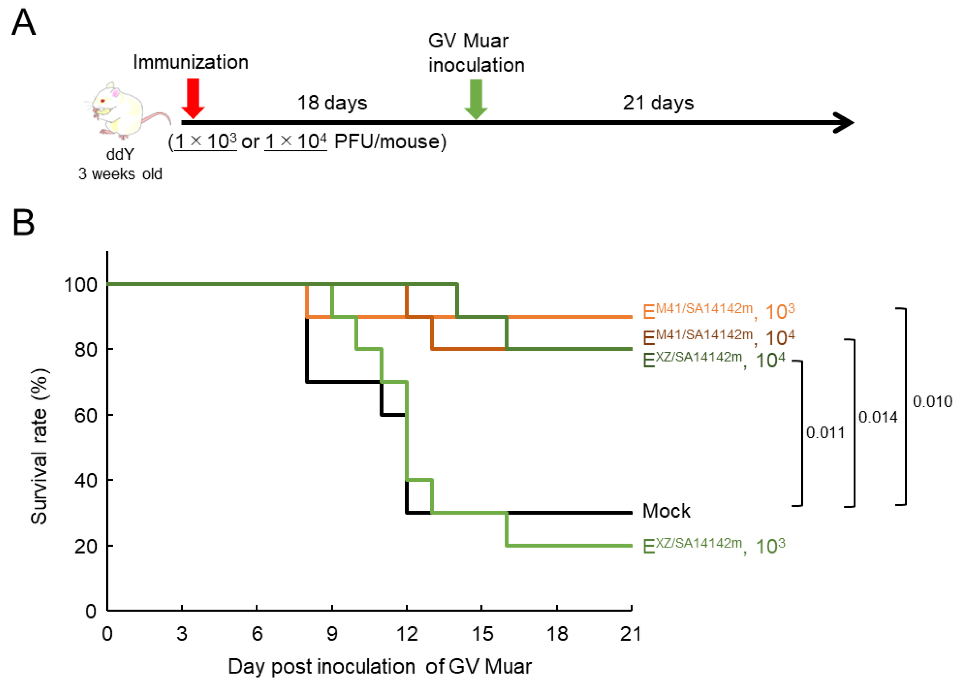
3.6. Protective Efficacy of EXZ/SA14142m and EM41.SA14142m Strains against GV Muar in Mice

Our results indicated that the mutant viruses EM41/SA14142m and EXZ/SA14142m display little or no virulence in mice, implying that these strains can be used as live attenuated vaccines against JEV infections. To investigate this possibility, mice inoculated once or twice with 1 × 10⁵ PFU of EM41/SA14142m and EXZ/SA14142m were inoculated i.p. with the GV Muar strain (Figure 6A). Before the Muar strain challenge, blood samples were collected from the mice, and PRNT50 against Muar was measured (Figure 6B and Table 2). Neutralizing antibodies against Muar were induced in most mice inoculated with the strains. However, PRNT50 was significantly higher in mice inoculated twice with EXZ/SA14142m. No obvious differences were observed in terms of neutralizing titers among the other three groups. No neutralization abilities (>1:10) were detected in three mouse sera, but antibodies against Muar were detected in the sera of the mice by IFAs (Table 2). All mice immunized with one or two doses of EM41/SA14142m or EXZSA14142m survived, while nine of ten mock-immunized mice died by day 16 after Muar challenge (Figure 6C). No evident loss in body weight was observed in the mutant-inoculated mouse groups (Figure S3).
Next, mice were inoculated with lower doses (1 × 10³ and 1 × 10⁴ PFU) of EM41/SA14142m or EXZ/SA14142m, and at 18 days after immunization, the mice were challenged i.p. with Muar (Figure 7A). One and two of ten mice immunized with 1 × 10³ and 1 × 10⁴ PFU of EM41/SA14142m, respectively, died, whereas eight and two of ten mice immunized with 1 × 10³ and 1 × 10⁴ PFU of EXZ/SA14142m, respectively, died (Figure 7B). Seven of ten mock-immunized mice died. Some mice with rapid and severe body weight loss survived, regardless of the virus used for immunization (Figure S4). This result suggested that the protective potential of EXZ/SA14142m against Muar may be weaker than that of EM41/SA14142m.

4. Discussion

In this study, we sought to develop a recombinant live attenuated JE vaccine that is effective against GV JEV infection using a reverse genetics system. In our previous study, we generated a recombinant GI-GV intertypic JEV EXZ0934, encoding the E protein of a recent GV isolate, XZ0934, using the GI strain Mie/41/2002 backbone [19]. We used EXZ0934 to generate a novel mutant virus EXZ/SA14142m. Ten amino acid substitutions related to the attenuation of the GIII JEV strain SA 14 were introduced in the E region of EXZ/SA14142m. In addition, we also produced a recombinant mutant virus, EM41/SA14142m, that was introduced to the same mutations in the E region of GI Mie/41/2002. The in vitro and in vivo properties of the mutant viruses were examined, and their ability to serve as a live attenuated vaccine against the GV JEV infection was evaluated. Previously, we generated a recombinant intertypic virus, rJEV-5NCMEXZ0934-M41, in which the 5′NCR-C-prM-E region was replaced with that of XZ0934 in the Mie/41/2002 backbone [21]. However, the prM protein is also involved in the high virulence of GV JEV; therefore, EXZ0934 was selected for this study.
Previous reports have shown that the introduction of multiple mutations found in the E region of SA 14-14-2 reduces the proliferative capacity of GI and GIII JEV [33,34,35,41]. Although EM41/SA14142m and EXZ/SA14142m also exhibited reduced growth capability compared with the parental viruses in Vero cells, the mutants maintained a peak infectious titer of approximately 107 PFU/mL (Figure 2B), indicating that viral replication was not critically impaired. However, the mutants presented marked decreases in proliferative ability in mouse neuroblastoma Neuro-2a and human neuroblastoma IMR-32 cells (Figure 2C,D). Moreover, no evident increase in infectious virus levels was observed in the culture supernatant of Neuro-2a cells inoculated with EXZ/SA14142m (Figure 2C). The growth rate of EXZ0934 was also lower than that of Mie/41/2002, and these intertypic viruses may be more sensitive to additional mutations than Mie/41/2002 in virus replication.
The mutants EXZ/SA14142m and EM41/SA14142m showed lower neuroinvasiveness than the parental strains in mice (Figure 3A). Infectious virus levels and genomic RNA levels in peripheral tissues and the brain were also lower in mice infected with the mutant viruses than in those infected with the parental viruses (Figure 5), suggesting that the low neuroinvasiveness of the mutants is due to the low proliferative potential of the mutants. In the neurovirulence analysis, all mice inoculated with the parental strains died within 6 days. In contrast, all mice inoculated with the mutant viruses survived until the end of the observation period without any symptoms (Figure 3B). This result indicated that the mutations substantially reduced the replicative ability and cytotoxicity of the parental viruses in central neurons.
In neutralization analysis using sera from surviving mice in Figure 3A, PRNT50 in sera inoculated with EXZ/SA14142m or EM41/SA14142m against GV Muar was lower than those in sera inoculated with EXZ0934 or Mie/41/2002 (Figure 4 and Table 1). These results could be attributed to the low proliferative potential of mutant viruses in mice. There was no significant difference in PRNT50 against Muar between EM41/SA14142m-inoculated mouse sera (<1:10–40, median 1:20) and EXZ/SA14142m-inoculated mouse sera (<1:10–80, median 1:80). PRNT50 against Mie/41/2002 was 1:40–160 (median 1:80) in EM41/SA14142m-inoculated mouse sera, whereas they were all <1:10 in EXZ/SA14142m-inoculated mouse sera. Furthermore, PRNT50 of EM41/SA14142m-inoculated serum against EM41/SA14142m was 1:80–1280 (median 1:320), whereas that of EXZ/SA14142m-inoculated serum against EXZ/SA14142m was <1:10–160 (median 1:30) (Table 1). These results raise the possibility that EXZ/SA14142m may have a weaker potential to induce neutralizing antibodies against JEV than EM41/SA14142m. The low growth capacity of EXZ/SA14142m may be associated with the low immunogenicity of the mutant virus in mice. The mutant viruses were used for immunization twice; PRNT50 against Muar was higher in EXZ/SA14142m-inoculated mouse sera (<1:10–320, median 1:320) than in EM41/SA14142m-inoculated mouse sera (1:10–160, median 1:40) (Table 2, Figure 6B), implying that the booster effect of EXZ/SA14142m was higher than that of EM41/SA14142m.
EXZ0934-inoculated mouse sera had a higher neutralizing titer against Muar than Mie/41/2002-inoculated sera, while Mie/41/2002-inoculated mouse sera had higher neutralizing titer against Mie/41/2002 than Muar-inoculated sera. These results are consistent with our previous findings that GI and GV JEV can be separated into antigenically distinct groups [19,24]. PRNT50 of EXZ0934-inoculated mouse sera against EXZ0934 was identical to that against Muar (Table 1), whereas PRNT50 of Mie/41/2002-infected and EM41/SA14142m-infected mouse sera against EXZ0934 appeared to be lower than those against Muar (Table 1). There is a difference of 6 amino acid residues in the E protein between Muar and XZ0934 (Figure S5), and the differences in E protein could influence the reactivity of sera with GV E proteins.
Our results demonstrated that a single inoculation with 1 × 10⁵ PFU of EXZ/SA14142m or EM41/SA14142m could block lethal GV Muar infection in mice (Figure 6). It is generally accepted that neutralizing antibodies with a PRNT50 of 1:10 or greater are sufficient to protect against the onset of JE [42]. However, the PRNT50 of sera from three mice inoculated with 1 × 10⁵ PFU of the mutant viruses against Muar were lower than 1:10 (Table 2). All mutant virus-inoculated mouse sera, including <1:10 of PRNT50 against Muar, were positive for antibodies against Muar in IFA (Table 2), indicating that all of the mice were immunized with the mutant viruses. Cellular immunity to non-structural proteins of JEV may also be induced in mutant virus-inoculated mice and have a suppressive effect on the pathogenesis caused by Muar infection [43,44,45]. Twenty percent of mice inoculated with 1 × 10⁴ PFU EM41/SA14142m in vitro and in of EM41/SA14142m or EXZ/SA14142m died; however, 10% of mice in the EM41/SA14142m group and 80% of mice in EXZ/SA14142m group died when 1 × 10³ PFU of the mutant viruses were inoculated (Figure 7B). As mentioned above, the growth ability of EXZ/SA14142m was lower than that of vivo, suggesting that EXZ/SA14142m does not amplify sufficiently to induce humoral and cellular immunity in mice infected with the virus at lower doses. It might be possible to recover the growth capacity and immunogenicity of EXZ/SA14142m by reducing the number of amino acid substitutions. Previous reports suggest that four amino acid residues at positions 107, 138, 176, and 244 of the 10 substitution sites play essential roles in the attenuation of neurovirulence and neuroinvasiveness of the SA 14 strain, but at least two at positions 177 and 264 seem to be dispensable for such attenuation [33,34,36,41]. A recombinant JEV with a minimum of four amino acid substitutions in the GV E protein may be a safe and highly efficacious live vaccine against GV JEV infection. The reverse genetics of GV JEV have already been established [18,46]. This method could be used to generate mutant GV JEV with the same amino acid substitutions in the E protein, and the viruses may represent highly proliferative but safe and effective live vaccine candidates. However, we previously showed that the growth of Muar is deficient in mouse neuroblastoma cells [19,22], indicating that the GV-based mutants would have low replicative potential in mouse-derived cells. Increasing the growth capacity of GVs by introducing new mutations might be required for GV-based live attenuated vaccine development. Our previous data showed that Muar could grow efficiently in human neuroblastoma-derived IMR-32 cells [21]. Thus the low growth ability of GV JEV in mouse-derived cells may not be critical for the development of live attenuated recombinant GV JE vaccines in humans.
Most amino acid residues in the 10 sites we introduced specific amino acid substitutions in this study are also conserved in JE serocomplex flaviviruses, the West Nile virus, Usutu virus, and Murray Valley encephalitis virus (Table S2). A previous report indicated that introducing all 10 substitutions into the attenuated West Nile virus strain W1806 resulted in defective virus propagation in mammalian cells [47]. ChimeriVax-WN, which has the prM-E region of West Nile virus in the live attenuated yellow fever vaccine strain 17D backbone, has three amino acid substitutions L107F, A316V (A315V in JEV), and K440R (K439R in JEV), and the mutations are critical for the reduction in neurovirulence of the chimeric virus [48]. However, a single E138K mutation in the E protein of West Nile virus does not attenuate its virulence [49]. Thus, the “multiple substitution method” could apply to the development of live attenuated vaccines for JE serocomplex flavivirus infections, but would also require careful selection of the positions introducing amino acid substitutions.
In this study, we used a mouse infection model to evaluate the protection and immunogenicity potency of the mutant viruses. However, a more comprehensive analysis using mice and non-human primates is needed to assess the safety and efficacy of the mutant virus against JEV infection before considering its application to humans.

Supplementary Materials

The following are available online at https://www.mdpi.com/article/10.3390/vaccines9101077/s1, Table S1: Comparison of the amino acid residues on 10 sites in JEV E protein. Table S2: Comparison of the amino acid residues at 10 sites in E protein of JEV, West Nile virus, Usutu virus, Murray valley encephalitis virus, and St. Louis encephalitis virus. Figure S1: Alignment of the nucleotide sequences of E region of recombinant JEV strains. Figure S2: Body weight of mice inoculated with recombinant JEV strains as shown in Figure 3A. Figure S3: Body weight of mice inoculated with recombinant JEV strains as shown in Figure 6C. Figure S4: Body weight of mice inoculated with recombinant JEV strains as shown in Figure 7B. Figure S5: Comparison of amino acid sequences of E protein of GV JEV Muar and XZ0934 strains.

Author Contributions

Conceptualization, S.T. (Shigeru Tajima); methodology, S.T. (Shigeru Tajima); validation, S.T. (Shigeru Tajima); investigation, S.T. (Shigeru Tajima); writing—original draft preparation, S.T. (Shigeru Tajima); writing—review and editing, S.T. (Shigeru Tajima), S.T. (Satoshi Taniguchi), E.N., T.M., T.I., M.S. and C.K.L., supervision, C.K.L.; project administration, S.T. (Shigeru Tajima), C.K.L.; funding acquisition, S.T. (Shigeru Tajima) and C.K.L. All authors have read and agreed to the published version of the manuscript.

Funding

This research was supported by the Research Program on Emerging and Re-emerging Infectious Diseases of the Japan Agency for Medical Research and Development (AMED) under Grant Number JP20fk0108123.

Institutional Review Board Statement

Experiments on mice were performed in accordance with the Guidelines for Animal Experiments Performed at the National Institute of Infectious Diseases (NIID), under approval (no. 119139, 120035, 120062, and 120117) from the Animal Welfare and Animal Care Committee of the NIID, Japan. All efforts were made to minimize pain and distress. The mice infected with JEV were observed daily for adverse reactions and signs of disease. For collection of organ samples, mice were euthanized using isoflurane.

Data Availability Statement

The data presented in this study are available on request from the corresponding author.

Acknowledgments

The authors thank Ken-ichi Shibasaki and Naoko Katsuta (National Institute of Infectious Diseases) and Niji Shiratori for their technical assistance.

Conflicts of Interest

The authors declare no conflict of interest. The authors had no role in the design of the study, collection, analyses, or interpretation of data, in the writing of the manuscript, or in the decision to publish the results.

References

  1. Tsai, T.F. New initiatives for the control of Japanese encephalitis by vaccination: Minutes of a WHO/CVI meeting, Bangkok, Thailand, 13–15 October 1998. Vaccine 2000, 18, 1–25. [Google Scholar] [CrossRef]
  2. Erlanger, T.E.; Weiss, S.; Keiser, J.; Utzinger, J.; Wiedenmayer, K. Past, present, and future of Japanese encephalitis. Emerg. Infect. Dis. 2009, 15, 1–7. [Google Scholar] [CrossRef] [PubMed]
  3. Campbell, G.L.; Hills, S.L.; Fischer, M.; Jacobson, J.A.; Hoke, C.H.; Hombach, J.M.; Marfin, A.A.; Solomon, T.; Tsai, T.F.; Tsu, V.D.; et al. Estimated global incidence of Japanese encephalitis: A systematic review. Bull. World Health Organ. 2011, 89, 766–774, 774A–774E. [Google Scholar] [CrossRef] [PubMed]
  4. Pierson, T.C.; Diamond, M.S. Flaviviruses. In Fields Virology, 6th ed.; David, M.K., Peter, M.H., Eds.; Lippincott Williams & Wilkins: Philadelphia, PA, USA, 2013; pp. 747–794. [Google Scholar]
  5. Uchil, P.D.; Satchidanandam, V. Phylogenetic analysis of Japanese encephalitis virus: Envelope gene based analysis reveals a fifth genotype, geographic clustering, and multiple introductions of the virus into the Indian subcontinent. Am. J. Trop Med. Hyg. 2001, 65, 242–251. [Google Scholar] [CrossRef] [PubMed]
  6. Solomon, T.; Ni, H.; Beasley, D.W.; Ekkelenkamp, M.; Cardosa, M.J.; Barrett, A.D. Origin and evolution of Japanese encephalitis virus in southeast Asia. J. Virol. 2003, 77, 3091–3098. [Google Scholar] [CrossRef] [PubMed] [Green Version]
  7. Gao, X.; Liu, H.; Wang, H.; Fu, S.; Guo, Z.; Liang, G. Southernmost Asia is the source of Japanese encephalitis virus (genotype 1) diversity from which the viruses disperse and evolve throughout Asia. PLoS Negl. Trop Dis. 2013, 7, e2459. [Google Scholar] [CrossRef] [Green Version]
  8. Schuh, A.J.; Ward, M.J.; Leigh Brown, A.J.; Barrett, A.D. Dynamics of the Emergence and Establishment of a Newly Dominant Genotype of Japanese Encephalitis Virus throughout Asia. J. Virol. 2014, 88, 4522–4532. [Google Scholar] [CrossRef] [Green Version]
  9. Yoshikawa, A.; Nabeshima, T.; Inoue, S.; Agoh, M.; Morita, K. Molecular and serological epidemiology of Japanese encephalitis virus (JEV) in a remote island of western Japan: An implication of JEV migration over the East China Sea. Trop Med. Health 2016, 44, 8. [Google Scholar] [CrossRef] [Green Version]
  10. Hameed, M.; Wahaab, A.; Nawaz, M.; Khan, S.; Nazir, J.; Liu, K.; Wei, J.; Ma, Z. Potential Role of Birds in Japanese Encephalitis Virus Zoonotic Transmission and Genotype Shift. Viruses 2021, 13, 357. [Google Scholar] [CrossRef]
  11. Schuh, A.J.; Guzman, H.; Tesh, R.B.; Barrett, A.D. Genetic diversity of Japanese encephalitis virus isolates obtained from the Indonesian archipelago between 1974 and 1987. Vector Borne. Zoonotic Dis. 2013, 13, 479–488. [Google Scholar] [CrossRef] [Green Version]
  12. Kuwata, R.; Torii, S.; Shimoda, H.; Supriyono, S.; Phichitraslip, T.; Prasertsincharoen, N.; Takemae, H.; Bautista, R.; Ebora, V.; Abella, J.A.C.; et al. Distribution of Japanese Encephalitis Virus, Japan and Southeast Asia, 2016–2018. Emerg. Infect. Dis. 2020, 26, 125–128. [Google Scholar] [CrossRef]
  13. Li, M.H.; Fu, S.H.; Chen, W.X.; Wang, H.Y.; Guo, Y.H.; Liu, Q.Y.; Li, Y.X.; Luo, H.M.; Da, W.; Duo Ji, D.Z.; et al. Genotype v Japanese encephalitis virus is emerging. PLoS Negl. Trop Dis. 2011, 5, e1231. [Google Scholar] [CrossRef]
  14. Kim, H.; Cha, G.W.; Jeong, Y.E.; Lee, W.G.; Chang, K.S.; Roh, J.Y.; Yang, S.C.; Park, M.Y.; Park, C.; Shin, E.H. Detection of Japanese encephalitis virus genotype V in Culex orientalis and Culex pipiens (Diptera: Culicidae) in Korea. PLoS ONE 2015, 10, e0116547. [Google Scholar] [CrossRef] [Green Version]
  15. Takhampunya, R.; Kim, H.C.; Tippayachai, B.; Kengluecha, A.; Klein, T.A.; Lee, W.J.; Grieco, J.; Evans, B.P. Emergence of Japanese encephalitis virus genotype V in the Republic of Korea. Virol. J. 2011, 8, 449. [Google Scholar] [CrossRef] [Green Version]
  16. Bae, W.; Kim, J.H.; Kim, J.; Lee, J.; Hwang, E.S. Changes of Epidemiological Characteristics of Japanese Encephalitis Viral Infection and Birds as a Potential Viral Transmitter in Korea. J. Korean Med. Sci. 2018, 33, e70. [Google Scholar] [CrossRef]
  17. Woo, J.H.; Jeong, Y.E.; Jo, J.E.; Shim, S.M.; Ryou, J.; Kim, K.C.; Lee, W.J.; Lee, J.Y. Genetic Characterization of Japanese Encephalitis Virus Genotype 5 Isolated from Patient, South Korea, 2015. Emerg. Infect. Dis. 2020, 26, 1002–1006. [Google Scholar] [CrossRef] [PubMed]
  18. de Wispelaere, M.; Frenkiel, M.P.; Despres, P. A Japanese Encephalitis virus genotype 5 molecular clone is highly neuropathogenic in a mouse model: Implication of the structural proteins region in virulence. J. Virol. 2015, 89, 5862–5875. [Google Scholar] [CrossRef] [PubMed] [Green Version]
  19. Tajima, S.; Yagasaki, K.; Kotaki, A.; Tomikawa, T.; Nakayama, E.; Moi, M.L.; Lim, C.K.; Saijo, M.; Kurane, I.; Takasaki, T. In vitro growth, pathogenicity and serological characteristics of the Japanese encephalitis virus genotype V Muar strain. J. Gen. Virol. 2015, 96, 2661–2669. [Google Scholar] [CrossRef]
  20. Cao, L.; Fu, S.; Gao, X.; Li, M.; Cui, S.; Li, X.; Cao, Y.; Lei, W.; Lu, Z.; He, Y.; et al. Low Protective Efficacy of the Current Japanese Encephalitis Vaccine against the Emerging Genotype 5 Japanese Encephalitis Virus. PLoS Negl. Trop Dis. 2016, 10, e0004686. [Google Scholar] [CrossRef] [PubMed]
  21. Tajima, S.; Shibasaki, K.I.; Taniguchi, S.; Nakayama, E.; Maeki, T.; Lim, C.K.; Saijo, M. E and prM proteins of genotype V Japanese encephalitis virus are required for its increased virulence in mice. Heliyon 2019, 5, e02882. [Google Scholar] [CrossRef] [Green Version]
  22. Tajima, S.; Taniguchi, S.; Nakayama, E.; Maeki, T.; Inagaki, T.; Lim, C.K.; Saijo, M. Amino Acid at Position 166 of NS2A in Japanese Encephalitis Virus (JEV) is Associated with In Vitro Growth Characteristics of JEV. Viruses 2020, 12, 709. [Google Scholar] [CrossRef] [PubMed]
  23. Maeki, T.; Tajima, S.; Kyaw, A.K.; Matsumoto, F.; Miura, K.; Yamashita, A.; Yoshikawa, A.; Negishi, K.; Noguchi, Y.; Tadokoro, K.; et al. Comparison of Neutralizing Antibody Titers against Japanese Encephalitis Virus Genotype V Strain with Those against Genotype I and III Strains in the Sera of Japanese Encephalitis Patients in Japan in 2016. Jpn. J. Infect. Dis. 2018, 71, 360–364. [Google Scholar] [CrossRef]
  24. Nguyen, T.T.T.; Tajima, S.; Ikeda, M.; Nguyen, T.T.; Le, T.T.H.; Pham, H.T.T.; Pham, D.Q.; Le, M.T.Q.; Maeki, T.; Taniguchi, S.; et al. Neutralization Potency of Sera from Vietnamese Patients with Japanese Encephalitis (JE) against Genotypes I and V JE Viruses. Jpn. J. Infect. Dis. 2019, 72, 115–117. [Google Scholar] [CrossRef] [PubMed] [Green Version]
  25. Li, X.; Ma, S.J.; Liu, X.; Jiang, L.N.; Zhou, J.H.; Xiong, Y.Q.; Ding, H.; Chen, Q. Immunogenicity and safety of currently available Japanese encephalitis vaccines: A systematic review. Hum. Vaccin. Immunother. 2014, 10, 3579–3593. [Google Scholar] [CrossRef] [PubMed] [Green Version]
  26. Satchidanandam, V. Japanese Encephalitis Vaccines. Curr. Treat Options Infect. Dis. 2020, 12, 375–386. [Google Scholar] [CrossRef]
  27. Monath, T.P.; Seligman, S.J.; Robertson, J.S.; Guy, B.; Hayes, E.B.; Condit, R.C.; Excler, J.L.; Mac, L.M.; Carbery, B.; Chen, R.T.; et al. Live virus vaccines based on a yellow fever vaccine backbone: Standardized template with key considerations for a risk/benefit assessment. Vaccine 2015, 33, 62–72. [Google Scholar] [CrossRef] [PubMed]
  28. Yu, Y. Phenotypic and genotypic characteristics of Japanese encephalitis attenuated live vaccine virus SA14-14-2 and their stabilities. Vaccine 2010, 28, 3635–3641. [Google Scholar] [CrossRef]
  29. Sumiyoshi, H.; Tignor, G.H.; Shope, R.E. Characterization of a highly attenuated Japanese encephalitis virus generated from molecularly cloned cDNA. J. Infect. Dis. 1995, 171, 1144–1151. [Google Scholar] [CrossRef]
  30. Ni, H.; Barrett, A.D. Molecular differences between wild-type Japanese encephalitis virus strains of high and low mouse neuroinvasiveness. J. Gen. Virol. 1996, 77, 1449–1455. [Google Scholar] [CrossRef]
  31. Zhao, Z.; Date, T.; Li, Y.; Kato, T.; Miyamoto, M.; Yasui, K.; Wakita, T. Characterization of the E-138 (Glu/Lys) mutation in Japanese encephalitis virus by using a stable, full-length, infectious cDNA clone. J. Gen. Virol. 2005, 86, 2209–2220. [Google Scholar] [CrossRef]
  32. Liang, J.J.; Liao, C.L.; Liao, J.T.; Lee, Y.L.; Lin, Y.L. A Japanese encephalitis virus vaccine candidate strain is attenuated by decreasing its interferon antagonistic ability. Vaccine 2009, 27, 2746–2754. [Google Scholar] [CrossRef]
  33. Gromowski, G.D.; Firestone, C.Y.; Whitehead, S.S. Genetic Determinants of Japanese Encephalitis Virus Vaccine Strain SA14-14-2 That Govern Attenuation of Virulence in Mice. J. Virol. 2015, 89, 6328–6337. [Google Scholar] [CrossRef] [Green Version]
  34. Yang, J.; Yang, H.; Li, Z.; Wang, W.; Lin, H.; Liu, L.; Ni, Q.; Liu, X.; Zeng, X.; Wu, Y.; et al. Envelope Protein Mutations L107F and E138K Are Important for Neurovirulence Attenuation for Japanese Encephalitis Virus SA14-14-2 Strain. Viruses 2017, 9, 20. [Google Scholar] [CrossRef] [Green Version]
  35. Zheng, X.; Zheng, H.; Tong, W.; Li, G.; Wang, T.; Li, L.; Gao, F.; Shan, T.; Yu, H.; Zhou, Y.; et al. Acidity/Alkalinity of Japanese Encephalitis Virus E Protein Residue 138 Alters Neurovirulence in Mice. J. Virol. 2018, 92, e00108-18. [Google Scholar] [CrossRef] [PubMed] [Green Version]
  36. Zhou, Y.; Wu, R.; Zhao, Q.; Chang, Y.F.; Wen, X.; Feng, Y.; Huang, X.; Wen, Y.; Yan, Q.; Huang, Y.; et al. Mutation of I176R in the E coding region weakens Japanese encephalitis virus neurovirulence, but not its growth rate in BHK-21 cells. Arch. Virol. 2018, 163, 1351–1355. [Google Scholar] [CrossRef] [PubMed]
  37. Anwar, M.N.; Wang, X.; Hameed, M.; Wahaab, A.; Li, C.; Sharma, M.; Pang, L.; Malik, M.I.; Liu, K.; Li, B.; et al. Phenotypic and Genotypic Comparison of a Live-Attenuated Genotype I Japanese Encephalitis Virus SD12-F120 Strain with Its Virulent Parental SD12 Strain. Viruses 2020, 12, 552. [Google Scholar] [CrossRef]
  38. Nerome, R.; Tajima, S.; Takasaki, T.; Yoshida, T.; Kotaki, A.; Lim, C.K.; Ito, M.; Sugiyama, A.; Yamauchi, A.; Yano, T.; et al. Molecular epidemiological analyses of Japanese encephalitis virus isolates from swine in Japan from 2002 to 2004. J. Gen. Virol. 2007, 88, 2762–2768. [Google Scholar] [CrossRef] [PubMed]
  39. Tajima, S.; Nerome, R.; Nukui, Y.; Kato, F.; Takasaki, T.; Kurane, I. A single mutation in the Japanese encephalitis virus E protein (S123R) increases its growth rate in mouse neuroblastoma cells and its pathogenicity in mice. Virology 2010, 396, 298–304. [Google Scholar] [CrossRef] [PubMed] [Green Version]
  40. Shirato, K.; Miyoshi, H.; Kariwa, H.; Takashima, I. Detection of West Nile virus and Japanese encephalitis virus using real-time PCR with a probe common to both viruses. J. Virol. Methods 2005, 126, 119–125. [Google Scholar] [CrossRef] [PubMed]
  41. Yun, S.I.; Song, B.H.; Kim, J.K.; Yun, G.N.; Lee, E.Y.; Li, L.; Kuhn, R.J.; Rossmann, M.G.; Morrey, J.D.; Lee, Y.M. A molecularly cloned, live-attenuated japanese encephalitis vaccine SA14-14-2 virus: A conserved single amino acid in the ij Hairpin of the Viral E glycoprotein determines neurovirulence in mice. PLoS Pathog. 2014, 10, e1004290. [Google Scholar] [CrossRef] [PubMed] [Green Version]
  42. Kurane, I.; Takasaki, T. Immunogenicity and protective efficacy of the current inactivated Japanese encephalitis vaccine against different Japanese encephalitis virus strains. Vaccine 2000, 18, 33–35. [Google Scholar] [CrossRef]
  43. Schlesinger, J.J.; Brandriss, M.W.; Walsh, E.E. Protection of mice against dengue 2 virus encephalitis by immunization with the dengue 2 virus non-structural glycoprotein NS1. J. Gen. Virol. 1987, 68, 853–857. [Google Scholar] [CrossRef] [PubMed]
  44. Putnak, J.R.; Schlesinger, J.J. Protection of mice against yellow fever virus encephalitis by immunization with a vaccinia virus recombinant encoding the yellow fever virus non-structural proteins, NS1, NS2a and NS2b. J. Gen. Virol. 1990, 71, 1697–1702. [Google Scholar] [CrossRef] [PubMed]
  45. Mishra, N.; Boudewijns, R.; Schmid, M.A.; Marques, R.E.; Sharma, S.; Neyts, J.; Dallmeier, K. A Chimeric Japanese Encephalitis Vaccine Protects against Lethal Yellow Fever Virus Infection without Inducing Neutralizing Antibodies. mBio 2020, 11, e02494-19. [Google Scholar] [CrossRef] [PubMed] [Green Version]
  46. Ishikawa, T.; Abe, M.; Masuda, M. Construction of an infectious molecular clone of Japanese encephalitis virus genotype V and its derivative subgenomic replicon capable of expressing a foreign gene. Virus. Res. 2015, 195, 153–161. [Google Scholar] [CrossRef]
  47. Yamshchikov, V.; Manuvakhova, M.; Rodriguez, E. Development of a human live attenuated West Nile infectious DNA vaccine: Suitability of attenuating mutations found in SA14-14-2 for WN vaccine design. Virology 2016, 487, 198–206. [Google Scholar] [CrossRef] [PubMed] [Green Version]
  48. Arroyo, J.; Miller, C.; Catalan, J.; Myers, G.A.; Ratterree, M.S.; Trent, D.W.; Monath, T.P. ChimeriVax-West Nile virus live-attenuated vaccine: Preclinical evaluation of safety, immunogenicity, and efficacy. J. Virol. 2004, 78, 12497–12507. [Google Scholar] [CrossRef] [PubMed] [Green Version]
  49. Kaiser, J.A.; Luo, H.; Widen, S.G.; Wood, T.G.; Huang, C.Y.; Wang, T.; Barrett, A.D.T. Japanese encephalitis vaccine-specific envelope protein E138K mutation does not attenuate virulence of West Nile virus. NPJ Vaccines 2019, 4, 50. [Google Scholar] [CrossRef]
Figure 1. (A) Schematic representation of the genomic structure of recombinant JEVs used in this study. Regions derived from GI Mie/41/2002 and GV XZ0934 strains are shown in blue and gray, respectively. Yellow lines indicate point mutation sites. Numbers: nucleotide positions in the Mie/41/2002 genome. (B) Enlargement of the E regions of rJEV-EXZ/SA14142m-M41 and rJEV-EM41/SA14142m-M41 genomes in (A). Ten amino acid substitution sites are indicated.
Vaccines 09 01077 g001
Figure 2. Growth properties of recombinant JEV strains. (A) Plaque phenotypes of Mie/41/2002, rJEV-EM41/SA14142m-M41 (EM41/SA14142m), rJEV-EXZ0934-M41 (EXZ0934), and rJEV-EXZ/SA14142m-M41 (EXZ/SA14142m) in Vero cells. (B,C) Growth curves measured for Mie/41/2002, EM41/SA14142m, EXZ0934, and EXZ/SA14142m in Vero (B), Neuro-2a (C), and IMR-32 (D) cells. Cells were plated into 6-well culture plates and infected with the JEV strains at a multiplicity of infection (MOI) of 0.1 PFU/cell. Values: means ± standard deviation from three independent inoculations. Significance (parental virus vs. mutant virus, day 3) was analyzed using the student’s t-test (** p < 0.01).
Vaccines 09 01077 g002
Figure 3. Murine neuroinvasiveness (A) and neurovirulence (B) of recombinant JEV strains. (A) Survival curve of mice inoculated intraperitoneally (i.p.) with 1 × 10⁵ PFU of Mie/41/2002 (n = 10), EM41/SA14142m (n = 10), EXZ0934 (n = 10), or EXZ/SA14142m (n = 10) and mice mock-inoculated (n = 10). (B) Survival rate of mice inoculated intracerebrally with 3 × 10³ PFU of Mie/41/2002 (n = 10), EM41/SA14142m (n = 10), EXZ0934 (n = 10), or EXZ/SA14142m (n = 10) and mice mock-inoculated (n = 10). Significant p values by log-rank test are also indicated.
Vaccines 09 01077 g003
Figure 4. Neutralization activity of serum taken from mice inoculated with recombinant JEV strains. Sera were collected from surviving mice inoculated with Mie/41/2002 (n = 8), EM41/SA14142m (E_M41/SA14142m, n = 10), EXZ0934 (E_XZ0934, n = 3), or EXZ/SA14142m (E_XZ/SA14142m, n = 10) and mice mock-inoculated (n = 10) as shown in Figure 3A, and used for measuring PRNT50 against JEV Muar (A) and Mie/41/2002 (B) strains. Dotted line: detection limit. Significance was analyzed using the Kruskal-Wallis test (* p < 0.05, ** p < 0.01, *** p < 0.001); NS: not statistically significant.
Vaccines 09 01077 g004
Figure 5. Levels of infectious virus (A,C) and viral RNA (B,D) at two (A,B) and five (C,D) days after inoculation of JEV-infected mice. Mice inoculated i.p. with 1 × 10⁴ PFU of Mie/41/2002 (n = 5), EM41/SA14142m (E_M41/SA14142m, n = 5), EXZ0934 (E_XZ0934, n = 5), or EXZ/SA14142m (E_XZ/SA14142m, n = 5) were euthanized at two or five days after inoculation, and serum, brain, and spleen samples were collected. Sera and tissue homogenates were used to quantify the infectious virus titer (PFU/mL or g) (A,C) and viral genome (genome copies/mL or g) (B,D). Dotted line: detection limit. Significance was analyzed using the Kruskal-Wallis test (* 0.01 < p < 0.05).
Vaccines 09 01077 g005
Figure 6. Protective capacity of high-titer recombinant JEV strains against GV Muar strain in mice. (A) Experimental timeline. For single-dose immunization groups, mice were inoculated i.p. with 1 × 10⁵ PFU of EM41/SA14142m (n = 10) or EXZ/SA14142m (n = 10), or mock-inoculated (n = 10). Double-dose immunization group mice were inoculated i.p. with 1 × 10⁵ PFU of EM41/SA14142m (n = 10) or EXZ/SA14142m (n = 10) and were immunized again two weeks after the first immunization. At five weeks after the initial immunization, mice were bled to determine serum PRNT50 and then inoculated i.p. with 1 × 10⁴ PFU of Muar strain and observed for 20 days. (B) Neutralization activity of serum taken from immunized mice against GV Muar. Dotted line: detection limit. Significance was analyzed using the Kruskal-Wallis test (* 0.01 < p < 0.05); NS: not statistically significant. (C) Survival curve of immunized mice after challenge with GV Muar strain. Significant p values by log-rank test are also indicated.
Vaccines 09 01077 g006
Figure 7. Protective ability of low-titer recombinant JEV strains against GV Muar strain in mice. (A) Experimental timeline. Mice were inoculated i.p. with 1 × 10³ PFU or 1 × 10⁴ PFU of EM41/SA14142m (n = 10) or EXZ/SA14142m (n = 10), or mock-inoculated (n = 10). At 18 days after immunization, mice were inoculated i.p. with 1 × 10⁴ PFU of Muar strain and observed for 21 days. (B) Survival curve of immunized mice after challenge with GV Muar strain. Significant p values by log-rank test are also indicated.
Vaccines 09 01077 g007
Table 1. Neutralization titers (PRNT50) of sera from mice infected with recombinant JEVs against GI, GV, and recombinant JEVs 1.
MouseMouse No.PRNT50 against:
Muar (GV)Mie/41/2002
(GI)
EXZ0934EXZ/SA14142mEM41/SA14142m
Mie/41/2002-inoculated group14016080NT 2NT
28032040
316062010
432064040
58064020
6160128020
716064020
816032040
median16048030
EM41/SA14142m-inoculated group1<1040<10NT160
2204010320
3<108010320
42016020640
54016020320
61040<1080
72040<10320
810160101280
92080<10320
1040160101280
median208010320
EXZ0934-inoculated group164040640NTNT
232080320
3640160640
median64080640
EXZ/SA14142m-inoculated group180<10160160NT
240<102080
3<10<10<10<10
420<101040
520<101020
610<102020
720<101010
880<104040
920<101020
1010<10<1040
median20<101530
1 Serum was recovered from mice 3 weeks after inoculation of the viruses and then used for PRNT. 2 NT, not tested.
Table 2. Neutralization titers (PRNT50) and results of indirect immunofluorescent analysis (IFA) of sera from mice inoculated with one or two doses of recombinant JEVs 1.
MouseMouse No.PRNT50 against MuarIFA (Serum Dilution Rate) 2
×20×200
EM41/SA14142m
1 dose
group
120+±
240++
3<10+±
480++
520++
640+
720+
820+
940++
1080++
median40
EM41/SA14142m
2 doses
group
140++
240+
3160+±
420++
540++
640++
740+
840++
940++
1010++
median40
EXZ/SA14142m
1 dose
group
1<10+
2160+±
310+
440++
520+±
620+
710+
820+±
910++
1020++
median20
EXZ/SA14142m
2 doses
group
1320++
2160++
380+±
4<10++
5320++
6320++
7320++
8320++
9160++
1040++
median320
1 Serum was recovered from mice 5 weeks after the initial inoculation of viruses and then used for PRNT against GV Muar strain and for IFA to stain Muar-infected Vero cells. 2 +, positive; −, negative; ±, uncertain.
Publisher’s Note: MDPI stays neutral with regard to jurisdictional claims in published maps and institutional affiliations.

