The Potential of Intraepithelial Lymphocytes as Immunological Targets for the Induction of Innate and Adaptive Memory Responses Through Mucosal Vaccination and Prime Boost

Abstract
1. Introduction
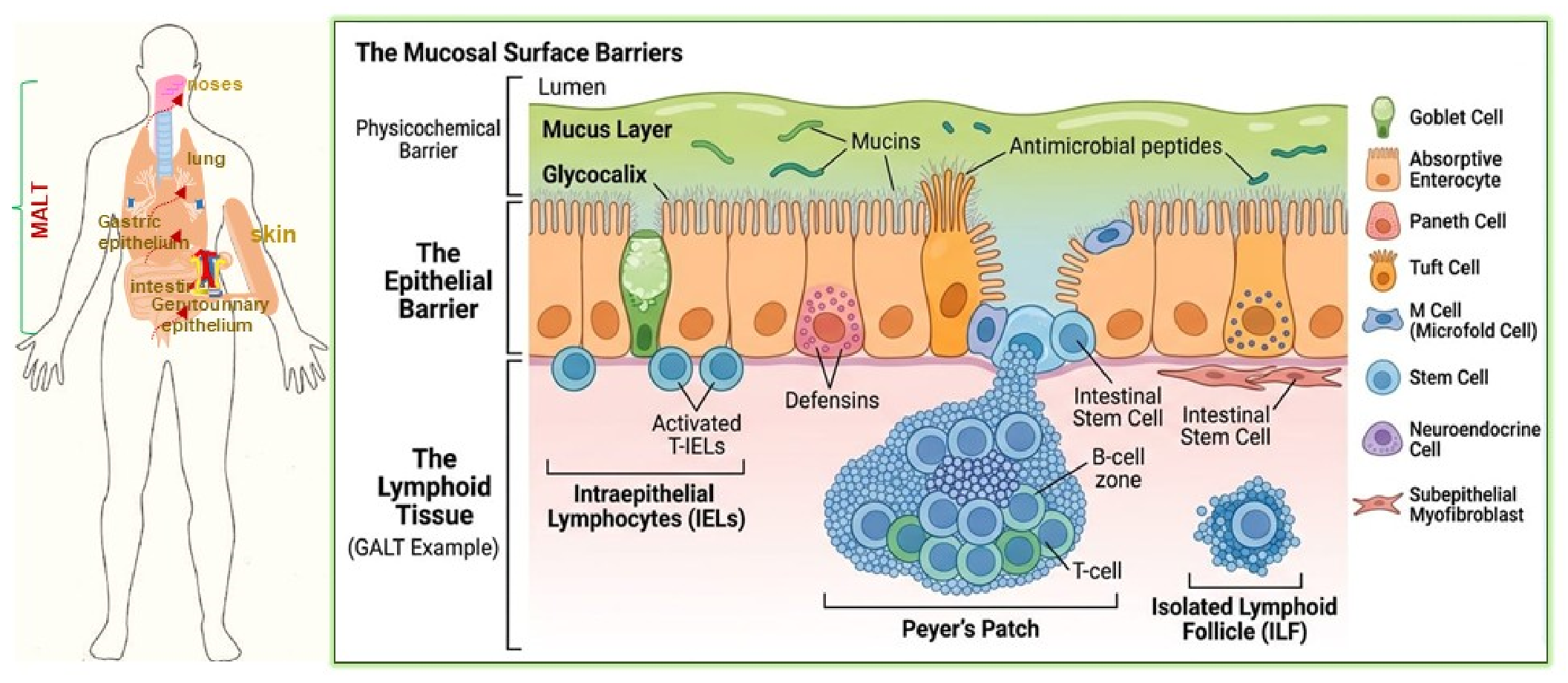
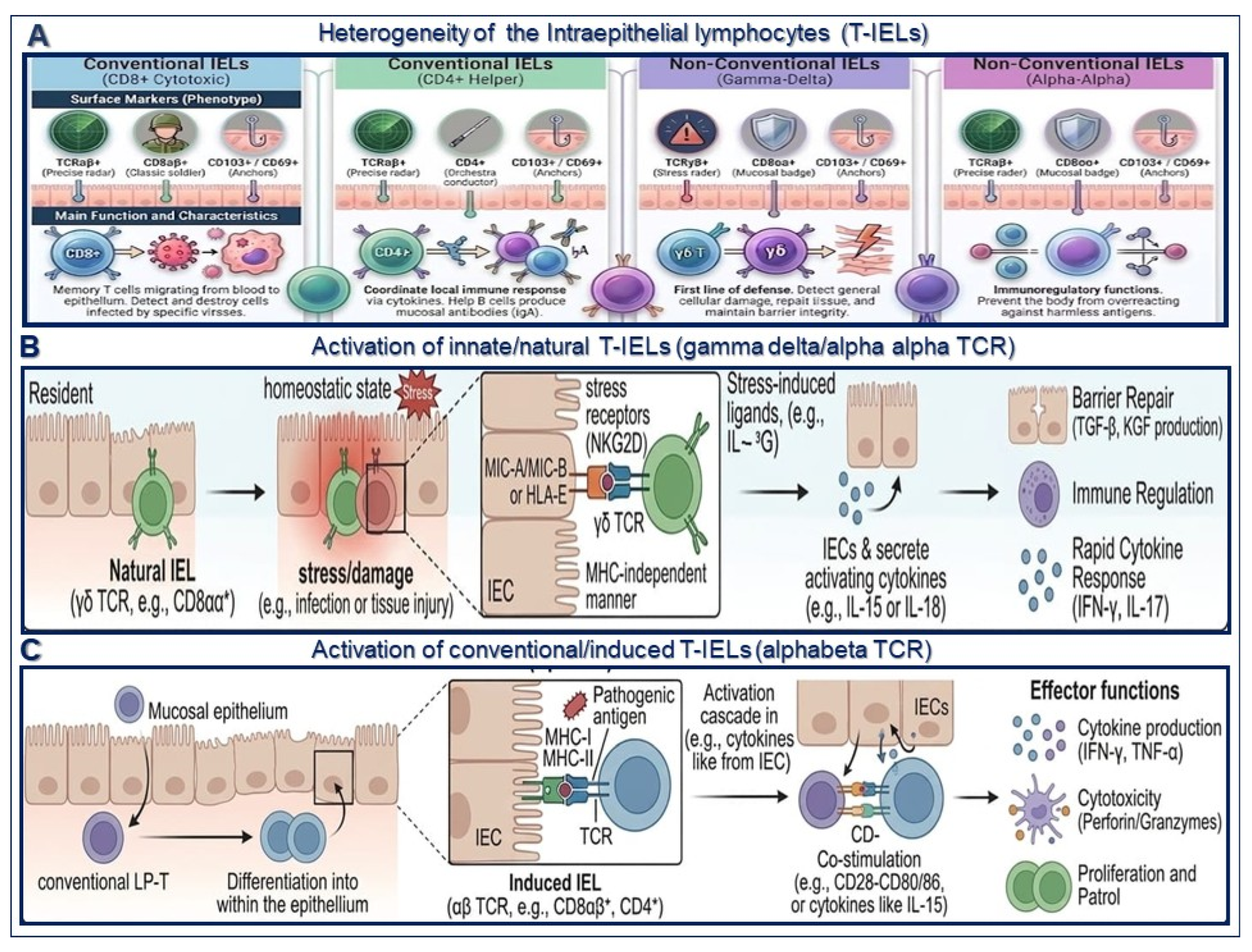
2. Heterogeneity of Intraepithelial Lymphocytes (T-IELs) in MALT
2.1. Heterogeneity of T-IELs in MALT That Depends of
2.1.1. -Infection Site
2.1.2. -Type of Pathogen
3. Mucosal Vaccination and Prime Boost Strategies for the Induction of Innate and Adaptive Memory
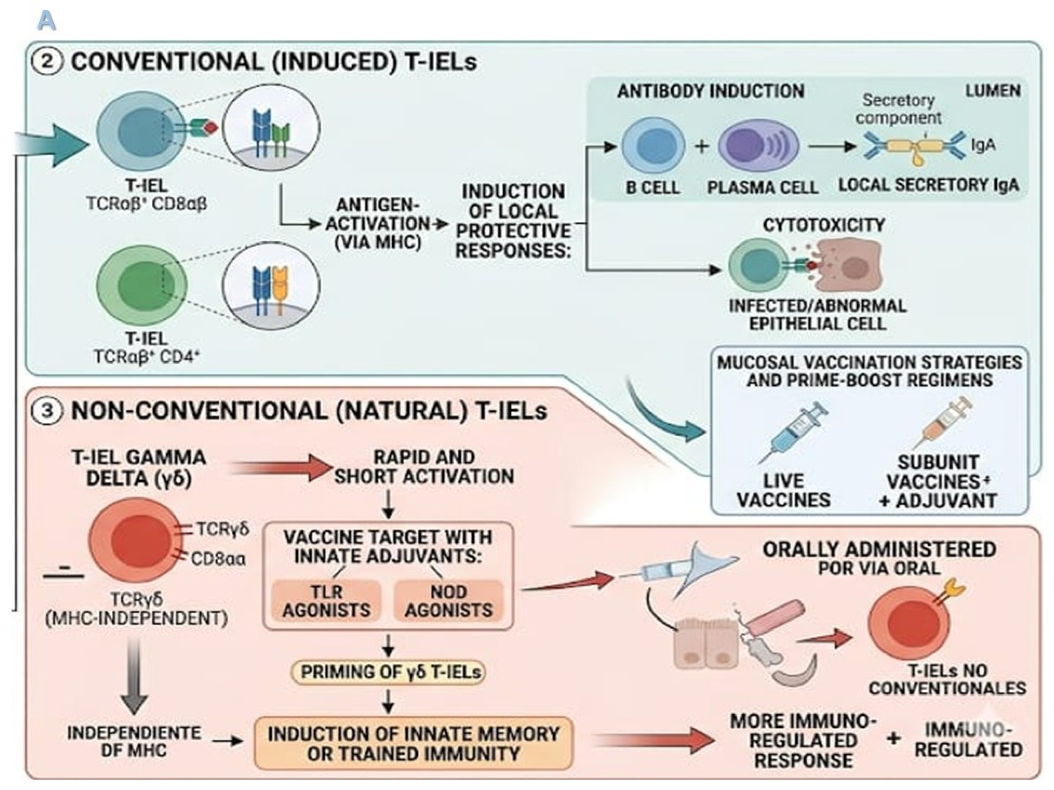
Targeting T-IELs Through Mucosal Vaccination Depends on How the Immune System Is Priming. To Perform This, the Type of Antigen, System Delivery and Route of Vaccination Play a Pivotal Role (Figure 3A)
- -
- Type of antigen. Antigens are used to mimic natural infection as live vectors and induce the proliferative burst and efficient effector and memory B and T cells in mucosal tissues. In addition, live vectors actively infect host cells or actively cross mucosal barriers (mimicking a natural infection), and provide massive amounts of endogenous danger signals (PAMPs). This triggers an immediate and robust innate immune cascade that drives intense T cell clonal expansion. A recent report showed that by mining the CD4+ T cell repertoire, it is possible to screen TB protective antigens, with an augmented and superior performance to the BCG vaccine. These antigens are also recognized in humans exposed to M. tuberculosis.
- -
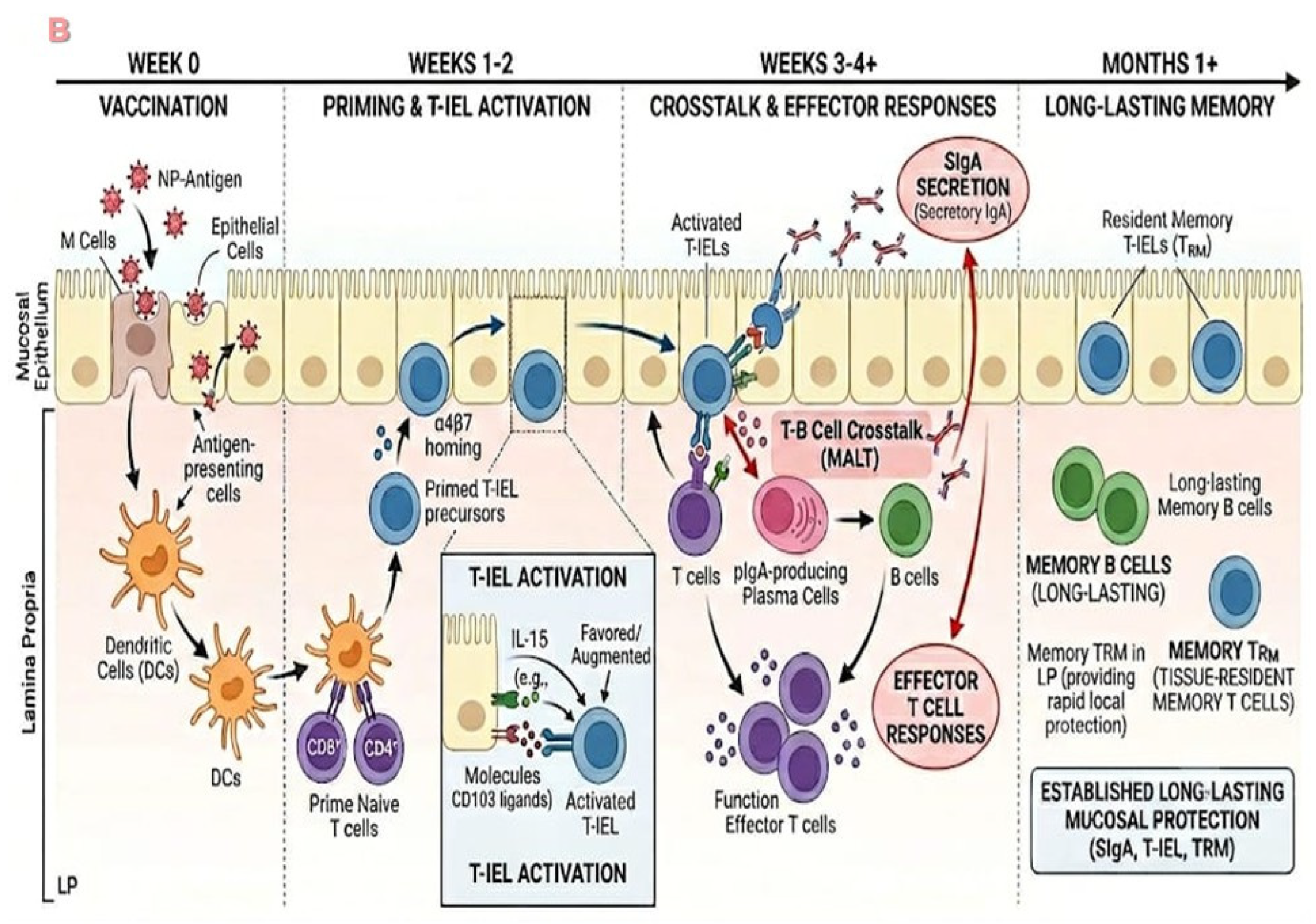
- System delivery. As a part of the vaccine formulation, a drug delivery formulation specifically targets the M cells on the mucosal epithelium of GALT [66,67,68,69,70] (Figure 3B). These Ag vaccine formulations utilize distinct immunological mechanisms but essentially rely on the basic and fundamental aims, that is, to mimic utmost natural infections (microbial or viral infectious disease) [71] and to activate the entire immune system, innate and adaptive, resulting in an inflammatory, and effector and memory immune response [53,71,72]. In the vaccine formulation, adjuvants on adenoviral-vectored (i.e., COVID-19 or measles) [73,74,75,76,77,78,79] multiepitope vaccines can also be used. The mechanism relies on a particle aided by the adjuvants (such as aluminum, chitosan, CpG polymers, ISCOMS, recombinant bacteria, or inactivated toxins) [76,78,80,81] toward passive uptake by mucosal antigen-presenting cells (APCs), leading to the induction of a pro-inflammatory response (IL-6, IL-12, and TNF-α), and then the activation of the cellular immune response, in terms of cytokines (IFN-γ, IL-4, IL-15, IL-18) for the differentiation of naïve CD4+ T cells; subsets of Th1, Th2, Th3, and Th17, for the activation of CD8+ T cells; and iNKT, NK, and TRM/IELs, for the differentiation of plasmacytoid B cells and the production of antibodies (sIgA) [69,79,82] (Figure 3B).
- -
- By other hand, mucosal vaccines depend heavily on the delivery platform such as viral vectors, bacterial vectors, and nanoparticles to activate and establish the TRM through mucosal vaccination [1,2,3,28,52,66,67,68,70,83]. Nanoparticles are used as live vectors to mimic natural infection and induce the proliferative burst and efficient effector and memory B and T cells in mucosal tissues. There is also a novel delivery system based on the yeast Saccharomyces cerevisiae (S. cerevisiae) for oral vaccine formulations. This system is safe and effective for delivering heterologous antigens (i.e., VP2 capsid protein) (PAMPS) as agonists of TLRs (PRRS) to induce systemic and mucosal immune responses, including antibody responses mainly of IgG and IgA [84]. This type of mucosal adjuvant candidate has been formulated as nano-carriers or nanoparticles based on chitosan or viral particles [11,12,80,81,84,85,86,87,88,89,90,91], a formulation that can be accessible and taken up by M cells, and transported to the inductive sites in the lung or GALT. Regarding dry powder vaccines, or drug formulations for immunization by the nasal route, there are several advantages to this delivery system: the dose, the cost, and, importantly, the priming and induction of systemic and mucosal immune responses [80,92]. Several reports in the literature have described the use of next-generation vaccine platforms as a delivery system, based on microbial organisms (i.e., Salmonella) as carriers of specific heterologous antigens (i.e., outer membrane vesicles) and molecule-like receptors (i.e., FcRN) for the induction of opsonizing antibodies and cellular immune response (CD4+ T cells) [93,94,95] (Figure 3B).
- -
- Route of vaccination. Vaccines administered by the mucosal route are sampled M or DCs cells in the lumen of the mucosal epithelium of MALT (gut, lung and urogenital tract) uptake and transported through mucus and across specialized epithelia to reach tissue-specific immune cells, lymphoid structures and secondary inductive sites in the lymph nodes (axial, close to BALT and lung) and Peyer’s patches in GALT. This results in the induction of protective and long-lasting memory immune responses of antibodies (mostly secretory IgA) (SIgA), and TRM [1,3,25,36,53]. Due to the heterologous boosting at local sites (mucosal administration versus intramuscular), secondary B cell responses are higher and show cross-reactivity compared to those after intramuscular boosting [96,97,98,99,100,101,102,103]. Intramuscular DNA vaccine priming and intranasal boosting with live attenuated virus influenza-vectored vaccine (LAIV) 1 induced migration of the systemic central memory T cells (TCM) via CXCR3-dependent chemotaxis to the local sites/MALT (lung mucosal epithelium) for differentiation into TRM, and increased CD8+ TRM cells in the lung [102,103]. In addition, priming and boosting for M. tuberculosis infection produced induction adaptive immune responses of CD8+ T cells and CD4+ T cells, while γδ IL-17-producing cells enable immune protective immune responses [96,99,104]. Of note, a vaccine formulation consisting of a nano-emulsion (NE) and an RNA-based RIG-I agonist (IVT), a protein-based SARS-CoV-2 vaccine, with priming by the parenteral route (intramuscular), initiates a route that can be rerouted by IN immunization, driving the maturation of B and T cellular immune responses, to a protective and memory local response against Orto poxviruses (OPXVs) and against SARS-CoV-2 [82,100,105,106,107]. In either case, an increase in anti-S IgG immune status ratio (ISR) [79,82] and effective production of neutralizing antibodies were observed in a murine model [100]. Moreover, using a prime boost regimen against the chicken infectious anemia virus [99], consisting of DNA priming and boosting with a recombinant protein, elicited higher antibody titers and significant amounts of IL2, IL4, and IFN-γ [101]. Studies have shown that it is possible to induce airway luminal T cell responses accompanied by the induction of Th17-mediated IL-17 and IL-23, which has been shown to protect mice challenged with M. tuberculosis [105,106,107], by combining the parenteral route of immunization with priming based on attenuated microorganisms, followed by IN boosting with recombinant proteins [79,82,108,109] (Figure 3B).
4. Remarks
- -
- The challenges and limitations of targeting T-IELs through mucosal vaccination and prime boost strongly are dictated by the nature, the dose and the formulation of the vaccine, to avoid inducing excessive inflammation or autoimmunity. In fact, the dichotomous role of T-IELs is to maintain homeostasis and tolerance at the mucosal epithelium. Usually upon mucosal vaccination, vaccine antigens should be optimized to be delivered as a particulate formulation to reach the inductive sites, under the premise that the role of vaccination is to mimic to the utmost the microbial/viral infectious disease.
- -
- How can these strategies really enhance proliferation, activation and memory responses? Upon oral/nasal vaccination of a particulate system (liposomes, viral particles, or nanoparticles), the innate immune cells, the APCs, M, epithelial cells and the arms of dendritic cells take the antigen up, transport it to the inductive sites and present it to T-IELs, which already are in a threshold activation state, ready to act.
- -
- How they can react to this antigen presentation, and to repeated antigen vaccination, will depend strongly on the dosification of the stimulus and the route of immunization. For example, heterologous prime boost protocols, IM/IN, or IN/IN, have been shown to induce protection against challenges of M. tuberculosis, Salmonella typhymurium, and other viruses, mostly due to the activation and induction of γδT-IEL Th-17 cells.
- -
- One of the hallmarks of the recent vaccine development is to take into account that vaccine candidates must mimic to the utmost the microbial/virus or the pathogens. Particulate formulations, based on liposomes or nanoparticles, are chosen to trigger and activate systemic and mucosal immunity; otherwise, they may reach the systemic compartments but not MALT.
- -
- The development of vaccines and immunotherapies has been accelerated by the use of artificial intelligence, speeding up the design, modeling, and prediction of effectiveness in preclinical animal models. Furthermore, recent reports have shown that mining the CD4 repertoire systematically screened for protective Tb antigens in preclinical models, which are also recognized in humans exposed to M. tuberculosis, which showed higher and enhanced protection than the BCG vaccine alone. These results provide a novel platform of trivalent mRNA–lipid nanoparticle (RNA-LNP) vaccines for clinical development.
Author Contributions
Funding
Data Availability Statement
Acknowledgments
Conflicts of Interest
References
- Leceta, J.; Del Campo, R.; Jordan, S.; Klose, C.S.N. Editorial: Immunoregulation at mucosal surfaces. Front. Immunol. 2022, 13, 983201. [Google Scholar] [CrossRef] [PubMed]
- Smith, P.; Blumberg, R.; MacDonald, T.; Society for Mucosal Immunology (Eds.) Principles of Mucosal Immunology, 2nd ed.; Garland Science: New York, NY, USA, 2020. [Google Scholar] [CrossRef]
- Allie, S.R.; Randall, T.D. Resident Memory B Cells. Viral Immunol. 2020, 33, 282–293. [Google Scholar] [CrossRef] [PubMed]
- Gozzi-Silva, S.C.; Teixeira, F.M.E.; Duarte, A.J.D.S.; Sato, M.N.; Oliveira, L.M. Immunomodulatory Role of Nutrients: How Can Pulmonary Dysfunctions Improve? Front. Nutr. 2021, 8, 674258. [Google Scholar] [CrossRef] [PubMed]
- Ansaldo, E.; Farley, T.K.; Belkaid, Y. Control of Immunity by the Microbiota. Annu. Rev. Immunol. 2021, 39, 449–479. [Google Scholar] [CrossRef] [PubMed]
- Ma, H.; Qiu, Y.; Yang, H. Intestinal intraepithelial lymphocytes: Maintainers of intestinal immune tolerance and regulators of intestinal immunity. J. Leukoc. Biol. 2021, 109, 339–347. [Google Scholar] [CrossRef] [PubMed]
- Han, M.; Rajput, C.; Ishikawa, T.; Jarman, C.R.; Lee, J.; Hershenson, M.B. Small Animal Models of Respiratory Viral Infection Related to Asthma. Viruses 2018, 10, 682. [Google Scholar] [CrossRef] [PubMed]
- Marozzi, M.; Parnigoni, A.; Negri, A.; Viola, M.; Vigetti, D.; Passi, A.; Karousou, E.; Rizzi, F. Inflammation, Extracellular Matrix Remodeling, and Proteostasis in Tumor Microenvironment. Int. J. Mol. Sci. 2021, 22, 8102. [Google Scholar] [CrossRef] [PubMed]
- Omahdi, Z.; Horikawa, Y.; Nagae, M.; Toyonaga, K.; Imamura, A.; Takato, K.; Teramoto, T.; Ishida, H.; Kakuta, Y.; Yamasaki, S. Structural insight into the recognition of pathogen-derived phosphoglycolipids by C-type lectin receptor DCAR. J. Biol. Chem. 2020, 295, 5807–5817. [Google Scholar] [CrossRef] [PubMed]
- Sadhu, S.; Kumar, S.; Mitra, D.K.; Joshi, B. Activated TLR2/4-positive T cells boost cell exhaustion during lepromatous leprosy infection via PD-1 upregulation. Heliyon 2022, 8, e11633. [Google Scholar] [CrossRef] [PubMed]
- Xie, B.; Wang, M.; Zhang, X.; Zhang, Y.; Qi, H.; Liu, H.; Wu, Y.; Wen, X.; Chen, X.; Han, M.; et al. Gut-derived memory gammadelta T17 cells exacerbate sepsis-induced acute lung injury in mice. Nat. Commun. 2024, 15, 6737. [Google Scholar] [CrossRef] [PubMed]
- Kumar, V. Pulmonary Innate Immune Response Determines the Outcome of Inflammation during Pneumonia and Sepsis-Associated Acute Lung Injury. Front. Immunol. 2020, 11, 1722. [Google Scholar] [CrossRef] [PubMed]
- Fischer, M.A.; Golovchenko, N.B.; Edelblum, K.L. γδ T cell migration: Separating trafficking from surveillance behaviors at barrier surfaces. Immunol. Rev. 2020, 298, 165–180. [Google Scholar] [CrossRef] [PubMed]
- Balaji, S.; Cholan, P.K.; Victor, D.J. An emphasis of T-cell subsets as regulators of periodontal health and disease. J. Clin. Transl. Res. 2021, 7, 648–656. [Google Scholar] [PubMed]
- Mintz, M.A.; Cyster, J.G. T follicular helper cells in germinal center B cell selection and lymphomagenesis. Immunol. Rev. 2020, 296, 48–61. [Google Scholar] [CrossRef] [PubMed]
- Planer, J.D.; Morrisey, E.E. After the Storm: Regeneration, Repair, and Reestablishment of Homeostasis Between the Alveolar Epithelium and Innate Immune System Following Viral Lung Injury. Annu. Rev. Pathol. 2023, 18, 337–359. [Google Scholar] [CrossRef] [PubMed]
- Dizaji Asl, K.; Mazloumi, Z.; Majidi, G.; Kalarestaghi, H.; Sabetkam, S.; Rafat, A. NK cell dysfunction is linked with disease severity in SARS-CoV-2 patients. Cell Biochem. Funct. 2022, 40, 559–568. [Google Scholar] [CrossRef] [PubMed]
- Li, Y.; Handley, S.A.; Baldridge, M.T. The dark side of the gut: Virome-host interactions in intestinal homeostasis and disease. J. Exp. Med. 2021, 218, e20201044. [Google Scholar] [CrossRef] [PubMed]
- Ivanov, I.I.; Tuganbaev, T.; Skelly, A.N.; Honda, K. T cell Responses to the Microbiota. Annu. Rev. Immunol. 2022, 40, 559–587. [Google Scholar] [CrossRef] [PubMed]
- Yu, Z.; Chen, J.; Liu, Y.; Meng, Q.; Liu, H.; Yao, Q.; Song, W.; Ren, X.; Chen, X. The role of potential probiotic strains Lactobacillus reuteri in various intestinal diseases: New roles for an old player. Front. Microbiol. 2023, 14, 1095555. [Google Scholar] [CrossRef] [PubMed]
- Mikami, Y.; Kuroda, E.; Kimura, S.; Hayatsu, M.; Watanabe, K.; Tsuda, H.; Nakamura, Y.; Sagawa, T.; Ichinose, T.; Takano, H.; et al. Bronchus-associated lymphoid tissue: A review of its development and function, including recent findings on the impact of environmental particulate exposure. Clin. Exp. Med. 2025, 26, 2. [Google Scholar] [CrossRef] [PubMed]
- Han, S. Unveiling an Important New Cell Type in the Lung: Microfold Cells. Am. J. Respir. Cell Mol. Biol. 2024, 70, 235–236. [Google Scholar] [CrossRef] [PubMed]
- Johnson, M.D.; Witherden, D.A.; Havran, W.L. The Role of Tissue-resident T Cells in Stress Surveillance and Tissue Maintenance. Cells 2020, 9, 686. [Google Scholar] [CrossRef] [PubMed]
- McArdle, S.; Seo, G.Y.; Kronenberg, M.; Mikulski, Z. Intravital Imaging of Intestinal Intraepithelial Lymphocytes. Bio-Protoc. 2023, 13, e4720. [Google Scholar] [CrossRef] [PubMed]
- Vandereyken, M.; James, O.J.; Swamy, M. Mechanisms of activation of innate-like intraepithelial T lymphocytes. Mucosal Immunol. 2020, 13, 721–731. [Google Scholar] [CrossRef] [PubMed]
- Nazmi, A.; McClanahan, K.G.; Olivares-Villagomez, D. Unconventional Intestinal Intraepithelial Lymphocytes in Health and Disease. Crit. Rev. Immunol. 2021, 41, 23–38. [Google Scholar] [CrossRef] [PubMed]
- Nie, J.; Carpenter, A.C.; Chopp, L.B.; Chen, T.; Balmaceno-Criss, M.; Ciucci, T.; Xiao, Q.; Kelly, M.C.; McGavern, D.B.; Belkaid, Y.; et al. The transcription factor LRF promotes integrin β7 expression by and gut homing of CD8αα+ intraepithelial lymphocyte precursors. Nat. Immunol. 2022, 23, 594–604. [Google Scholar] [CrossRef] [PubMed]
- Contreras, A.V.; Wiest, D.L. Development of γδ T Cells: Soldiers on the Front Lines of Immune Battles. Methods Mol. Biol. 2023, 2580, 71–88. [Google Scholar] [CrossRef] [PubMed]
- Lai, A.Y.; Patel, A.; Brewer, F.; Evans, K.; Johannes, K.; González, L.E.; Yoo, K.J.; Fromm, G.; Wilson, K.; Schreiber, T.H.; et al. Cutting Edge: Bispecific γδ T Cell Engager Containing Heterodimeric BTN2A1 and BTN3A1 Promotes Targeted Activation of Vγ9Vδ2+ T Cells in the Presence of Costimulation by CD28 or NKG2D. J. Immunol. 2022, 209, 1475–1480. [Google Scholar] [CrossRef] [PubMed]
- Sun, S.; Li, E.; Zhao, G.; Tang, J.; Zuo, Q.; Cai, L.; Xu, C.; Sui, C.; Ou, Y.; Liu, C.; et al. Respiratory mucosal vaccination of peptide-poloxamine-DNA nanoparticles provides complete protection against lethal SARS-CoV-2 challenge. Biomaterials 2023, 292, 121907. [Google Scholar] [CrossRef] [PubMed]
- Lei, H.; Alu, A.; Yang, J.; Ren, W.; He, C.; Lan, T.; He, X.; Yang, L.; Li, J.; Wang, Z.; et al. Intranasal administration of a recombinant RBD vaccine induces long-term immunity against Omicron-included SARS-CoV-2 variants. Signal Transduct. Target. Ther. 2022, 7, 159. [Google Scholar] [CrossRef] [PubMed]
- Dijkgraaf, F.E.; Kok, L.; Schumacher, T.N.M. Formation of Tissue-Resident CD8+ T-Cell Memory. Cold Spring Harb. Perspect. Biol. 2021, 13, a038117. [Google Scholar] [CrossRef] [PubMed]
- Wijeyesinghe, S.; Beura, L.K.; Pierson, M.J.; Stolley, J.M.; Adam, O.A.; Ruscher, R.; Steinert, E.M.; Rosato, P.C.; Vezys, V.; Masopust, D. Expansible residence decentralizes immune homeostasis. Nature 2021, 592, 457–462. [Google Scholar] [CrossRef] [PubMed]
- Crowl, J.T.; Heeg, M.; Ferry, A.; Milner, J.J.; Omilusik, K.D.; Toma, C.; He, Z.; Chang, J.T.; Goldrath, A.W. Tissue-resident memory CD8+ T cells possess unique transcriptional, epigenetic, and functional adaptations to different tissue environments. Nat. Immunol. 2022, 23, 1121–1131. [Google Scholar] [CrossRef] [PubMed]
- Palgen, J.L.; Feraoun, Y.; Dzangué-Tchoupou, G.; Joly, C.; Martinon, F.; Le Grand, R.; Beignon, A.S. Optimize Prime/Boost Vaccine Strategies: Trained Immunity as a New Player in the Game. Front. Immunol. 2021, 12, 612747. [Google Scholar] [CrossRef] [PubMed]
- Kuraoka, M.; Yeh, C.H.-Y.; Bajic, G.; Kotaki, R.; Song, S.H.; Windsor, I.; Harrsion, S.C.; Kelsoe, G. Recall of B cell memory depends on relative locations of prime and boost immunization. Sci. Immunol. 2022, 7, eabn5311. [Google Scholar] [CrossRef] [PubMed]
- Chowdhury, R.; Valainis, J.R.; Dubey, M.; von Boehmer, L.; Sola, E.; Wilhelmy, J.; Guo, J.; Kask, O.; Ohanyan, M.; Sun, M.; et al. NK-like CD8+ gammadelta T cells are expanded in persistent Mycobacterium tuberculosis infection. Sci. Immunol. 2023, 8, eade3525. [Google Scholar] [CrossRef] [PubMed]
- Zhou, D.-Y.; Bao, C.-F.; Zhou, G. Intraepithelial lymphocytes in human oral diseases. Front. Immunol. 2025, 16, 1597088. [Google Scholar] [CrossRef] [PubMed]
- Li, G.Q.; Xia, J.; Zeng, W.; Luo, W.; Liu, L.; Zeng, X.; Cao, D. The intestinal γd T cells: Functions in the gut and in the distant organs. Front. Immunol. 2023, 14, 1206299. [Google Scholar] [CrossRef] [PubMed]
- Dickson, K.B.; Stadnyk, A.W.; Zhou, J.; Lehmann, C. Mucosal immunity: Lessons from the lower respiratory and small intestinal epithelia. Biomedicines 2025, 13, 1052. [Google Scholar] [CrossRef] [PubMed]
- Bordoni, D.; Fazio, A. Crosstalk between T cell gene regulation and intestinal epithelial cells: Insights into mucosal immunity. In Advances in Genetics; Elsevier: Amsterdam, The Netherlands, 2025. [Google Scholar] [CrossRef] [PubMed]
- Visnyaiová, K.; Varga, I.; Feitscherova, C.; Pavlikova, L.; Zahumensky, J.; Mikušova, R. Morphology of the immune cells in the wall of the human uterine tube and their possible impact on reproduction—Uterine tube as a possible immune privileged organ. Front. Cell Dev. Biol. 2024, 12, 1325565. [Google Scholar] [CrossRef] [PubMed]
- Lockhart, A.; Mucida, D.; Bilate, A.M. Intraepithelial Lymphocytes of the Intestine. Annu. Rev. Immunol. 2024, 42, 289–316. [Google Scholar] [CrossRef] [PubMed]
- Yilmaz, F.; Atay, K.; Çirkin, G.; Sanmak, E. The impact of gastric Helicobacter pylori infection on duodenal mucosa: New evidence on the alteration of intraepithelial lymphocytes. Am. J. Clin. Pathol. 2025, 164, 443–454. [Google Scholar] [CrossRef] [PubMed]
- Livanos, A.E.; Jha, D.; Cossarini, F.; Gonzalez-Reiche, A.S.; Tokuyama, M.; Aydillo, T.; Parigi, T.L.; Ladinsky, M.S.; Ramo, I.; Dunleavy, K.; et al. Intestinal host response to SARS-CoV-2 infection and COVID-19 outcomes in patients with gastrointestinal symptoms. Gastroenterology 2021, 160, 2435–2450.e34. [Google Scholar] [CrossRef] [PubMed]
- Lehmann, M.; Allers, K.; Heldt, C.; Meinhardt, J.; Schmidt, F.; Rodriguez-Sillke, Y.; Kunkel, D.; Schumann, M.; Chotima Böttcher Stahl-Hennig, C.H.; Elezkurtag, S.; et al. Human small intestinal infection by SARS-CoV-2 is characterized by a mucosal infiltration with activated CD8+ T cells. Mucosal Immunol. 2021, 14, 1381–1392. [Google Scholar] [CrossRef] [PubMed]
- Flores, E.Y.; Hume, A.J.; Olejnik, J.; Mithal, A.; D’Amico, A.; Yang, M.; Bawa, P.; Wang, F.; O’Connell, A.K.; Tseng, A.; et al. Filovirus infection disrupts epithelial barrier function and ion transport in human iPSC-derived gut organoids. PLoS Pathog. 2025, 21, e1013698. [Google Scholar] [CrossRef] [PubMed]
- Perez-Valencia, L.J.; Vannella, K.M.; Ramos-Benitez, M.J.; Sun, J.; Abu-Asab, M.; Dorward, D.W.; Awad, K.S.; Platt, A.; Jacobson, E.; Kindrachuk, J.; et al. Ebola virus shed glycoprotein is toxic to human T, B, and natural killer lymphocytes. iScience 2023, 26, 107323. [Google Scholar] [CrossRef] [PubMed]
- Von Massow, G.; Oh, S.; Lam, A.; Gustafsson, K. Gamma Delta T Cells and Their Involvement in COVID-19 Virus Infections. Front. Immunol. 2021, 12, 741218. [Google Scholar] [CrossRef] [PubMed]
- Parsa, R.; London, M.; Rezende de Castro, T.B.; Reis, B.; Buissant des Amorie, J.; Smith, J.G.; Mucida, D. Newly recruited intraepithelial Ly6A+CCR9+CD4+ T cells protect against enteric viral infection. Immunity 2022, 55, 1234–1249. [Google Scholar] [CrossRef] [PubMed]
- Eleftheriotis, G.; Tsounis, E.P.; Aggeletopoulou, I.; Dousdampanis, P.; Triantos, C.; Mouzaki, A.; Marangos, M.; Assimakopoulos, S.F. Alterations in gut immunological barrier in SARS-CoV-2 infection and their prognostic potential. Front. Immunol. 2023, 14, 1129190. [Google Scholar] [CrossRef] [PubMed]
- Al-Nemrawi, N.K.; Darweesh, R.S.; Al-Shriem, L.A.; Al-Qawasmi, F.S.; Emran, S.O.; Khafajah, A.S.; Abu-Dalo, M.A. Polymeric Nanoparticles for Inhaled Vaccines. Polymers 2022, 14, 4450. [Google Scholar] [CrossRef] [PubMed]
- Matsumoto, R.; Ogata, K.; Takahashi, D.; Kinashi, Y.; Yamada, T.; Morita, R.; Tanaka, K.; Hattori, K.; Endo, M.; Fujimura, Y.; et al. AP-1B regulates interactions of epithelial cells and intraepithelial lymphocytes in the intestine. Cell. Mol. Life Sci. 2024, 81, 425. [Google Scholar] [CrossRef] [PubMed]
- Rani, S.B.E.; Diaz, F.E.; Maina, T.W.; Corbett, R.J.; Tuggle, C.H.K.; McGill, J.L. Evidence of innate training in bovine γδ T cells following subcutaneous BCG administration. Front. Immunol. 2024, 15, 1423843. [Google Scholar] [CrossRef] [PubMed]
- Jia, L.; Wu, G.; Alonso, S.; Zhao, C.; Lemenze, A.; Lam, Y.Y.; Zhao, L.; Edelblum, K.L. A transmissible γδ intraepithelial lymphocyte hyperproliferative phenotype is associated with the intestinal microbiota and confers protection against acute infection. Mucosal Immunol. 2022, 15, 772–788. [Google Scholar] [CrossRef] [PubMed]
- Suen, T.K.; Al, B.; Scarpa, A.; Dorhoi, A.; Netea, M.G.; Palcek, K. A dual nature of γδ T cell immune memory responses. eLife 2025, 14, e104887. [Google Scholar] [CrossRef] [PubMed]
- Netea, M.G.; Domínguez- Andreés, J.; Barreiro, L.B.; Chavakis, T.; Divangahi, M.; Fuchs, E.; Joosten, L.A.B.; van der Meer, J.W.M.; Mhlanga, M.M.; Mulder, W.J.M.; et al. Defining trained immunity and its role in health and disease. Nat. Rev. Immunol. 2020, 20, 375–388. [Google Scholar] [CrossRef] [PubMed]
- Guo, J.; Chowdhury, R.R.; Mallajosyula, V.; Xie, J.; Dubey, M.; Liu, Y.; Li, J.; Wei, Y.; Palanski, B.A.; Wang, C.; et al. γδ T cell antigen receptor polyspecificity enables T cell responses to a broad range of immune challenges. Proc. Natl. Acad. Sci. USA 2024, 121, 2315592121. [Google Scholar] [CrossRef] [PubMed]
- Serrano, R.; Lettau, M.; Zarobkiewicz, M.; Wesch, D.; Peters, C.; Kabelitz, D. Stimulatory and inhibitory activity of STING ligands on tumor- reactive human gamma/delta T cells. Oncoimmunology 2022, 11, 2030021. [Google Scholar] [CrossRef] [PubMed]
- Bevington, S.L.; Fiancette, R.; Gajdasik, D.W.; Keane, P.; Soley, J.K.; Willis, C.M.; Coleman, D.J.L.; Withers, D.R.; Cockerill, P.N. Stable Epigenetic Programming of Effector and Central Memory CD4 T Cells Occurs Within 7 Days of Antigen Exposure In Vivo. Front. Immunol. 2021, 12, 642807. [Google Scholar] [CrossRef] [PubMed]
- Mittelstaedt, N.N.; Becker, A.L.; de Freitas, D.N.; Zanin, R.F.; Stein, R.T.; Duarte de Souza, A.P. DNA Methylation and Immune Memory Response. Cells 2021, 10, 2943. [Google Scholar] [CrossRef] [PubMed]
- Hassert, M.; Harty, J.T. Tissue resident memory T cells- A new benchmark for the induction of vaccine-induced mucosal immunity. Front. Immunol. 2022, 13, 1039194. [Google Scholar] [CrossRef] [PubMed]
- Zhou, Y.; Qu, J.; Sun, X.; Yue, Z.; Liu, Y.; Zhao, K.; Yang, F.; Feng, J.; Pan, X.; Jin, Y.; et al. Delivery of spike-RBD by bacterial type three secretion system for SARS-CoV-2 vaccine development. Front. Immunol. 2023, 14, 1129705. [Google Scholar] [CrossRef] [PubMed]
- Lublin, A.; Katz, C.; Gruzdev, N.; Yadid, I.; Bloch, I.; Farnoushi, Y.; Simanov, L.; Berkowitz, A.; Elyahu, D.; Pitcovski, J.; et al. Protection against avian coronavirus conferred by oral vaccination with live bacteria secreting LTB-fused viral proteins. Vaccine 2022, 40, 726–733. [Google Scholar] [CrossRef] [PubMed]
- Peng, Z.; Cao, D.Y.; Wu, H.Y.; Saito, S. Immunization with a Bacterial Lipoprotein Establishes an Immuno-Protective Response with Upregulation of Effector CD4+ T Cells and Neutrophils Against Methicillin-Resistant Staphylococcus aureus Infection. Pathogens 2020, 9, 138. [Google Scholar] [CrossRef] [PubMed]
- McCright, C.J.; Maisel, K. Engineering drug delivery systems to overcome mucosal barriers for immunotherapy and vaccination. Tissue Barriers 2020, 8, 1695476. [Google Scholar] [CrossRef] [PubMed]
- Saggese, A.; Baccigalupi, L.; Donadio, G.; Ricca, E.; Isticato, R. The Bacterial Spore as a Mucosal Vaccine Delivery System. Int. J. Mol. Sci. 2023, 24, 10880. [Google Scholar] [CrossRef] [PubMed]
- Tang, W.; Zhang, Y.; Zhu, G. Pulmonary delivery of mucosal nanovaccines. Nanoscale 2022, 14, 263–276. [Google Scholar] [CrossRef] [PubMed]
- Vidal, S.J.; Lasrado, N.; Tostanoski, L.H.; Chaudari, J.; Mbiwan, E.R.; Neka, G.D.; Strutton, E.A.; Espinoza, P.A.A.; Sellers, D.; Barrett, J.; et al. Mining the CD4 antigen repertoire for next generation tuberculosis vaccines. Cell 2025, 188, 6791–6803.e13. [Google Scholar] [CrossRef] [PubMed]
- Freire, H.H.; Roe, E.F.; Collier, J.H. Expanding opportunities to engineer mucosal vaccination with biomaterials. Biomater. Sci. 2023, 11, 1625–1647. [Google Scholar] [CrossRef] [PubMed]
- Sargazi, S.; Arshad, R.; Ghamari, R.; Rahdar, A.; Bakhshi, A.; Karkan, S.F.; Ajalli, N.; Bilal, M.; Díez-Pascual, A.M. SiRNA-based nanotherapeutics as emerging modalities for immune-mediated diseases: A preliminary review. Cell Biol. Int. 2022, 46, 1320–1344. [Google Scholar] [CrossRef] [PubMed]
- Hartwell, B.L.; Melo, M.B.; Xiao, P.; Lemnios, A.A.; Li, N.; Chang, J.Y.H.; Yu, J.; Gebre, M.S.; Chang, A.; Maiorino, L.; et al. Intranasal vaccination with lipid-conjugated immunogens promotes antigen transmucosal uptake to drive mucosal and systemic immunity. Sci. Transl. Med. 2022, 14, eabn1413. [Google Scholar] [CrossRef] [PubMed]
- Afkhami, S.; Kang, A.; Jeyanathan, V.; Xing, Z.; Jeyanathan, M. Adenoviral-vectored next-generation respiratory mucosal vaccines against COVID-19. Curr. Opin. Virol. 2023, 61, 101334. [Google Scholar] [CrossRef] [PubMed]
- Guan, X.; Verma, A.K.; Liu, Q.; Palacios, M.; Oddle, A.E.; Perlman, S.; Du, L. Glycosylated Receptor-Binding-Domain-Targeting Mucosal Vaccines Protect Against SARS-CoV-2 Omicron and MERS-CoV. Vaccines 2025, 13, 293. [Google Scholar] [CrossRef] [PubMed]
- Lei, H.; Alu, A.; Yang, J.; He, C.; Shi, J.; Hong, W.; Peng, D.; Zhang, Y.; Liu, J.; Qin, F.; et al. Intranasal Inoculation of Cationic Crosslinked Carbon Dots-Adjuvanted Respiratory Syncytial Virus F Subunit Vaccine Elicits Mucosal and Systemic Humoral and Cellular Immunity. MedComm 2025, 6, e70146. [Google Scholar] [CrossRef] [PubMed]
- Zhao, Y.; Liu, J.; Peng, C.H.; Guo, S.H.; Wang, B.; Longping, C.; Wang, Y.; Tang, H.; Liu, L.; Pan, Q.; et al. Cross-protection against homo and heterologous influenza viruses via intranasal administration of an HA chimeric multiepitope nanoparticle vaccine. J. Nanobiotechnol. 2025, 23, 77. [Google Scholar] [CrossRef] [PubMed]
- Peng, C.; Tang, F.; Wang, J.; Cheng, P.; Wang, L.; Gong, W. Immunoinformatic-Based Multi-Epitope Vaccine Design for Co-Infection of Mycobacterium tuberculosis and SARS-CoV-2. J. Pers. Med. 2023, 13, 116. [Google Scholar] [CrossRef] [PubMed]
- Nakahashi, O.R.; Mori, H.; Yuki, Y.; Machita, T.; Kataka, Y.; Umemoto, S.H.; Uchida, Y.; Yamanoue, T.; Sawada, S.H.-I.; Ishige, K.; et al. Cationic nanogel-based nasal therapeutic HPV vaccine prevents the development of cervical cancer. Sci. Transl. Med. 2025, 17, eado8840. [Google Scholar] [CrossRef] [PubMed]
- Radaelli, A.; Zanotto, C.; Brambilla, C.; Adami, T.; Paolini, F.; Venuti, A.; Manuka, A.; Mehmeti, I.; De Giuli Morghen, C. Different immunogens and prime-boost vaccination strategies affect the efficacy of recombinant candidate vaccines against pathogenic orthopoxviruses. Virol. J. 2024, 21, 282. [Google Scholar] [CrossRef] [PubMed]
- Kazemi, M.; Madani, R.; Aghamaali, M.R.; Emami, T.; Golchinfar, F.; Heshmati, L. Preparation and Characterization of Nanoliposome Containing Isolated VP1 Protein of Foot and Mouth Disease Virus as a Model of Vaccine. Arch. Razi Inst. 2022, 77, 37–44. [Google Scholar] [CrossRef] [PubMed]
- Harrell, J.E.; Kurtz, J.R.; Bauer, D.L.; Prior, J.T.; Gellings, P.S.; Morici, L.A.; McLachlan, J.B. An Outer Membrane Vesicle-Adjuvanted Oral Vaccine Protects Against Lethal, Oral Salmonella Infection. Pathogens 2021, 10, 616. [Google Scholar] [CrossRef] [PubMed]
- Jeyanathan, M.; Vaseghi-Shanjani, M.; Afkhami, S.; Grondin, J.A.; Kang, A.; D’Agostino, M.R.; Yao, Y.; Jain, S.; Zganiacz, A.; Kroezen, Z.; et al. Parenteral BCG vaccine induces lung-resident memory macrophages and trained immunity via the gut-lung axis. Nat. Immunol. 2022, 23, 1687–1702. [Google Scholar] [CrossRef] [PubMed]
- Xu, J.; Dai, W.; Wang, Z.; Chen, B.; Li, Z.; Fan, X. Intranasal vaccination with chitosan-DNA nanoparticles expressing pneumococcal surface antigen a protects mice against nasopharyngeal colonization by Streptococcus pneumoniae. Clin. Vaccine Immunol. 2011, 18, 75–81. [Google Scholar] [CrossRef] [PubMed]
- Cao, P.; Han, F.Y.; Grøndahl, L.; Xu, Z.P.; Li, L. Enhanced Oral Vaccine Efficacy of Polysaccharide-Coated Calcium Phosphate Nanoparticles. ACS Omega 2020, 5, 18185–18197. [Google Scholar] [CrossRef] [PubMed]
- Wang, N.; Wei, C.; Zhang, Z.; Liu, T.; Wang, T. Aluminum Nanoparticles Acting as a Pulmonary Vaccine Adjuvant-Delivery System (VADS) Able to Safely Elicit Robust Systemic and Mucosal Immunity. J. Inorg. Organomet. Polym. Mater. 2020, 30, 4203–4217. [Google Scholar] [CrossRef] [PubMed]
- J, L.A.A.; Pa, P.; Seng, C.Y.; Rhee, J.H.; Lee, S.E. Protein nanocages: A new frontier in mucosal vaccine delivery and immune activation. Hum. Vaccine Immunother. 2025, 21, 2492906. [Google Scholar] [CrossRef] [PubMed]
- Cazzola, M.; Ora, J.; Calzetta, L.; Rogliani, P.; Matera, M.G. The future of inhalation therapy in chronic obstructive pulmonary disease. Curr. Res. Pharmacol. Drug Discov. 2022, 3, 100092. [Google Scholar] [CrossRef] [PubMed]
- Feng, F.; Wen, Z.; Chen, J.; Yuan, Y.; Wang, C.; Sun, C. Strategies to Develop a Mucosa-Targeting Vaccine against Emerging Infectious Diseases. Viruses 2022, 14, 520. [Google Scholar] [CrossRef] [PubMed]
- Parriott, J.E.; Stewart, J.P.; Smith, D.D.; Curran, S.M.; Bauer, C.D.; Wyatt, T.A.; Phillips, J.A.; Lyden, E.; Thiele, G.M.; Vetro, J.A. Surface Modification of Biodegradable Microparticles with the Novel Host-Derived Immunostimulant CPDI-02 Significantly Increases Short-Term and Long-Term Mucosal and Systemic Antibodies against Encapsulated Protein Antigen in Young Naïve Mice after Respiratory Immunization. Pharmaceutics 2022, 14, 1843. [Google Scholar] [CrossRef] [PubMed]
- Dałek, P.; Drabik, D.; Wołczańska, H.; Foryś, A.; Jagas, M.; Jędruchniewicz, N.; Przybyło, M.; Witkiewicz, W.; Langner, M. Bioavailability by design—Vitamin D3 liposomal delivery vehicles. Nanomedicine 2022, 43, 102552. [Google Scholar] [CrossRef] [PubMed]
- Zhang, C.; Zhang, P.Q.; Guo, S.; Zhao, Z.; Wang, G.X.; Zhu, B. Dual-Targeting Polymer Nanoparticles Efficiently Deliver DNA Vaccine and Induce Robust Prophylactic Immunity against Spring Viremia of Carp Virus Infection. Microbiol. Spectr. 2022, 10, e0308522. [Google Scholar] [CrossRef] [PubMed]
- Li, H.; Hua, D.; Qu, Q.; Cao, H.; Feng, Z.; Liu, N.; Huang, J.; Zhang, L. Oral Immunization with Recombinant Saccharomyces cerevisiae Expressing Viral Capsid Protein 2 of Infectious Bursal Disease Virus Induces Unique Specific Antibodies and Protective Immunity. Vaccines 2023, 11, 1849. [Google Scholar] [CrossRef] [PubMed]
- Jia, Z.; Ma, C.; Yang, X.; Pan, X.; Li, G.; Ma, D. Oral Immunization of Recombinant Lactococcus lactis and Enterococcus faecalis Expressing Dendritic Cell Targeting Peptide and Hexon Protein of Fowl Adenovirus 4 Induces Protective Immunity Against Homologous Infection. Front. Vet. Sci. 2021, 8, 632218. [Google Scholar] [CrossRef] [PubMed]
- Kong, D.; Pan, H.; Wu, H.; Chen, J. Engaging Broader Stakeholders to Accelerate Group a Streptococcus Vaccine Development. Vaccines 2025, 13, 734. [Google Scholar] [CrossRef] [PubMed]
- Coria, L.M.; Pueblas Castro, C.; Bruno, L.; Nakaya, H.I.; Pasquevich, K.A.; Cassataro, J. Discovery of protease inhibitors from bacteria as novel adjuvants for oral vaccine formulations. Front. Immunol. 2025, 16, 1679540. [Google Scholar] [CrossRef] [PubMed]
- Laher, F.; Bekker, L.G.; Garrett, N.; Lazarus, E.M.; Gray, G.E. Review of preventative HIV vaccine clinical trials in South Africa. Arch. Virol. 2020, 165, 2439–2452. [Google Scholar] [CrossRef] [PubMed]
- Liu, Z.; Lu, L.; Jiang, S. Application of “B+1” heterologous boosting strategy for preventing infection of SARS-CoV-2 variants with resistance to broad-spectrum coronavirus vaccines. Emerg. Microbes Infect. 2023, 12, 2192817. [Google Scholar] [CrossRef] [PubMed]
- Taus, E.; Hofmann, C.; Ibarrondo, F.J.; Gong, L.S.; Hausner, M.A.; Fulcher, J.A.; Krogstad, P.; Kitchen, S.G.; Ferbas, K.G.; Tobin, N.H.; et al. Persistent memory despite rapid contraction of circulating T cell responses to SARS-CoV-2 mRNA vaccination. Front. Immunol. 2023, 14, 1100594. [Google Scholar] [CrossRef] [PubMed]
- Launay, O.; Thill, P. Heterologous prime boost COVID 19 vaccination. Infect. Dis. Now 2022, 52, S7–S8. [Google Scholar] [CrossRef] [PubMed]
- Xu, H.; Yue, M.; Zhou, R.; Wang, P.; Wong, M.Y.; Wang, J.; Huang, H.; Chen, B.; Mo, Y.; Tam, R.C.; et al. A Prime-Boost Vaccination Approach Induces Lung Resident Memory CD8+ T Cells Derived from Central Memory T Cells That Prevent Tumor Lung Metastasis. Cancer Res. 2024, 84, 3173–3188. [Google Scholar] [CrossRef] [PubMed]
- Sharifi Aliabadi, L.; Karami, M.; Barkhordar, M.; Hashemi Nazari, S.S.; Kavousi, A.; Ahmadvand, M.; Vaezi, M. Homologous versus Heterologous prime-boost COVID-19 Vaccination in autologous hematopoietic stem cell transplantation recipients: A blinded randomized controlled trial. Front. Immunol. 2023, 14, 1237916. [Google Scholar] [CrossRef] [PubMed]
- Chiu, N.C.; Chi, H.; Tu, Y.K.; Huang, Y.N.; Tai, Y.L.; Weng, S.L.; Chang, L.; Huang, D.T.; Huang, F.Y.; Lin, C.Y. To mix or not to mix? A rapid systematic review of heterologous prime-boost covid-19 vaccination. Expert Rev. Vaccines 2021, 20, 1211–1220. [Google Scholar] [CrossRef] [PubMed]
- Cottrell, C.A.; Hu, X.; Lee, J.H.; Skog, P.; Luo, S.; Flynn, C.T.; McKenney, K.R.; Hurtado, J.; Kalyuzhniy, O.; Liguori, A.; et al. Heterologous prime-boost vaccination drives early maturation of HIV broadly neutralizing antibody precursors in humanized mice. Sci. Transl. Med. 2024, 16, eadn0223. [Google Scholar] [CrossRef] [PubMed]
- Liu, L.; Yin, M.; Li, Y.; Su, H.; Fang, L.; Sun, X.; Chang, S.; Zhao, P.; Wang, Y. DNA Prime and Recombinant Protein Boost Vaccination Confers Chickens with Enhanced Protection against Chicken Infectious Anemia Virus. Viruses 2022, 14, 2115. [Google Scholar] [CrossRef] [PubMed]
- Arroyo-Díaz, N.M.; Bachus, H.; Papillion, A.; Randall, T.D.; Akther, J.; Rosenberg, A.F.; León, B.; Ballesteros-Tato, A. Interferon-gamma production by Tfh cells is required for CXCR3+ pre-memory B cell differentiation and subsequent lung-resident memory B cell responses. Immunity 2023, 56, 2358–2372.e5. [Google Scholar] [CrossRef] [PubMed]
- Li, F.; Dang, W.; Du, Y.; Xu, X.; He, P.; Zhou, Y.; Zhu, B. Tuberculosis Vaccines and T cell Immune Memory. Vaccines 2024, 12, 483. [Google Scholar] [CrossRef] [PubMed]
- Kaveh, D.A.; Garcia-Pelayo, M.C.; Bull, N.C.; Sanchez-Cordon, P.J.; Spiropoulos, J.; Hogarth, P.J. Airway delivery of both a BCG prime and adenoviral boost drives CD4 and CD8 T cells into the lung tissue parenchyma. Sci. Rep. 2020, 10, 18703. [Google Scholar] [CrossRef] [PubMed]
- Kirk, N.M.; Huang, Q.; Vrba, S.; Rahman, M.; Block, A.M.; Murphy, H.; White, D.W.; Namugenyi, S.B.; Ly, H.; Tischler, A.D.; et al. Recombinant Pichinde viral vector expressing tuberculosis antigens elicits strong T cell responses and protection in mice. Front. Immunol. 2023, 14, 1127515. [Google Scholar] [CrossRef] [PubMed]
- Niu, L.; Li, H.; Guo, J.; Jia, Z.; Chang, Q.; Liu, N.; Zhang, S.; Ge, J. Gut commensal Lactobacillus strain induces a balanced trained immunity phenotype to enhance vaccine efficacy through JAK-STAT-SOCS pathway. Probiot. Antimicrob. Proteins 2026, 18, 4315–4338. [Google Scholar] [CrossRef] [PubMed]


Disclaimer/Publisher’s Note: The statements, opinions and data contained in all publications are solely those of the individual author(s) and contributor(s) and not of MDPI and/or the editor(s). MDPI and/or the editor(s) disclaim responsibility for any injury to people or property resulting from any ideas, methods, instructions or products referred to in the content. |
© 2026 by the authors. Licensee MDPI, Basel, Switzerland. This article is an open access article distributed under the terms and conditions of the Creative Commons Attribution (CC BY) license.
Share and Cite
Guerrero-Manríquez, G.G.; De León-Nava, M.A. The Potential of Intraepithelial Lymphocytes as Immunological Targets for the Induction of Innate and Adaptive Memory Responses Through Mucosal Vaccination and Prime Boost. Vaccines 2026, 14, 579. https://doi.org/10.3390/vaccines14070579
Guerrero-Manríquez GG, De León-Nava MA. The Potential of Intraepithelial Lymphocytes as Immunological Targets for the Induction of Innate and Adaptive Memory Responses Through Mucosal Vaccination and Prime Boost. Vaccines. 2026; 14(7):579. https://doi.org/10.3390/vaccines14070579
Chicago/Turabian StyleGuerrero-Manríquez, Gloria G., and Marco A. De León-Nava. 2026. "The Potential of Intraepithelial Lymphocytes as Immunological Targets for the Induction of Innate and Adaptive Memory Responses Through Mucosal Vaccination and Prime Boost" Vaccines 14, no. 7: 579. https://doi.org/10.3390/vaccines14070579
APA StyleGuerrero-Manríquez, G. G., & De León-Nava, M. A. (2026). The Potential of Intraepithelial Lymphocytes as Immunological Targets for the Induction of Innate and Adaptive Memory Responses Through Mucosal Vaccination and Prime Boost. Vaccines, 14(7), 579. https://doi.org/10.3390/vaccines14070579