Article

Immunization with centrin-Deficient Leishmania braziliensis Does Not Protect against Homologous Challenge

by
Francys Avendaño-Rangel
1,2,
Gabriela Agra-Duarte
1,
Pedro B. Borba
1,
Valdomiro Moitinho
1,
Leslye T. Avila
1,
Larissa O. da Silva
1,
Sayonara M. Viana
1,
Rohit Sharma
1,
Sreenivas Gannavaram
3,
Hira L. Nakhasi
3 and
Camila I. de Oliveira
1,2,4,*
1
Instituto Gonçalo Moniz, FIOCRUZ, Salvador 40296-710, BA, Brazil
2
Programa de Pós-Graduação em Ciências da Saúde, Faculdade de Medicina da Bahia, Universidade Federal da Bahia, Salvador 40170-110, BA, Brazil
3
Division of Emerging and Transfusion Transmitted Diseases, CBER, FDA, Silver Spring, MD 20993, USA
4
Instituto Nacional de Ciência e Tecnologia em Doenças Tropicais (INCT-DT), Salvador 40296-710, BA, Brazil
*
Author to whom correspondence should be addressed.
Vaccines 2024, 12(3), 310; https://doi.org/10.3390/vaccines12030310
Submission received: 6 February 2024 / Revised: 5 March 2024 / Accepted: 13 March 2024 / Published: 15 March 2024

Abstract

:
Immunization with various Leishmania species lacking centrin induces robust immunity against a homologous and heterologous virulent challenge, making centrin mutants a putative candidate for a leishmaniasis vaccine. Centrin is a calcium-binding cytoskeletal protein involved in centrosome duplication in higher eukaryotes and Leishmania spp. lacking centrin are unable to replicate in vivo and are non-pathogenic. We developed a centrin-deficient Leishmania braziliensis (LbCen−/−) cell line and confirmed its impaired survival following phagocytosis by macrophages. Upon experimental inoculation into BALB/c mice, LbCen−/− failed to induce lesions and parasites were rapidly eliminated. The immune response following inoculation with LbCen−/− was characterized by a mixed IFN-γ, IL-4, and IL-10 response and did not confer protection against L. braziliensis infection, distinct from L. major, L. donovani, and L mexicana centrin-deficient mutants. A prime-boost strategy also did not lead to a protective immune response against homologous challenge. On the contrary, immunization with centrin-deficient L. donovani (LdonCen−/−) cross-protected against L. braziliensis challenge, illustrating the ability of LdonCen−/− to induce the Th1-dominant protective immunity needed for leishmaniasis control. In conclusion, while centrin deficiency in L. braziliensis causes attenuation of virulence, and disrupts the ability to cause disease, it fails to stimulate a protective immune response.

1. Introduction

Leishmaniasis is a parasitic disease caused by protozoans of the genus Leishmania that are transmitted by sand fly insects. There are several clinical presentations of leishmaniasis which vary according to the parasite species. The leishmaniases cause high morbidity and mortality worldwide and are an important public health problem [1]. In 2021, Brazil reported 15,023 cases of cutaneous/mucosal leishmaniasis [2] caused mainly by Leishmania braziliensis. Although different vaccine formulations have conferred protection against Leishmania spp. in animal models [3], to this date, there are no leishmaniasis vaccines approved for human use.
Leishmania infection can stimulate a protective response that depends upon the generation of IFN-γ-producing CD4+ T cells, leading to macrophage activation and subsequent parasite killing [4]. On the other hand, disease development is mainly driven by the production of non-protective Th2-associated cytokines IL-4, IL-10, IL-13, and TGF-β [5,6]. Durable protective immunity acquired following leishmanization led to the development of several live attenuated Leishmania strains as prophylactic vaccines [7]. The possibility of using genetically modified Leishmania spp. as candidate vaccines has expanded given the feasibility of large-scale and cost-effective production [8]. Adaptation of CRISPR/Cas9 methods to Leishmania genome editing enabled the development of marker-free Leishmania strains [9]. Research towards the deletion of genes related to parasite survival and/or virulence is promising, as these modifications maintain the antigenic repertoire of wild-type counterparts and thus the ability to induce a broad-spectrum immune response while eliminating the pathology associated with infection [10]. Centrin 1 is a calcium-binding protein that plays a fundamental role in centrosome duplication [11]. Centrin 1 was initially characterized in L. donovani as a homolog of human centrin 2, and infection of mice with centrin-deficient L. donovani (LdCen−/−) induces a multifunctional T-cell response that significantly reduces the parasite load upon virulent challenge, conferring protection against disease [12].
L. braziliensis has been little explored in terms of vaccine development despite its importance as the causative agent of mucosal and disseminated leishmaniasis, two severe clinical manifestations of leishmaniasis [13]. Given the promising results obtained with centrin-deficient Leishmania spp., we developed a centrin-deficient L. braziliensis cell line (LbCen−/−) [14]. We previously showed that centrin deletion did not cause off-target mutations and that LbCen−/− axenic amastigotes become multinucleated cells and display impaired survival in macrophages similar to other centrin-deleted Leishmania species [12,15,16]. In vivo, LbCen−/− failed to induce cutaneous lesions in BALB/c mice [14]. Herein, we explored LbCen−/− as a vaccine candidate against cutaneous leishmaniasis caused by L. braziliensis. Although LbCen−/− is safe and non-pathogenic, it did not induce a protective immune response, differently from what was observed in mice that healed from a primary infection with Leishmania braziliensis wild type (LbWT) or mice inoculated with centrin-deficient L. donovani (LdonCen−/−). Our results point out the differences in the ability of attenuated Leishmania spp. to induce protective immune responses, as well as the divergent mechanisms of protection that may be necessary, collectively highlighting the hurdles of vaccine development against some species causing American Tegumentary Leishmaniasis.

2. Materials and Methods

2.1. Parasite Culture

L. braziliensis wild-type (LbWT) parasites (strain MHOM/BR/01/BA788) were grown in Schneider’s insect medium (Sigma Aldrich) supplemented with 20% heat-inactivated FBS, 2 mM of glutamine, 100 U/mL of penicillin, and 100 mg/mL of streptomycin (all from Thermo Scientific) at 26 °C. Centrin-deficient L. braziliensis (LbCen−/−) [16] was maintained in Schneider’s insect medium (Sigma Aldrich) supplemented with 20% heat-inactivated FBS, 2 mM of glutamine, 100 U/mL of penicillin, and 100 mg/mL of streptomycin (all from Thermo Scientific) and supplemented with neomycin (50 µg/mL) (Promega) and puromycin (10 µg/mL) (Sigma).

2.2. Bone Marrow-Derived Macrophage (BMDM) Culture and In Vitro Exposure to LbCen−/−

Bone marrow cells were isolated from femurs and tibias of BALB/c mice and cultured with RPMI 1640 medium (Sigma-Aldrich) supplemented with 10% fetal bovine serum (FBS), 100 U/mL of penicillin, 100 mg/mL of streptomycin, and 2.5% HEPES (all from Invitrogen) and further complemented with conditioned media from the mouse fibroblast cell line L929 (ThermoFisher). After differentiation, bone marrow-derived macrophages (BMDMs) were seeded at 3 × 10⁵ cells/500 μL/coverslip per well in 24-well plates. BMDM monolayers were washed to remove non-adherent cells and infected with stationary-phase promastigotes LbWT or LbCen−/− at a 10:1 parasite/host cell ratio in supplemented RPMI 1640/well. Plates were incubated at 37 °C under 5% CO2 for 6 h or 24 h. Coverslips were extensively washed to remove any non-internalized Leishmania and were methanol-fixed and stained with hematoxylin and eosin (H&E). Leishmania uptake and internalized parasites were assessed by observing 200 macrophages, and the number of cells with and without Leishmania as well as the total number of intracellular parasites were counted by optical microscopy (magnification ×1000). Alternatively, coverslips were washed after 24 h of infection and further cultured in 500 μL of supplemented RPMI 1640/well at 37 °C and 5% CO2 for a total of 72 h. Then, cells were washed and cultured in supplemented Schneider’s insect medium for 5 days at 26 °C. Promastigote numbers were determined at days 3 and 5 by counting in a hemocytometer.

2.3. Nitric Oxide and Cytokine Quantification

BMDMs were primed or not with lipopolysaccharide (LPS) (100 ng/mL) (Invitrogen) and infected with LbWT or LbCen−/− as described above for 24 h. Cell culture supernatants were collected after 24 h for nitric oxide quantification using a Griess reagent [17]. Cytokine levels (IL-10, TNF-a, IL-12p70, and IL-1B) were determined by sandwich ELISA, following the manufacturer’s instructions (Invitrogen).

2.4. Safety and Efficacy of LbCen−/− in BALB/c Mice

BALB/c mice (n = 10) were inoculated in the right ear dermis with LbCen−/− (3 × 10⁶ stationary-phase promastigotes), using a 27G needle. Ear thickness was measured weekly using a digital caliper (Thermo Scientific). Parasite load was evaluated at the inoculation site (ear) and in draining lymph nodes (dLNs), two and five weeks later (five mice per time point), by limiting dilution analysis, as previously described [18].
To evaluate protection, mice (n = 12) were inoculated with LbCen−/−, as described above. Five weeks later, mice were challenged in the left ear dermis with 105 LbWT. Controls consisted of age-matched naïve mice (n = 10) infected with 105 LbWT. Parasite load was determined five weeks after challenge (6 mice per time point), as described above.

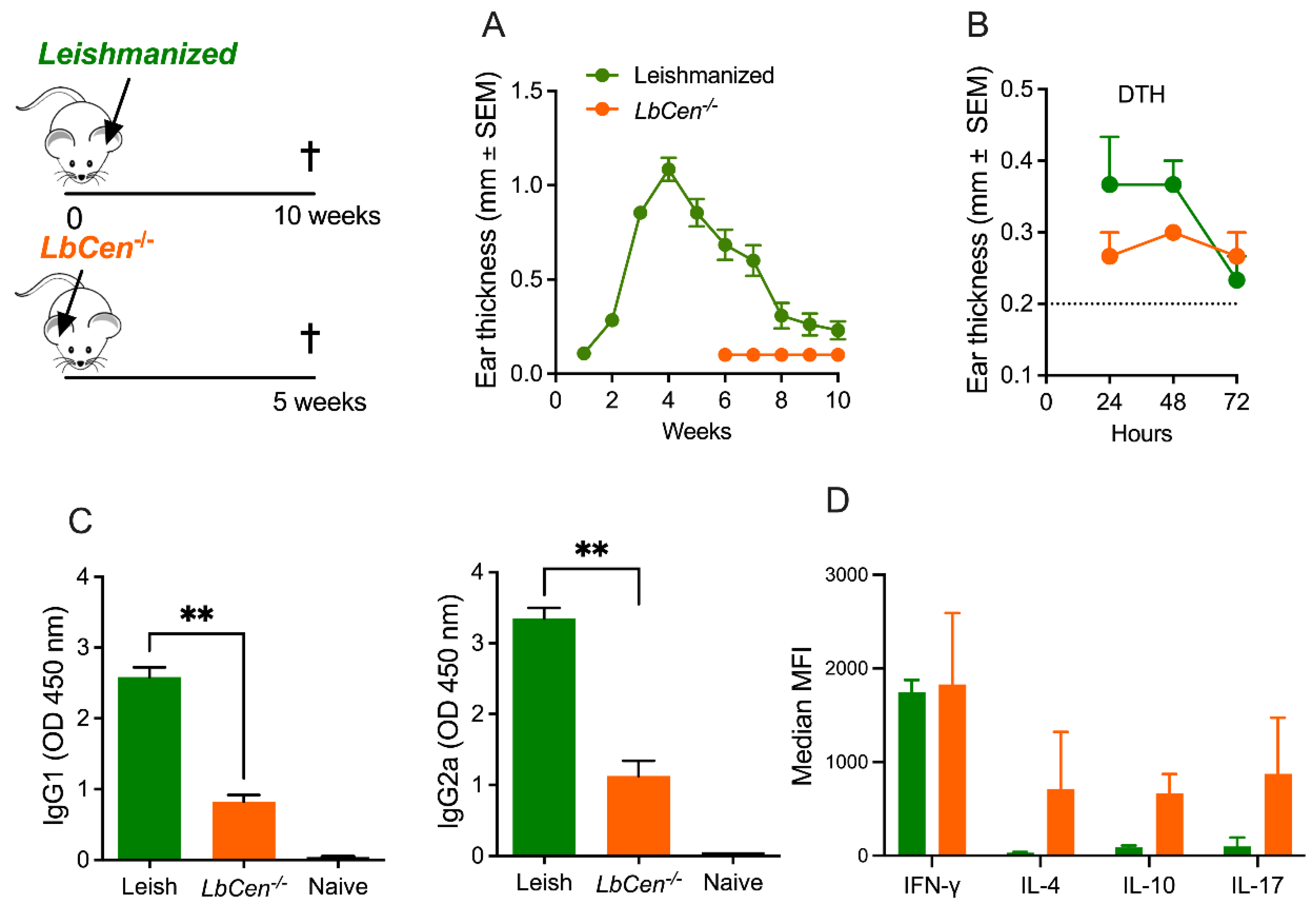
2.5. Leishmanization

BALB/c mice (n = 12) were inoculated in the right ear dermis with LbWT (105 stationary-phase promastigotes), using a 27G needle. Ear thickness was measured weekly using a digital caliper (Thermo Scientific). Ten weeks later, after lesion healing, we evaluated the cellular and humoral response. Delayed-Type Hypersensitivity (DTH) was evaluated (n = 6 mice) by inoculation of L. braziliensis SLA (10 ug/mL), prepared as described elsewhere [19], in the left ear dermis. Ear induration was measured 24, 48, and 72 h later, using a digital caliper (Thermo Scientific). For the humoral response (n = 6 mice), MaxiSorp 96-plates were coated with Soluble Leishmania Antigen (SLA) (10 ug/mL) and incubated overnight at 4 °C. Plates were washed with PBS 1X +0.05% Tween 20 (PBS-T) and blocked with 5% non-fat milk in PBS-T for 2 h at room temperature. Sera were diluted 1/100 in PBS-T +5% non-fat milk and were incubated overnight at 4 °C. The plates were washed, incubated with IgG1 or IgG2a HRP-conjugated antibodies (Invitrogen) (1/1000 in PBS-T +5% non-fat milk) for 2 h at room temperature. The plates were washed, TMB solution (Invitrogen) was added, and reactions were stopped with H2SO4 2N. The plates were read at 450 nm using a FilterMax F3 Multi-mode Microplate Reader (Molecular Devices). SoftMax Pro Software (Version 6.3) was used to obtain an indirect measure of antibody titers in serum samples. Sera from naïve mice were used as controls.
Ten weeks after the primary LbWT infection, healed (leishmanized) mice (n = 6) were challenged with 105 stationary-phase LbWT promastigotes, inoculated in the left ear dermis. Controls consisted of age-matched naïve mice (n = 6) inoculated with stationary-phase LbWT promastigotes. Parasite load was determined five weeks after challenge, as described above, with 6 mice per time point. To evaluate the cellular response, dLN cells (1 × 10⁶) were seeded on 24 well-plates and stimulated with SLA (10 ug/mL) or ConA (1 ug/mL) or left unstimulated, in supplemented RPMI. Cells were incubated at 37 °C and 5% CO2 for 48 h and cytokine production was assayed in culture supernatants by a Luminex Assay (MILLIPLEX MAP Mouse Cytokine/Chemokine Magnetic Bead Panel, Merck Millipore, Cat#MCYTOMAG-70K), following the manufacturer’s instructions. Data acquisition was performed with the Luminex LX200 (Merck Millipore). Mean Fluorescent Intensity was calculated against standards using the Milliplex Analyst software Version 5.1.

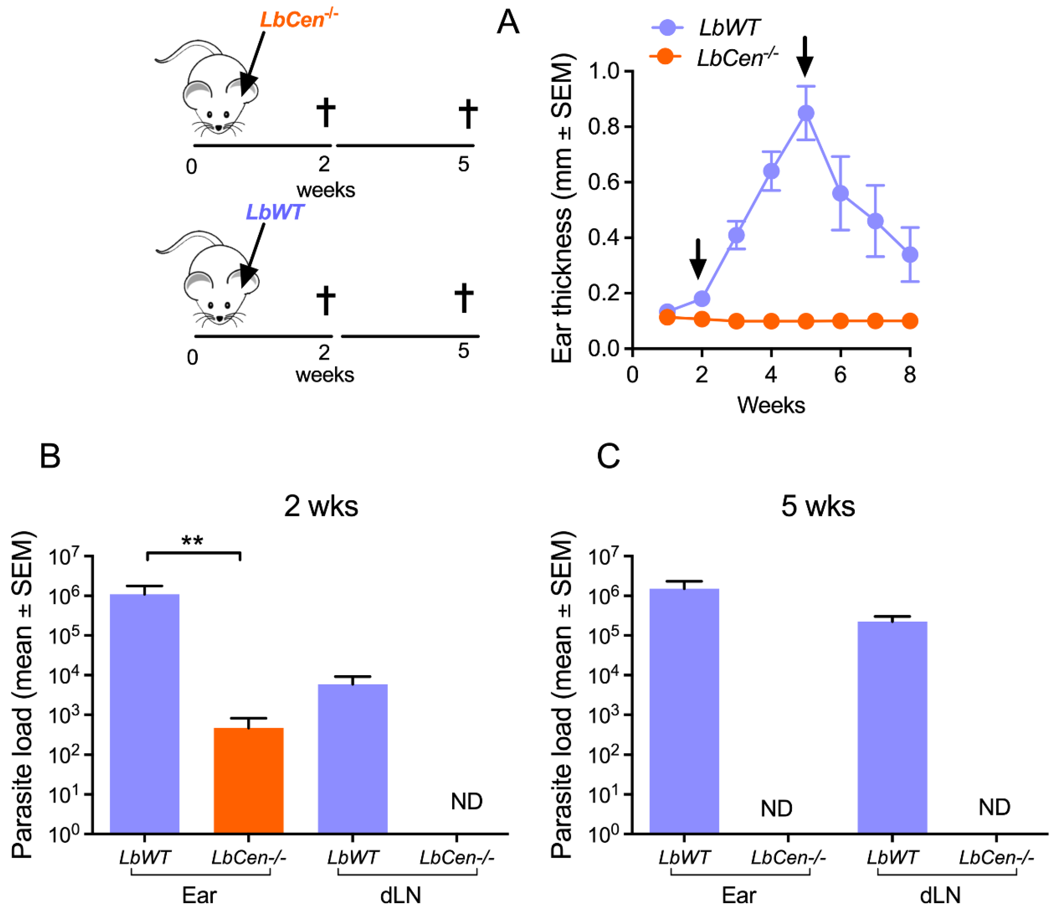
2.6. Immunization with LbCen−/− and Challenge with LbWT

BALB/c mice (n = 6) were inoculated in the right ear dermis with LbCen−/− (3 × 10⁶ stationary-phase promastigotes) or LbWT (105 stationary-phase promastigotes) using a 27G needle. Ear thickness was measured weekly using a digital caliper (Thermo Scientific). The cellular response (DTH, n = 3 mice; re-stimulation of dLN cells with SLA, n = 3 mice) and humoral immune response (IgG1 and IgG2a, n = 6 mice) were evaluated as described above at the following time points: ten weeks after the healing of the primary infection with LbWT (leishmanization) and four weeks after inoculation with LbCen−/−.

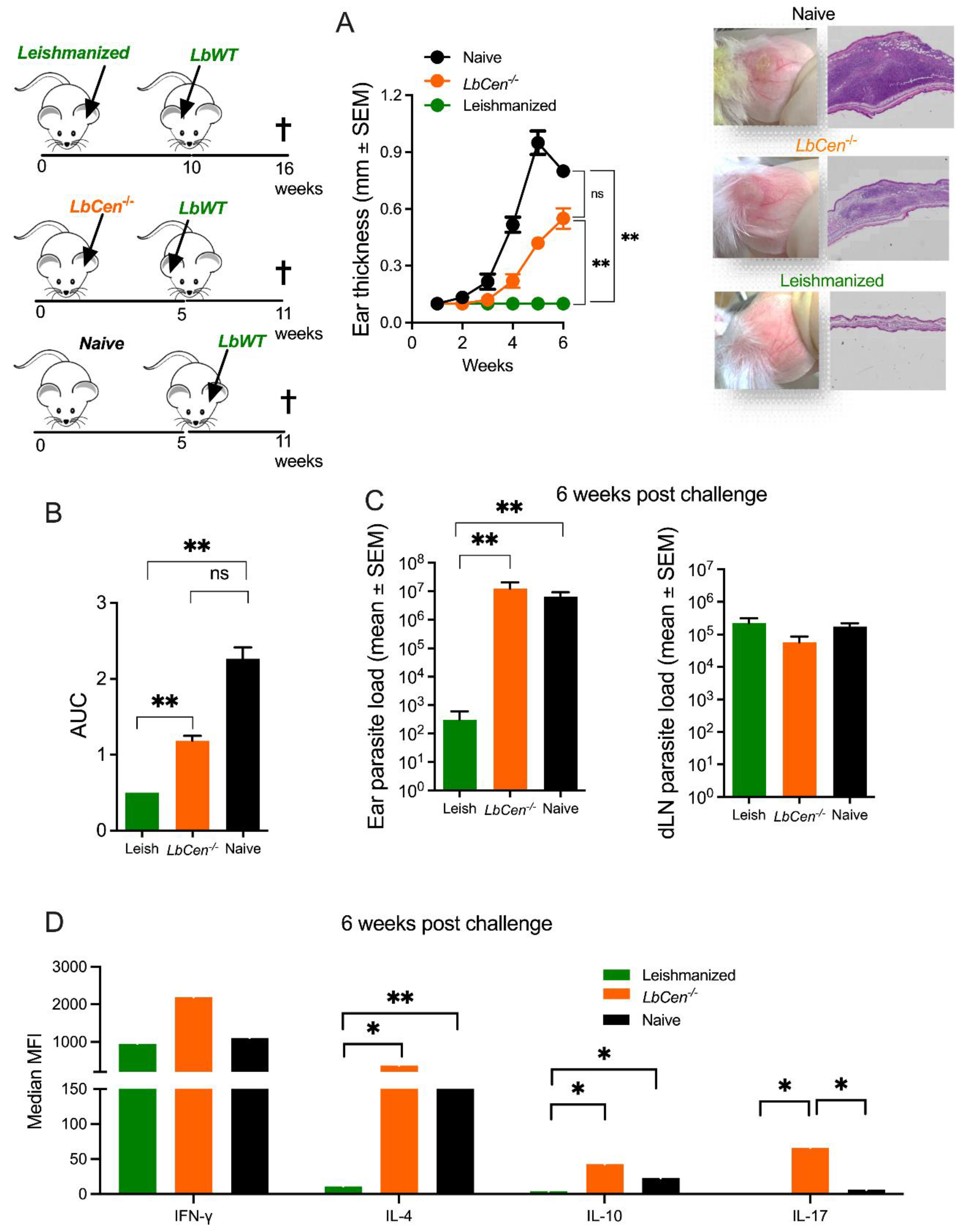
2.7. Efficacy of Immunization with LbCen−/− versus Leishmanization

BALB/c mice (n = 6 per group) were either leishmanized or immunized with LbCen−/− as described above. Ten weeks after leishmanization and six weeks after immunization, mice were challenged with LbWT (105 stationary-phase promastigotes). Controls consisted of age-matched naïve mice (n = 6) inoculated with stationary-phase LbWT promastigotes. Six weeks post challenge, mice were euthanized for the determination of parasite load and cytokine production in dLN cells. Challenged ears were excised and fixed in 10% formalin. Tissues were embedded in paraffin, cut, and stained with hematoxylin and eosin (H&E). Slides were examined under a light microscope and images were obtained using a Slide scanner Olympus VS110 system and photographs were obtained using OlyVIA Software (version 3.8).

2.8. Primary and Booster Immunization

BALB/c mice (n = 8) were inoculated in the right ear dermis with LbCen−/− (3 × 10⁶ stationary-phase promastigotes), and two weeks later, the mice were boosted at the same site. Two weeks after the booster inoculation, the cellular response (DTH and re-stimulation of dLN cells with SLA) was evaluated as described above. The frequency of central memory T cells (TCMs) was determined by flow cytometry. Briefly, dLN cells (5 × 10⁵) were incubated with Fc Block (BD Biosciences, clone 2.4G2) for 5 min at 4 °C and then stained with CD4 (eBioscience, clone GK1.5), CD44 (BD Pharmingen), and CD62L (BD Pharmingen) antibodies in the dark for 20 min at 4 °C. The cells were washed in PBS and resuspended in PBS containing Hoechst 33258 at 5 ug/mL. Acquisition was performed using an LSRFortessa™ flow cytometer (BD Biosciences) and analyzed using FlowJo Software version 10. Isotype controls for each antibody used under similar conditions indicated specific binding of the test antibody. Electronic compensation was performed with single-stained cells with individual mAbs used in the test samples.
To evaluate the efficacy of the prime-boost strategy, mice (n = 8) were immunized as described and, five weeks later, were challenged with LbWT (105 stationary-phase promastigotes). Controls consisted of age-matched naïve mice (n = 8) inoculated with stationary-phase LbWT promastigotes. Lesion development was measured weekly. Five weeks after the challenge, parasite load, cytokine response in dLN cells stimulated with SLA, and the frequency of TCM were determined, as described above.

2.9. Cross-Protection: Immunization with LdonCen−/− and Challenge with LbWT

BALB/c mice (n = 6) were inoculated in the right ear dermis with 3 × 10⁶ stationary-phase LbCen−/− or with metacyclic LdonCen−/− via the tail vein. Five weeks later, mice were challenged with LbWT (105 stationary-phase promastigotes) in the left ear dermis. Controls consisted of age-matched naïve mice (n = 6). Ear thickness was measured weekly. Six weeks post challenge, parasite load was determined by limiting-dilution analysis, as described above.

2.10. Statistical Analysis

The course of disease was plotted individually for mice in all experimental and control groups. Comparisons between two groups were performed using Mann–Whitney non-parametric t-tests and comparisons among more than two groups were performed using the Kruskal–Wallis method. Data are presented as the mean ± error of the mean. Analyses were conducted using Prism (V.8.0, GraphPad) and a p-value ≤ 0.05 was considered significant.

3. Results

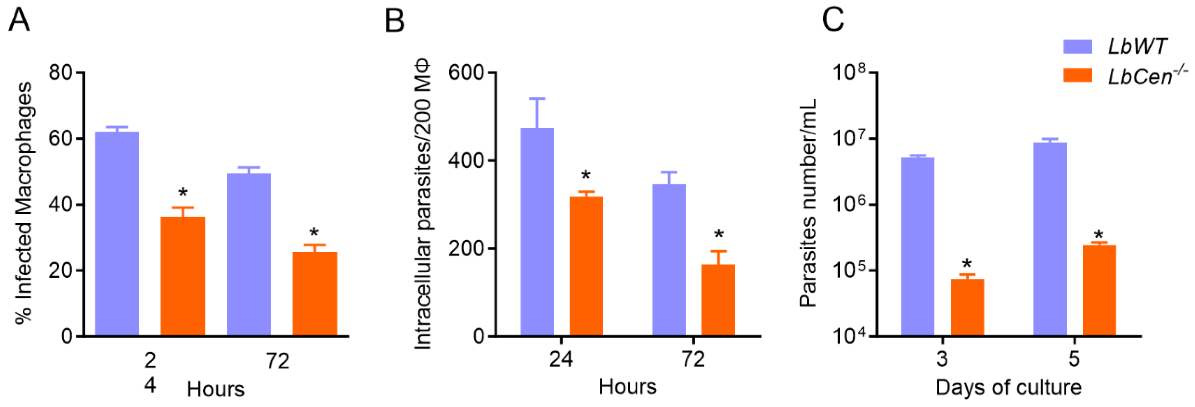
3.1. LbCen−/− Parasites Exhibit Impaired Growth and Survival in BMDMs

BMDMs were infected with LbCen−/− or LbWT and, after 24 h and 72 h, the number of infected macrophages and intracellular parasites was determined. Exposure to LbCen−/− led to significantly reduced survival (p < 0.05) compared to LbWT, both at 24 h and 72 h (Figure 1A,B). After 72 h, the BMDMs were washed and cultured in a supplemented Schneider medium to determine the number of recovered parasites. At both time points, we observed a significantly decreased (p < 0.05) recovery of LbCen−/− compared to LbWT (Figure 1C). This outcome was not due to impaired uptake by the host cell, as the number of BMDMs harboring LbCen−/− or LbWT was similar after 6 h of exposure (Supplemental Figure S1A,B) as was the number of intracellular amastigotes. These results confirm that lack of centrin in L. braziliensis impairs survival and replication within the host cell.

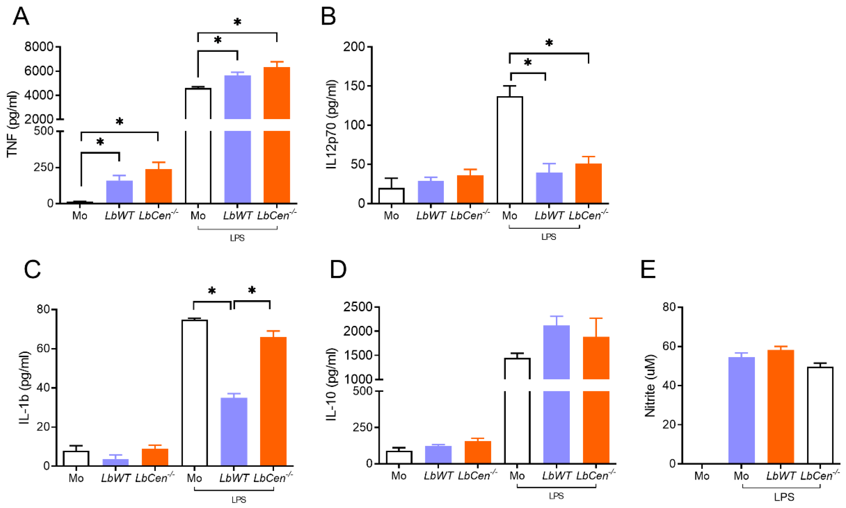
3.2. Macrophage Exposure to LbCen−/− or LbWT Induces Similar Cytokine Response

BMDMs were exposed to LbCen−/− or LbWT in the presence or absence of LPS, and cytokine production was evaluated after 24 h. Levels of TNF were similar in both LbCen−/− and LbWT infections and significantly higher (p < 0.05) compared to controls (Figure 2A). Levels of IL-12p70 were also similar for LbCen−/− and LbWT and significantly lower (p < 0.05) compared to controls stimulated with LPS (Figure 2B). Exposure to LbWT impaired IL-1β secretion, whereas LbCen−/− did not (Figure 2C). IL-10 levels (Figure 2D) and NO production were not changed in comparison to controls (Figure 2E). Taken together, these results show that the macrophages exposed to LbWT or LbCen−/− parasites produce a similar innate immune response, except for IL-1β.

3.3. Immunization with LbCen−/− Does Not Cause Lesions and Is Safe

The deletion of centrin affects amastigote growth, and mice inoculated with centrin-deficient Leishmania do not develop disease [15,16]. Deletion of the centrin in L. braziliensis reproduced this avirulent phenotype, as BALB/c inoculated with LbCen−/− parasites failed to develop cutaneous lesions (Figure 3A), whereas inoculation of LbWT leads to lesions that develop and heal spontaneously [13]. Two weeks after inoculation, the parasite load was significantly lower (p < 0.05) in the ears of mice inoculated with LbCen−/− compared to LbWT (Figure 3B). Viable parasites were recovered from the inoculation site for both LbWT and LbCen−/− groups at 2 weeks p.i., but only from the LbWT group and not from the LbCen−/− group from the dLNs 2 weeks p.i. (Figure 3B). Five weeks post inoculation, LbCen−/− parasites were not detected at either the inoculation site or in dLNs (Figure 3C), whereas in LbWT-infected mice, parasites were detected at both sites.

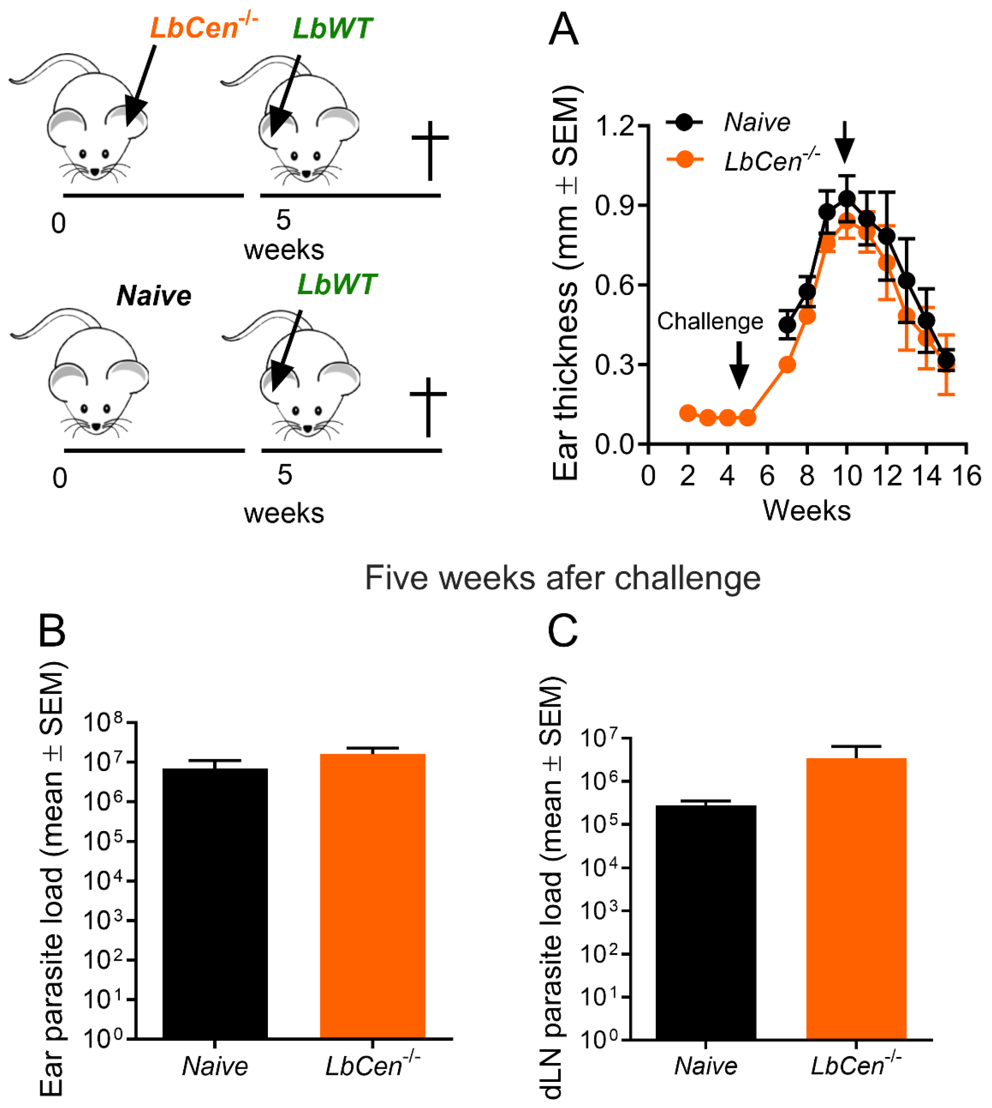
3.4. Immunization with LbCen−/− Does Not Protect against LbWT Infection

To evaluate the protective capacity of LbCen−/−, we immunized BALB/c mice with LbCen−/− and, 5 weeks later, challenged them with LbWT. Surprisingly, immunization with LbCen−/− did not protect against LbWT infection (Figure 4A). Mice developed cutaneous lesions that were comparable to those observed in naïve controls upon a challenge infection with LbWT at week 10 (Figure 4A). In a separate experiment, after 5 weeks of immunization followed by five weeks of WT challenge, there was no difference in the parasite load in the two groups, both at the inoculation site and in dLNs (Figure 4B and Figure 4C, respectively). Thus, a lack of centrin in L. braziliensis renders parasites unable to cause pathology but incapable of conferring protection against infection with wild-type L. braziliensis, differently from the centrin deletion mutation of other Leishmania spp. [12,15,16].

3.5. Immunization with LbCen−/− Cannot Recapitulate the Immune Response Induced by a Healed Primary Infection with LbWT

Leishmanization, known as the deliberate inoculation of Leishmania parasites to provide protective immunity to re-infection, is effective and has been successfully tested in pre-clinical animal models [20,21] and in humans [22]. We thus evaluated the immune response in mice that healed from a primary infection with LbWT. Following inoculation of LbWT, BALB/c mice developed lesions that peaked at around week 5 and spontaneously healed by week 10 (Supplemental Figure S2A). These primary infected and healed mice mounted a DTH response against SLA (Supplemental Figure S2B) and produced both IgG1 and IgG2a (Supplemental Figure S2C). Upon re-infection of healed mice with LbWT, lesions did not develop (Supplemental Figure S2D), differently from the control (naïve) mice. This protective outcome is accompanied by a significantly lower (p < 0.01) parasite load at the inoculation site but not in dLNs (Supplemental Figure S2E). Mice that healed from a primary infection also produced significantly (p < 0.01) lower levels of IFN-γ, IL-4, and IL-10 (p < 0.01) (Supplemental Figure S2F) compared to control (naïve) mice. Thus, experimental infection with LbWT induces disease development that is spontaneously cured in BALB/c mice, and healed mice are immune to challenge.
Next, we wanted to compare the immune response of leishmanized versus immunized animals. As observed before, LbCen−/− does not cause lesion development, differently from LbWT (Figure 5A). Although the DTH responses in the two groups were not significantly different (Figure 5B), IgG1 and IgG2a levels (Figure 5C) were significantly higher (p < 0.01) in leishmanized mice compared to immunized animals. Moreover, while IFN-γ production was similar in the two groups, IL-4, IL-10 and IL-17 levels were higher in mice immunized with LbCen−/− (Figure 5D). These results showed that the immune responses that developed upon inoculation with LbCen−/− were not Th1-dominant, contrary to that developed by mice that healed from a primary infection with LbWT.
Given the immune responses developed by mice immunized with LbCen−/− versus leishmanized mice, we compared the outcome of a challenge with LbWT in these two settings. At the peak of lesion development, tissue sections from the ears of LbCen−/−-immunized mice showed signs of chronic inflammation, features not observed in leishmanized mice (Figure 6A), yet not as prominent as those observed in the naïve mice. On the contrary, leishmanized mice showed only mild signs of inflammation. Leishmanized mice presented a significantly lower (p < 0.01) disease burden (AUC) compared to mice inoculated with LbCen−/− or control (naïve) mice (Figure 6B) and significantly lower (p < 0.01) parasite load at the inoculation site, compared to LbCen−/−-inoculated or control (naïve) mice (Figure 7C). While the three experimental groups displayed similar levels of IFN-γ, detected upon stimulation of dLN cells (Figure 6D), the production of IL-4, IL-10, and IL-17 was significantly higher in mice inoculated with LbCen−/− (Figure 6D). Thus, we can speculate that the cellular recruitment induced by immunization with LbCen−/− may have favored parasite establishment. Taken together, these results demonstrate that inoculation with LbCen−/− cannot recapitulate the immune profile induced by leishmanization and, as such, is incapable of conferring protection against challenge with L. braziliensis.

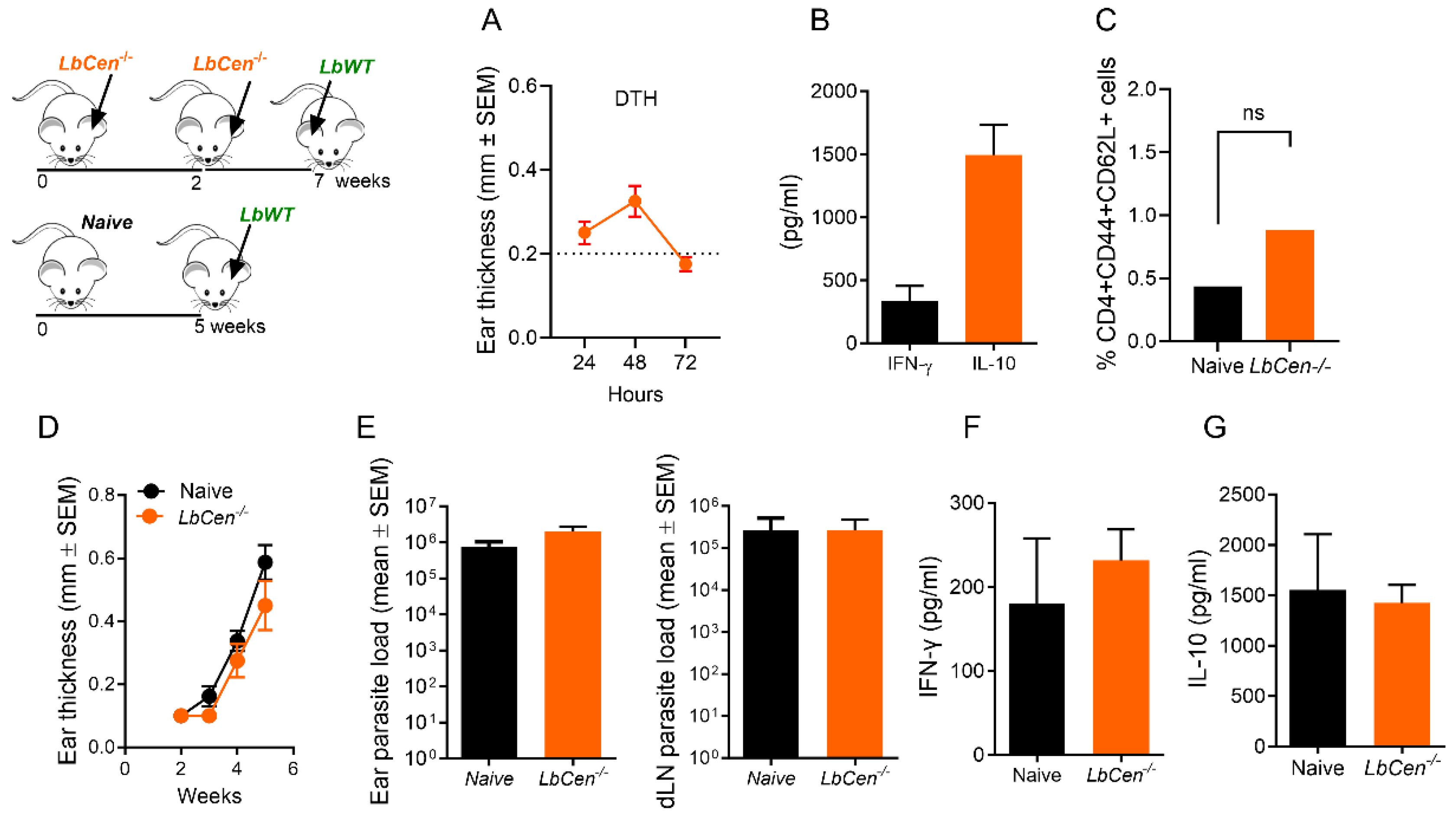
3.6. Priming/Boosting Induces Prolonged Parasite Persistence

For most vaccine strategies, multiple immunizations are required to induce efficient protection [23]. Although this has not been necessary in studies performed with Leishmania spp. lacking centrin [15,16], we investigated whether a second immunization with LbCen−/− could boost the primary immune response and, hence, alter the outcome of infection after challenge. Priming and boosting with LbCen−/− parasites continued to be safe as mice remained lesion-free (data not shown). The cellular response, measured by DTH (Figure 7A), was similar to that observed with a single immunization (Figure 5B). The recall response also showed that while IFN-γ was produced, IL-10 was detected at comparatively higher levels (Figure 7B). Two immunizations with LbCen−/− were also not capable of generating a significant increase in CD4+ T central memory cells (CD4+CD44+CD62L+) (Figure 7C), as seen upon a single immunization with LmexCen−/− [16]. Given that a booster inoculation with LbCen−/− did not alter the immune response observed earlier, we expected that the outcome of a challenge infection would also be the same. Indeed, mice receiving two inoculations with LbCen−/− continued to be susceptible to a challenge infection with LbWT (Figure 7D), with lesions developing similarly to the control, naïve mice. The parasite load in the two groups was similar (Figure 7E). Lastly, no significant differences were observed in the production of IFN-γ (Figure 7F) or IL-10 (Figure 7G), comparing mice primed and boosted with LbCen−/− and control, naïve mice. Collectively, our results show that a booster LbCen−/− immunization does not alter the lack of protection observed upon a single immunization.

3.7. Immunization with LdonCen−/− Cross-Protects against LbWT

Previously, it was shown that immunization with LdonCen−/− induces cross-protection against L. braziliensis injected subcutaneously [12]. We sought to investigate whether a similar outcome would be observed using the L. braziliensis strain employed in the generation of our centrin-deleted mutant, using an intradermal model of infection. We thus compared the outcome of immunization with LdonCen−/− versus LbCen−/− followed by a challenge with LbWT in the ear dermis. We confirmed that immunization with LdonCen−/− protects against intradermal inoculation of LbWT, differently from LbCen−/− (Figure 8A). Protection was paralleled by a significantly lower parasite (p < 0.01) load at the inoculation site and in dLNs (Figure 8B), suggesting that the immune response induced by LdonCen−/− controls parasite load even at distal sites.

4. Discussion

The current therapeutic options for leishmaniasis remain limited because of toxicity, drug administration challenges, treatment failure, and drug resistance [24]. Protection against leishmaniasis can be achieved through leishmanization [25], but this method presents safety concerns. To this end, attenuated Leishmania spp. cell lines are an attractive alternative, as safety issues can be overcome while the delivery of the near-complete antigen repertoire of the parasite is achieved. Indeed, attenuated Leishmania lacking the centrin 1 gene is being intensively explored in the context of vaccination against visceral [12] and cutaneous leishmaniasis [15,16]. Herein, we explored this possibility by using a centrin-1-deficient L. braziliensis [14], aimed at the development of a vaccine against American Tegumentary Leishmaniasis.
While deletion of the centrin 1 gene in L. braziliensis did not alter the ability of promastigotes to successfully enter the host cell, intracellular LbCen−/− proliferation was significantly reduced. In addition, exposure of macrophages to either LbCen−/− or LbWT led to similar outcomes regarding the production of innate mediators such as TNF, IL-12, IL-10, or NO. Infection of dendritic cells with L. braziliensis resulted in elevated IL-12 production [26], an effect also observed in vivo [27]. Also in vivo, inflammasome activation was shown to be involved in restricting Leishmania growth [28]. Herein, IL-1β was significantly increased upon infection with LbCen−/− compared to LbWT. We can speculate that LbCen−/− may have induced inflammasome activation and, hence, reduced parasite replication.
Inoculation of BALB/c mice with LbCen−/− was deemed safe as parasites did not induce lesion development, corroborating results obtained with centrin-1-deficient Leishmania spp. We next evaluated its ability to confer protection against LbWT. To our surprise, immunization with LbCen−/− failed to inhibit lesion development following challenge with live LbWT, contrary to what has been published for counterparts in the Leishmania subgenus, L. donovani [12], L. major [15], and L. mexicana [16]. Importantly, protection has been achieved even in the context of sand-fly-transmitted parasites [15], considered the gold standard of challenge model in leishmaniasis.
Protective immunity against L. major requires low levels of the persisting antigen [29,30]. Herein, LbCen−/− parasites were detected two weeks after intradermal inoculation into BALB/c mice but were rapidly cleared and could not be retrieved at a later time point (five weeks). These results differ from those reported for LdonCen−/− [12], LmajorCen−/− [15], and LmexCen−/− [16], the latter also employed in intradermal infections, in which centrin-deficient parasites were detected five weeks post inoculation, indicating their enhanced ability to survive in the vertebrate host, compared to LbCen−/−. Data from immunosuppressed mouse models showed persistence of LmCen−/− parasites 15 weeks post inoculation, suggesting that a low level of parasites may be important for acquiring protective immunity [15]. Moreover, mice primed and boosted with LbCen−/− continued to show susceptibility to infection, indicating that centrin-deficient L. braziliensis is an extremely attenuated strain, leading to rapid clearance by BALB/c mice.
We showed that BALB/c mice that heal from a primary infection with LbWT develop protective immunity against re-infection, analogous to leishmanization models. In this experimental demonstration of leishmanization, immunity was associated with elevated IgG1, IG2a levels, and IFN-γ production by dLN cells, all paralleled by a significantly decreased parasite load in challenged mice. In mice inoculated with LbCen−/−, an opposite result was observed: mice mounted a Th2-biased immune response, with elevated levels IL-4, IL-10, and IL-17 and lower levels of IgG1 and IgG2a antibodies. Although IFN-γ production was similar comparing LbCen−/−-immunized and leishmanized mice, IL-4 and IL-10 were only upregulated in mice inoculated with LbCen−/−. In L. major-infected BALB/c mice, the early production of IL-4 drives a Th2 response and susceptibility to disease, and treatment with anti-IL-4 or the use of IL-4-deficient mice abrogates this effect [31,32]. In C57BL/6 mice that heal from a primary infection with L. major, IL-10 is necessary for the long-term parasite persistence, and a sterile cure can be achieved in mice lacking both IL-4 and IL-10 [33]. Interestingly, immunity conferred by LmexCen−/− was mediated mainly by a reduction in IL-4 and IL-10 levels rather than an increase in IFN-γ, thus enabling the control of infection and inhibiting disease development [16]. Herein, in the same genetic background (BALB/c), immunization with LbCen−/− induced an opposing effect: upregulation in IL-4, IL-10, and IL-17. In comparison to L. major, L. braziliensis induces significantly lower expression of IL-4, IL-10, and IL-13 in the early phase of infection in BALB/c mice [27]. In C57BL/6 mice infected with LbWT, authors observed an expansion of IFN-γ-, IL-10-, and IL-17-producing CD4+ T cells at the time when lesions peaked (four weeks). By eight weeks post infection, when lesions healed, the frequency of these populations decreased, suggesting that the fine balance between pro-inflammatory and regulatory cytokines is linked to controlled L. braziliensis infection [26]. In C57BL/6 mice infected with L. infantum, IL-17A and IFN-γ potentiated NO production, restricting parasite growth in macrophages [34]. We thus suggest that although IFN-γ is induced by immunization with LbCen−/−, the unabated IL-4 and IL-10 production accompanied by sustained IL-17 are associated with a lack of protection.

5. Conclusions

We highlight that the deletion of centrin in L. braziliensis replicated the phenotype observed in L. mexicana, L. major, and L. donovani, with the major difference that a mixed immune response, with elevated IL-4 and IL-10 production, was observed. This cannot be attributed to the presence of Leishmania RNA Virus (LRV) [35], as the strain of L. braziliensis used in this study does not carry LRV (unpublished). While immunization with LmexCen−/− promotes changes in the host cell leading to M1 polarization [36], LmajorCen−/− modulates tryptophan metabolism, promoting the production of IL-12 rather than IL-10 and TGF-β [37]. It remains to be investigated if the metabolic immune regulation in LbCen−/− infection disfavors the replication of parasites, rapidly clearing the infection. Alternatively, other innate immune responses observed in LdonCen−/− may fail to materialize in LbCen−/−, resulting in a lack of protection. The divergent mechanisms of protection induced by LmajorCen−/− and LmexCen−/− highlight the need for fine tuning vaccination strategies to control American Tegumentary Leishmaniasis. It remains to be tested if LmajorCen−/−, a dermotropic strain, can confer protection against L. braziliensis challenge. Such studies will illustrate the common correlates of protection that could be targeted in a pan-leishmania vaccine.

Supplementary Materials

The following supporting information can be downloaded at: https://www.mdpi.com/article/10.3390/vaccines12030310/s1, Figure S1: Uptake of LbCen−/− and LbWT; Figure S2: A primary infection with L. braziliensis confers immunity against subsequent challenge.

Author Contributions

Conceptualization, F.A.-R. and C.I.d.O.; methodology, F.A.-R., G.A.-D., P.B.B., V.M., L.T.A., L.O.d.S., S.M.V. and R.S.; formal analysis, F.A.-R., P.B.B., S.M.V. and C.I.d.O.; investigation, F.A.-R., G.A.-D., P.B.B., L.O.d.S. and S.M.V.; resources, R.S., S.G. and H.L.N.; writing—original draft, F.A.-R. and C.I.d.O.; writing—review and editing, F.A.-R., S.G., H.L.N. and C.I.d.O.; funding acquisition, C.I.d.O. All authors have read and agreed to the published version of the manuscript.

Funding

This work was supported by grants from IGM-Fiocruz Bahia and by the Brazilian Ministry of Science, Technology, and Innovation, Instituto Nacional de Ciência e Tecnologia de Doenças Tropicais (INCT-DT) #465229/2014-0—CNPq, Fundação de Amparo à Pesquisa do Estado da Bahia (FAPESB) INC 0003/2019. C.I.d.O. is a senior researcher at CNPq. F.A.-R., P.B.B. and S.M.V. received a fellowship from Coordenação de Aperfeiçoamento de Pessoal de Nível Superior—Brasil (CAPES)—Finance Code 001. L.T.A. received a fellowship from CNPq.

Institutional Review Board Statement

Six- to eight-week-old female BALB/c mice were obtained from the IGM-FIOCRUZ animal facility and maintained under pathogen-free conditions. All animal experimentation was conducted following the Guidelines for Animal Experimentation established by the Brazilian Council for Animal Experimentation Control (CONCEA). All procedures involving animals were approved by the local Institutional Review Board for Animal Care and Experimentation (CEUA-015/2019-IGM/FIOCRUZ).

Data Availability Statement

The data presented in this study are available on request from the corresponding author.

Conflicts of Interest

Our contributions are an informal communication and represent our own best judgement. These comments do not bind or obligate the FDA.

References

  1. Burza, S.; Croft, S.L.; Boelaert, M. Leishmaniasis. Lancet 2018, 15, 951–970. [Google Scholar] [CrossRef]
  2. Pan American Health Organization. LEISHMANIASES Epidemiological Report on the Region of the Americas. Available online: https://iris.paho.org/bitstream/handle/10665.2/56831/PAHOCDEVT220021_eng.pdf?sequence=1&isAllowed=y (accessed on 2 February 2023).
  3. Sundar, S.; Singh, B. Identifying vaccine targets for anti-leishmanial vaccine development. Expert. Rev. Vaccines 2014, 13, 489–505. [Google Scholar] [CrossRef] [PubMed]
  4. Mauël, J.; Ransijn, A.; Buchmüller-Rouiller, Y. Killing of Leishmania parasites in activated murine macrophages is based on an L-arginine-dependent process that produces nitrogen derivatives. J. Leukoc. Biol. 1991, 49, 73–82. [Google Scholar] [CrossRef] [PubMed]
  5. Cummings, H.E.; Tuladhar, R.; Satoskar, A.R. Cytokines and their STATs in cutaneous and visceral leishmaniasis. J. Biomed. Biotechnol. 2010, 2010, 294389. [Google Scholar] [CrossRef]
  6. Ikeogu, N.M.; Akaluka, G.N.; Edechi, C.A.; Salako, E.S.; Onyilagha, C.; Barazandeh, A.F.; Uzonna, J.E. Leishmania Immunity: Advancing Immunotherapy and Vaccine Development. Microorganisms 2020, 8, 1201. [Google Scholar] [CrossRef] [PubMed]
  7. Moreira, P.O.L.; Nogueira, P.M.; Monte-Neto, R.L. Next-generation Leishmanization: Revisiting Molecular Targets for Selecting Genetically Enigneered Live-Attenuated Leishmania. Microorganisms 2023, 11, 1043. [Google Scholar] [CrossRef]
  8. Velez, R.; Gállego, M. Commercially approved vaccines for canine leishmaniosis: A review of available data on their safety and efficacy. Trop. Med. Int. Health 2020, 25, 540–557. [Google Scholar] [CrossRef] [PubMed]
  9. Zhang, W.W.; Matlashewski, G. CRISPR-Cas9-Mediated Genome Editing in Leishmania donovani. mBio 2015, 6, e00861. [Google Scholar] [CrossRef]
  10. Saljoughian, N.; Taheri, T.; Rafati, S. Live vaccination tactics: Possible approaches for controlling visceral leishmaniasis. Front. Immunol. 2014, 31, 134. [Google Scholar] [CrossRef]
  11. Wiech, H.; Geier, B.M.; Paschke, T.; Spang, A.; Grein, K.; Steinkötter, J.; Melkonian, M.; Schiebel, E. Characterization of green alga, yeast, and human centrins. Specific subdomain features determine functional diversity. J. Biol. Chem. 1996, 271, 22453–22461. [Google Scholar] [CrossRef]
  12. Selvapandiyan, A.; Dey, R.; Nylen, S.; Duncan, R.; Sacks, D.; Nakhasi, H.L. Intracellular replication-deficient Leishmania donovani induces long lasting protective immunity against visceral leishmaniasis. J. Immunol. 2009, 183, 1813–1820. [Google Scholar] [CrossRef]
  13. de Moura, T.R.; Novais, F.O.; Oliveira, F.; Clarêncio, J.; Noronha, A.; Barral, A.; Brodskyn, C.; de Oliveira, C.I. Toward a novel experimental model of infection to study American cutaneous leishmaniasis caused by Leishmania braziliensis. Infect. Immun. 2005, 73, 5827–5834. [Google Scholar] [CrossRef]
  14. Sharma, R.; Avendaño-Rangel, F.; Reis-Cunha, J.L.; Marques, L.P.; Figueira, C.P.; Borba, P.B.; Viana, S.M.; Beneke, T.; Bartholomeu, D.C.; de Oliveira, C.I. Targeted Deletion of Centrin in Leishmania braziliensis Using CRISPR-Cas9-Based Editing. Front. Cell Infect. Microbiol. 2022, 11, 790418. [Google Scholar] [CrossRef] [PubMed]
  15. Zhang, W.W.; Karmakar, S.; Gannavaram, S.; Dey, R.; Lypaczewski, P.; Ismail, N.; Siddiqui, A.; Simonyan, V.; Oliveira, F.; Coutinho-Abreu, I.V.; et al. A second generation leishmanization vaccine with a markerless attenuated Leishmania major strain using CRISPR gene editing. Nat. Commun. 2020, 11, 3461. [Google Scholar] [CrossRef] [PubMed]
  16. Volpedo, G.; Pacheco-Fernandez, T.; Holcomb, E.A.; Zhang, W.W.; Lypaczewski, P.; Cox, B.; Fultz, R.; Mishan, C.; Verma, C.; Huston, R.H.; et al. Centrin-deficient Leishmania mexicana confers protection against New World cutaneous leishmaniasis. NPJ Vaccines 2022, 7, 32. [Google Scholar] [CrossRef] [PubMed]
  17. Ding, A.H.; Nathan, C.F.; Stuehr, D.J. Release of reactive nitrogen intermediates and reactive oxygen intermediates from mouse peritoneal macrophages. Comparison of activating cytokines and evidence for independent production. J. Immunol. 1988, 141, 2407–2412. [Google Scholar] [CrossRef] [PubMed]
  18. Novais, F.O.; Carvalho, L.P.; Graff, J.W.; Beiting, D.P.; Ruthel, G.; Roos, D.S.; Betts, M.R.; Goldschmidt, M.H.; Wilson, M.E.; de Oliveira, C.I.; et al. Cytotoxic T cells mediate pathology and metastasis in cutaneous leishmaniasis. PLoS Pathog. 2013, 9, e1003504. [Google Scholar] [CrossRef] [PubMed]
  19. Reed, S.G.; Badaró, R.; Masur, H.; Carvalho, E.M.; Lorenco, R.; Lisboa, A.; Teixeira, R.; Johnson, W.D., Jr.; Jones, T.C. Selection of a skin test antigen for American visceral leishmaniasis. Am. J. Trop. Med. Hyg. 1986, 35, 79–85. [Google Scholar] [CrossRef]
  20. Selvapandiyan, A.; Debrabant, A.; Duncan, R.; Muller, J.; Salotra, P.; Sreenivas, G.; Salisbury, J.L.; Nakhasi, H.L. Centrin gene disruption impairs stage-specific basal body duplication and cell cycle progression in Leishmania. J. Biol. Chem. 2004, 279, 25703–25710. [Google Scholar] [CrossRef]
  21. Zaph, C.; Uzonna, J.; Beverley, S.M.; Scott, P. Central memory T cells mediate long-term immunity to Leishmania major in the absence of persistent parasites. Nat. Med. 2004, 10, 1104–1110. [Google Scholar] [CrossRef] [PubMed]
  22. Peters, N.C.; Kimblin, N.; Secundino, N.; Kamhawi, S.; Lawyer, P.; Sacks, D.L. Vector Transmission of Leishmania Abrogates Vaccine-Induced Protective Immunity. PLOS Pathog. 2009, 5, e1000484. [Google Scholar] [CrossRef] [PubMed]
  23. Mohebali, M.; Nadim, A.; Khamesipour, A. An overview of leishmanization experience: A successful control measure and a tool to evaluate candidate vaccines. Acta Trop. 2019, 200, 105173. [Google Scholar] [CrossRef] [PubMed]
  24. Lu, S. Heterologous prime-boost vaccination. Curr. Opin. Immunol. 2009, 21, 346–351. [Google Scholar] [CrossRef] [PubMed]
  25. McGwire, B.S.; Satoskar, A.R. Leishmaniasis: Clinical Syndromes and Treatment. QJM 2014, 107, 7–14. [Google Scholar] [CrossRef]
  26. Khamesipour, A.; Dowlati, Y.; Asilian, A.; Hashemi-Fesharki, R.; Javadi, A.; Noazin, S.; Modabber, F. Leishmanization: Use of an old method for evaluation of candidate vaccines against leishmaniasis. Vaccine 2005, 23, 3642–3648. [Google Scholar] [CrossRef] [PubMed]
  27. Vargas-Inchaustegui, D.A.; Xin, L.; Soong, L. Leishmania braziliensis infection induces dendritic cell activation, ISG15 transcription, and the generation of protective immune responses. J. Immunol. 2008, 180, 7537–7545. [Google Scholar] [CrossRef] [PubMed]
  28. Rocha, F.J.; Schleicher, U.; Mattner, J.; Alber, G.; Bogdan, C. Cytokines, signaling pathways, and effector molecules required for the control of Leishmania (Viannia) braziliensis in mice. Infect. Immun. 2007, 75, 3823–3832. [Google Scholar] [CrossRef]
  29. Lima-Junior, D.S.; Costa, D.L.; Carregaro, V.; Cunha, L.D.; Silva, A.L.N.; Mineo, T.W.P.; Gutierrez, F.R.S.; Bellio, M.; Bortoluci, K.R.; Flavell, R.A.; et al. Inflammasome-derived IL-1β production induces nitric oxide-mediated resistance to Leishmania. Nat. Med. 2013, 19, 909–915. [Google Scholar] [CrossRef]
  30. Uzonna, J.E.; Wei, G.; Yurkowski, D.; Bretscher, P. Immune elimination of Leishmania major in mice: Implications for immune memory, vaccination, and reactivation disease. J. Immunol. 2001, 167, 6967–6974. [Google Scholar] [CrossRef]
  31. Belkaid, Y.; Piccirillo, C.A.; Mendez, S.; Shevach, E.M.; Sacks, D.L. CD4+CD25+ regulatory T cells control Leishmania major persistence and immunity. Nature 2002, 420, 502–507. [Google Scholar] [CrossRef]
  32. Chatelain, R.; Varkila, K.; Coffman, R.L. IL-4 induces a Th2 response in Leishmania major-infected mice. J. Immunol. 1992, 148, 1182–1187. [Google Scholar] [CrossRef]
  33. Kopf, M.; Brombacher, F.; Köhler, G.; Kienzle, G.; Widmann, K.H.; Lefrang, K.; Humborg, C.; Ledermann, B.; Solbach, W. IL-4-deficient Balb/c mice resist infection with Leishmania major. J. Exp. Med. 1996, 184, 1127–1136. [Google Scholar] [CrossRef] [PubMed]
  34. Belkaid, Y.; Hoffmann, K.F.; Mendez, S.; Kamhawi, S.; Udey, M.C.; Wynn, T.A.; Sacks, D.L. The role of interleukin (IL)-10 in the persistence of Leishmania major in the skin after healing and the therapeutic potential of anti-IL-10 receptor antibody for sterile cure. J. Exp. Med. 2001, 194, 1497–1506. [Google Scholar] [CrossRef] [PubMed]
  35. Nascimento, M.S.; Carregaro, V.; Lima-Júnior, D.S.; Costa, D.L.; Ryffel, B.; Duthie, M.S.; de Jesus, A.; de Almeida, R.P.; da Silva, J.S. Interleukin 17A acts synergistically with interferon γ to promote protection against Leishmania infantum infection. J. Infect. Dis. 2015, 211, 1015–1026. [Google Scholar] [CrossRef] [PubMed]
  36. Hartley, M.A.; Ronet, C.; Zangger, H.; Beverley, S.M.; Fasel, N. Leishmania RNA virus: When the host pays the toll. Front. Cell Infect. Microbiol. 2012, 2, 99. [Google Scholar] [CrossRef]
  37. Oljuskin, T.; Azodi, N.; Volpedo, G.; Bhattacharya, P.; Markle, H.L.; Hamano, S.; Matlashewski, G.; Satoskar, A.R.; Gannavaram, S.; Nakhasi, H.L. Leishmania major centrin knock-out parasites reprogram tryptophan metabolism to induce a pro-inflammatory response. iScience 2023, 26, 107593. [Google Scholar] [CrossRef]
Figure 1. Impaired survival of LbCen−/− in murine macrophages. Bone marrow-derived macrophages (BMDMs) were infected with LbWT or LbCen−/− promastigotes. After 24 h, cells were washed and stained with H&E (at 24 h or 72 h). The percentage of infection (A) and the number of amastigotes per 200 macrophages (B) were determined by optical microscopy. Alternatively, infected macrophages were washed (72 h) and cultured in Schneider medium. Three or five days later, the number of promastigotes was evaluated (C). Results are presented as means ± SEM and are pooled from two independent experiments, each performed in quadruplicate. * p < 0.05.
Vaccines 12 00310 g001
Figure 2. Innate immune response in LbCen−/−-infected macrophages. Bone marrow-derived macrophages (BMDMs) were primed with LPS (100 ng/mL) and were later infected with LbWT or LbCen−/− promastigotes. Controls (Mo) were left unprimed. After 24 h, culture supernatants were collected and the presence of TNF (A), IL-10 (B), IL-1B (C), and IL-10 (D) was determined by ELISA. (E) Nitrite concentration was evaluated by Griess reaction. Results are presented as mean ± SEM and are from a representative experiment, performed in quadruplicate. * p < 0.05.
Vaccines 12 00310 g002
Figure 3. Safety of LbCen−/− upon experimental infection. BALB/c mice were inoculated with LbWT or LbCen−/− promastigotes. (A) Lesion development was measured weekly. Parasite load was measured in dLNs two (B) and five (C) weeks post challenge. Results are expressed as means ± SEM and are from one representative experiment, performed with five mice. ** p < 0.01. ND, not detected.
Vaccines 12 00310 g003
Figure 4. Efficacy of LbCen−/− against L. braziliensis. BALB/c mice were inoculated with LbCen−/− stationary promastigotes in the ear dermis. Five weeks later, mice were challenged with LbWT and (A) lesion development was measured weekly. (B) Parasite load was measured five weeks after challenge in the ear (B) and dLNs (C). Results are expressed as means ± SEM and are from one representative experiment performed with six mice.
Vaccines 12 00310 g004
Figure 5. Immune response induced by LbCen−/− versus a primary L. braziliensis infection. BALB/c mice were primary infected with LbWT and left to heal or were inoculated with LbCen−/− promastigotes. (A) Lesion development was measured weekly. (B) DTH response to SLA, serology (C) IgG1 and IgG2a, and (D) cytokine production in dLNs were measured ten weeks after mice healed from the primary infection (LbWT) or four weeks after inoculation with LbCen−/−. Results are expressed as means ± SEM and are from one representative experiment, performed with five mice. ** p < 0.01.
Vaccines 12 00310 g005
Figure 6. Efficacy of leishmanization vs. immunization with LbCen−/−. BALB/c mice were inoculated with LbWT or LbCen−/− promastigotes. Healed (LbWT) or immunized (LbCen−/−) mice were challenged with LbWT. Controls consisted of age-matched naïve mice. (A) Lesion development was measured weekly and histopathological analysis of ears was performed six weeks post challenge. (B) Areas under the curves (AUCs) of ear thicknesses shown in (A). (C) Parasite load in ears and draining lymph nodes six weeks post challenge. (D) Cytokine response in draining lymph nodes six weeks post challenge. Results are expressed as means ± SEM and are from one representative experiment, performed with six mice. * p < 0.05; ** p < 0.01. ns, not significant.
Vaccines 12 00310 g006
Figure 7. Prime boost with LbCen−/− cannot improve immunity against L. braziliensis. BALB/c mice were inoculated with 3 × 10⁶ LbCen−/− promastigotes and, two weeks later, mice received a booster inoculation. (A) DTH response and (B) cytokine response in dLNs cells following re-stimulation with SLA. (C) Frequency of CD4+ T central memory cells (CD4+CD44+CD62L+). Evaluations were performed two weeks after the boost inoculation. Mice were later challenged with 105 LbWT promastigotes and lesion development was measured weekly (D). (E) Parasite load, (F) cytokine production in DLN cells, and frequency of CD4+ T central memory cells (G) were determined five weeks after challenge. Results are expressed as means ± SEM and are from one representative experiment, performed with eight mice. ns, not significant, dotted line represents baseline ear thickness.
Vaccines 12 00310 g007
Figure 8. Protective immunity of LdCen−/− against L. braziliensis. Mice were immunized with LdCen−/− promastigotes and, five weeks later, mice were challenged with LbWT promastigotes. Lesion development was monitored weekly (A). Parasite load in ears and draining lymph nodes (B) was determined by limiting dilution five weeks post challenge. Results are expressed as means ± SEM and are from one experiment, performed with six mice. * p < 0.05; ** p < 0.01. ns, not significant.
Vaccines 12 00310 g008
Disclaimer/Publisher’s Note: The statements, opinions and data contained in all publications are solely those of the individual author(s) and contributor(s) and not of MDPI and/or the editor(s). MDPI and/or the editor(s) disclaim responsibility for any injury to people or property resulting from any ideas, methods, instructions or products referred to in the content.

